Embed Size (px)
Citation preview

Chapter 7: Membrane Structure and Function We are going to cover the structure and
function of the plasma membrane, including how molecules get in and out of cells.
We are also going to cover surface modifications.

Chapter 7: Membrane Models: Early Observations Lipid-soluble molecules entered cells more
rapidly than water-soluble molecules. Chemical analysis revealed the membrane
contained phospholipids.• Nonpolar tails directed inward, polar heads
outward.

Chapter 7: Membrane Model
1972, Singer and Nicolson introduced the fluid-mosaic model of membrane structure.
1. Plasma membrane is phospholipid bilayer in which protein molecules are partially or wholly embedded.
2. Embedded proteins are scattered throughout membrane in irregular pattern, depending on the membrane.

Membrane structure has 2 components:• 1. Lipids
• 2. Proteins
• phospholipids spontaneously arrange in bilayer due to amphipathic nature.

Glycolipids Cholesterol Glycoproteins
• Glycolipids and proteins occur only on outside and cytoskeletal filaments attach to proteins on the inside surface.
Plasma membrane is asymetrical

Chapter 7: Fluidity of the Plasma Membrane At body temperature, the phospholipid bilayer
has consistency of olive oil. Greater concentration of unsaturated fatty acid
residues, the more fluid the bilayer. Evolutionary adaptations and lipid composition. Fluidity allows cells to be pliable. Some proteins are held in place by cytoskeletal
filaments; most drift.

Membrane proteins determine most of membrane’s functions.
Channel proteins (hydrophilic) allow particular molecules to cross membrane.
•Carrier proteins selectively interact with a specific molecule so it can cross membrane
•Ex: Na+-K+ pump

Chapter 7: Mosaic Quality of the Membrane Receptor proteins are shaped so a specific
molecule (like a hormone) can bind to it. Enzymatic proteins catalyze specific
metabolic reactions.

Chapter 7: Cell-Cell Recognition Membrane carbohydrate chains of glycolipids and
glycoproteins identify cell.• Chains may vary by # of sugars.
• Chains vary in branching.
• Sequence of sugars in chains vary.
In development, different type cells develop their own carbohydrate chains, allowing tissues and cells to sort themselves out in the embryo.
Blood type

Plasma membrane is differentially permeable; only certain molecules can pass.
Permeable membrane allows all molecules to pass through.
Impermeable membrane allows none to pass
Semipermeable membrane allows some molecules to pass through.

Chapter 7: Types of Membranes and Transport Small non-charged lipid molecules (alcohol,
oxygen) pass freely through membrane. Small polar molecules (CO2, H2O) pass
through on concentration gradient. Macromolecules cannot cross a membrane. Ions and charged molecules have difficulty
crossing part of bilayer.

Chapter 7: Types of Membranes and Transport Passive Transport moves molecules across
membrane without use of energy by cell.• Includes diffusion and facilitated transport.
Active Transport uses energy (ATP) to move molecules across membrane.• Includes exocytosis, endocytosis, and
pinocytosis.

Chapter 7: Diffusion and Osmosis Diffusion = molecules move
from high conc. to low conc. (i.e. down concentration gradient) (show class diffusion model)
• A solution contains a solute, usually a solid, and a solvent, usually a liquid.
Lipid-soluble molecules (alcohol) and gases readily diffuse.

Osmosis is the diffusion of water across a differentially permeable membrane.
Osmotic pressure is hydrostatic pressure, or pressure that develops in the cell due to osmosis.
Osmosis is constant process in life.• Ex: water absorbed in large
intestine, retained by kidneys, and taken up by blood.

Tonicity is strength of a solution in relationship to osmosis, determining movement of water into or out of cell.
Isotonic - solute conc. of 2 solutions are equal.

Hypertonic - solute conc. of 1 solution is greater than another solution.
Hypotonic - solute conc. of 1 solution is less than another solution.

Transport by Carrier Proteins Carrier proteins are membrane
proteins that combine with and transport only 1 type of molecule.
Facilitated transport is passive transport of specific solutes down their conc. Gradient, facilitated by carrier proteins.• Move glucose and amino
acids (aa’s).
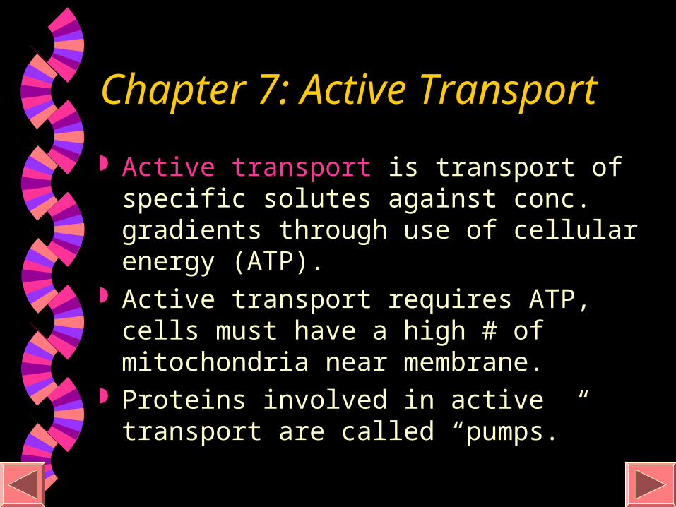
Chapter 7: Active Transport
Active transport is transport of specific solutes against conc. gradients through use of cellular energy (ATP).
Active transport requires ATP, cells must have a high # of mitochondria near membrane.
Proteins involved in active transport are called “pumps.”

Figure 7.18-1
EXTRACELLULARFLUID
[Na] high[K] low
[Na] low[K] high
CYTOPLASM
Na
Na
Na
1

Figure 7.18-2
EXTRACELLULARFLUID
[Na] high[K] low
[Na] low[K] high
CYTOPLASM
Na
Na
Na
1 2
Na
Na
Na
PATP
ADP
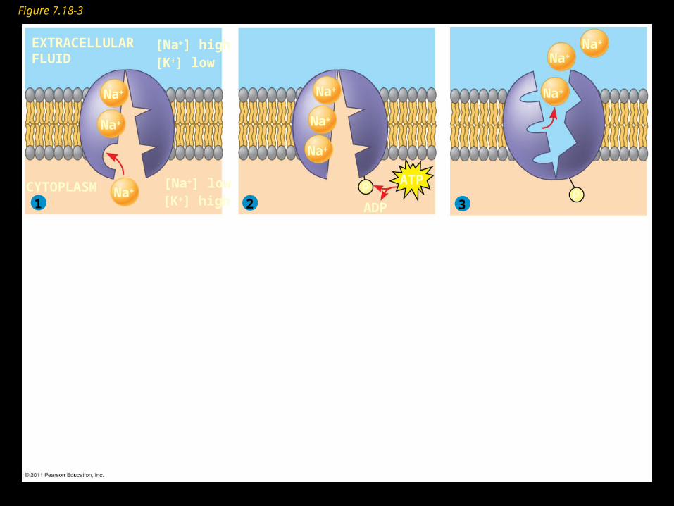
Figure 7.18-3
EXTRACELLULARFLUID
[Na] high[K] low
[Na] low[K] high
CYTOPLASM
Na
Na
Na
1 2 3
Na
Na
Na
Na
Na
Na
PP
ATP
ADP

Figure 7.18-4
EXTRACELLULARFLUID
[Na] high[K] low
[Na] low[K] high
CYTOPLASM
Na
Na
Na
1 2 3
4
Na
Na
Na
Na
Na
Na
K
K
PP
PP i
ATP
ADP

Figure 7.18-5
EXTRACELLULARFLUID
[Na] high[K] low
[Na] low[K] high
CYTOPLASM
Na
Na
Na
1 2 3
45
Na
Na
Na
Na
Na
Na
K
K
K
K
PP
PP i
ATP
ADP

Figure 7.18-6
EXTRACELLULARFLUID
[Na] high[K] low
[Na] low[K] high
CYTOPLASM
Na
Na
Na
1 2 3
456
Na
Na
Na
Na
Na
Na
K
K
K
K
K
K
PP
PP i
ATP
ADP

Figure 7.19Passive transport Active transport
Diffusion Facilitated diffusion ATP

Membrane-Assisted Transport In exocytosis, a vesicle often formed by
golgi apparatus fuses with the plasma membrane as secretion occurs.• Ex: method insulin leaves

Membrane-Assisted Transport During endocytosis, cells take in substances
by vesicle formation as membrane pinches off.

•Pinocytosis occurs when vesicles form around a liquid or very small particles.
•Ex: Blood cells and plant root cells•Phagocytosis is used when material is too large to be taken in endocytosis (like food particles)

Receptor-mediated endocytosis occurs when specific macromolecules bind to membrane receptors.• Macromolecule that binds to receptor is called a
ligand.

That’s right..Chapter 7 is over...Now on to the first lab.
You can cheer at any time