Embed Size (px)
DESCRIPTION
Chapter 7 Membrane Structure and Function. Plasma Membrane Rap. Overview: Life at the Edge. The plasma membrane is the boundary that separates the living cell from its surroundings - PowerPoint PPT Presentation
Citation preview
Plasma Membrane Rap
Chapter 7Membrane Structure and
Function
The plasma membrane is the boundary that separates the living cell from its surroundings
The plasma membrane exhibits selective permeability, allowing some substances to cross it more easily than others
In this chapter, you will learn how cellular membranes control the passage of substances.
Overview: Life at the Edge
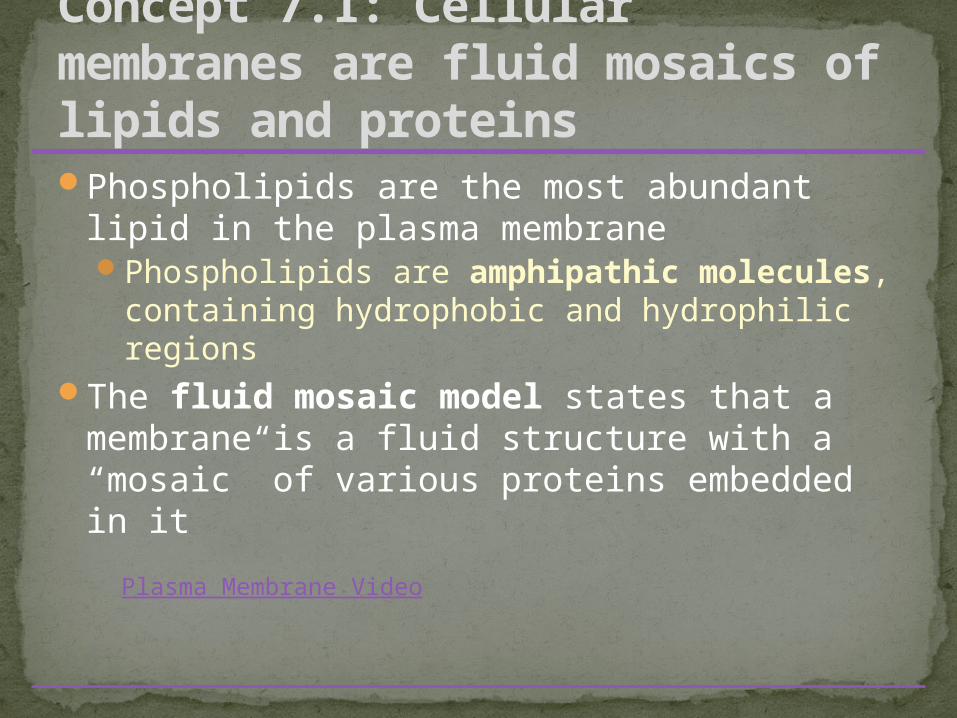
Phospholipids are the most abundant lipid in the plasma membranePhospholipids are amphipathic molecules,
containing hydrophobic and hydrophilic regions
The fluid mosaic model states that a membrane is a fluid structure with a “mosaic” of various proteins embedded in it
Concept 7.1: Cellular membranes are fluid mosaics of lipids and proteins
Plasma Membrane Video
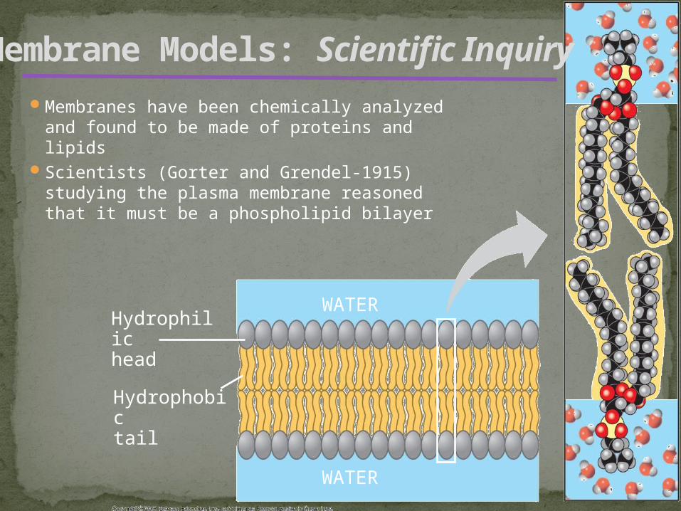
Membranes have been chemically analyzed and found to be made of proteins and lipids
Scientists (Gorter and Grendel-1915) studying the plasma membrane reasoned that it must be a phospholipid bilayer
Membrane Models: Scientific Inquiry
Hydrophilichead
WATER
Hydrophobictail
WATER
Davson & Danielli (1935) proposed a sandwich model in which the phospholipid bilayer lies between two layers of globular proteins
Later studies found problems with this model, particularly the placement of membrane proteins, which have hydrophilic and hydrophobic regions
J. Singer & G. Nicolson (1972) proposed that the membrane is a mosaic of proteins dispersed within the bilayer, with only the hydrophilic regions exposed to water (fluid mosaic model)
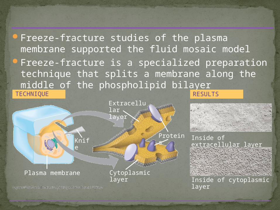
Freeze-fracture studies of the plasma membrane supported the fluid mosaic model
Freeze-fracture is a specialized preparation technique that splits a membrane along the middle of the phospholipid bilayer
Inside of cytoplasmic layer
TECHNIQUE
Extracellularlayer
KnifeProteins Inside of extracellular layer
RESULTS
Cytoplasmic layerPlasma membrane
Phospholipids in the plasma membrane can move within the bilayer
Most of the lipids, and some proteins, drift laterally
Rarely does a molecule flip-flop transversely across the membrane
The Fluidity of Membranes
As temperatures cool, membranes switch from a fluid state to a solid state
The temperature at which a membrane solidifies depends on the types of lipids
Membranes rich in unsaturated fatty acids are more fluid that those rich in saturated fatty acids
Membranes must be fluid to work properly; they are usually about as fluid as salad oil
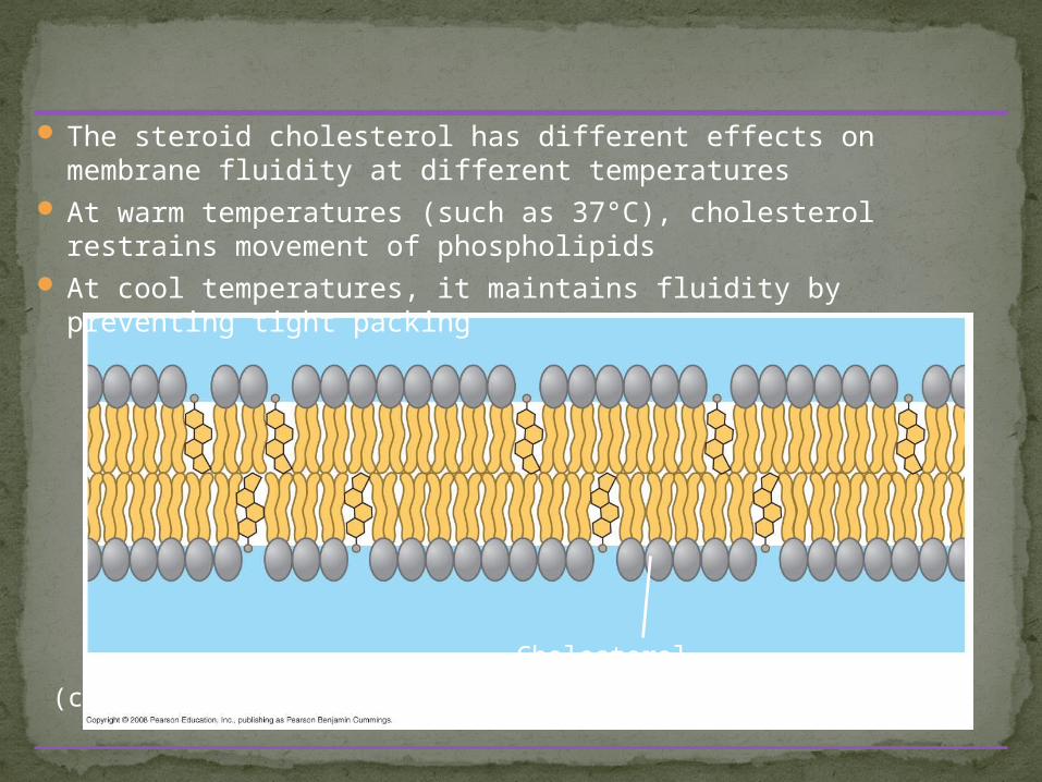
The steroid cholesterol has different effects on membrane fluidity at different temperatures
At warm temperatures (such as 37°C), cholesterol restrains movement of phospholipids
At cool temperatures, it maintains fluidity by preventing tight packing
Cholesterol
(c) Cholesterol within the animal cell membrane
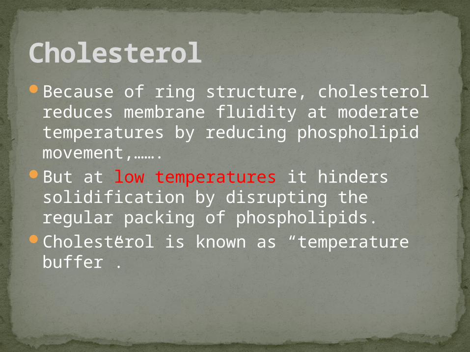
Because of ring structure, cholesterol reduces membrane fluidity at moderate temperatures by reducing phospholipid movement,…….
But at low temperatures it hinders solidification by disrupting the regular packing of phospholipids.
Cholesterol is known as “temperature buffer”.
Cholesterol
Some proteins in the plasma membrane can drift within the bilayer
Proteins are much larger than lipids and move more slowly
To investigate whether membrane proteins move, researchers fused a mouse cell and a human cell
Experiment: Do membrane Proteins Move?
A membrane is a collage of different proteins embedded in the fluid matrix of the lipid bilayer
Proteins determine most of the membrane’s specific functions
The two sides of a membrane have different protein and lipid compositions.
Membrane Proteins and Their Functions
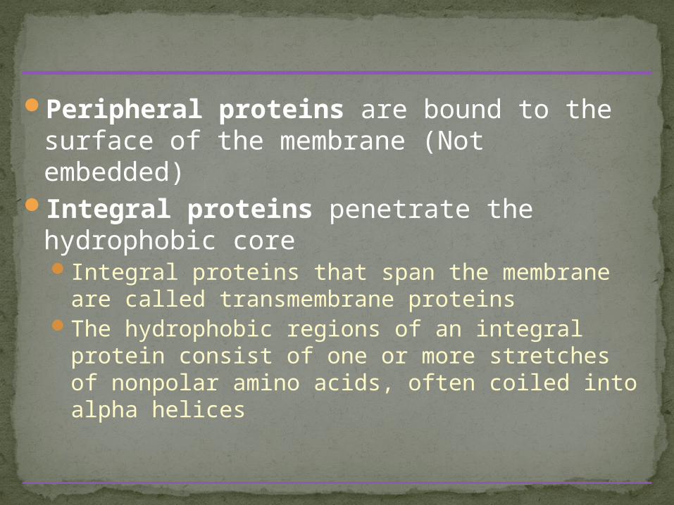
Peripheral proteins are bound to the surface of the membrane (Not embedded)
Integral proteins penetrate the hydrophobic core Integral proteins that span the membrane are
called transmembrane proteinsThe hydrophobic regions of an integral protein
consist of one or more stretches of nonpolar amino acids, often coiled into alpha helices
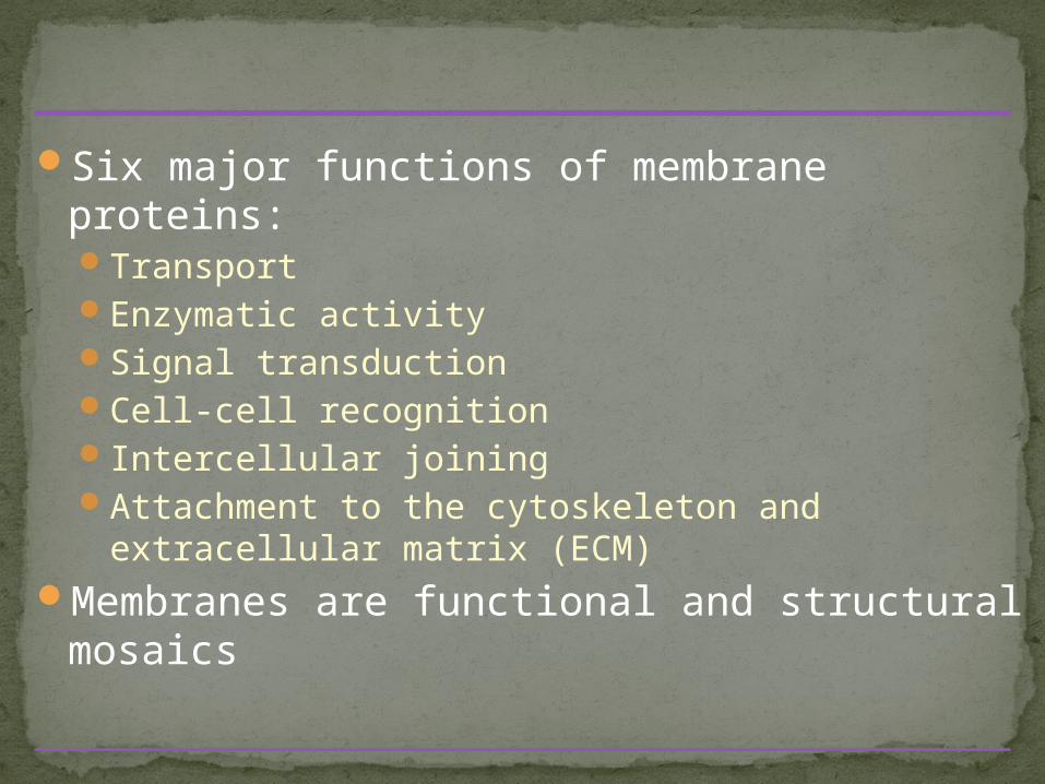
Six major functions of membrane proteins:TransportEnzymatic activitySignal transductionCell-cell recognitionIntercellular joiningAttachment to the cytoskeleton and extracellular
matrix (ECM)Membranes are functional and structural mosaics
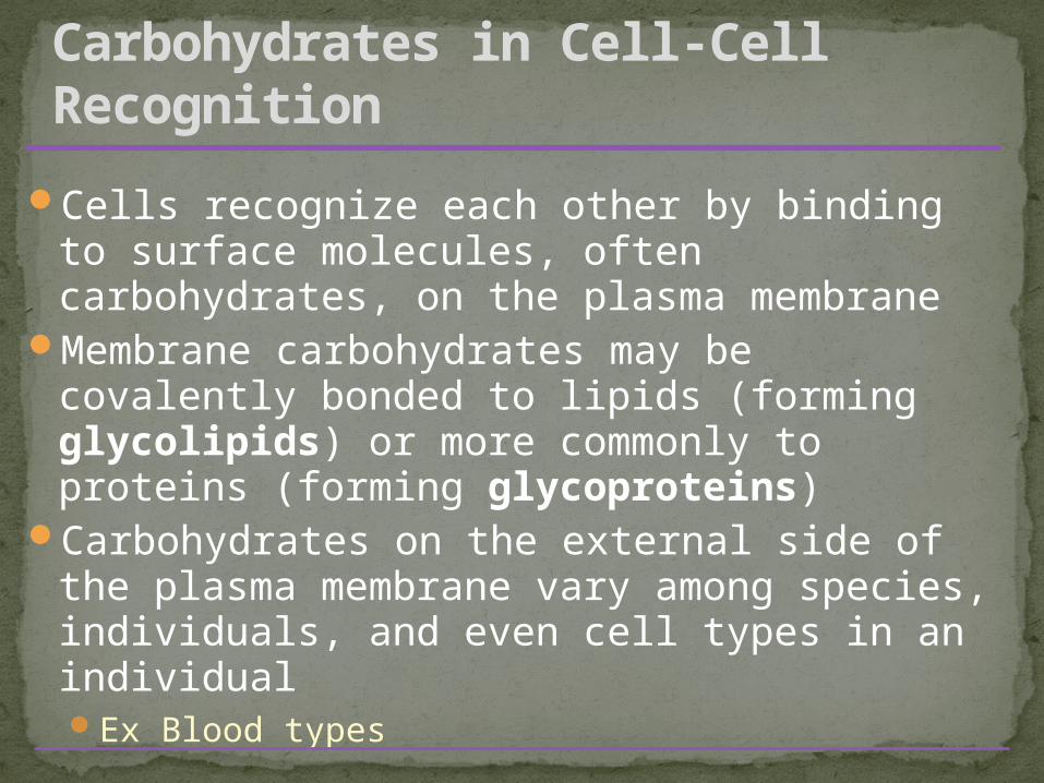
Cells recognize each other by binding to surface molecules, often carbohydrates, on the plasma membrane
Membrane carbohydrates may be covalently bonded to lipids (forming glycolipids) or more commonly to proteins (forming glycoproteins)
Carbohydrates on the external side of the plasma membrane vary among species, individuals, and even cell types in an individualEx Blood types
The Role of Membrane Carbohydrates in Cell-Cell Recognition
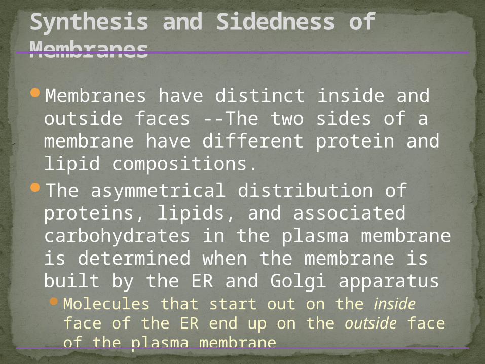
Membranes have distinct inside and outside faces --The two sides of a membrane have different protein and lipid compositions.
The asymmetrical distribution of proteins, lipids, and associated carbohydrates in the plasma membrane is determined when the membrane is built by the ER and Golgi apparatusMolecules that start out on the inside face of
the ER end up on the outside face of the plasma membrane
Synthesis and Sidedness of Membranes
A cell must exchange materials with its surroundings, a process controlled by the plasma membrane
Plasma membranes are selectively permeable, regulating the cell’s molecular trafficSize and polarity of the moleculeTransport proteins in the membrane
Concept 7.2: Membrane structure results in selective permeability
Hydrophobic (nonpolar) molecules, such as hydrocarbons, can dissolve in the lipid bilayer and pass through the membrane rapidlyHydrocarbons, CO2 and O2
Polar molecules, such as sugars, do not cross the membrane easilyC6H12O6, or charged molecules
The Permeability of the Lipid Bilayer
Transport proteins allow passage of hydrophilic substances across the membrane
Some transport proteins, called channel proteins, have a hydrophilic channel that certain molecules or ions can use as a tunnel
Channel proteins called aquaporins facilitate the passage of water
Other transport proteins, called carrier proteins, bind to molecules and change shape to shuttle them across the membrane
A transport protein is specific for the substance it moves
Transport Proteins
Diffusion is the tendency for molecules to spread out evenly into the available space
Although each molecule moves randomly, diffusion of a population of molecules may exhibit a net movement in one direction
At dynamic equilibrium, as many molecules cross one way as cross in the other directionThis is not static – even at equilibrium molecules are still
moving.
Concept 7.3: Passive transport is diffusion of a substance across a membrane with no energy investment
Substances diffuse down their concentration gradient, the difference in concentration of a substance from one area to another
No work must be done to move substances down the concentration gradientO2 gets into cells this way for cellular respiration
The diffusion of a substance across a biological membrane is passive transport because it requires no energy from the cell to make it happen
The concentration of one molecule has no effect on the movement of other molecules
Osmosis is the diffusion of water across a selectively permeable membrane
The direction of osmosis is determined only by a difference in total solute concentration.
Water diffuses across a membrane from the region of lower solute concentration to the region of higher solute concentration(or you can think [high water] to [low water]
Effects of Osmosis on Water Balance
Tonicity is the ability of a solution to cause a cell to gain or lose waterIsotonic solution: Solute concentration is the
same as that inside the cell; no net water movement across the plasma membrane
Hypertonic solution: Solute concentration is greater than that inside the cell; cell loses water
Hypotonic solution: Solute concentration is less than that inside the cell; cell gains water
Water Balance of Cells Without Walls
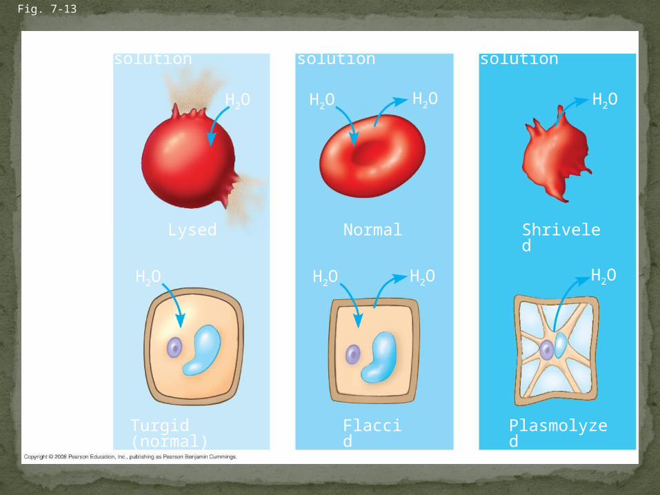
Fig. 7-13
Hypotonic solution
(a) Animal cell
(b) Plant cell
H2O
Lysed
H2O
Turgid (normal)
H2O
H2O
H2O
H2O
Normal
Isotonic solution
Flaccid
H2O
H2O
Shriveled
Plasmolyzed
Hypertonic solution
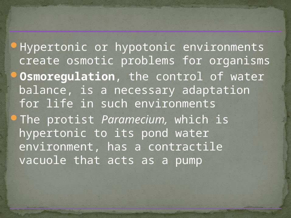
Hypertonic or hypotonic environments create osmotic problems for organisms
Osmoregulation, the control of water balance, is a necessary adaptation for life in such environments
The protist Paramecium, which is hypertonic to its pond water environment, has a contractile vacuole that acts as a pump
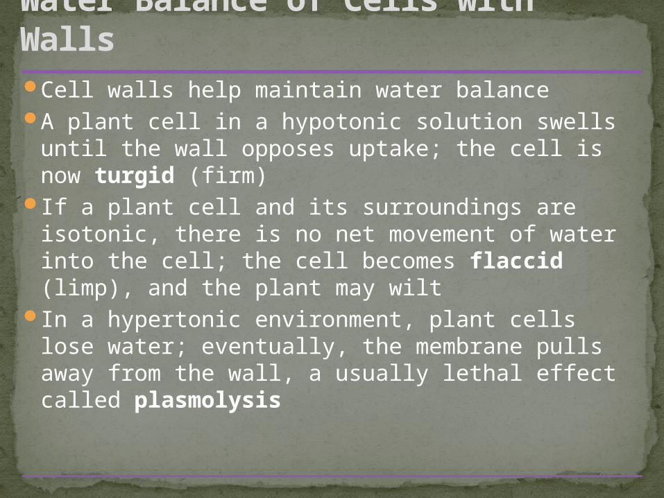
Cell walls help maintain water balanceA plant cell in a hypotonic solution swells until
the wall opposes uptake; the cell is now turgid (firm)
If a plant cell and its surroundings are isotonic, there is no net movement of water into the cell; the cell becomes flaccid (limp), and the plant may wilt
In a hypertonic environment, plant cells lose water; eventually, the membrane pulls away from the wall, a usually lethal effect called plasmolysis
Water Balance of Cells with Walls
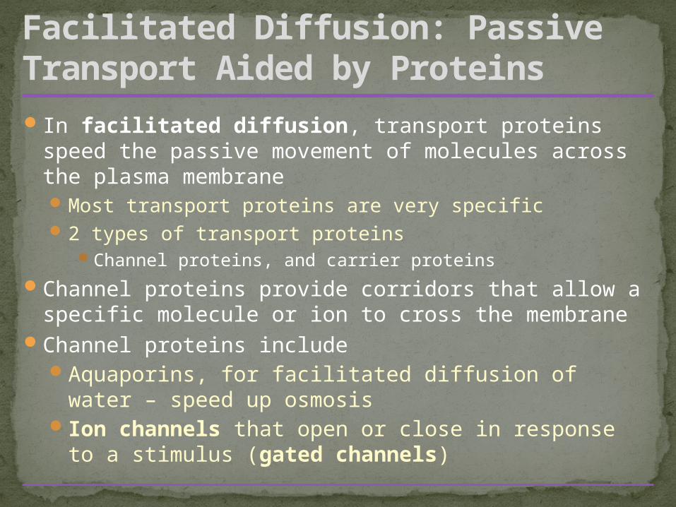
In facilitated diffusion, transport proteins speed the passive movement of molecules across the plasma membraneMost transport proteins are very specific2 types of transport proteins
Channel proteins, and carrier proteins
Channel proteins provide corridors that allow a specific molecule or ion to cross the membrane
Channel proteins includeAquaporins, for facilitated diffusion of water –
speed up osmosisIon channels that open or close in response to a
stimulus (gated channels)
Facilitated Diffusion: Passive Transport Aided by Proteins
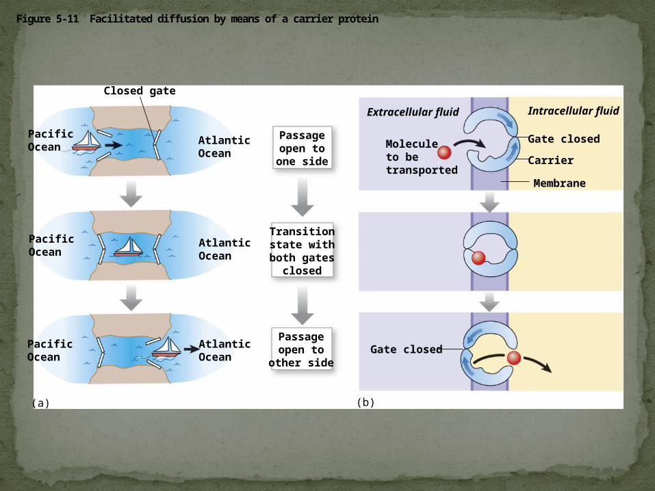
Figure 5-11 Facilitated diffusion by means of a carrier protein
Membrane
PacificOcean
AtlanticOcean
PacificOcean
AtlanticOcean
PacificOcean
AtlanticOcean
Closed gate
Transitionstate withboth gates
closed
Passageopen toone side
Passageopen to
other side
Intracellular fluid
Moleculeto betransported
Gate closed
Gate closed
Carrier
Extracellular fluid
(a) (b)
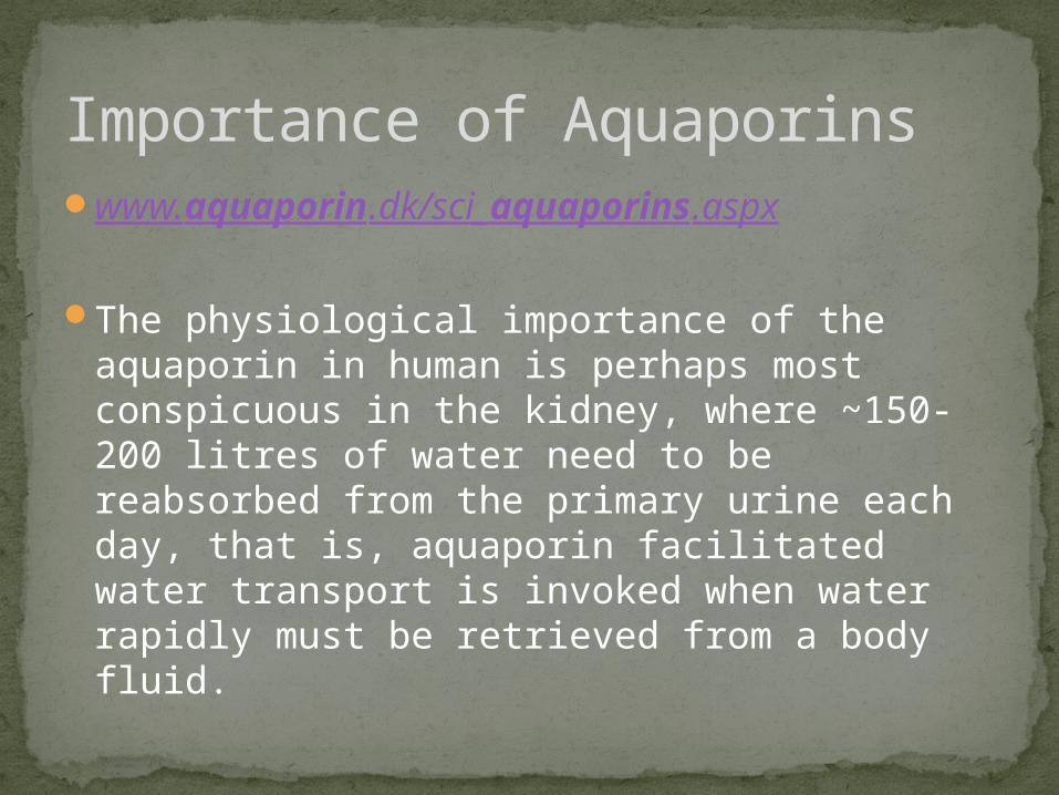
www.aquaporin.dk/sci_aquaporins.aspx
The physiological importance of the aquaporin in human is perhaps most conspicuous in the kidney, where ~150-200 litres of water need to be reabsorbed from the primary urine each day, that is, aquaporin facilitated water transport is invoked when water rapidly must be retrieved from a body fluid.
Importance of Aquaporins
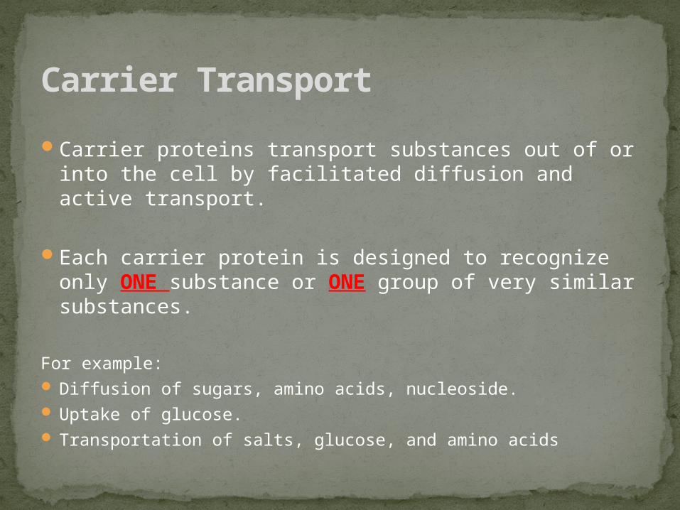
Carrier proteins transport substances out of or into the cell by facilitated diffusion and active transport.
Each carrier protein is designed to recognize only ONE substance or ONE group of very similar substances.
For example: Diffusion of sugars, amino acids, nucleoside. Uptake of glucose. Transportation of salts, glucose, and amino acids
Carrier Transport
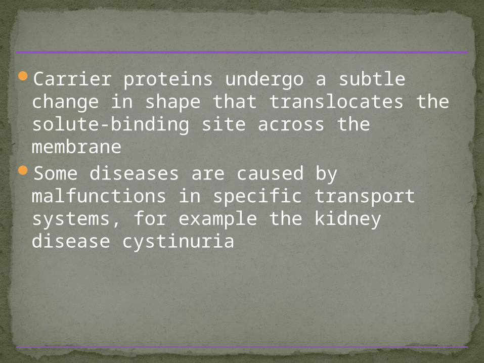
Carrier proteins undergo a subtle change in shape that translocates the solute-binding site across the membrane
Some diseases are caused by malfunctions in specific transport systems, for example the kidney disease cystinuria
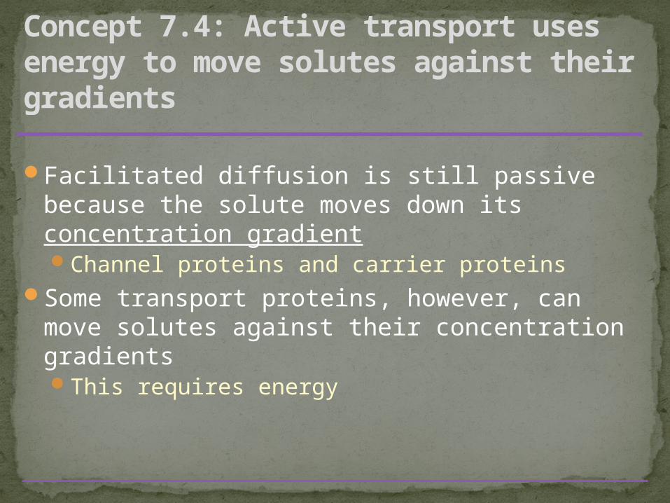
Facilitated diffusion is still passive because the solute moves down its concentration gradientChannel proteins and carrier proteins
Some transport proteins, however, can move solutes against their concentration gradientsThis requires energy
Concept 7.4: Active transport uses energy to move solutes against their gradients
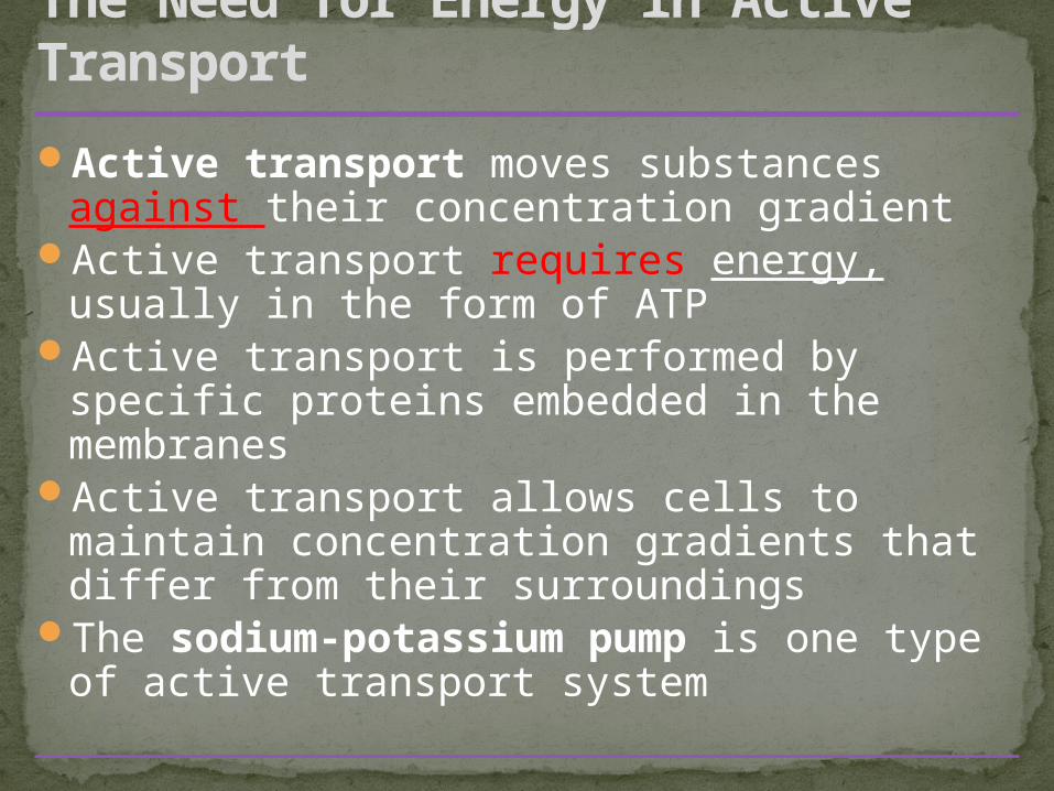
Active transport moves substances against their concentration gradient
Active transport requires energy, usually in the form of ATP
Active transport is performed by specific proteins embedded in the membranes
Active transport allows cells to maintain concentration gradients that differ from their surroundings
The sodium-potassium pump is one type of active transport system
The Need for Energy in Active Transport
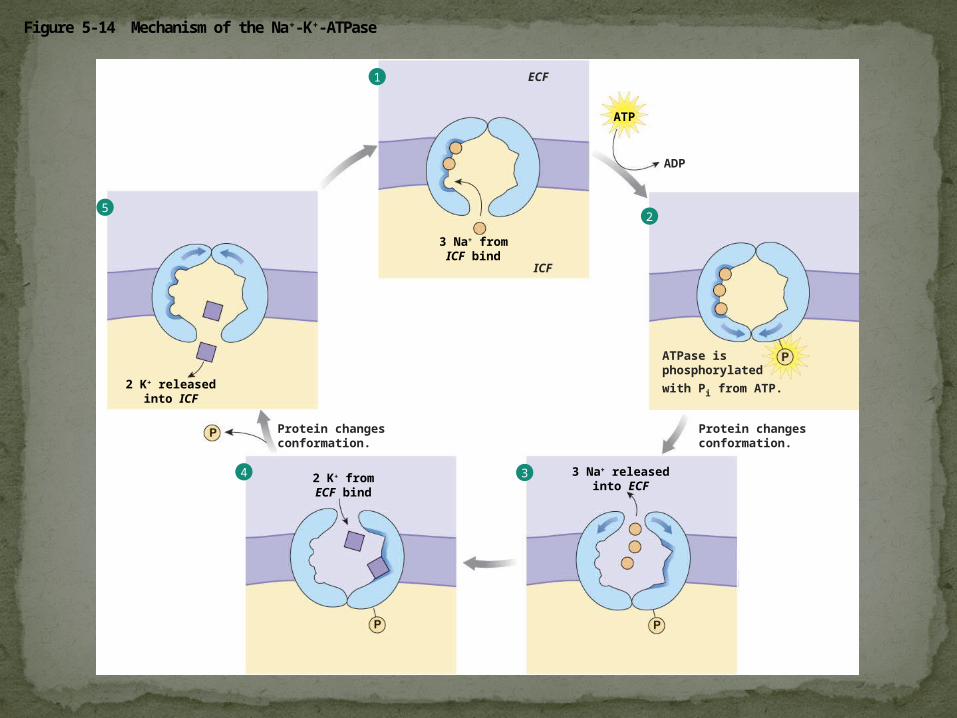
Figure 5-14 Mechanism of the Na+-K+-ATPase
ADP
ATP
ATPase isphosphorylated
with Pi from ATP.
Protein changesconformation.
ICF
ECF
Protein changesconformation.
3 Na+ fromICF bind
3 Na+ releasedinto ECF
2 K+ fromECF bind
2 K+ releasedinto ICF
1
25
4 3
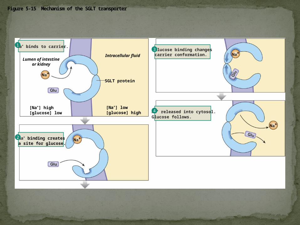
Figure 5-15 Mechanism of the SGLT transporter
[Na+] low[glucose] high
SGLT protein
Lumen of intestineor kidney
Intracellular fluidGlucose binding changescarrier conformation.
Na+ binds to carrier.
[Na+] high[glucose] low
Na+ binding createsa site for glucose.
Na+ released into cytosol. Glucose follows.
13
4
2
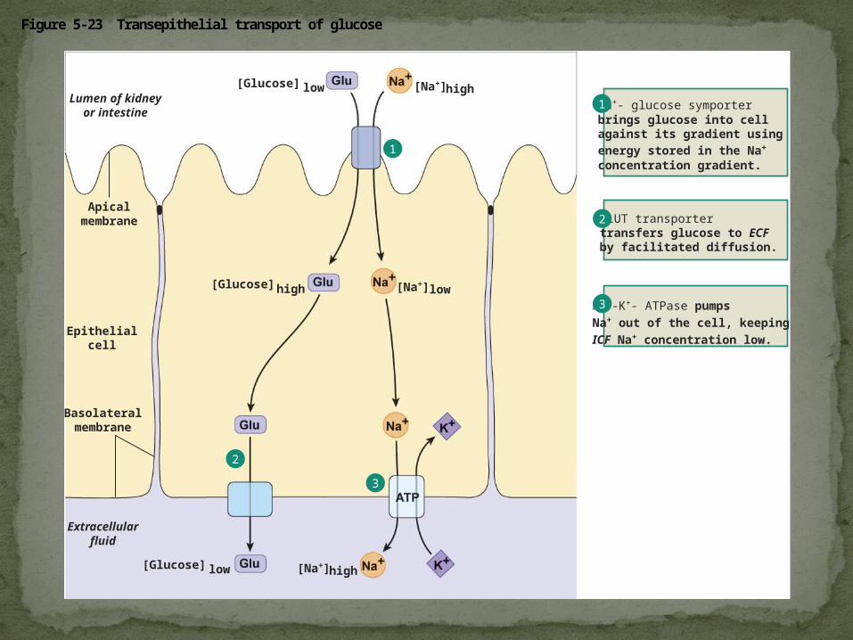
Figure 5-23 Transepithelial transport of glucose
[Glucose] low
[Glucose] high
[Glucose] low
[Na+]high
[Na+]low
[Na+]high
Apicalmembrane
Basolateralmembrane
Extracellularfluid
Lumen of kidneyor intestine
Epithelialcell
GLUT transportertransfers glucose to ECF by facilitated diffusion.
Na+- glucose symporterbrings glucose into cell against its gradient using energy stored in the Na+
concentration gradient.
Na+-K+- ATPase pumps Na+ out of the cell, keeping ICF Na+ concentration low.
1
2
3
1
2
3
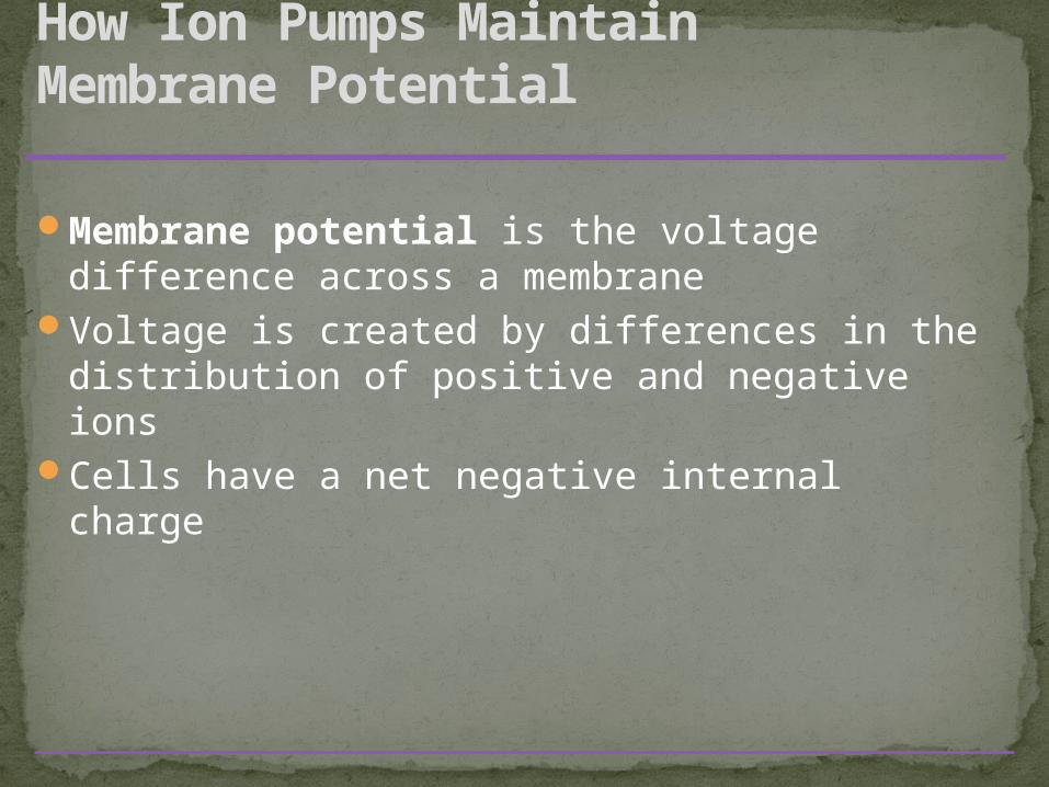
Membrane potential is the voltage difference across a membrane
Voltage is created by differences in the distribution of positive and negative ions
Cells have a net negative internal charge
How Ion Pumps Maintain Membrane Potential
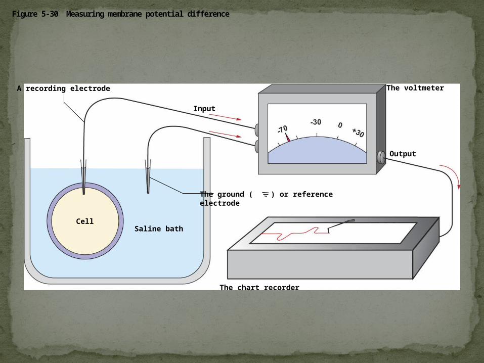
Figure 5-30 Measuring membrane potential difference
The voltmeter
Cell
The chart recorder
Saline bath
A recording electrode
Input
The ground ( ) or referenceelectrode
Output
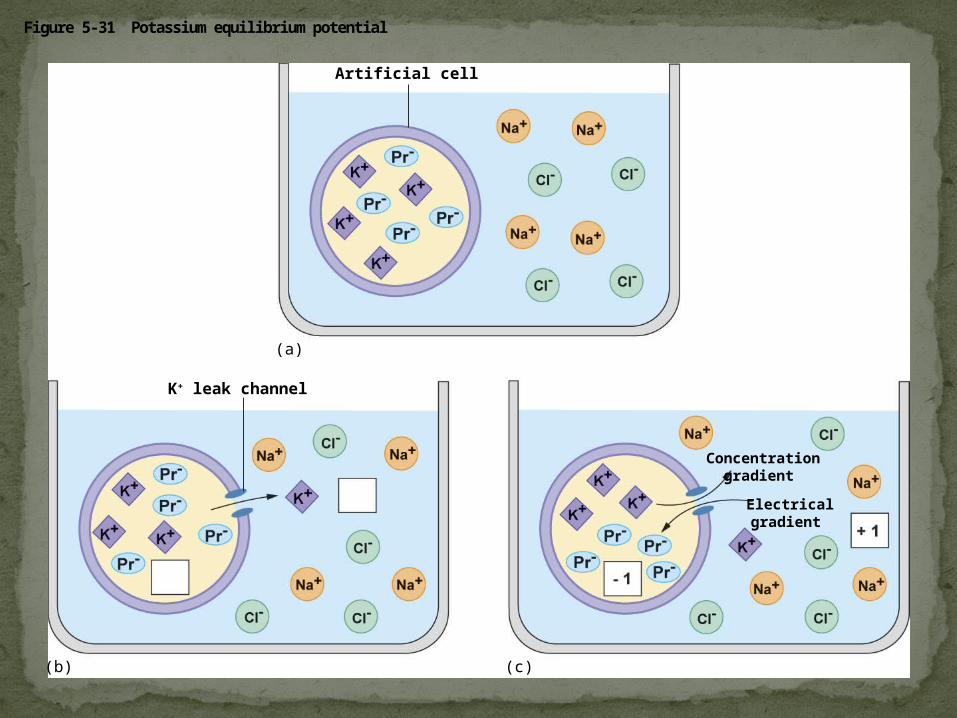
Figure 5-31 Potassium equilibrium potential
Artificial cell
Concentrationgradient
Electricalgradient
(a)
(b) (c)
K+ leak channel
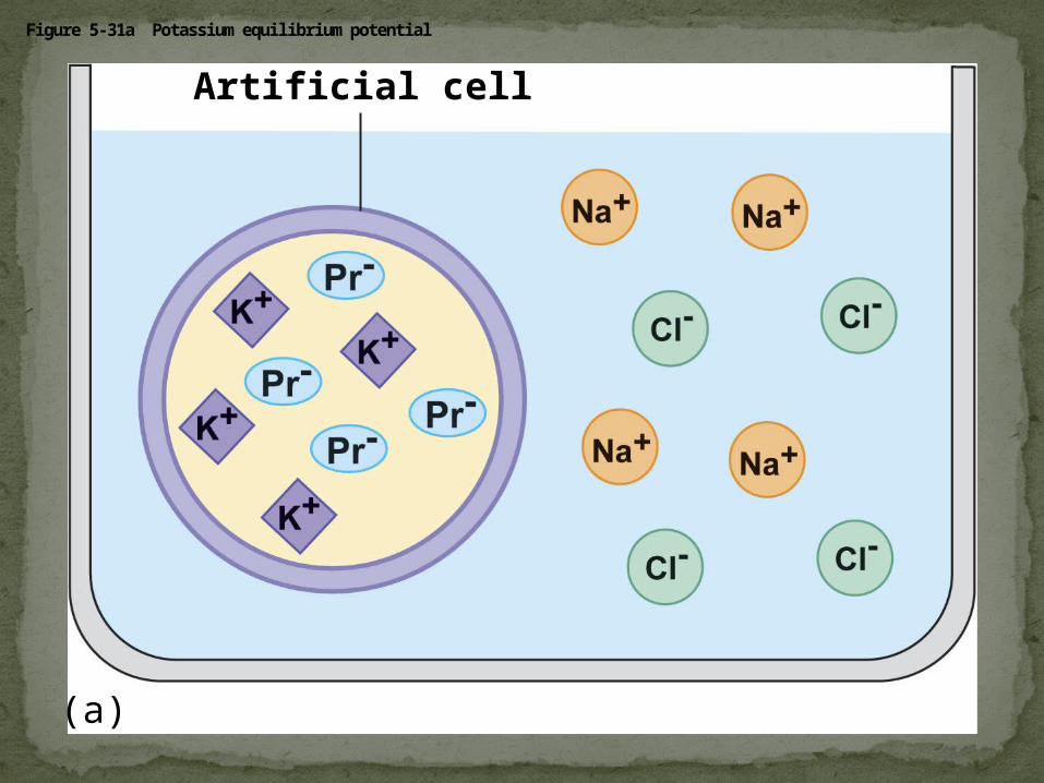
Figure 5-31a Potassium equilibrium potential
Artificial cell
(a)
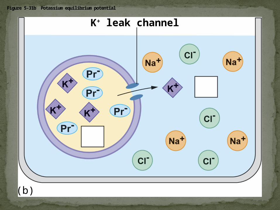
Figure 5-31b Potassium equilibrium potential
(b)
K+ leak channel
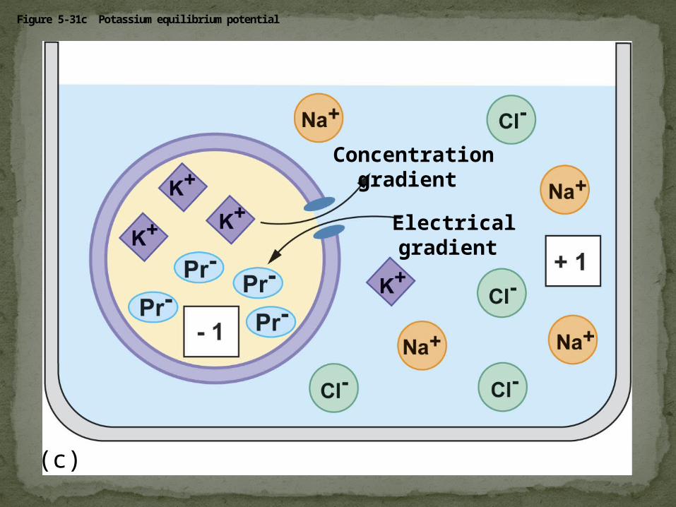
Figure 5-31c Potassium equilibrium potential
Concentrationgradient
Electricalgradient
(c)
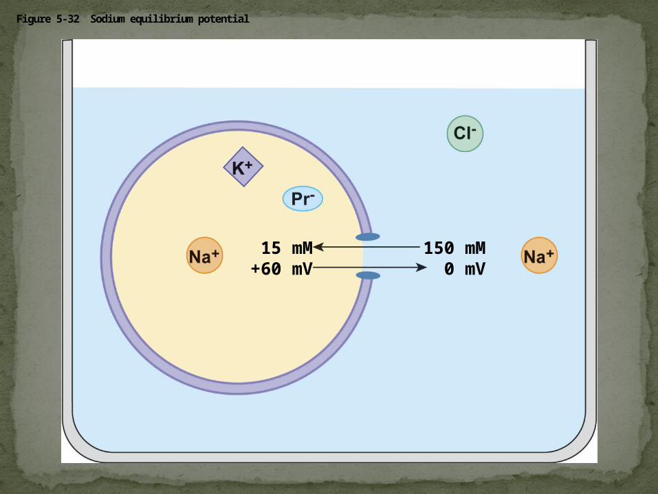
Figure 5-32 Sodium equilibrium potential
150 mM0 mV
15 mM+60 mV
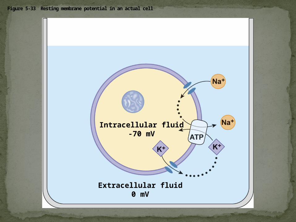
Figure 5-33 Resting membrane potential in an actual cell
Extracellular fluid0 mV
Intracellular fluid-70 mV
Two combined forces, collectively called the electrochemical gradient, drive the diffusion of ions across a membrane:A chemical force (the ion’s concentration
gradient)An electrical force (the effect of the membrane
potential on the ion’s movement)
An electrogenic pump is a transport protein that generates voltage across a membraneEX: Sodium-Potassium pump – major electrogenic
pump of animals3Na+out 2K+ in = overall1 positive charge to
the extracellular fluidThe main electrogenic pump of plants, fungi,
and bacteria is a proton pumpThese pumps generate voltage across
membranes which stores energy for use for the cell
Cotransport occurs when active transport of a solute indirectly drives transport of another solute
Plants commonly use the gradient of hydrogen ions generated by proton pumps to drive active transport of nutrients into the cell.
Cotransport: Coupled Transport by a Membrane Protein
Small molecules and water enter or leave the cell through the lipid bilayer or by transport proteins
Large molecules, such as polysaccharides and proteins, cross the membrane in bulk via vesicles
Bulk transport requires energy
Concept 7.5: Bulk transport across the plasma membrane occurs by exocytosis and endocytosis
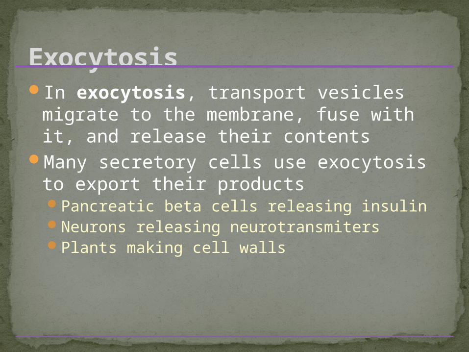
In exocytosis, transport vesicles migrate to the membrane, fuse with it, and release their contents
Many secretory cells use exocytosis to export their productsPancreatic beta cells releasing insulinNeurons releasing neurotransmitersPlants making cell walls
Exocytosis
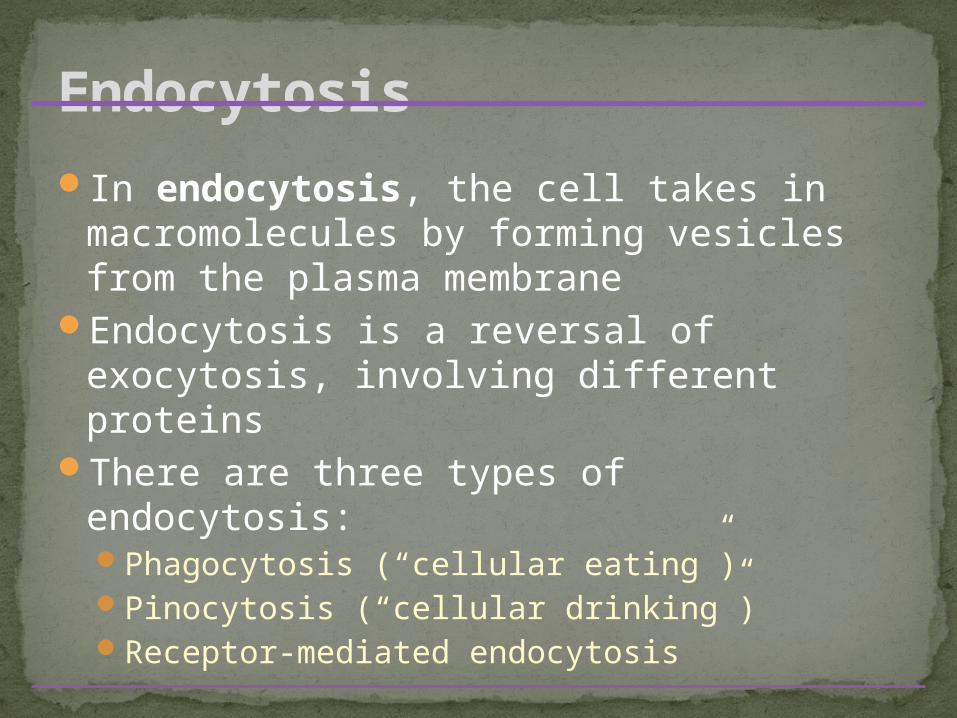
In endocytosis, the cell takes in macromolecules by forming vesicles from the plasma membrane
Endocytosis is a reversal of exocytosis, involving different proteins
There are three types of endocytosis:Phagocytosis (“cellular eating”)Pinocytosis (“cellular drinking”)Receptor-mediated endocytosis
Endocytosis
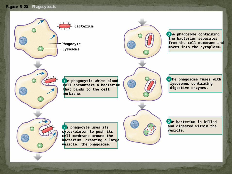
Figure 5-20 Phagocytosis
Bacterium
Lysosome
Phagocyte
The phagocytic white bloodcell encounters a bacteriumthat binds to the cellmembrane.
The phagocyte uses itscytoskeleton to push itscell membrane around thebacterium, creating a largevesicle, the phagosome.
The phagosome containingthe bacterium separatesfrom the cell membrane andmoves into the cytoplasm.
The phagosome fuses withlysosomes containing digestive enzymes.
The bacterium is killedand digested within thevesicle.
1
2
3
4
5
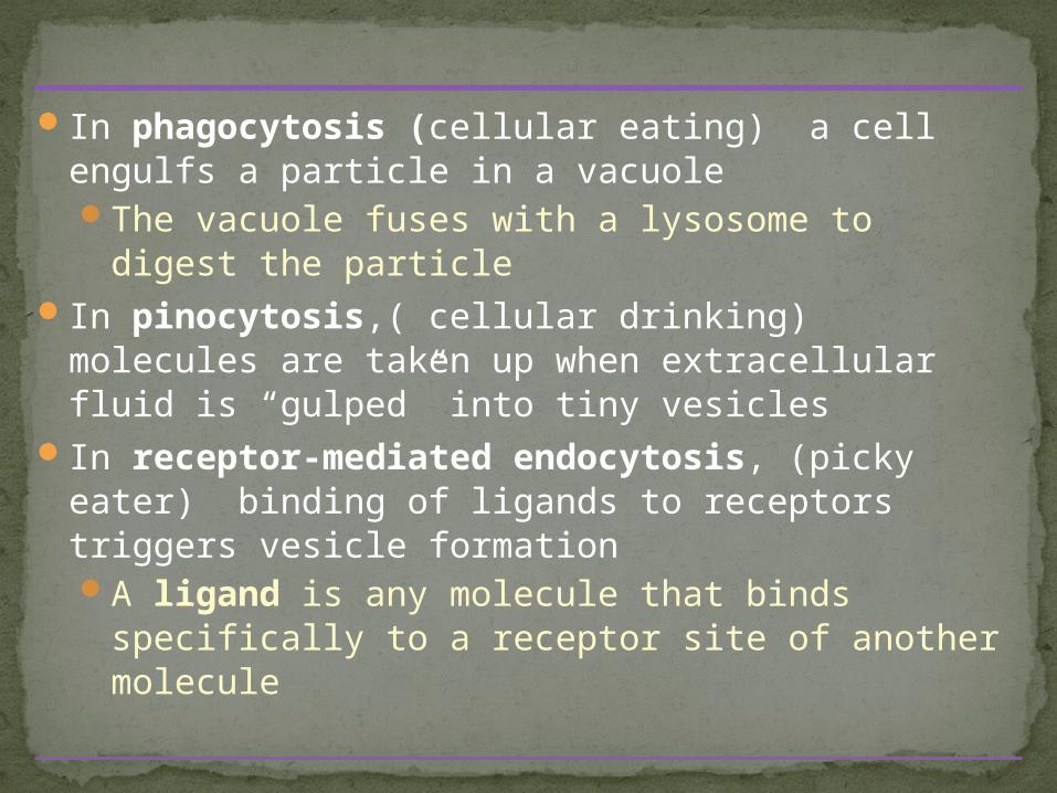
In phagocytosis (cellular eating) a cell engulfs a particle in a vacuoleThe vacuole fuses with a lysosome to digest the
particleIn pinocytosis,( cellular drinking) molecules are
taken up when extracellular fluid is “gulped” into tiny vesicles
In receptor-mediated endocytosis, (picky eater) binding of ligands to receptors triggers vesicle formationA ligand is any molecule that binds specifically
to a receptor site of another molecule
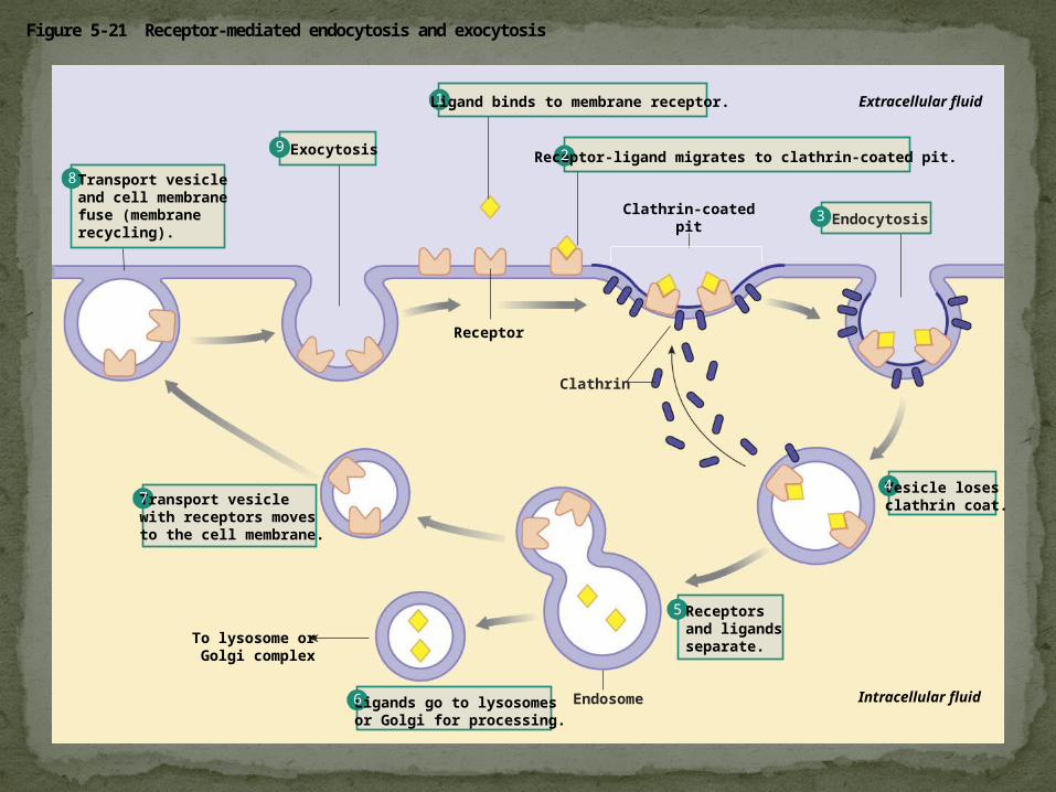
Figure 5-21 Receptor-mediated endocytosis and exocytosis
Ligand binds to membrane receptor.
Clathrin-coatedpit
Receptor
Extracellular fluid
Intracellular fluid
To lysosome orGolgi complex
Receptor-ligand migrates to clathrin-coated pit.
Endocytosis
Vesicle losesclathrin coat.
Ligands go to lysosomesor Golgi for processing.
Transport vesiclewith receptors moves to the cell membrane.
Transport vesicleand cell membranefuse (membranerecycling).
Exocytosis
Clathrin
Endosome
1
2
3
4
5
6
7
8
9
Receptorsand ligands separate.
1. Define the following terms: amphipathic molecules, aquaporins, diffusion
2. Explain how membrane fluidity is influenced by temperature and membrane composition
3. Distinguish between the following pairs or sets of terms: peripheral and integral membrane proteins; channel and carrier proteins; osmosis, facilitated diffusion, and active transport; hypertonic, hypotonic, and isotonic solutions
You should now be able to:
4. Explain how transport proteins facilitate diffusion
5. Explain how an electrogenic pump creates voltage across a membrane, and name two electrogenic pumps
6. Explain how large molecules are transported across a cell membrane
You should now be able to: