Embed Size (px)
Citation preview

Lecture Presentations by
Nicole Tunbridge and
Kathleen Fitzpatrick
Chapter 7
Cell Structure
and Function
© 2018 Pearson Education Ltd.

The Fundamental Units of Life
▪ All organisms are made of cells
▪ The cell is the simplest collection of matter
that can be alive
▪ All cells are related by their descent from earlier cells
▪ Cells can differ substantially from one another but
share common features
© 2018 Pearson Education Ltd.

© 2018 Pearson Education Ltd.
Figure 7.1

© 2018 Pearson Education Ltd.
Figure 7.1a
40 μm

Concept 7.1: Biologists use microscopes and
the tools of biochemistry to study cells
▪ Cells are usually too small to be seen by the naked
eye
© 2018 Pearson Education Ltd.

Microscopy
▪ Microscopes are used to visualize cells
▪ In a light microscope (LM), visible light is passed
through a specimen and then through glass lenses
▪ Lenses refract (bend) the light so that the image is
magnified
© 2018 Pearson Education Ltd.

▪ Three important parameters of microscopy:
▪ Magnification, the ratio of an object’s image
size to its real size
▪ Resolution, the measure of the clarity of the image, or
the minimum distance of two distinguishable points
▪ Contrast, visible differences in brightness between
parts of the sample
© 2018 Pearson Education Ltd.

© 2018 Pearson Education Ltd.
Figure 7.2
10 m
1 m
0.1 m
1 cm
1 mm
100 μm
10 μm
1 μm
Frog egg
Human height
Length of somenerve andmuscle cells
Chicken egg
Human egg
Most plant andanimal cells
NucleusMost bacteriaMitochondrion
Smallest bacteriaViruses
Ribosomes
Proteins
Lipids
Small molecules
Atoms
Un
aid
ed
eye
LM
Super-resolution
microscopy
EM
100 nm
10 nm
1 nm
0.1 nm

© 2018 Pearson Education Ltd.
Figure 7.2a
10 m
1 m
0.1 m
1 cm
Frog egg
LM
Human egg
Human height
Un
aid
ed
eye
Length of somenerve andmuscle cells
Chicken egg
1 mm
100 μm

© 2018 Pearson Education Ltd.
Figure 7.2b
100 μm
10 μm
1 μm
Most plant andanimal cells
NucleusMost bacteria
Mitochondrion
LM
100 nm Smallest bacteria
Viruses
Ribosomes
Super-resolution
microscopy
10 nmProteins
Lipids
Small molecules
Atoms
1 nm
0.1 nm
EM
© 2018 Pearson Education Ltd.
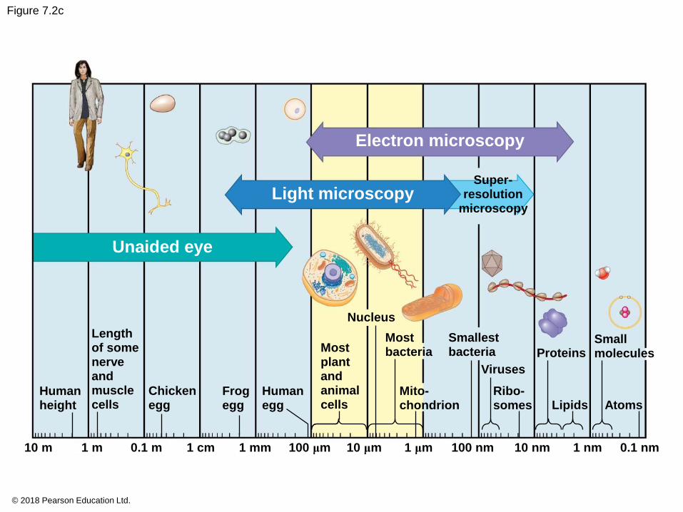
Figure 7.2c
Electron microscopy
Light microscopy
Unaided eye
Super-resolution
microscopy
Nucleus
Lengthof somenerveandmusclecells
Humanheight
Chickenegg
Mostplantandanimalcells
10 μm
Mostbacteria
Smallestbacteria
SmallmoleculesProteins
Viruses
Ribo-somes Lipids Atoms
Frogegg
Humanegg
Mito-chondrion
1 μm10 m 1 m 0.1 m 1 cm 1 mm 100 μm 100 nm 10 nm 1 nm 0.1 nm

▪ Light microscopes can magnify effectively to about
1,000 times the size of the actual specimen
▪ Various techniques enhance contrast and enable
cell components to be stained or labeled
▪ The resolution of standard light microscopy is too
low to study organelles, the membrane-enclosed
structures in eukaryotic cells
© 2018 Pearson Education Ltd.

© 2018 Pearson Education Ltd.
Figure 7.3
Brightfield(unstainedspecimen)
50 μmBrightfield(stained specimen)
Phase-contrast Differentialinterference contrast(Nomarski)
50 μ
m
Fluorescence10 μm
Confocal(without)
Confocal (with)
Deconvolution
Super-resolution(without)
Super-resolution(with)
1μ
m
Scanningelectronmicroscopy (SEM)
2 μmTransmissionelectronmicroscopy (TEM)
2 μm
10μ
m

▪ Two basic types of electron microscopes (EMs)
are used to study subcellular structures
▪ Scanning electron microscopes (SEMs) focus a
beam of electrons onto the surface of a specimen,
providing images that look 3-D
▪ Transmission electron microscopes (TEMs) focus
a beam of electrons through a specimen
▪ TEMs are used mainly to study the internal structure
of cells
© 2018 Pearson Education Ltd.

▪ Recent advances in light microscopy:
▪ Labeling individual cells with fluorescent markers
improve the level of detail that can be seen
▪ Confocal microscopy and deconvolution microscopy
provide sharper images of three-dimensional tissues
and cells
▪ New techniques for labeling cells improve resolution
▪ Super-resolution microscopy allows one to distinguish
structures as small as 10–20 nm across
© 2018 Pearson Education Ltd.

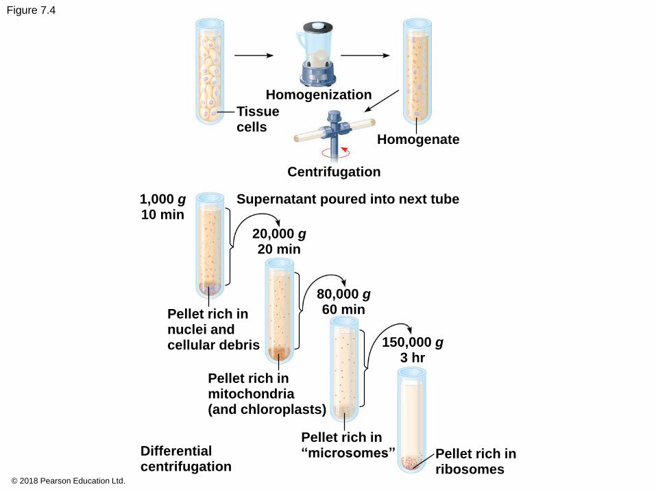
Cell Fractionation
▪ Cell fractionation takes cells apart and
separates the major organelles from one another
▪ Centrifuges fractionate cells into their
component parts
▪ Cell fractionation enables scientists to determine the
functions of organelles
▪ Biochemistry and cytology help correlate cell
function with structure
© 2018 Pearson Education Ltd.
© 2018 Pearson Education Ltd.
Figure 7.4
Homogenization
Tissuecells
Homogenate
Centrifugation
1,000 g10 min
Supernatant poured into next tube
20,000 g20 min
80,000 g60 min
150,000 g3 hr
Pellet rich innuclei andcellular debris
Pellet rich inmitochondria(and chloroplasts)
Differentialcentrifugation
Pellet rich in“microsomes” Pellet rich in
ribosomes

© 2018 Pearson Education Ltd.
Figure 7.4a
Homogenization
Tissuecells
Homogenate
Centrifugation
© 2018 Pearson Education Ltd.
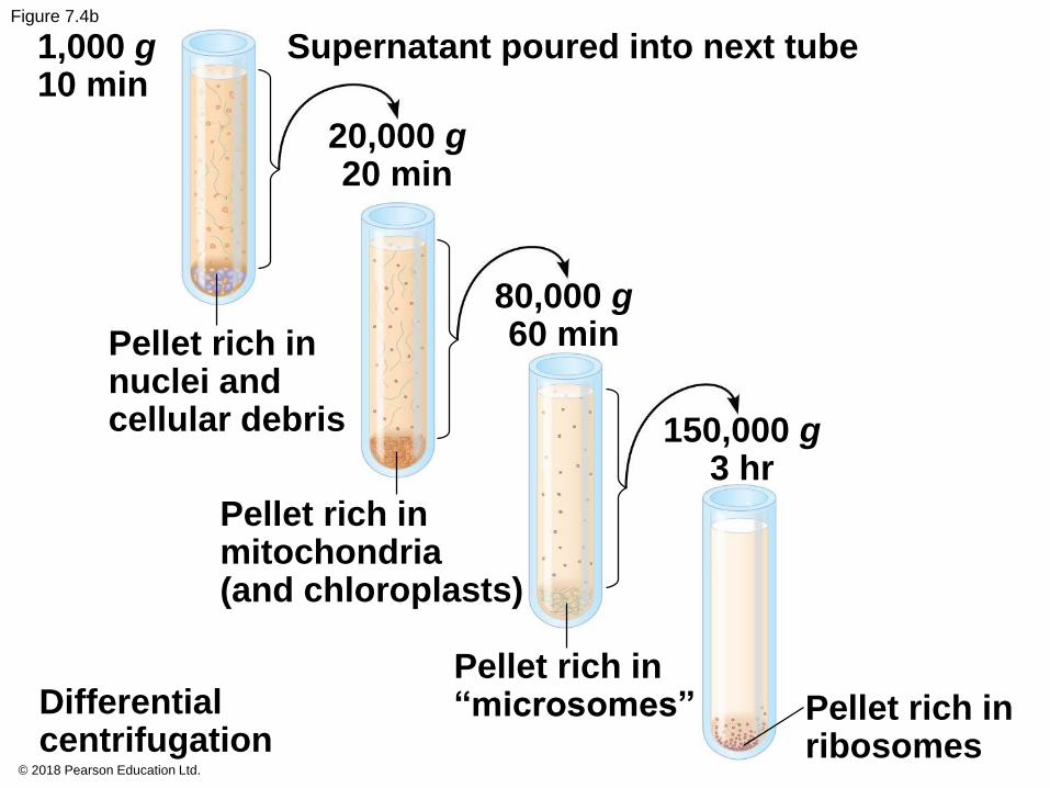
Figure 7.4b
1,000 g10 min
Supernatant poured into next tube
20,000 g20 min
Pellet rich innuclei andcellular debris
80,000 g60 min
150,000 g3 hr
Pellet rich inmitochondria(and chloroplasts)
Differentialcentrifugation
Pellet rich in“microsomes” Pellet rich in
ribosomes

Concept 7.2: Eukaryotic cells have
internal membranes that compartmentalize
their functions
▪ The basic structural and functional unit of every
organism is one of two types of cells: prokaryotic or
eukaryotic
▪ Only organisms of the domains Bacteria and
Archaea consist of prokaryotic cells
▪ Protists, fungi, animals, and plants all consist of
eukaryotic cells
© 2018 Pearson Education Ltd.
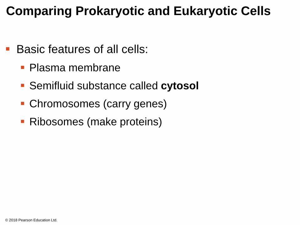
Comparing Prokaryotic and Eukaryotic Cells
▪ Basic features of all cells:
▪ Plasma membrane
▪ Semifluid substance called cytosol
▪ Chromosomes (carry genes)
▪ Ribosomes (make proteins)
© 2018 Pearson Education Ltd.

▪ Prokaryotic cells are characterized by having
▪ No nucleus
▪ DNA in an unbound region called the nucleoid
▪ No membrane-bound organelles
▪ Cytoplasm bound by the plasma membrane
© 2018 Pearson Education Ltd.

© 2018 Pearson Education Ltd.
Figure 7.5
Fimbriae
Nucleoid
Ribosomes
Plasma membrane
Bacterialchromosome
Cell wall
Glycocalyx
0.5 μm
Flagella
(a) A typical rod-shapedbacterium
(b) A thin section through thebacterium Corynebacteriumdiphtheriae (colorized TEM)
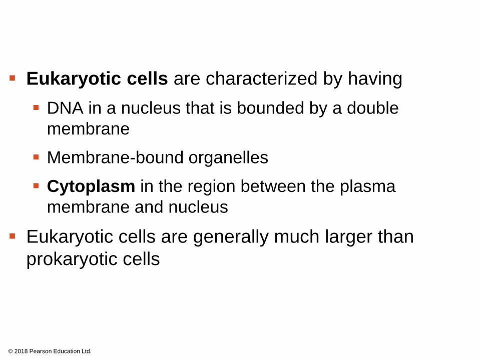
▪ Eukaryotic cells are characterized by having
▪ DNA in a nucleus that is bounded by a double
membrane
▪ Membrane-bound organelles
▪ Cytoplasm in the region between the plasma
membrane and nucleus
▪ Eukaryotic cells are generally much larger than
prokaryotic cells
© 2018 Pearson Education Ltd.

▪ The plasma membrane is a selective barrier that
allows sufficient passage of oxygen, nutrients, and
waste to service the volume of every cell
© 2018 Pearson Education Ltd.
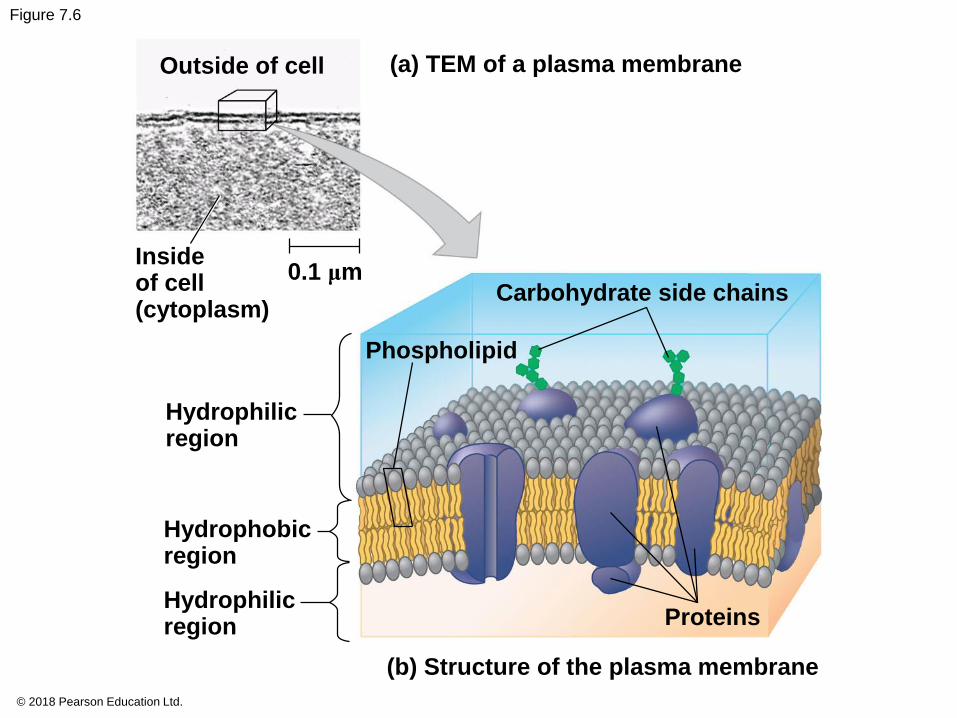
© 2018 Pearson Education Ltd.
Figure 7.6
Outside of cell (a) TEM of a plasma membrane
Insideof cell(cytoplasm)
0.1 μmCarbohydrate side chains
Phospholipid
Hydrophilicregion
Hydrophobicregion
Hydrophilicregion Proteins
(b) Structure of the plasma membrane
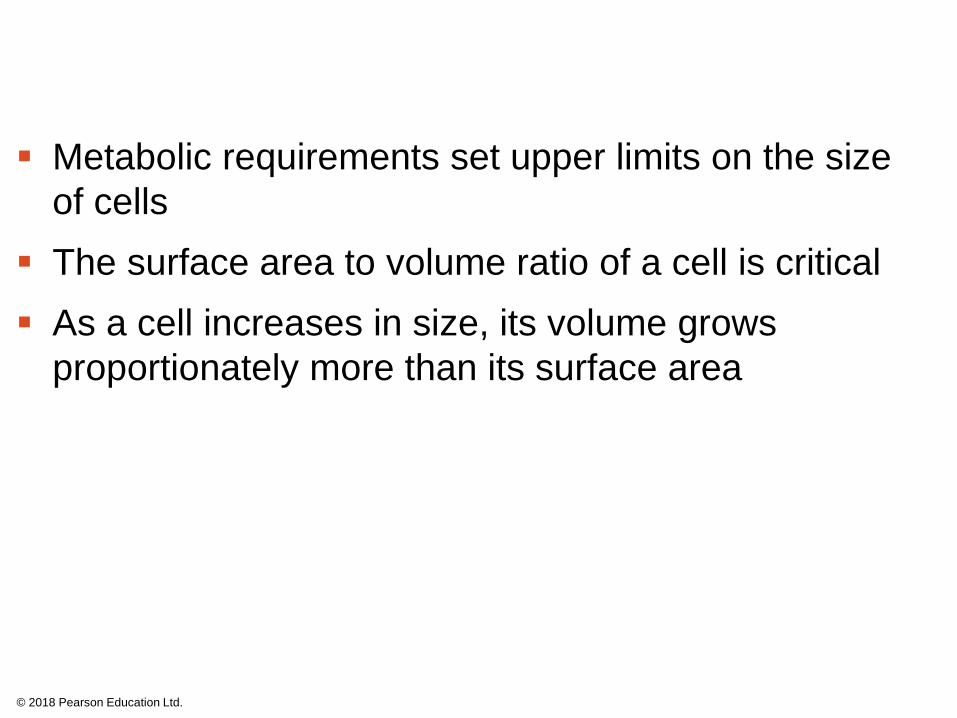
▪ Metabolic requirements set upper limits on the size
of cells
▪ The surface area to volume ratio of a cell is critical
▪ As a cell increases in size, its volume grows
proportionately more than its surface area
© 2018 Pearson Education Ltd.

© 2018 Pearson Education Ltd.
Figure 7.7
Surface area increases whiletotal volume remains constant
5
1
1
Total surface area[sum of the surface areas(height × width) of all boxsides× number of boxes]
Total volume[height × width × length× number of boxes]
Surface-to-volume(S-to-V) ratio[surface area ÷ volume]
6 150 750
1 125 125
6 1.2 6
A Panoramic View of the Eukaryotic Cell
▪ A eukaryotic cell has internal membranes that divide
the cell into compartments—the organelles
▪ The basic fabric of biological membranes is a double
layer of phospholipids and other lipids
▪ Plant and animal cells have most of the same
organelles
© 2018 Pearson Education Ltd.

© 2018 Pearson Education Ltd.
Figure 7.8a
ENDOPLASMICRETICULUM (ER)
Rough ER
Flagellum
Centrosome
Plasmamembrane
CYTOSKELETON:
Microfilaments
Intermediate filaments
Microtubules
MicrovilliRibosomes
Golgi apparatus
Peroxisome
Lysosome
Mitochondrion
Nuclearenvelope
Nucleolus
Chromatin
Smooth ER
NUCLEUS

© 2018 Pearson Education Ltd.
Figure 7.8b
Nuclearenvelope
NUCLEUSNucleolus
ChromatinRough ER
Smooth ER
Ribosomes
Golgiapparatus
Central vacuole
Microfilaments
MicrotubulesCYTOSKELETON
Mitochondrion
Peroxisome
Plasmamembrane
Cell wall
Wall of adjacent cell
Chloroplast
Plasmodesmata
Concept 7.3: The eukaryotic cell’s genetic
instructions are housed in the nucleus and
carried out by the ribosomes
▪ The nucleus contains most of the DNA in a
eukaryotic cell
▪ Ribosomes use the information from the DNA to
make proteins
© 2018 Pearson Education Ltd.

The Nucleus: Information Central
▪ The nucleus contains most of the cell’s genes and is
usually the most conspicuous organelle
▪ The nuclear envelope encloses the nucleus,
separating it from the cytoplasm
▪ The nuclear envelope is a double membrane; each
membrane consists of a lipid bilayer
© 2018 Pearson Education Ltd.

© 2018 Pearson Education Ltd.
Figure 7.9
1 μm Nucleus
Nucleus
Nucleolus
Chromatin
Nuclear envelope:
Outer membrane
Inner membrane
Nuclear pore
RoughER
Surface ofnuclear envelope(TEM)
Porecomplex
Ribosome
0.2
5 μ
m Close-upof nuclearenvelope
0.5μ
m
Chromatin
Pore complexes (TEM) Nuclear lamina (TEM)

© 2018 Pearson Education Ltd.
Figure 7.9a
Nucleus
Nucleolus
Chromatin
Nuclear envelope:Outer membrane
Inner membrane
Nuclear pore
Rough ER
Porecomplex
Ribosome
Close-upof nuclearenvelope
Chromatin

© 2018 Pearson Education Ltd.
Figure 7.9b
1 μm
Nuclear envelope:
Outer membrane
Inner membrane
Nuclear pore
Surface of nuclear envelope (TEM)

© 2018 Pearson Education Ltd.
Figure 7.9c
0.2
5μ
m
Pore complexes (TEM)

© 2018 Pearson Education Ltd.
Figure 7.9d
0.5μ
m
Nuclear lamina (TEM)

▪ Pores, lined with a structure called a pore complex,
regulate the entry and exit of molecules from the
nucleus
▪ The nuclear size of the envelope is lined by the
nuclear lamina, which is composed of proteins and
maintains the shape of the nucleus
© 2018 Pearson Education Ltd.
▪ In the nucleus, DNA is organized into discrete units
called chromosomes
▪ Each chromosome contains one DNA molecule
associated with proteins, called chromatin
▪ Chromatin condenses to form discrete
chromosomes as a cell prepares to divide
▪ The nucleolus is located within the nucleus and is
the site of ribosomal RNA (rRNA) synthesis
© 2018 Pearson Education Ltd.

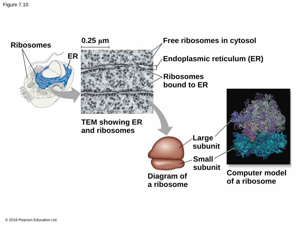
Ribosomes: Protein Factories
▪ Ribosomes are complexes made of ribosomal RNA
and protein
▪ Ribosomes carry out protein synthesis in two
locations:
▪ In the cytosol (free ribosomes)
▪ On the outside of the endoplasmic reticulum or the
nuclear envelope (bound ribosomes)
© 2018 Pearson Education Ltd.
© 2018 Pearson Education Ltd.
Figure 7.10
Ribosomes
ER
0.25 μm Free ribosomes in cytosol
Endoplasmic reticulum (ER)
Ribosomesbound to ER
TEM showing ERand ribosomes
Largesubunit
Smallsubunit
Diagram ofa ribosome
Computer modelof a ribosome
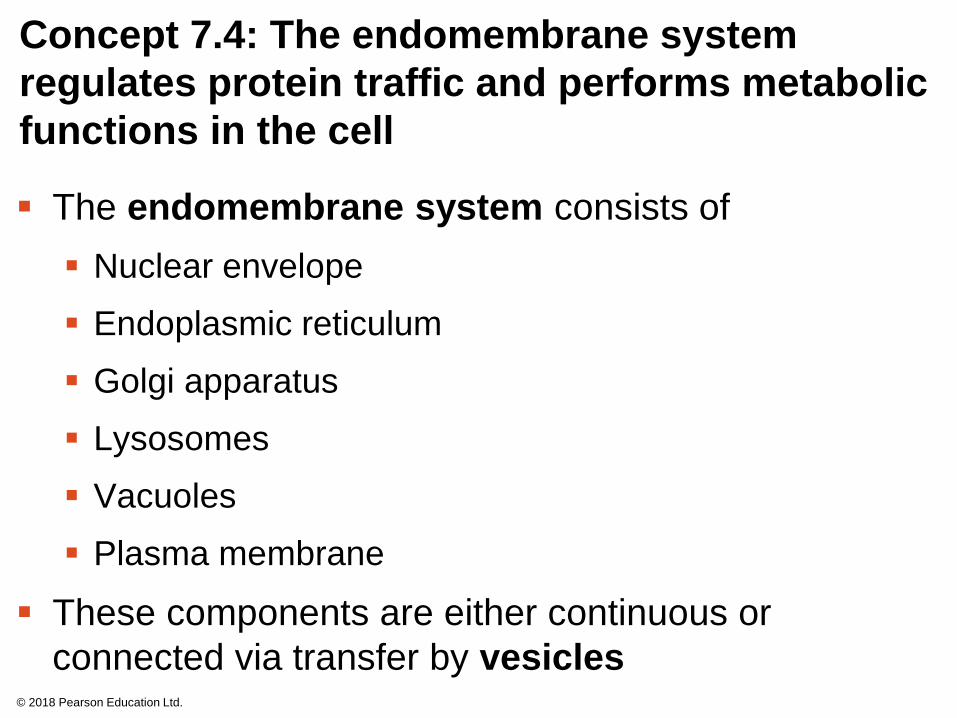
Concept 7.4: The endomembrane system
regulates protein traffic and performs metabolic
functions in the cell
▪ The endomembrane system consists of
▪ Nuclear envelope
▪ Endoplasmic reticulum
▪ Golgi apparatus
▪ Lysosomes
▪ Vacuoles
▪ Plasma membrane
▪ These components are either continuous or
connected via transfer by vesicles© 2018 Pearson Education Ltd.

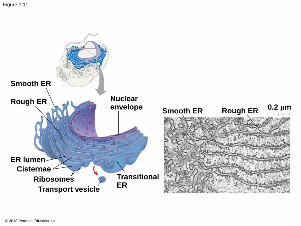
The Endoplasmic Reticulum: Biosynthetic
Factory
▪ The endoplasmic reticulum (ER) accounts for
more than half of the total membrane in many
eukaryotic cells
▪ The ER membrane is continuous with the nuclear
envelope
▪ There are two distinct regions of ER:
▪ Smooth ER, which lacks ribosomes
▪ Rough ER, whose surface is studded with ribosomes
© 2018 Pearson Education Ltd.
© 2018 Pearson Education Ltd.
Figure 7.11
Smooth ER
Rough ER Nuclearenvelope 0.2 μm
Smooth ER Rough ER
ER lumen
Cisternae
Ribosomes
Transport vesicle
TransitionalER

Functions of Smooth ER
▪ The smooth ER
▪ Synthesizes lipids
▪ Metabolizes carbohydrates
▪ Detoxifies drugs and poisons
▪ Stores calcium ions
© 2018 Pearson Education Ltd.

Functions of Rough ER
▪ The rough ER
▪ Has bound ribosomes, which secrete glycoproteins
(proteins covalently bonded to carbohydrates)
▪ Distributes transport vesicles, secretory proteins
surrounded by membranes
▪ Is a membrane factory for the cell
© 2018 Pearson Education Ltd.

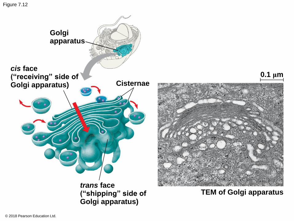
The Golgi Apparatus: Shipping and
Receiving Center
▪ The Golgi apparatus consists of flattened
membranous sacs called cisternae
▪ The Golgi apparatus
▪ Modifies products of the ER
▪ Manufactures certain macromolecules
▪ Sorts and packages materials into transport vesicles
© 2018 Pearson Education Ltd.
© 2018 Pearson Education Ltd.
Figure 7.12
Golgiapparatus
cis face(“receiving” side ofGolgi apparatus)
0.1 μm
Cisternae
trans face(“shipping” side ofGolgi apparatus)
TEM of Golgi apparatus

Lysosomes: Digestive Compartments
▪ A lysosome is a membranous sac of hydrolytic
enzymes that can digest macromolecules
▪ Lysosomal enzymes work best in the acidic
environment inside the lysosome
▪ Hydrolytic enzymes and lysosomal membranes are
made by rough ER and then transferred to the Golgi
apparatus for further processing
© 2018 Pearson Education Ltd.

▪ Some types of cell can engulf another cell by
phagocytosis; this forms a food vacuole
▪ A lysosome fuses with the food vacuole and digests
the molecules
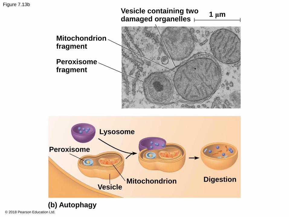
▪ Lysosomes also use enzymes to recycle the
cell’s own organelles and macromolecules,
a process called autophagy
© 2018 Pearson Education Ltd.

© 2018 Pearson Education Ltd.
Figure 7.13
Nucleus 1 μm
Mitochondrionfragment
Peroxisomefragment
Vesicle containingtwo damagedorganelles
1 μm
Lysosome
Digestiveenzymes
Plasmamembrane
Foodvacuole
(a) Phagocytosis
Lysosome
Peroxisome
Digestion
Lysosome
Mitochondrion
(b) Autophagy
DigestionVesicle

© 2018 Pearson Education Ltd.
Figure 7.13a
Nucleus 1 μm
Lysosome
Digestiveenzymes
LysosomePlasmamembrane
Food vacuole
Digestion
(a) Phagocytosis
© 2018 Pearson Education Ltd.
Figure 7.13b
Vesicle containing twodamaged organelles
Mitochondrionfragment
Peroxisomefragment
1 μm
Lysosome
Peroxisome
Vesicle
(b) Autophagy
Mitochondrion Digestion

Vacuoles: Diverse Maintenance Compartments
▪ Vacuoles are large vesicles derived from the ER
and Golgi apparatus
▪ Vacuoles perform a variety of functions in different
kinds of cells
© 2018 Pearson Education Ltd.

▪ Food vacuoles are formed by phagocytosis
▪ Contractile vacuoles, found in many freshwater
protists, pump excess water out of cells
▪ Central vacuoles, found in many mature plant cells,
hold organic compounds and water
© 2018 Pearson Education Ltd.

© 2018 Pearson Education Ltd.
Figure 7.14
Central vacuole
Cytosol
Nucleus
Cell wall
Chloroplast
Centralvacuole
5 μm

The Endomembrane System: A Review
▪ The endomembrane system is a complex and
dynamic player in the cell’s compartmental
organization
© 2018 Pearson Education Ltd.

© 2018 Pearson Education Ltd.
Figure 7.15
Nucleus
Nuclearenvelope
Rough ER
Smooth ER
cis Golgi
trans Golgi
Plasmamembrane

Concept 7.5: Mitochondria and chloroplasts
change energy from one form to another
▪ Mitochondria are the sites of cellular respiration,
a metabolic process that uses oxygen to
generate ATP
▪ Chloroplasts, found in plants and algae, are the
sites of photosynthesis
▪ Peroxisomes are oxidative organelles
© 2018 Pearson Education Ltd.
The Evolutionary Origins of Mitochondria and
Chloroplasts
▪ Mitochondria and chloroplasts have similarities with
bacteria:
▪ Enveloped by a double membrane
▪ Contain free ribosomes and circular DNA molecules
▪ Grow and reproduce somewhat independently
in cells
▪ These similarities led to the endosymbiont theory
© 2018 Pearson Education Ltd.

▪ The endosymbiont theory suggests that an early
ancestor of eukaryotes engulfed an oxygen-using
nonphotosynthetic prokaryotic cell
▪ The engulfed cell formed a relationship with the host
cell, becoming an endosymbiont
▪ The endosymbionts evolved into mitochondria
▪ At least one of these cells may have then taken up a
photosynthetic prokaryote, which evolved into a
chloroplast
© 2018 Pearson Education Ltd.

© 2018 Pearson Education Ltd.
Figure 7.16
Endoplasmicreticulum
Nuclearenvelope
Nucleus
Engulfing of oxygen-using nonphotosyntheticprokaryote, whichbecomes a mitochondrion
Ancestor ofeukaryotic cells (host cell)
Mitochondrion
Chloroplast
At leastone cell
Engulfing ofphotosyntheticprokaryote
MitochondrionNonphotosynthetic
eukaryote
Photosynthetic eukaryote

Mitochondria: Chemical Energy Conversion
▪ Mitochondria are found in nearly all eukaryotic cells
▪ They have a smooth outer membrane and an inner
membrane folded into cristae
▪ The inner membrane creates two compartments:
intermembrane space and mitochondrial matrix
▪ Some metabolic steps of cellular respiration are
catalyzed in the mitochondrial matrix
▪ Cristae present a large surface area for enzymes
that synthesize ATP
© 2018 Pearson Education Ltd.

© 2018 Pearson Education Ltd.
Figure 7.17
Mitochondrion
Intermembrane space
Outermembrane
DNA
Freeribosomesin themitochondrialmatrix
Innermembrane
Cristae
Matrix0.1 μm
MitochondrialDNA
Nuclear DNA
10 μm
Mitochondria
(a) Diagram and TEM of mitochondrion (b) Network of mitochondria inEuglena (LM)
Chloroplasts: Capture of Light Energy
▪ Chloroplasts contain the green pigment chlorophyll,
as well as enzymes and other molecules that
function in photosynthesis
▪ Chloroplasts are found in leaves and other green
organs of plants and in algae
© 2018 Pearson Education Ltd.

© 2018 Pearson Education Ltd.
Figure 7.18
Chloroplast
Stroma
RibosomesInner and outer
membranes
50 μm
DNA
Thylakoid
(a) Diagram and TEM of chloroplast
1 μm
Chloroplasts(red)
(b) Chloroplasts in an algalcell
Intermembrane space
Granum