Embed Size (px)
Citation preview
Chapter 46: Skeletal, Muscular, and the Integument
46-1 The Human Body Plan
46-2 Skeletal System
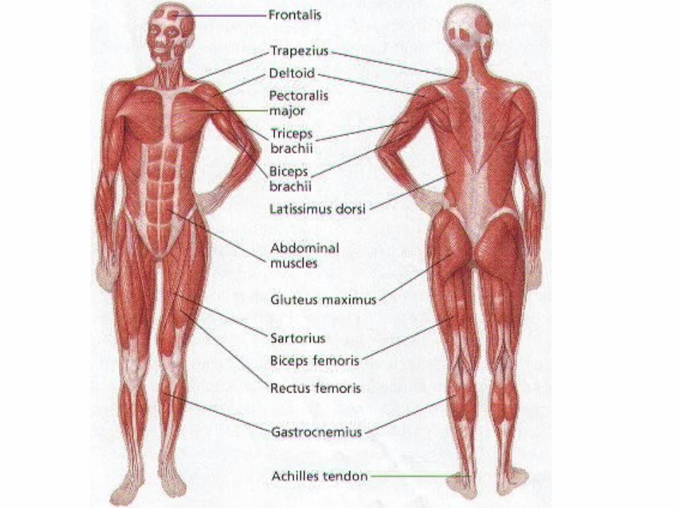
46-3 Muscular System
46-4 Integumentary System
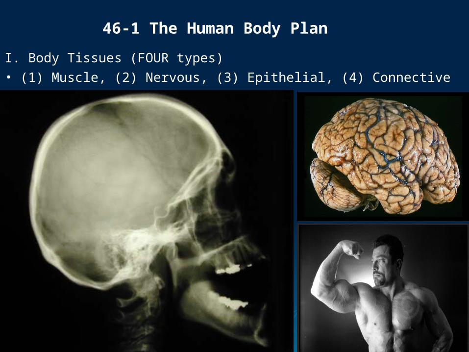
I. Body Tissues (FOUR types)• (1) Muscle, (2) Nervous, (3) Epithelial, (4) Connective
46-1 The Human Body Plan
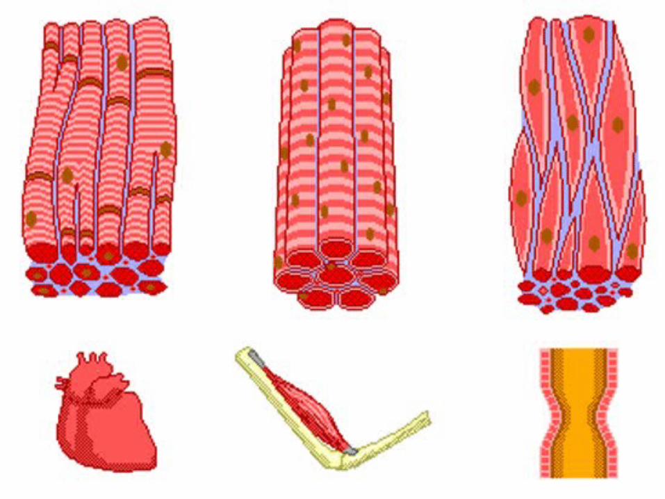
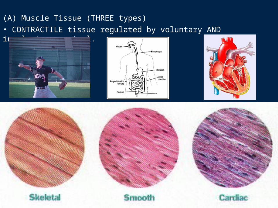
(A) Muscle Tissue (THREE types)• CONTRACTILE tissue regulated by voluntary AND involuntary control.
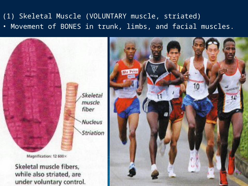
(1) Skeletal Muscle (VOLUNTARY muscle, striated)• Movement of BONES in trunk, limbs, and facial muscles.
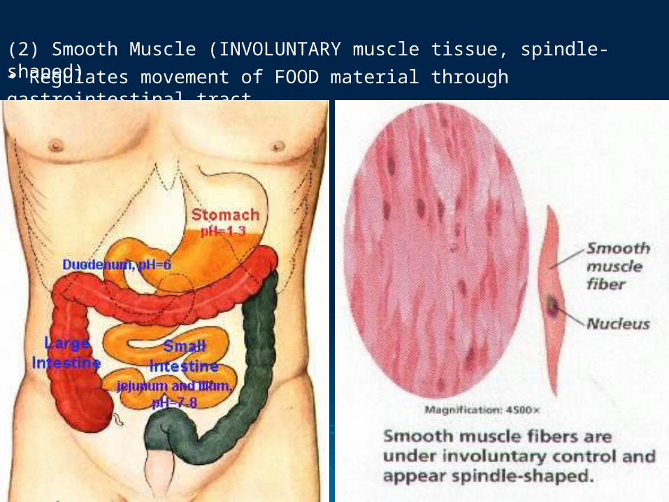
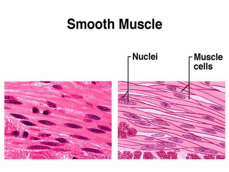
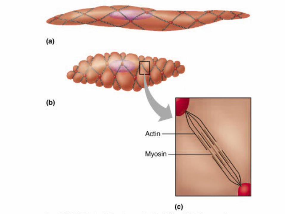
(2) Smooth Muscle (INVOLUNTARY muscle tissue, spindle-shaped)• Regulates movement of FOOD material through gastrointestinal tract.
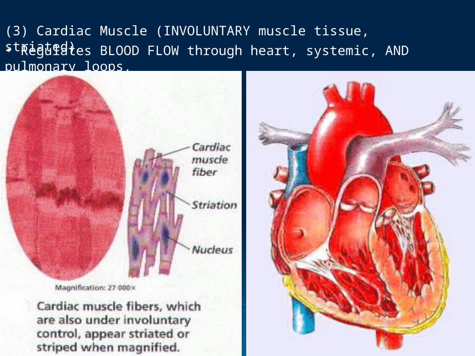
(3) Cardiac Muscle (INVOLUNTARY muscle tissue, striated)• Regulates BLOOD FLOW through heart, systemic, AND pulmonary loops.

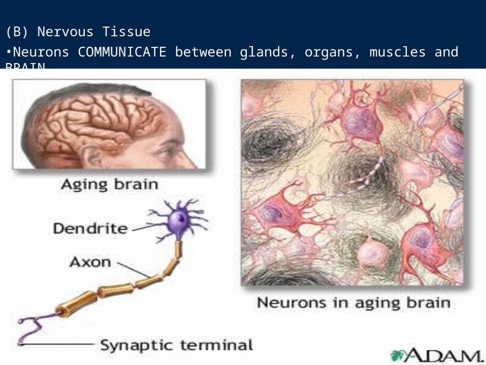
(B) Nervous Tissue •Neurons COMMUNICATE between glands, organs, muscles and BRAIN.
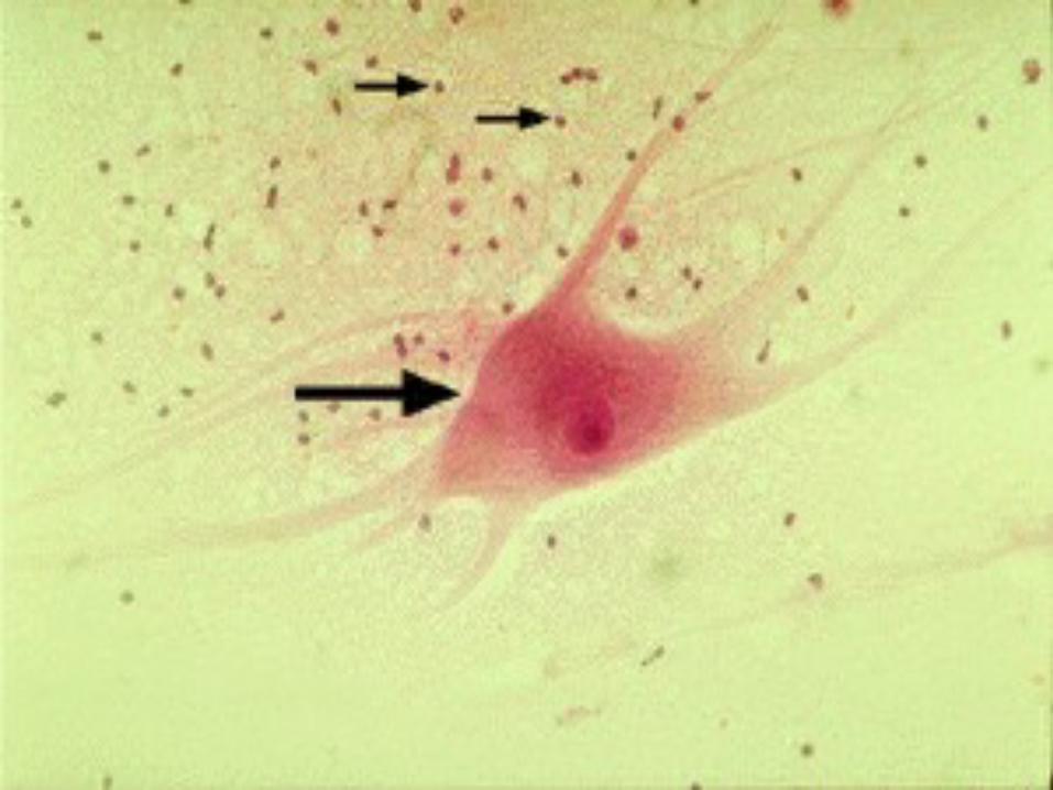
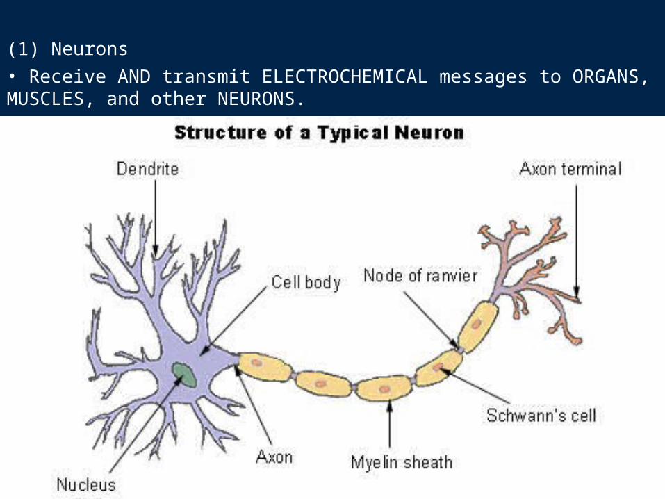
(1) Neurons• Receive AND transmit ELECTROCHEMICAL messages to ORGANS, MUSCLES, and other NEURONS.
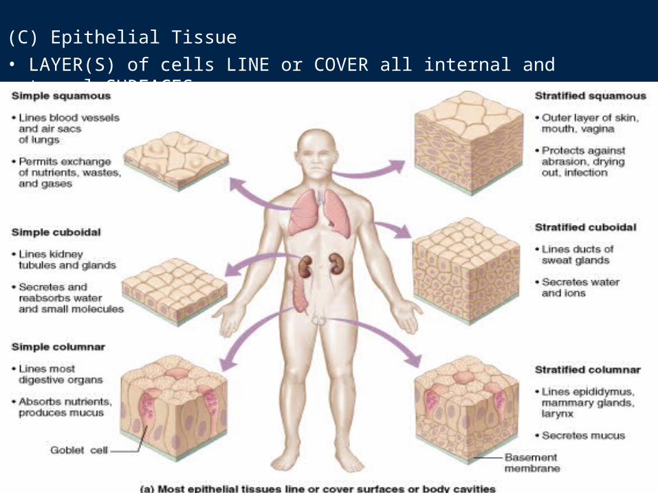
(C) Epithelial Tissue• LAYER(S) of cells LINE or COVER all internal and external SURFACES.
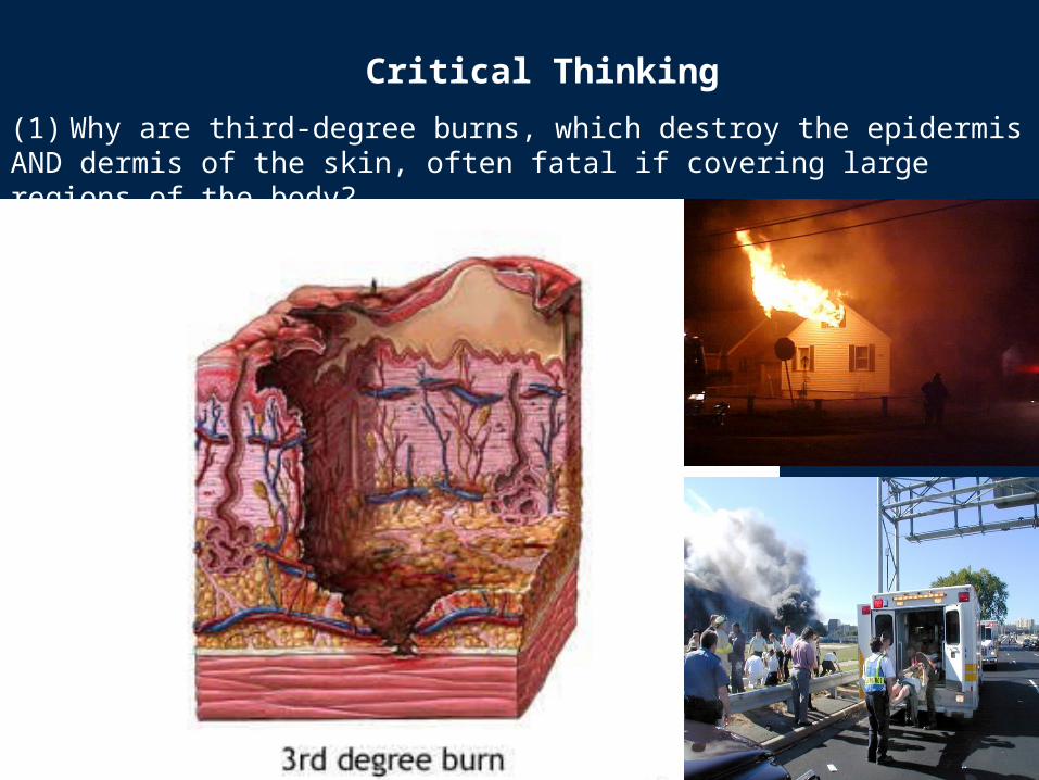
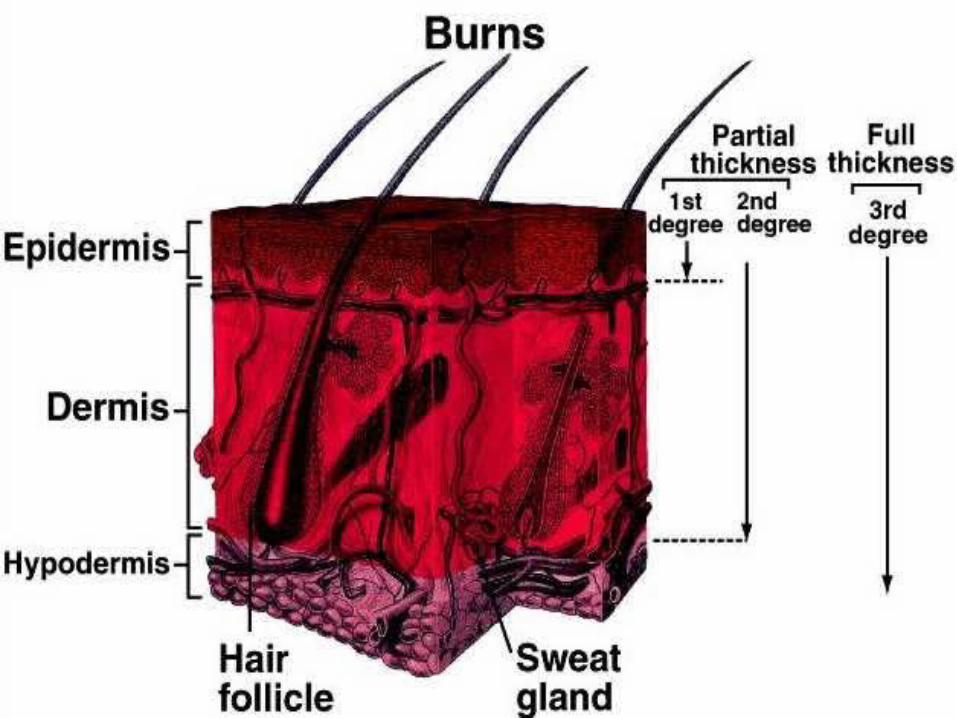
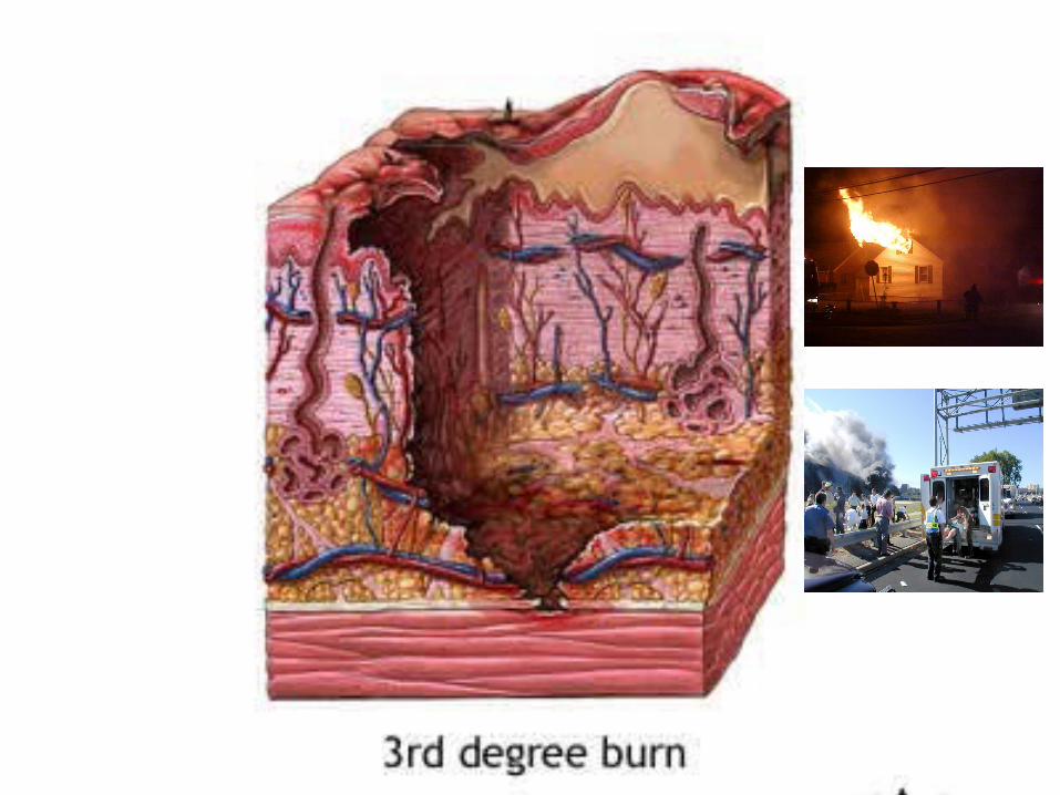
(1) Why are third-degree burns, which destroy the epidermis AND dermis of the skin, often fatal if covering large regions of the body?
Critical Thinking
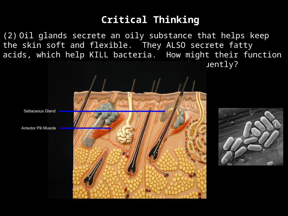
(2) Oil glands secrete an oily substance that helps keep the skin soft and flexible. They ALSO secrete fatty acids, which help KILL bacteria. How might their function be affected if you wash your skin TOO frequently?
Critical Thinking
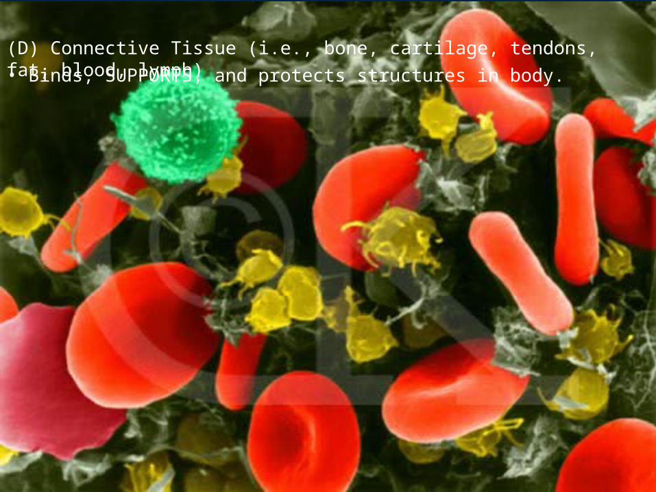
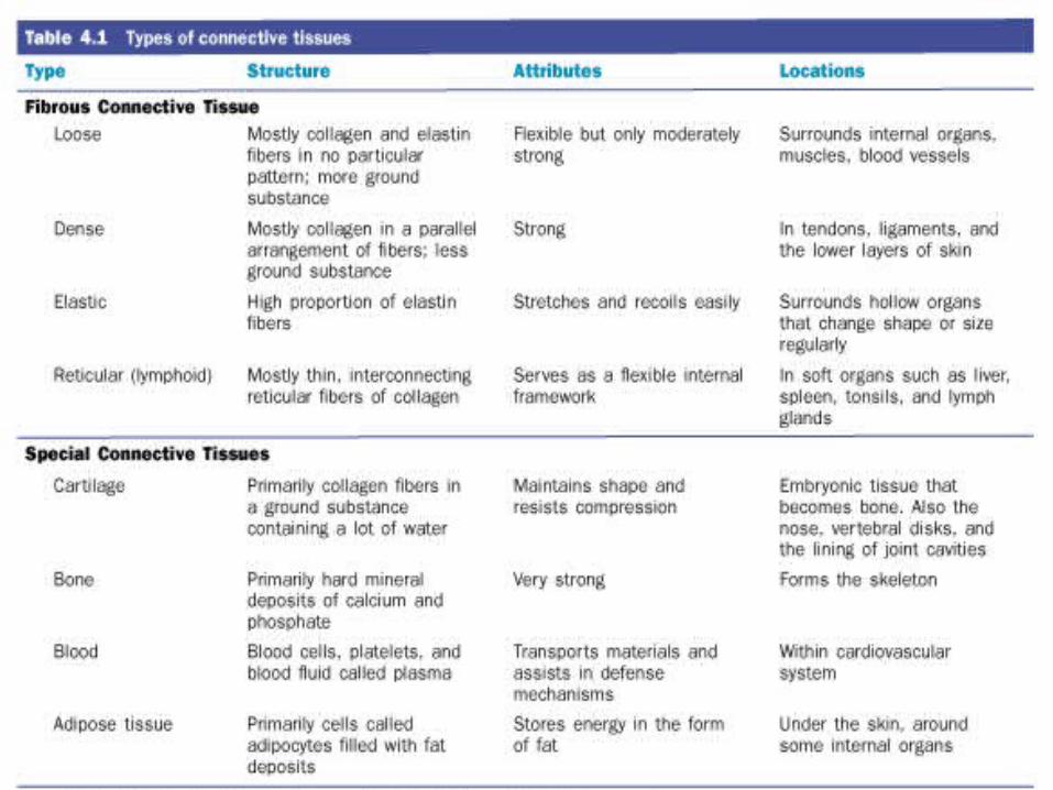
(D) Connective Tissue (i.e., bone, cartilage, tendons, fat, blood, lymph)• Binds, SUPPORTS, and protects structures in body.
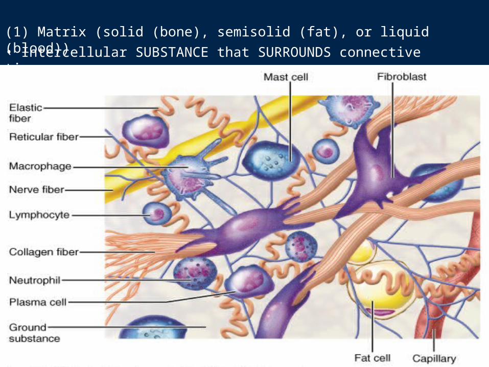
(1) Matrix (solid (bone), semisolid (fat), or liquid (blood))• Intercellular SUBSTANCE that SURROUNDS connective tissue.
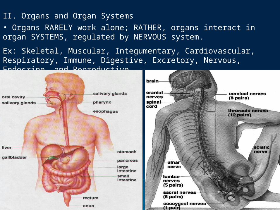
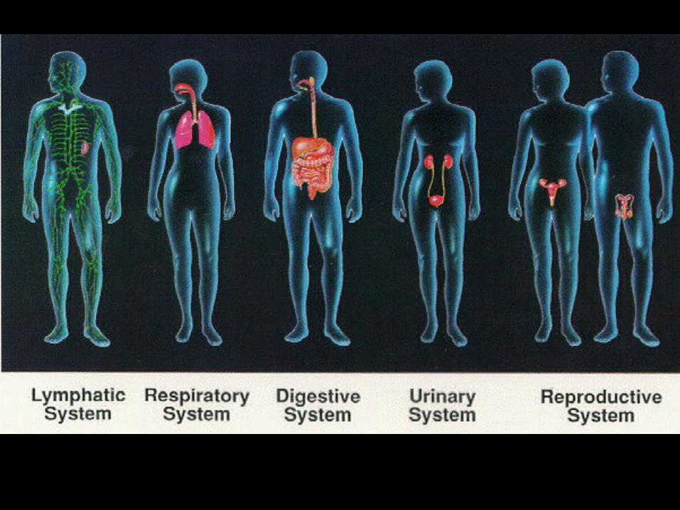
II. Organs and Organ Systems• Organs RARELY work alone; RATHER, organs interact in organ SYSTEMS, regulated by NERVOUS system.
Ex: Skeletal, Muscular, Integumentary, Cardiovascular, Respiratory, Immune, Digestive, Excretory, Nervous, Endocrine, and Reproductive.
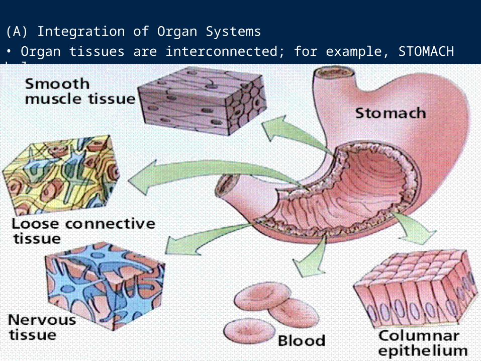
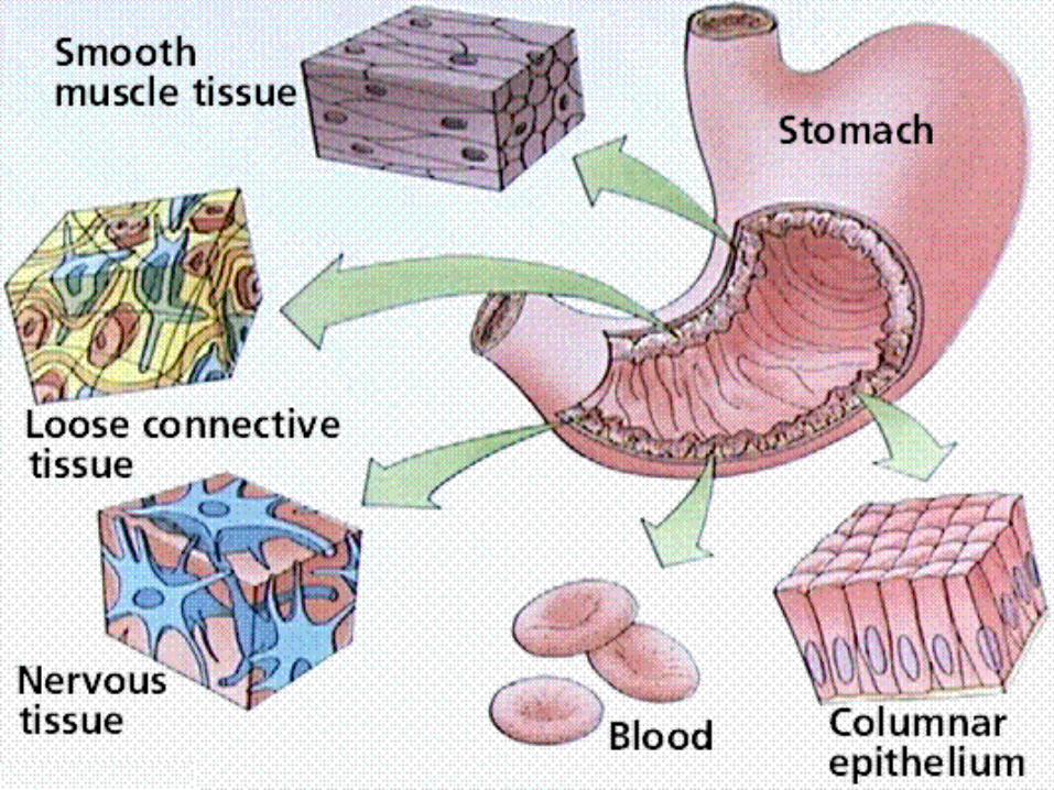
(A) Integration of Organ Systems• Organ tissues are interconnected; for example, STOMACH below.
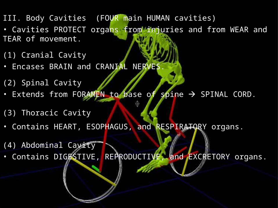
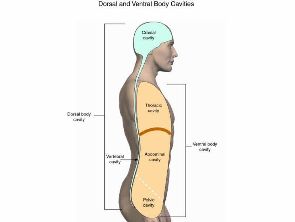
III. Body Cavities (FOUR main HUMAN cavities)• Cavities PROTECT organs from injuries and from WEAR and TEAR of movement.
(1) Cranial Cavity• Encases BRAIN and CRANIAL NERVES.
(2) Spinal Cavity• Extends from FORAMEN to base of spine SPINAL CORD.
(3) Thoracic Cavity
• Contains HEART, ESOPHAGUS, and RESPIRATORY organs.
(4) Abdominal Cavity• Contains DIGESTIVE, REPRODUCTIVE, and EXCRETORY organs.


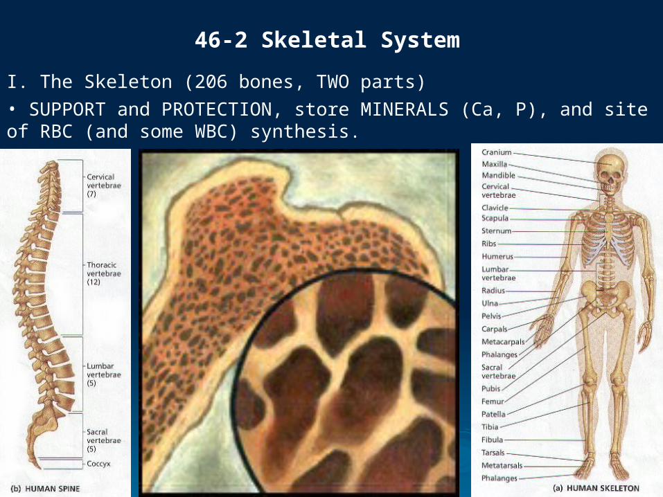
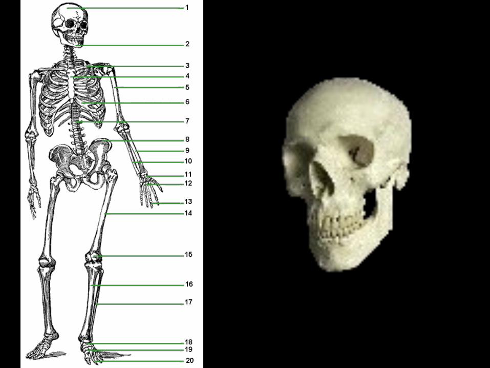
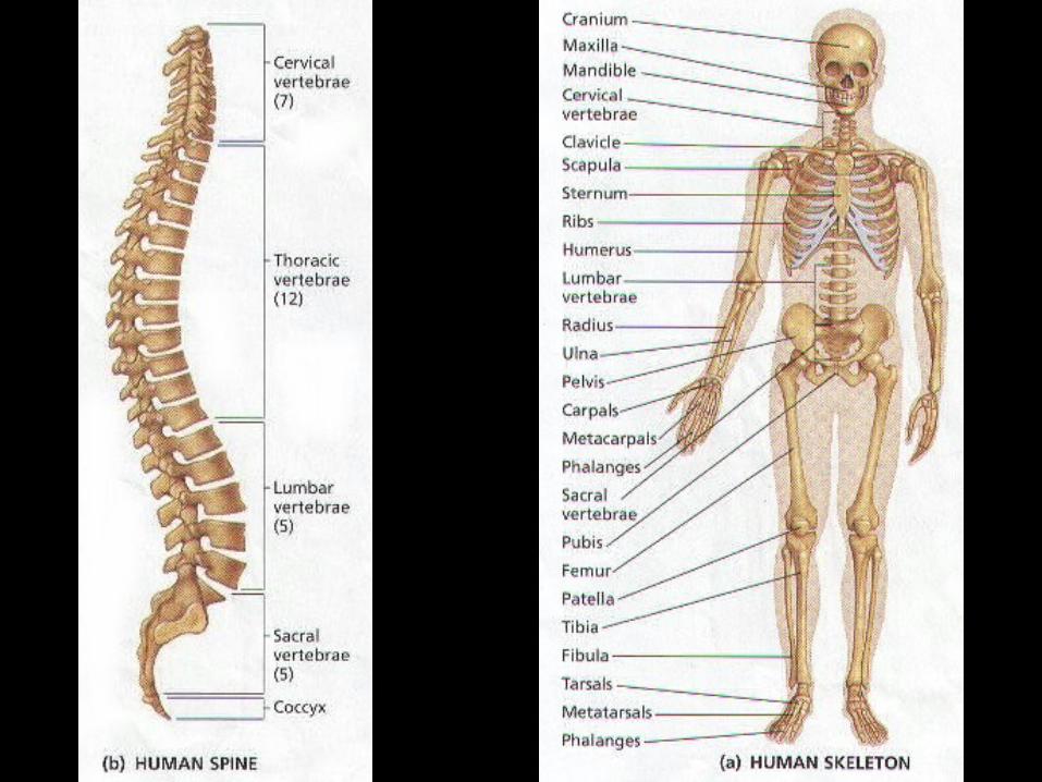
I. The Skeleton (206 bones, TWO parts)• SUPPORT and PROTECTION, store MINERALS (Ca, P), and site of RBC (and some WBC) synthesis.
46-2 Skeletal System
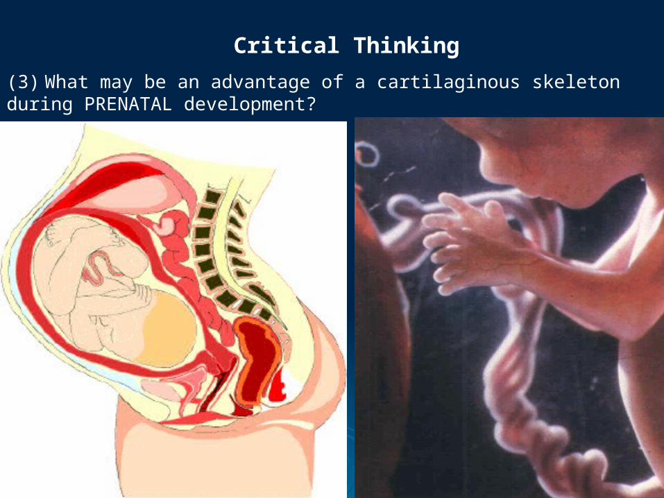
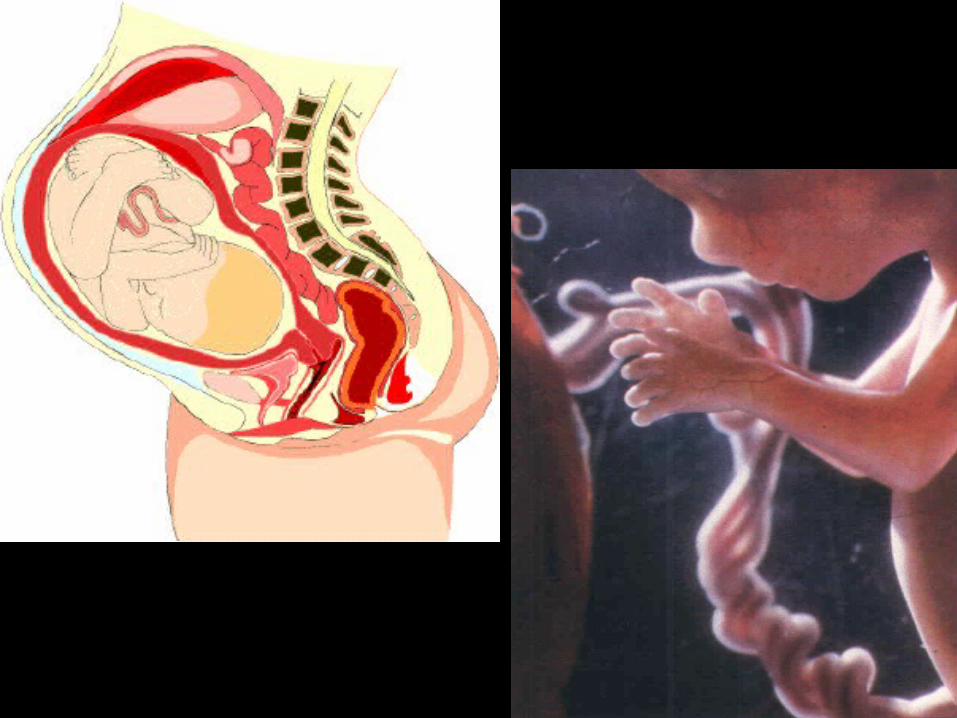
(3) What may be an advantage of a cartilaginous skeleton during PRENATAL development?
Critical Thinking
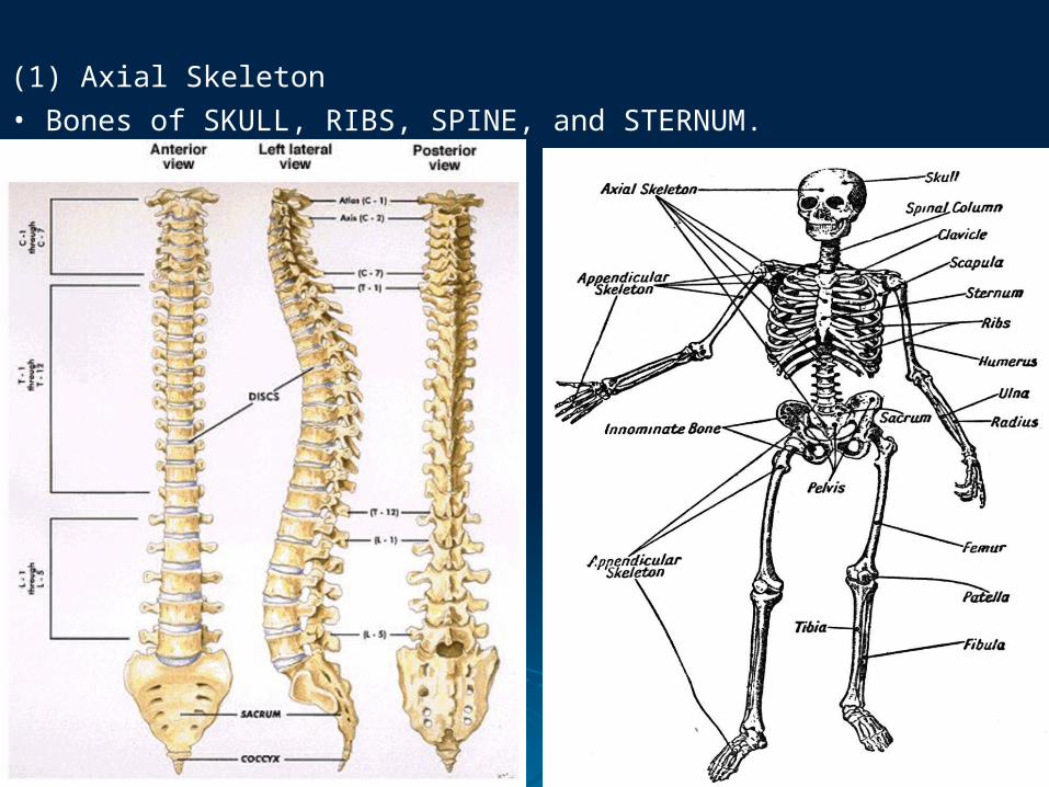
(1) Axial Skeleton• Bones of SKULL, RIBS, SPINE, and STERNUM.
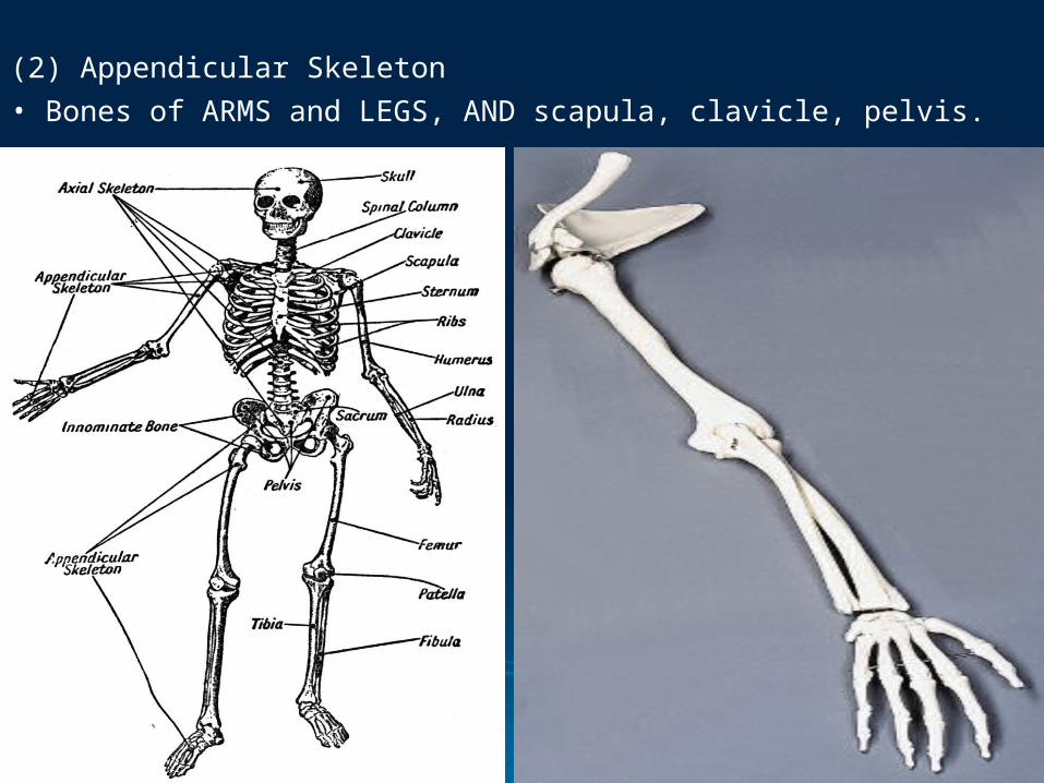
(2) Appendicular Skeleton• Bones of ARMS and LEGS, AND scapula, clavicle, pelvis.
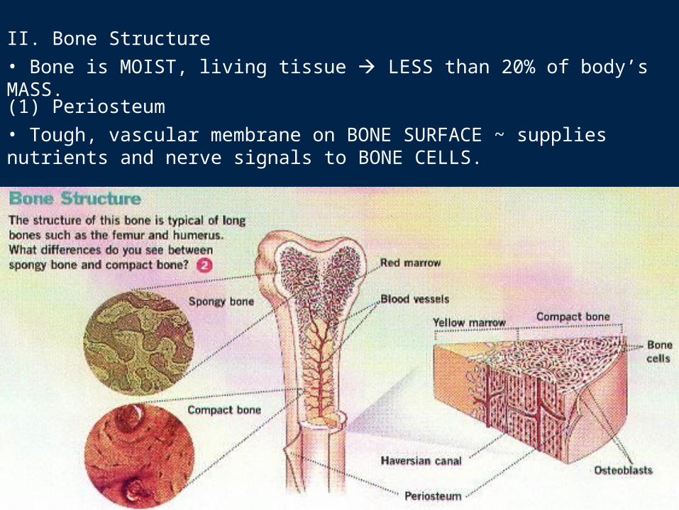
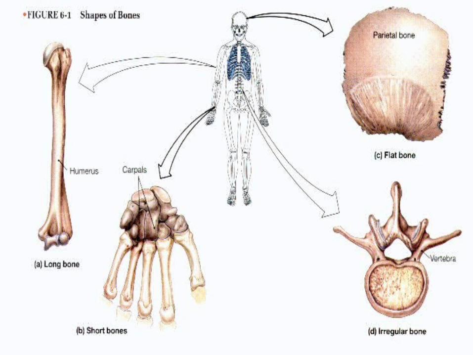
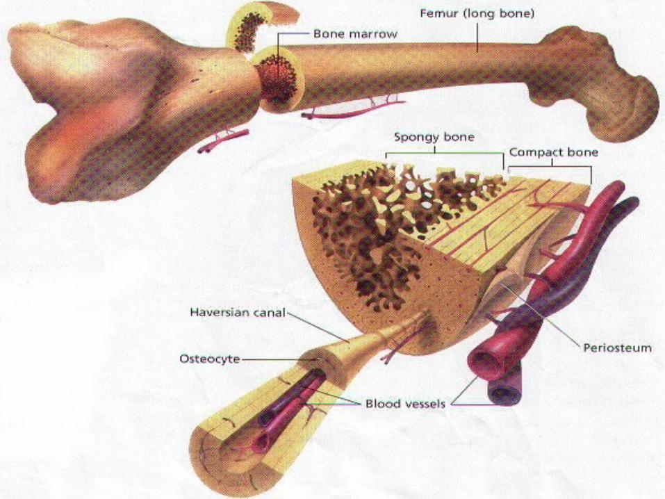
II. Bone Structure• Bone is MOIST, living tissue LESS than 20% of body’s MASS.
(1) Periosteum• Tough, vascular membrane on BONE SURFACE ~ supplies nutrients and nerve signals to BONE CELLS.

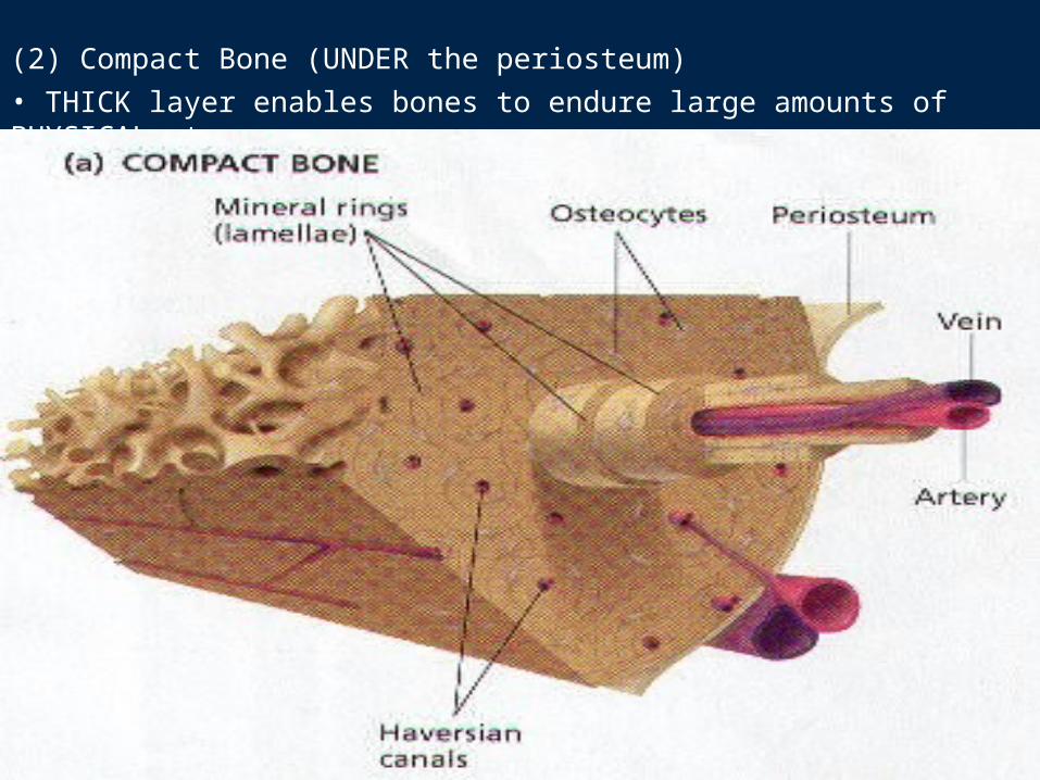
(2) Compact Bone (UNDER the periosteum)• THICK layer enables bones to endure large amounts of PHYSICAL stress.
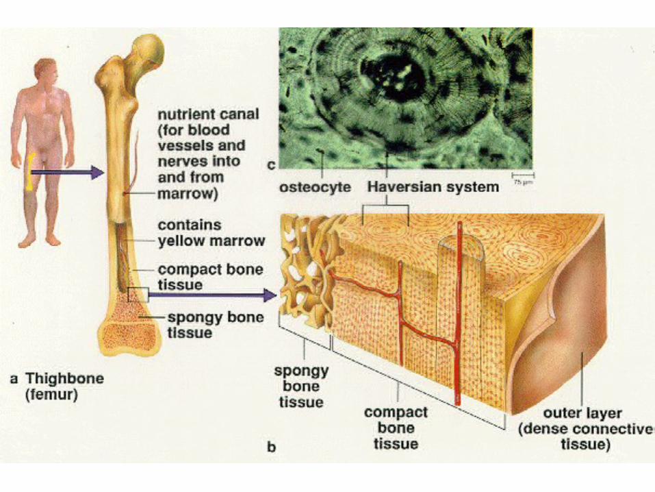
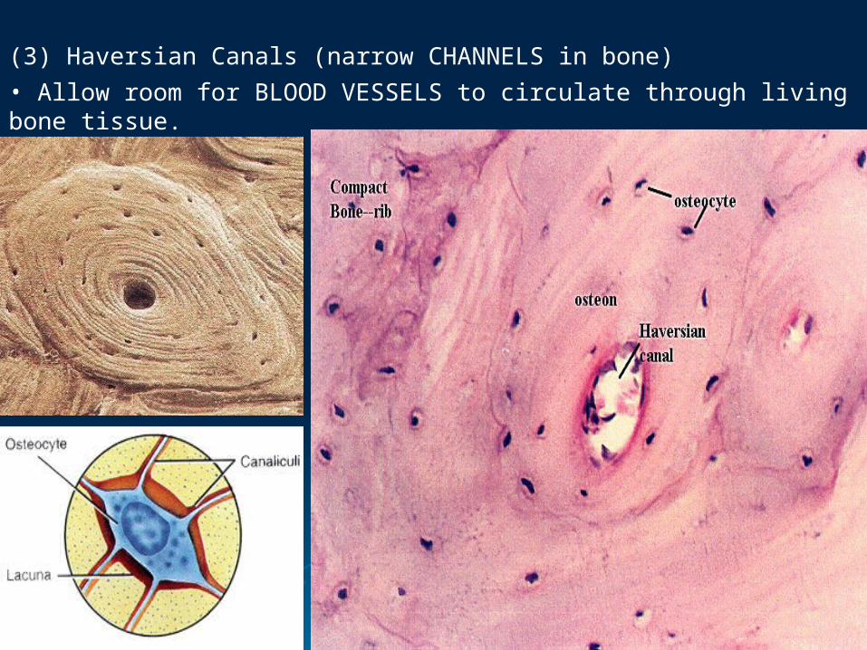
(3) Haversian Canals (narrow CHANNELS in bone)• Allow room for BLOOD VESSELS to circulate through living bone tissue.
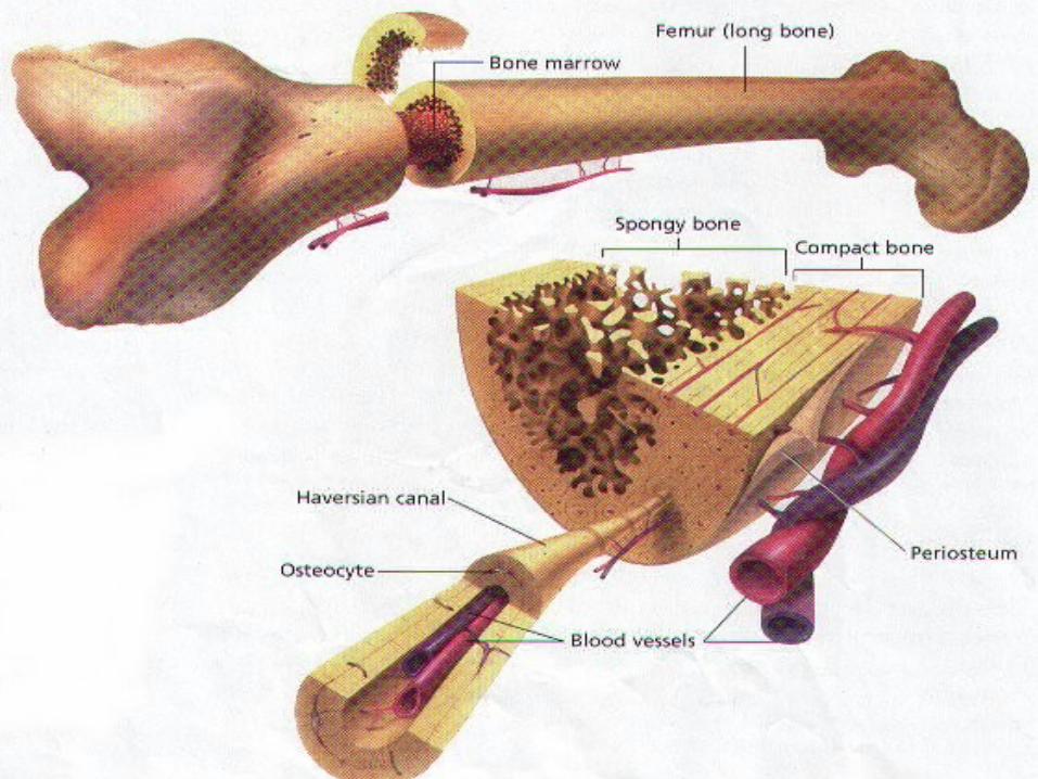
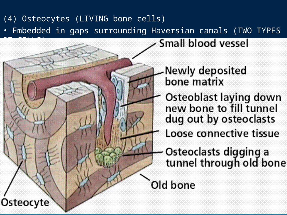
(4) Osteocytes (LIVING bone cells)• Embedded in gaps surrounding Haversian canals (TWO TYPES OF CELLS).
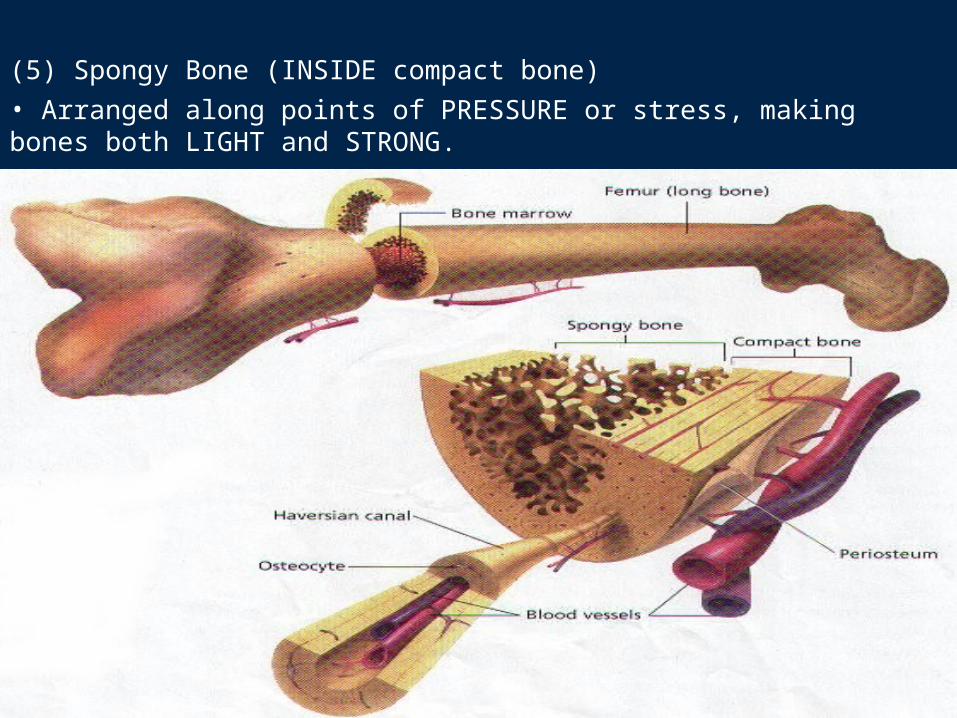
(5) Spongy Bone (INSIDE compact bone)• Arranged along points of PRESSURE or stress, making bones both LIGHT and STRONG.
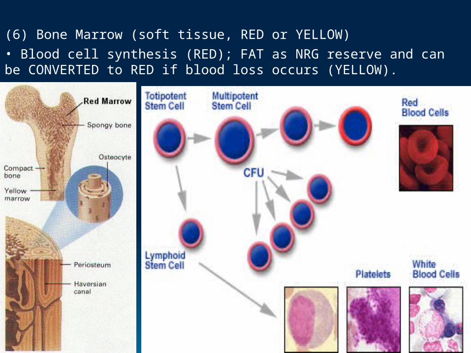
(6) Bone Marrow (soft tissue, RED or YELLOW)• Blood cell synthesis (RED); FAT as NRG reserve and can be CONVERTED to RED if blood loss occurs (YELLOW).
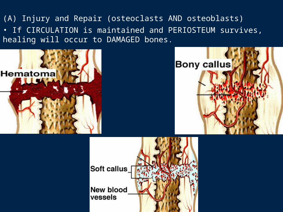
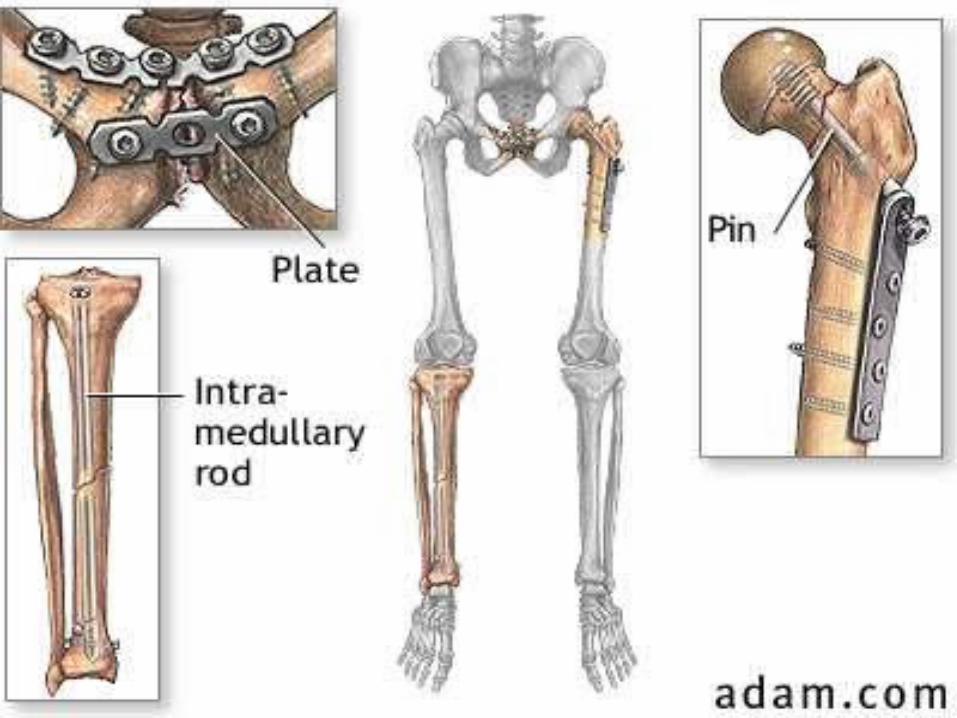
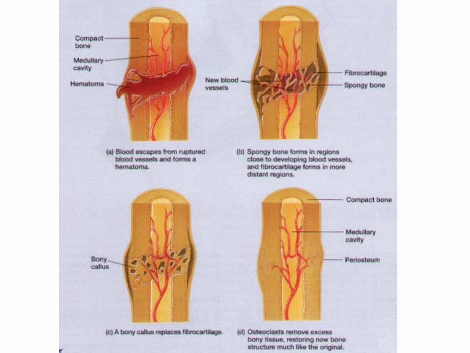
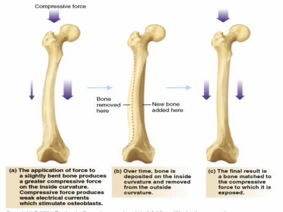
(A) Injury and Repair (osteoclasts AND osteoblasts)• If CIRCULATION is maintained and PERIOSTEUM survives, healing will occur to DAMAGED bones.
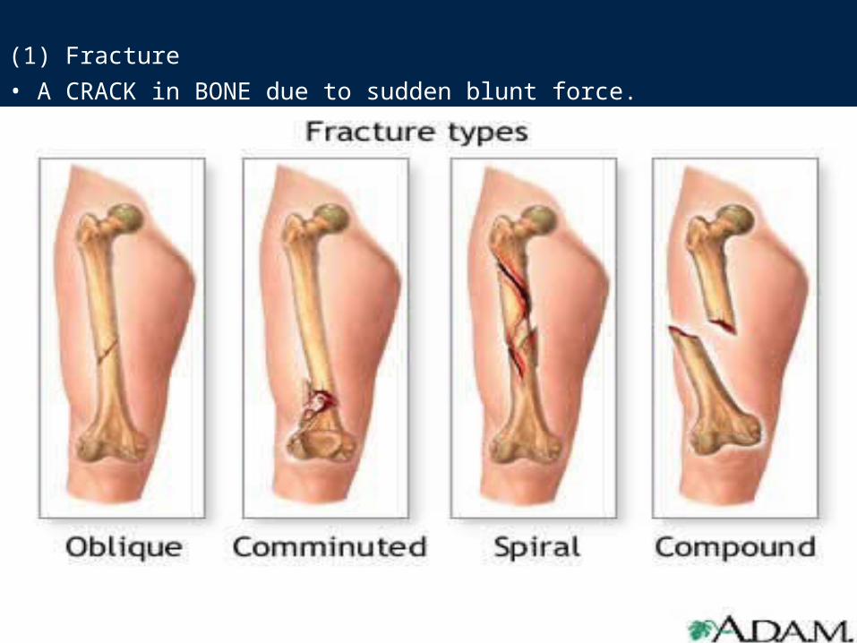
(1) Fracture• A CRACK in BONE due to sudden blunt force.
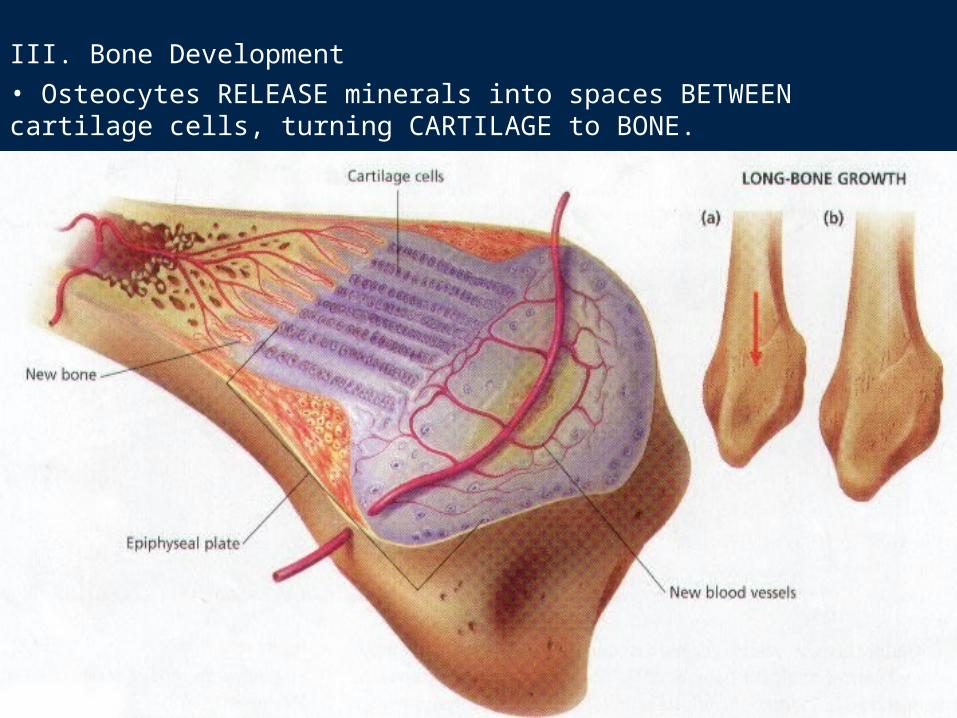
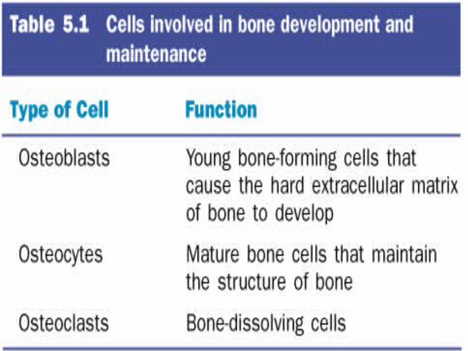
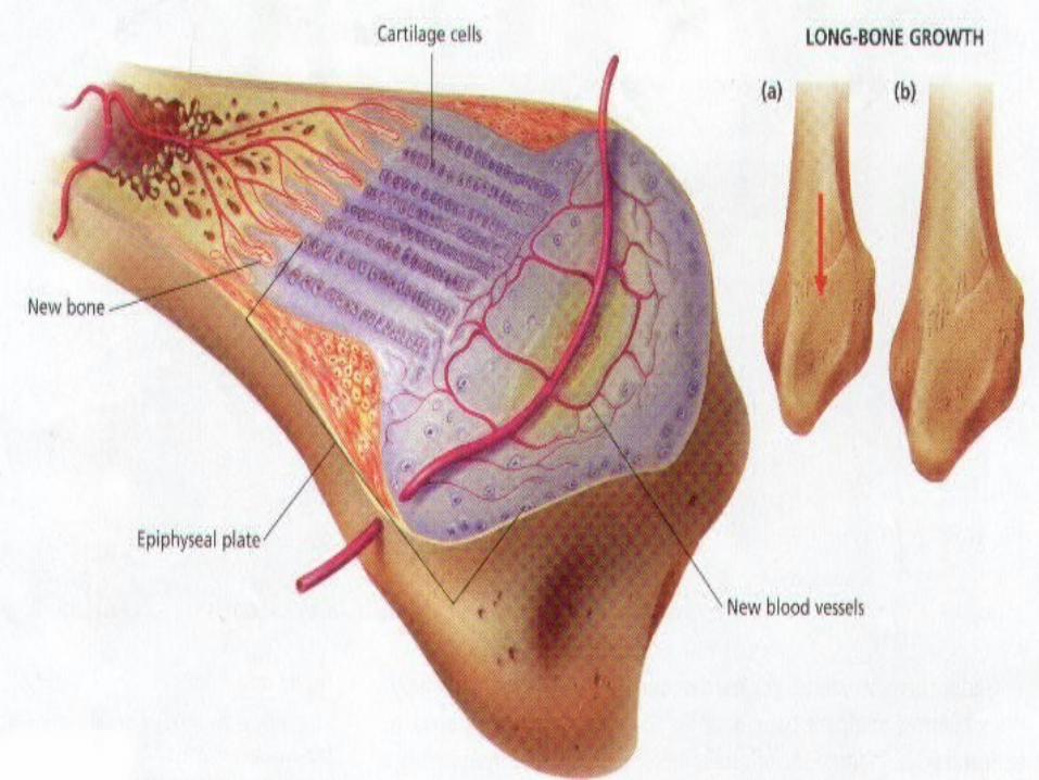
III. Bone Development• Osteocytes RELEASE minerals into spaces BETWEEN cartilage cells, turning CARTILAGE to BONE.
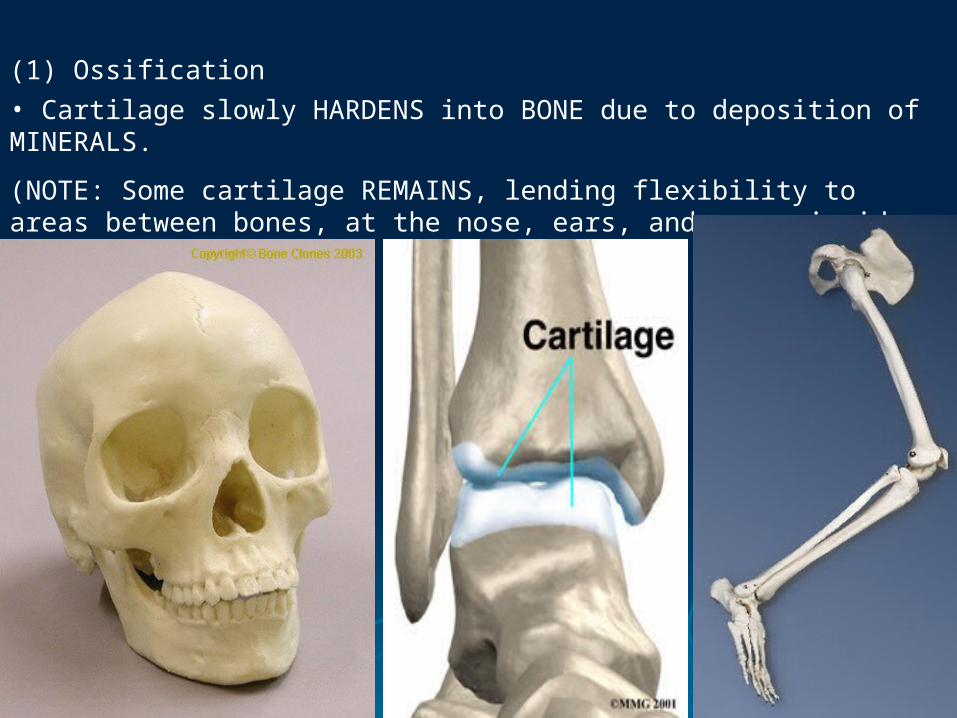
(1) Ossification• Cartilage slowly HARDENS into BONE due to deposition of MINERALS.
(NOTE: Some cartilage REMAINS, lending flexibility to areas between bones, at the nose, ears, and areas inside the trachea).

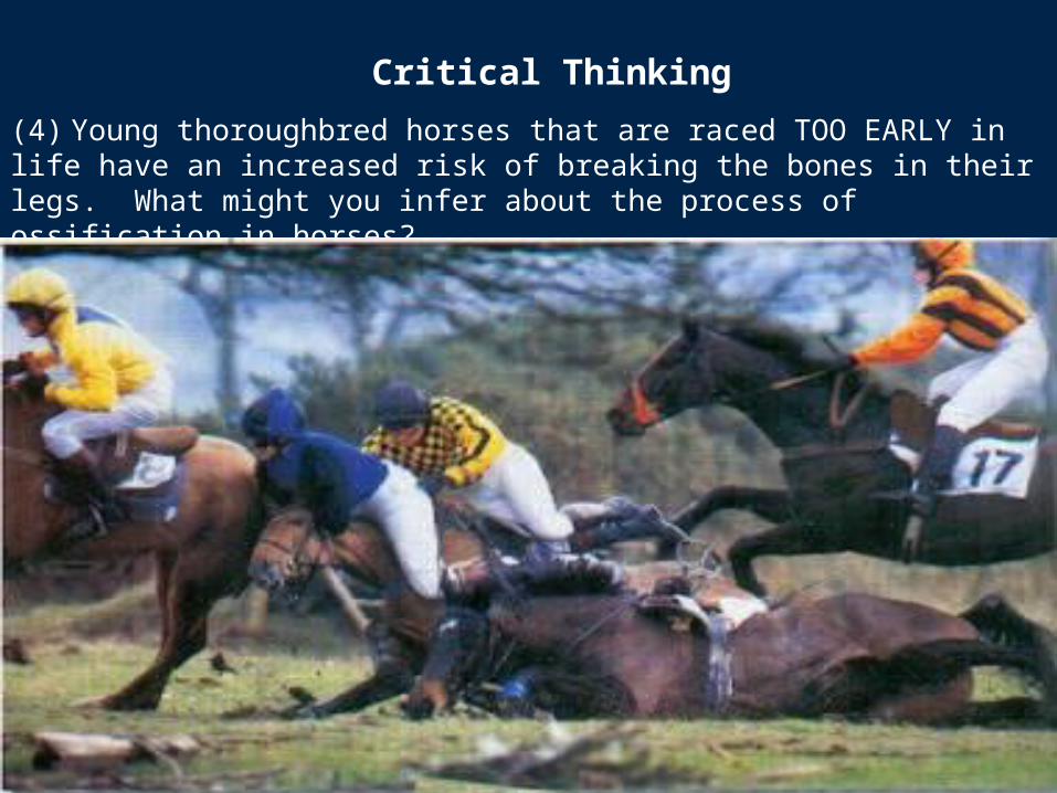
(4) Young thoroughbred horses that are raced TOO EARLY in life have an increased risk of breaking the bones in their legs. What might you infer about the process of ossification in horses?
Critical Thinking
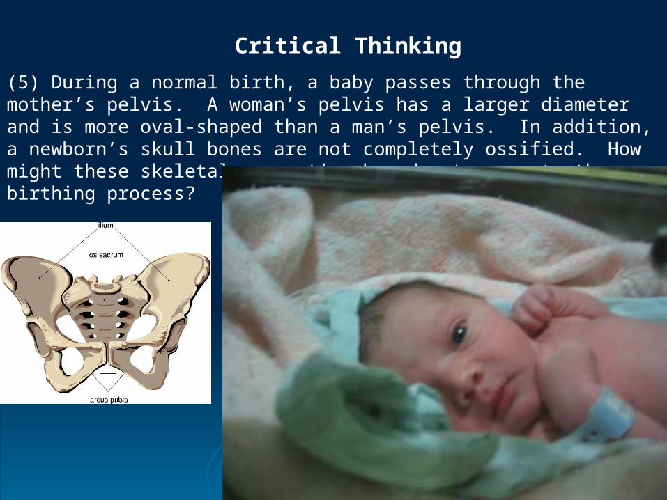
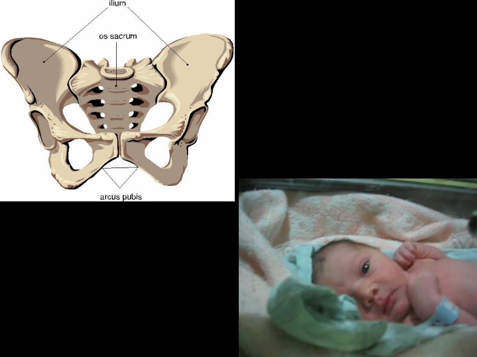
(5) During a normal birth, a baby passes through the mother’s pelvis. A woman’s pelvis has a larger diameter and is more oval-shaped than a man’s pelvis. In addition, a newborn’s skull bones are not completely ossified. How might these skeletal properties be advantageous to the birthing process?
Critical Thinking
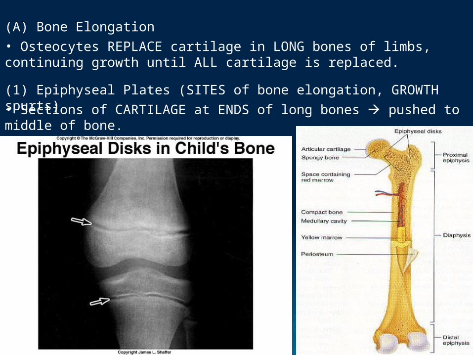
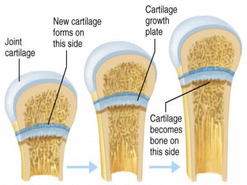
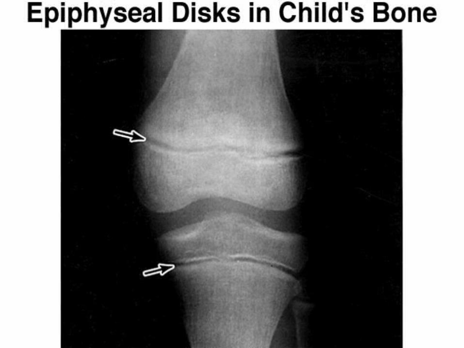
(A) Bone Elongation• Osteocytes REPLACE cartilage in LONG bones of limbs, continuing growth until ALL cartilage is replaced.
(1) Epiphyseal Plates (SITES of bone elongation, GROWTH spurts)• Sections of CARTILAGE at ENDS of long bones pushed to middle of bone.
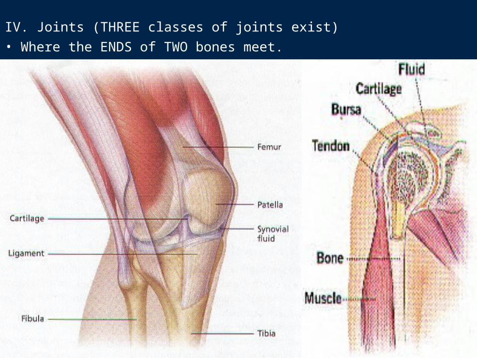
IV. Joints (THREE classes of joints exist)• Where the ENDS of TWO bones meet.
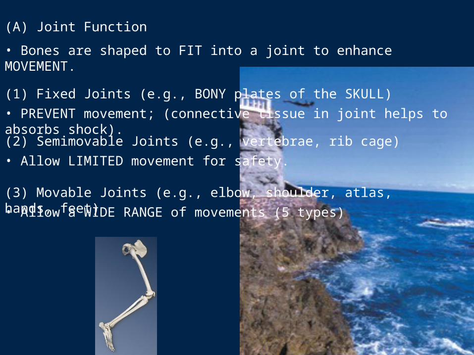
(A) Joint Function
• Bones are shaped to FIT into a joint to enhance MOVEMENT.
(1) Fixed Joints (e.g., BONY plates of the SKULL)• PREVENT movement; (connective tissue in joint helps to absorbs shock).(2) Semimovable Joints (e.g., vertebrae, rib cage)• Allow LIMITED movement for safety.
(3) Movable Joints (e.g., elbow, shoulder, atlas, hands, feet)• Allow a WIDE RANGE of movements (5 types)
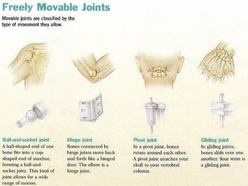
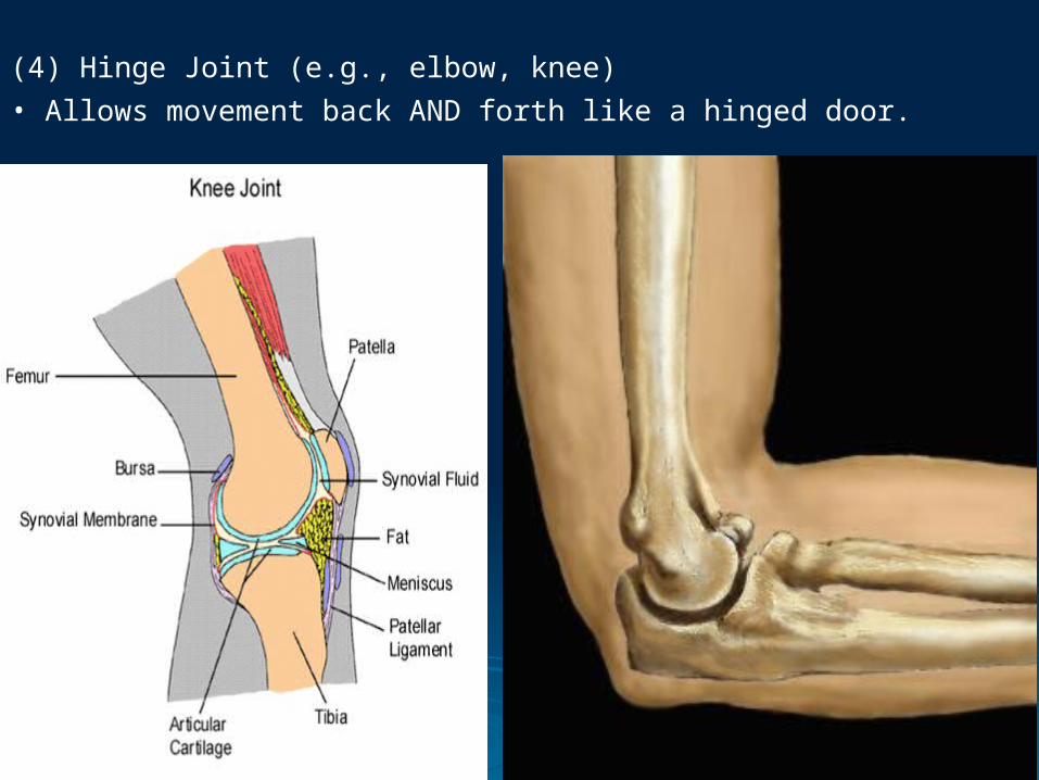
(4) Hinge Joint (e.g., elbow, knee)• Allows movement back AND forth like a hinged door.
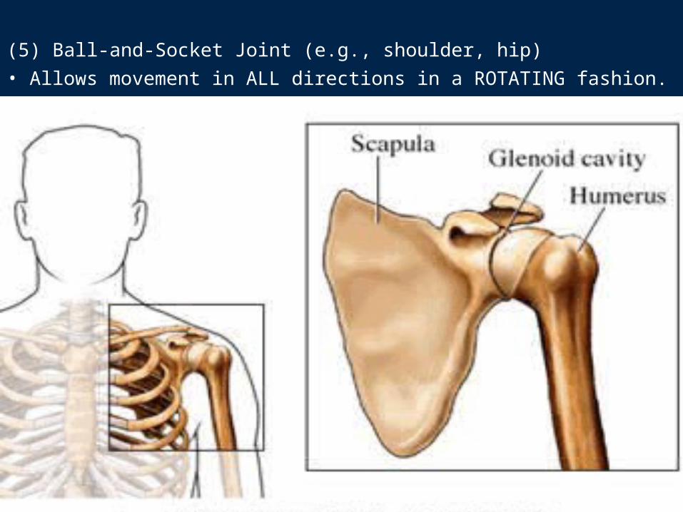
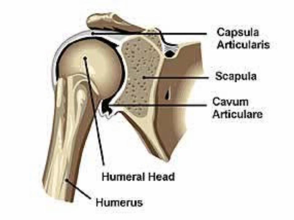
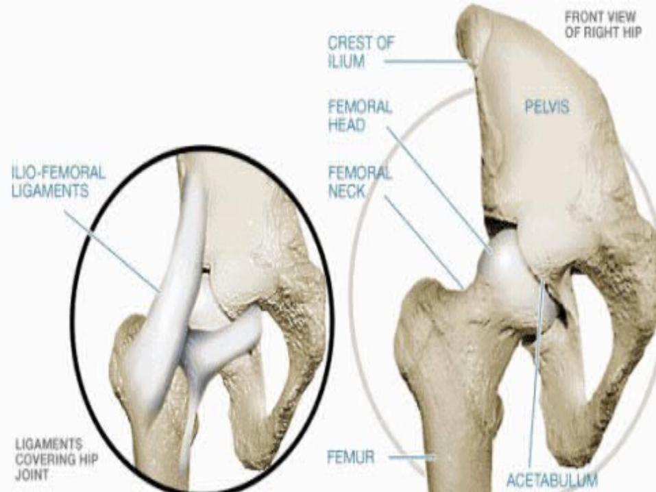
(5) Ball-and-Socket Joint (e.g., shoulder, hip) • Allows movement in ALL directions in a ROTATING fashion.
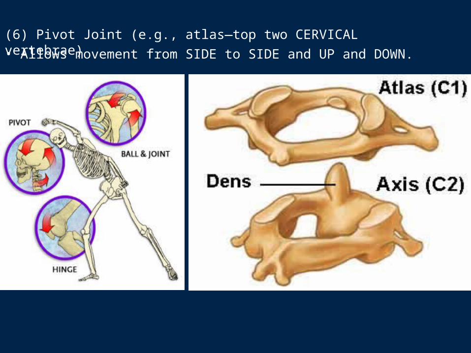
(6) Pivot Joint (e.g., atlas—top two CERVICAL vertebrae)• Allows movement from SIDE to SIDE and UP and DOWN.
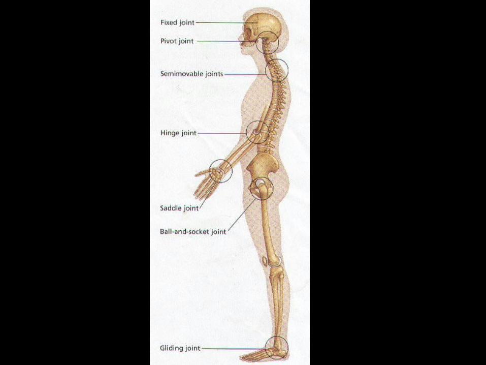
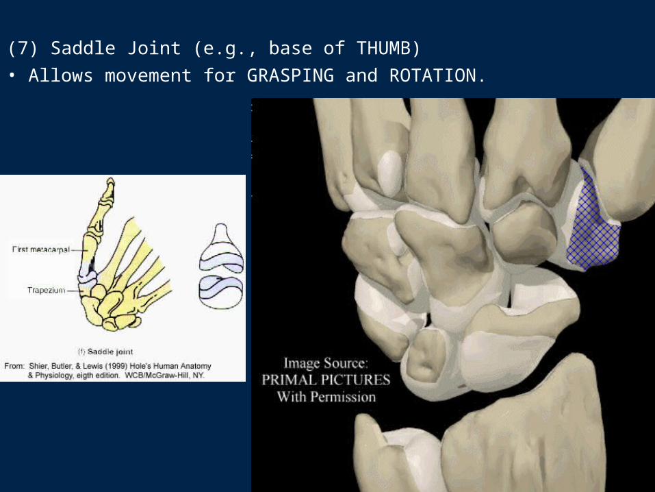
(7) Saddle Joint (e.g., base of THUMB)• Allows movement for GRASPING and ROTATION.
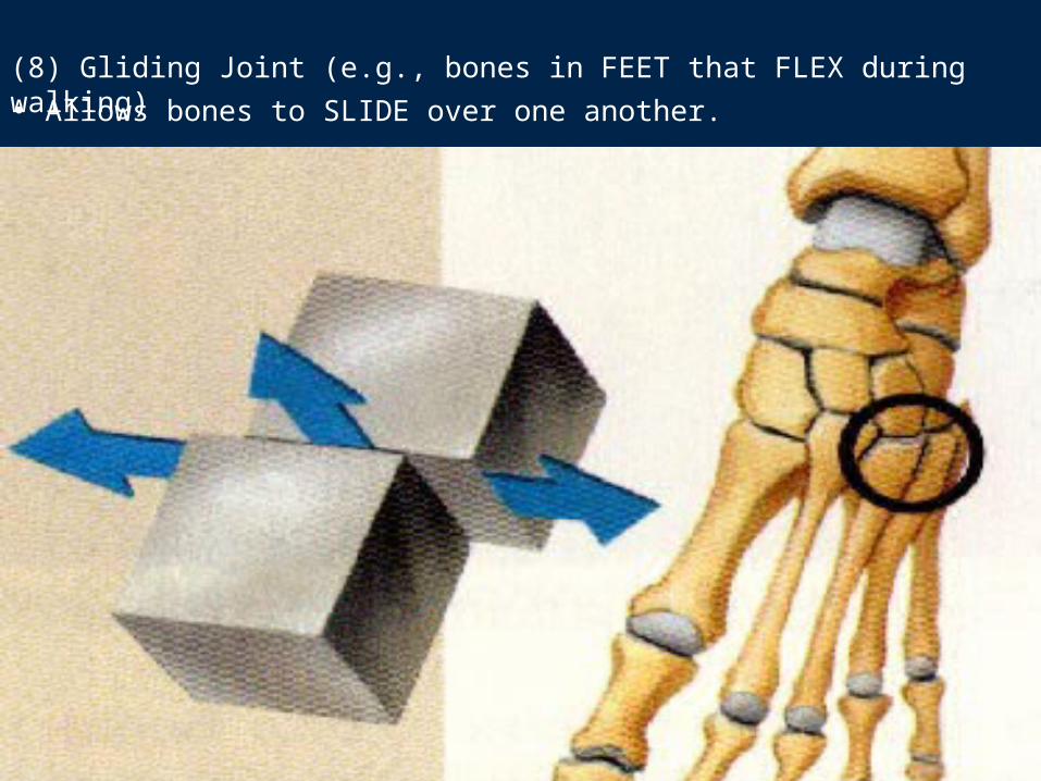
(8) Gliding Joint (e.g., bones in FEET that FLEX during walking)• Allows bones to SLIDE over one another.
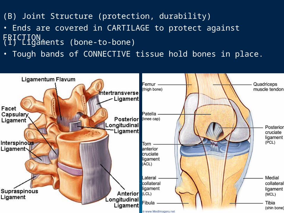
(B) Joint Structure (protection, durability)• Ends are covered in CARTILAGE to protect against FRICTION.
(1) Ligaments (bone-to-bone)• Tough bands of CONNECTIVE tissue hold bones in place.
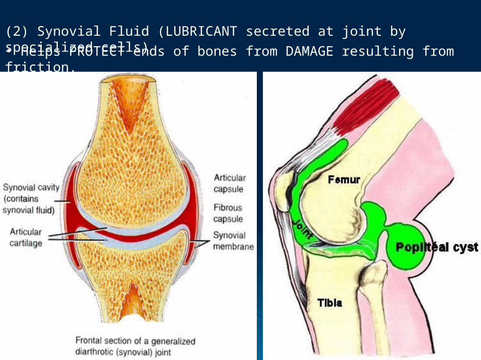
(2) Synovial Fluid (LUBRICANT secreted at joint by specialized cells)• Helps PROTECT ends of bones from DAMAGE resulting from friction.
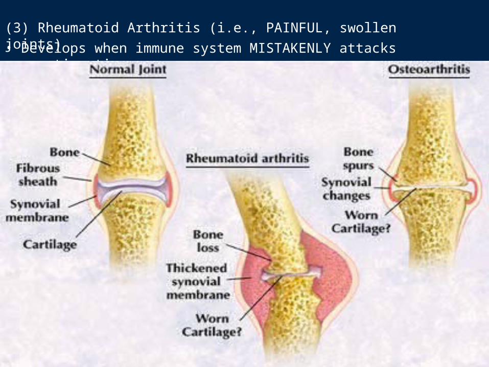
(3) Rheumatoid Arthritis (i.e., PAINFUL, swollen joints)• Develops when immune system MISTAKENLY attacks connective tissues.
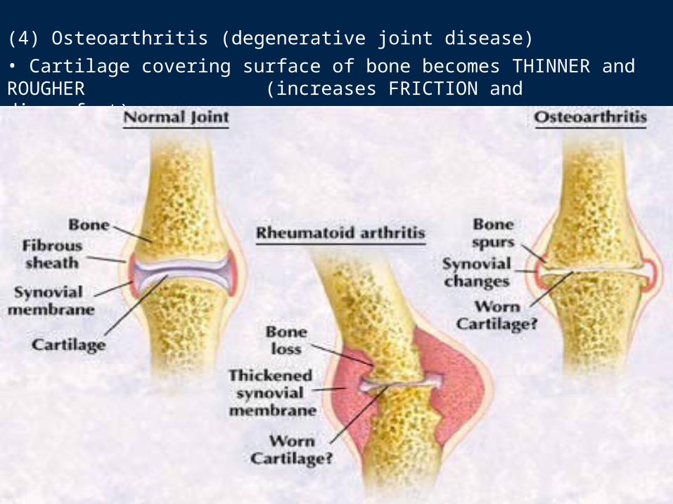
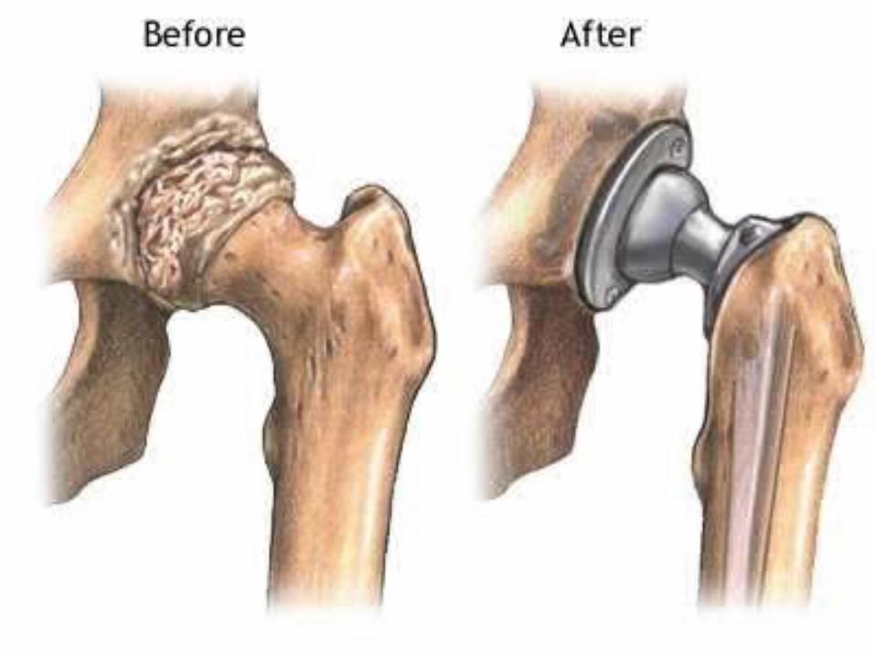
(4) Osteoarthritis (degenerative joint disease)• Cartilage covering surface of bone becomes THINNER and ROUGHER (increases FRICTION and discomfort).
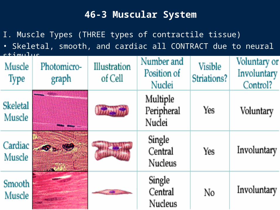
I. Muscle Types (THREE types of contractile tissue)• Skeletal, smooth, and cardiac all CONTRACT due to neural stimulus.
46-3 Muscular System
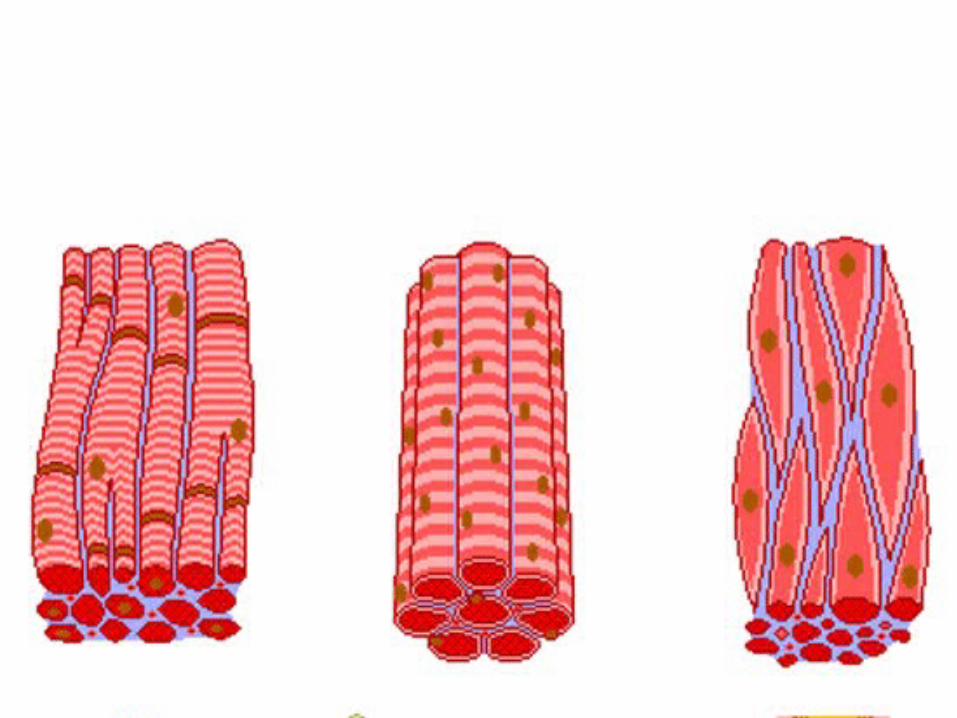

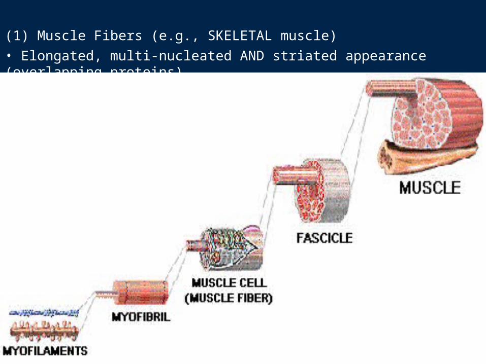
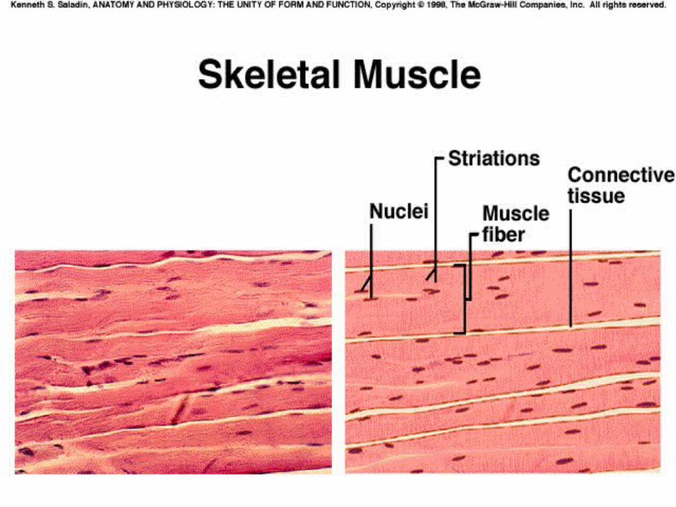
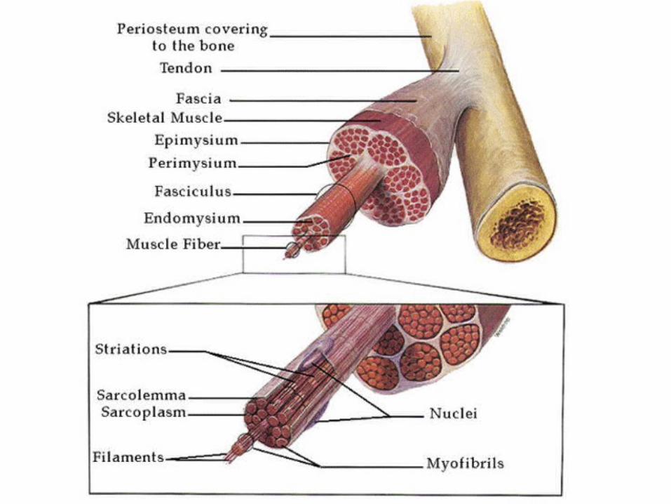
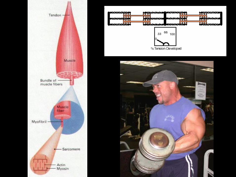
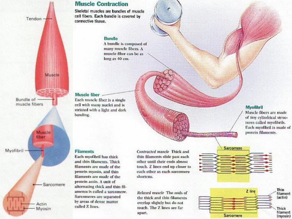
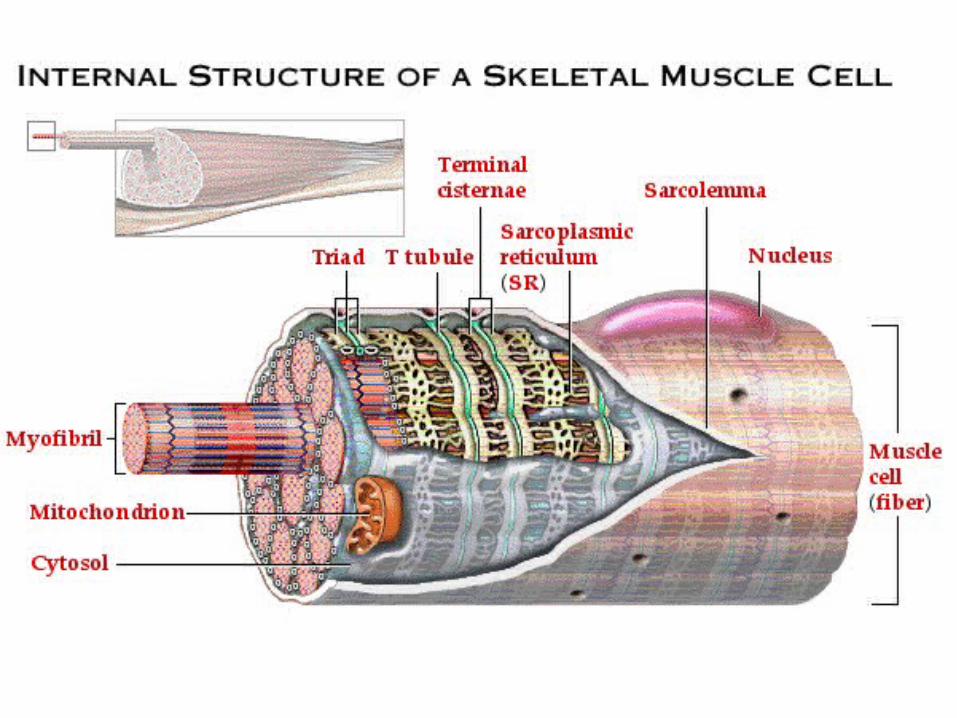
(1) Muscle Fibers (e.g., SKELETAL muscle)• Elongated, multi-nucleated AND striated appearance (overlapping proteins).

(2) Striations (in skeletal AND cardiac muscle, NOT smooth) • Light-dark STRIPES (between Z lines) present in skeletal muscle.
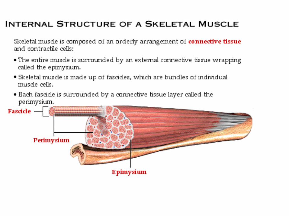
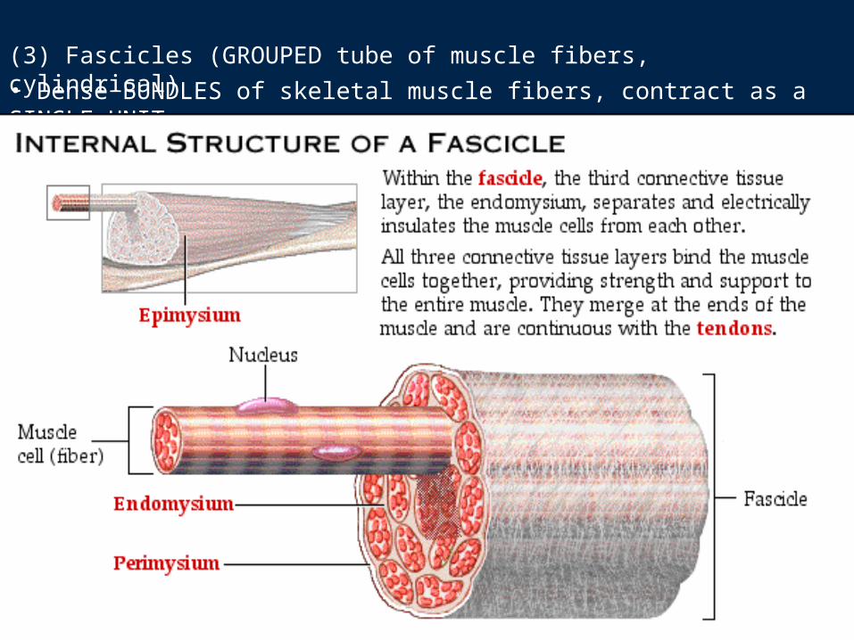
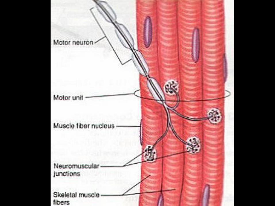
(3) Fascicles (GROUPED tube of muscle fibers, cylindrical)• Dense BUNDLES of skeletal muscle fibers, contract as a SINGLE UNIT.
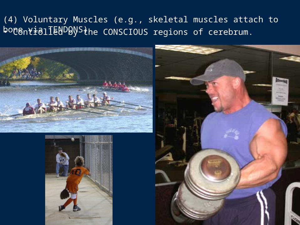
(4) Voluntary Muscles (e.g., skeletal muscles attach to bone via TENDONS)• Controlled by the CONSCIOUS regions of cerebrum.
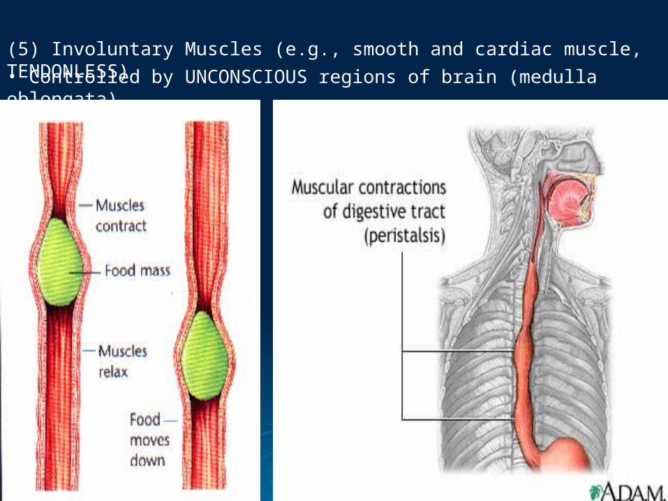
(5) Involuntary Muscles (e.g., smooth and cardiac muscle, TENDONLESS)• Controlled by UNCONSCIOUS regions of brain (medulla oblongata).
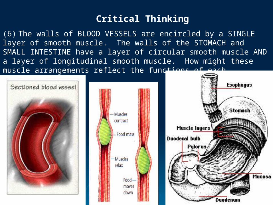
(6) The walls of BLOOD VESSELS are encircled by a SINGLE layer of smooth muscle. The walls of the STOMACH and SMALL INTESTINE have a layer of circular smooth muscle AND a layer of longitudinal smooth muscle. How might these muscle arrangements reflect the functions of each structure?
Critical Thinking
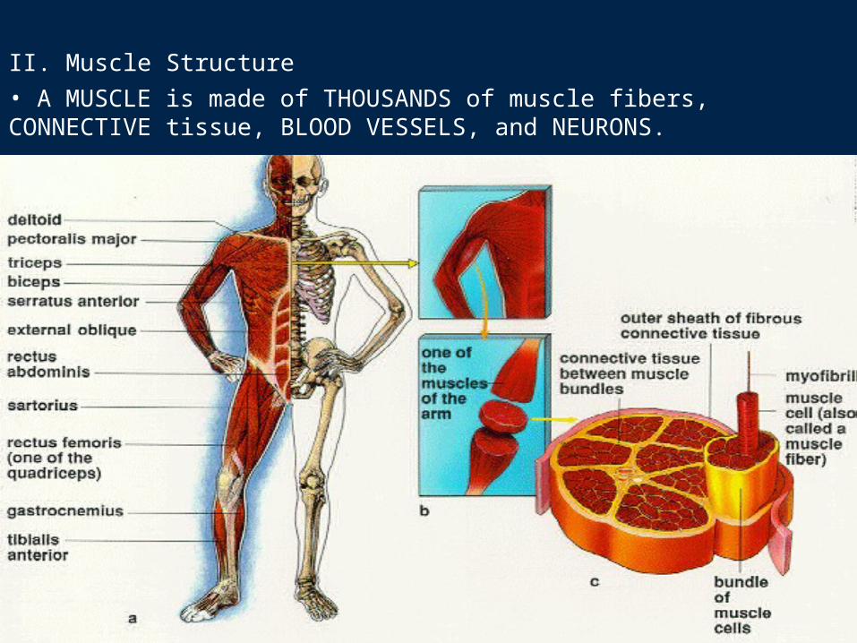
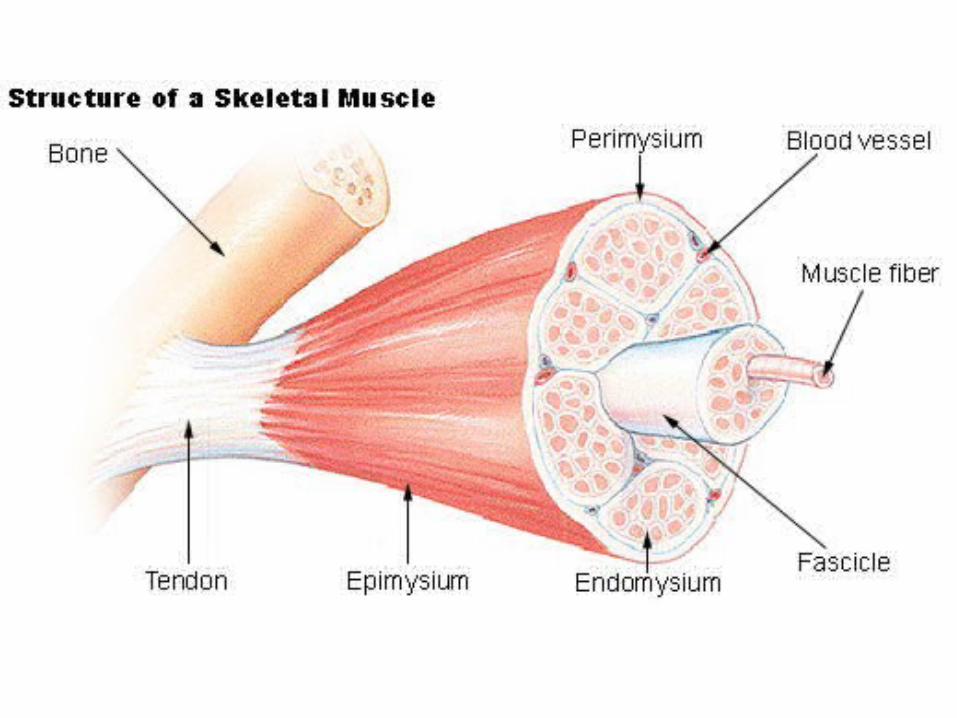
II. Muscle Structure• A MUSCLE is made of THOUSANDS of muscle fibers, CONNECTIVE tissue, BLOOD VESSELS, and NEURONS.
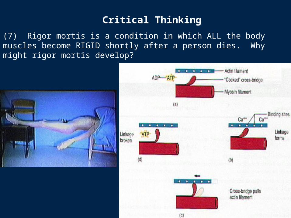
(7) Rigor mortis is a condition in which ALL the body muscles become RIGID shortly after a person dies. Why might rigor mortis develop?
Critical Thinking
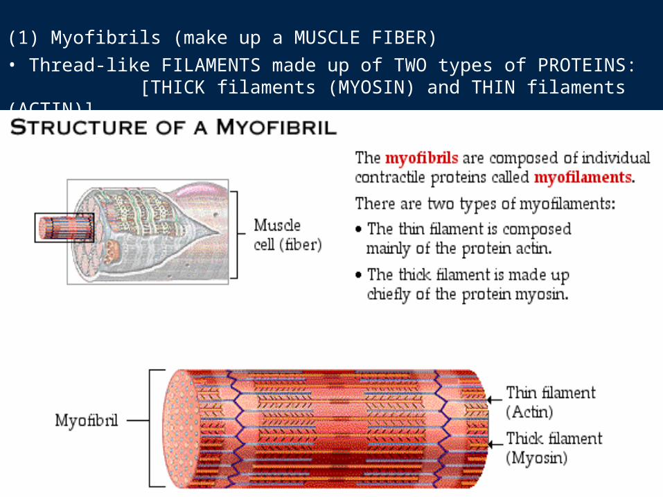
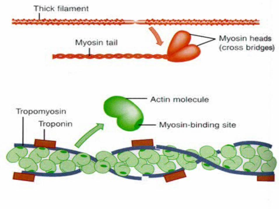
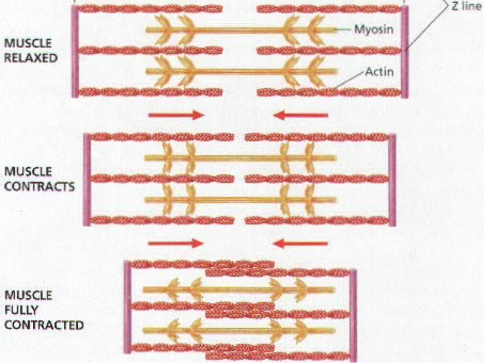
(1) Myofibrils (make up a MUSCLE FIBER)• Thread-like FILAMENTS made up of TWO types of PROTEINS:
[THICK filaments (MYOSIN) and THIN filaments (ACTIN)].
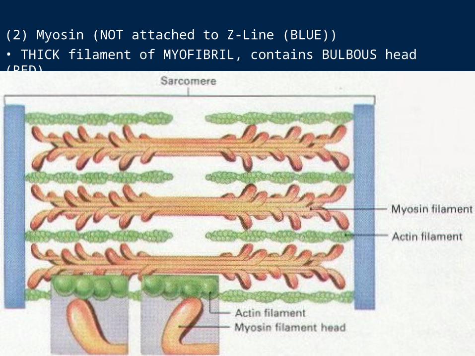
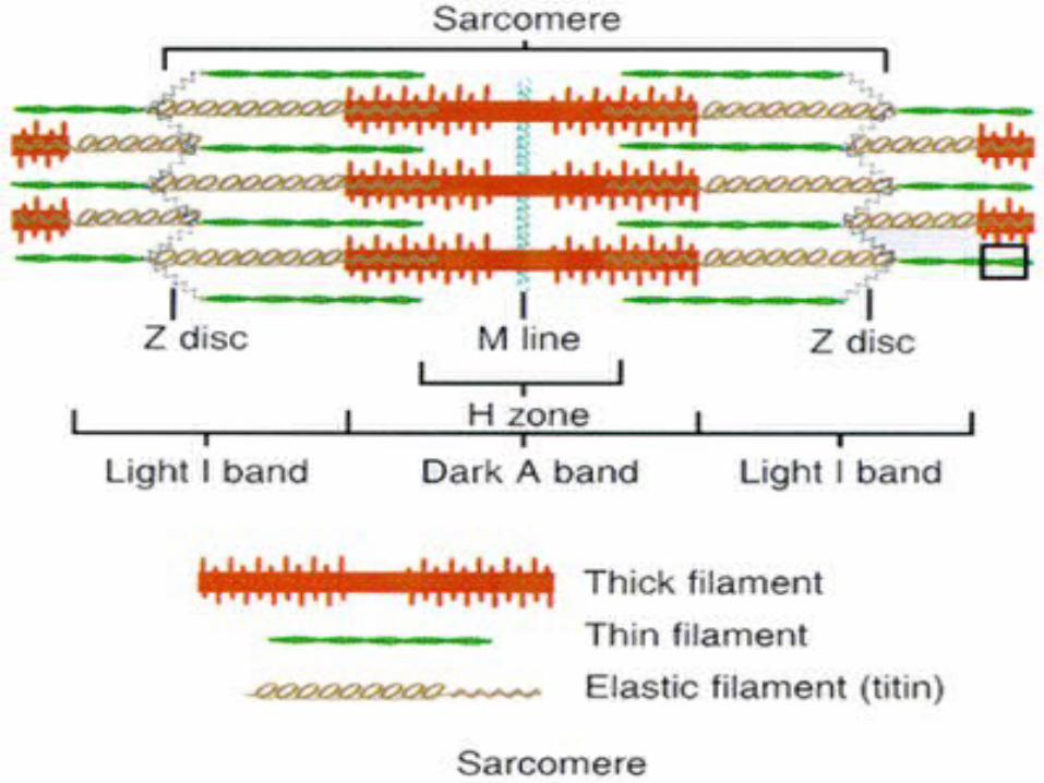
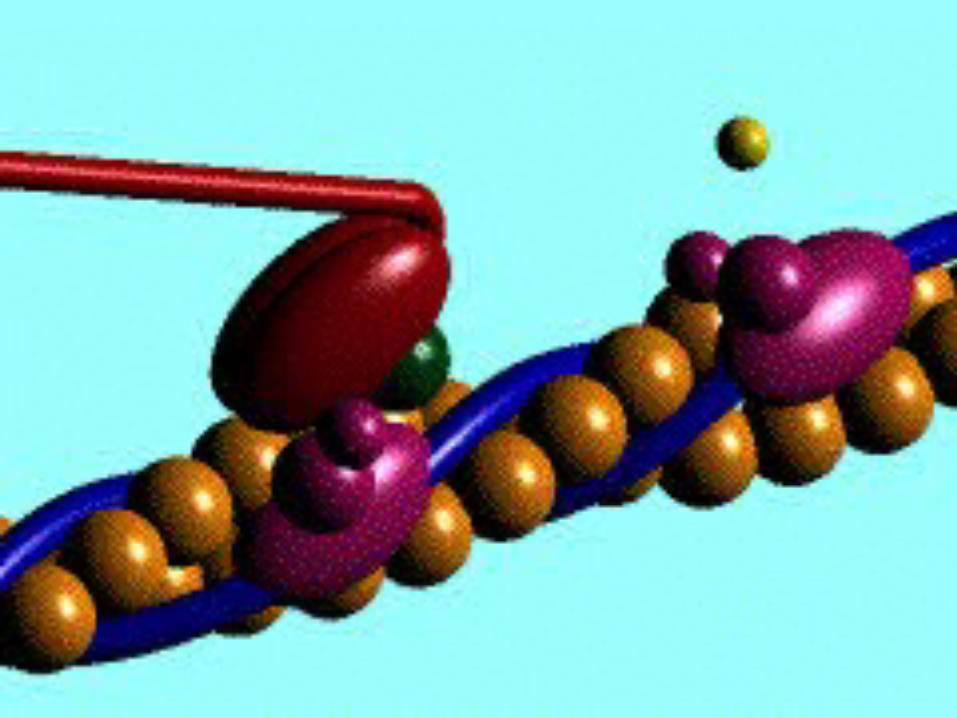
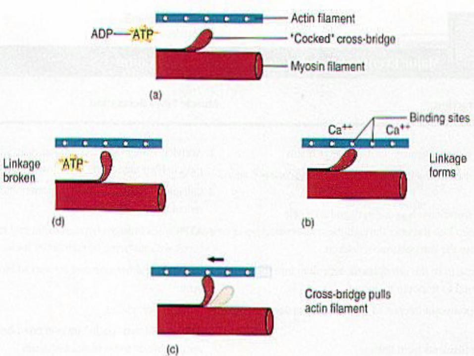
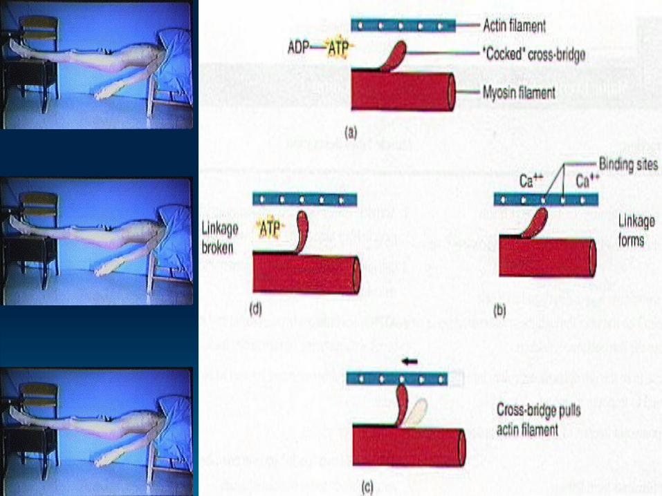
(2) Myosin (NOT attached to Z-Line (BLUE)) • THICK filament of MYOFIBRIL, contains BULBOUS head (RED).
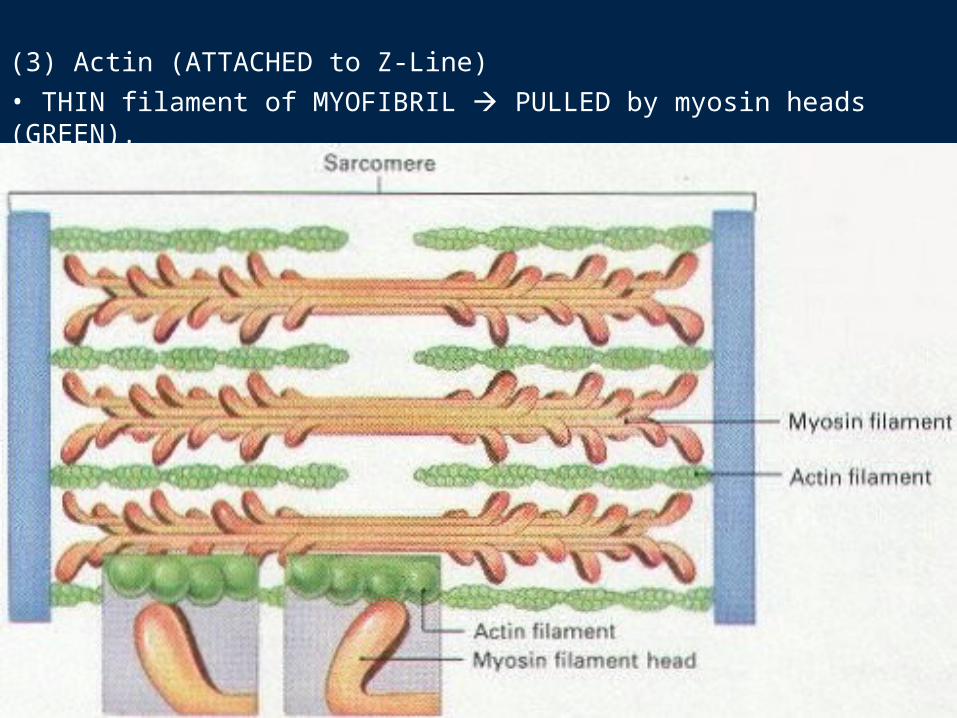
(3) Actin (ATTACHED to Z-Line)• THIN filament of MYOFIBRIL PULLED by myosin heads (GREEN).
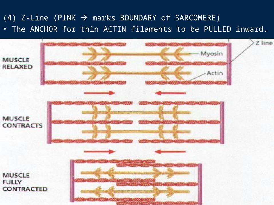
(4) Z-Line (PINK marks BOUNDARY of SARCOMERE)• The ANCHOR for thin ACTIN filaments to be PULLED inward.
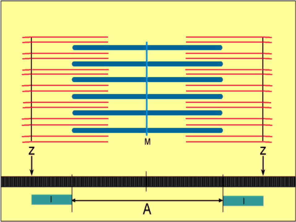
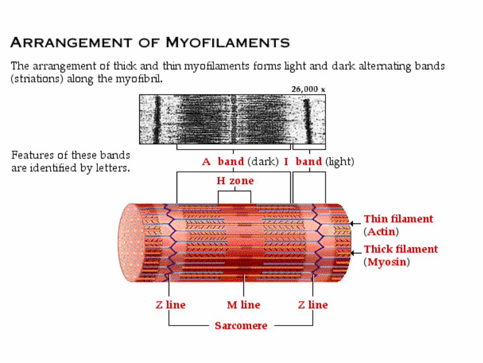
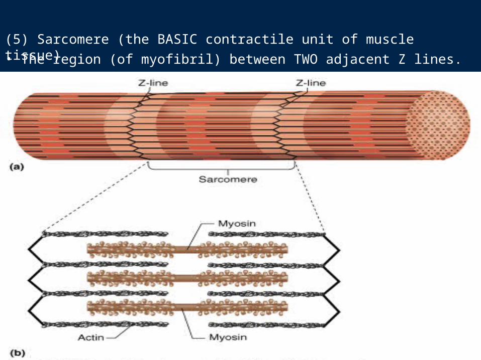
(5) Sarcomere (the BASIC contractile unit of muscle tissue)• The region (of myofibril) between TWO adjacent Z lines.
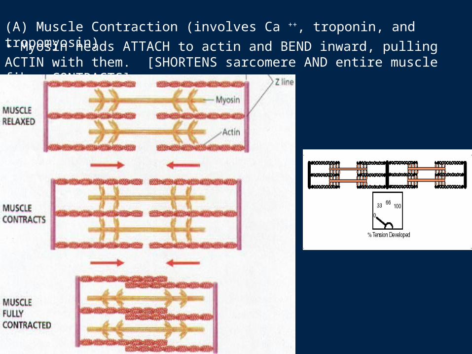
(A) Muscle Contraction (involves Ca ++, troponin, and tropomyosin)• Myosin heads ATTACH to actin and BEND inward, pulling ACTIN with them. [SHORTENS sarcomere AND entire muscle fiber CONTRACTS].
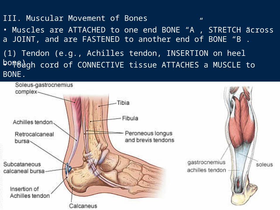
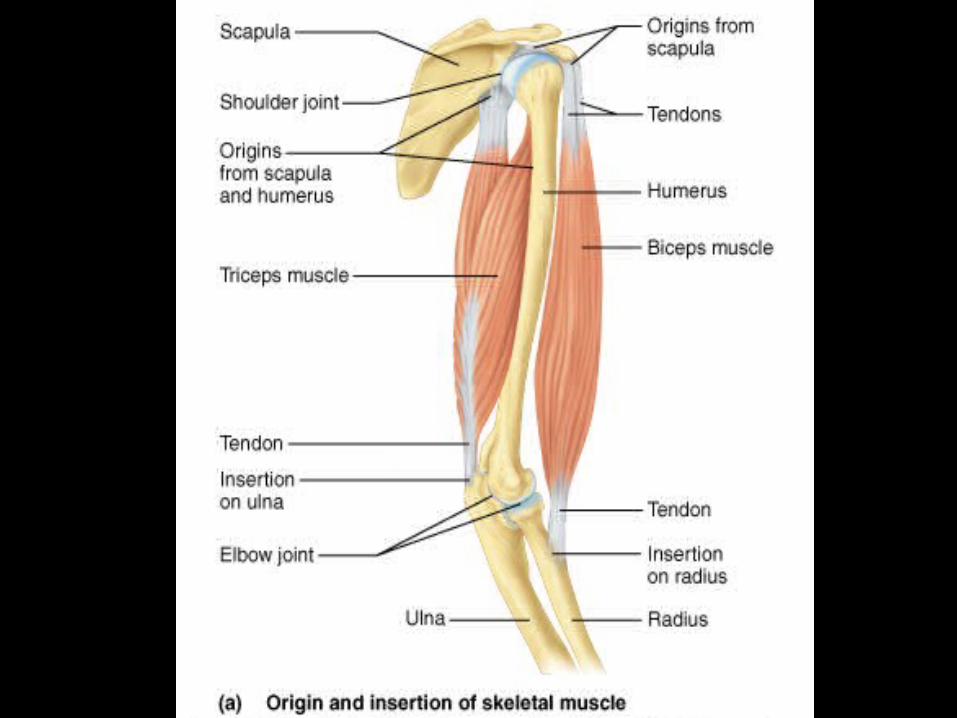
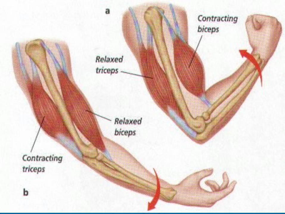
III. Muscular Movement of Bones• Muscles are ATTACHED to one end BONE “A”, STRETCH across a JOINT, and are FASTENED to another end of BONE “B”.
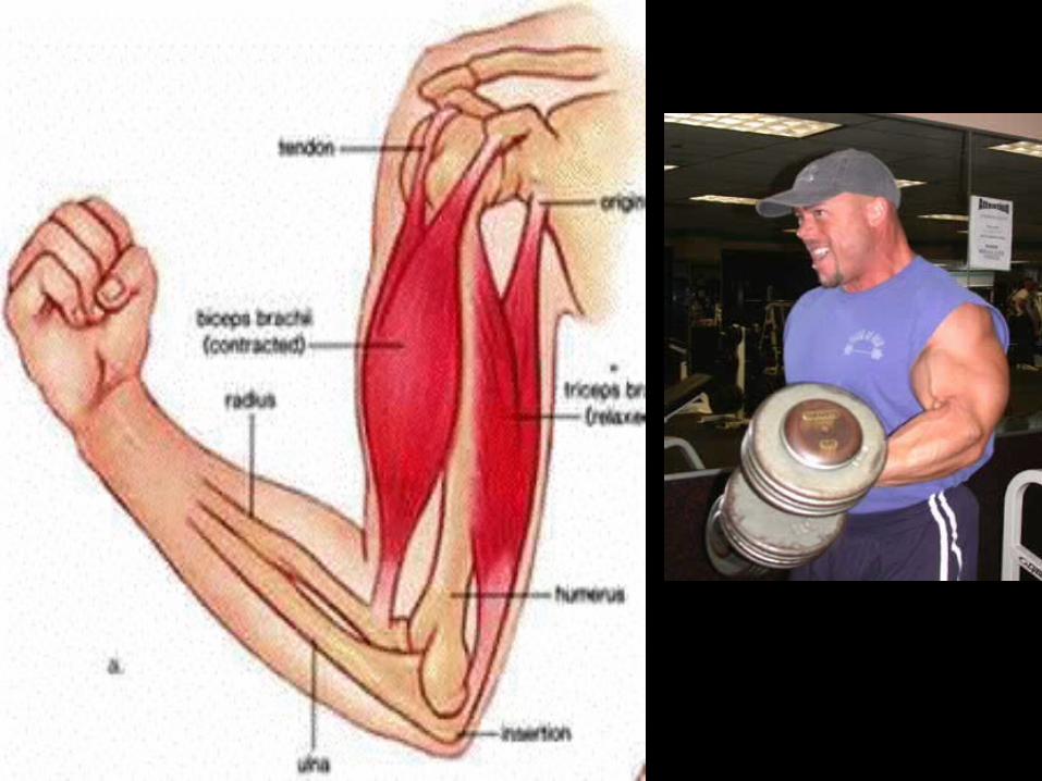
(1) Tendon (e.g., Achilles tendon, INSERTION on heel bone)• Tough cord of CONNECTIVE tissue ATTACHES a MUSCLE to BONE.
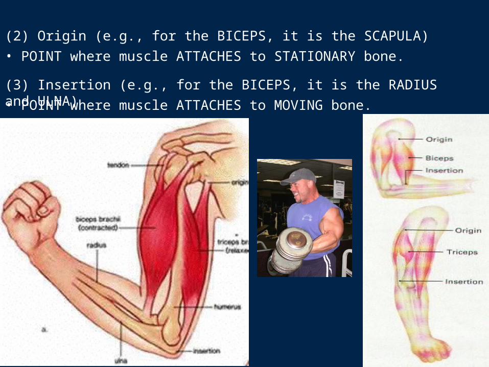
(2) Origin (e.g., for the BICEPS, it is the SCAPULA)• POINT where muscle ATTACHES to STATIONARY bone.
(3) Insertion (e.g., for the BICEPS, it is the RADIUS and ULNA)• POINT where muscle ATTACHES to MOVING bone.
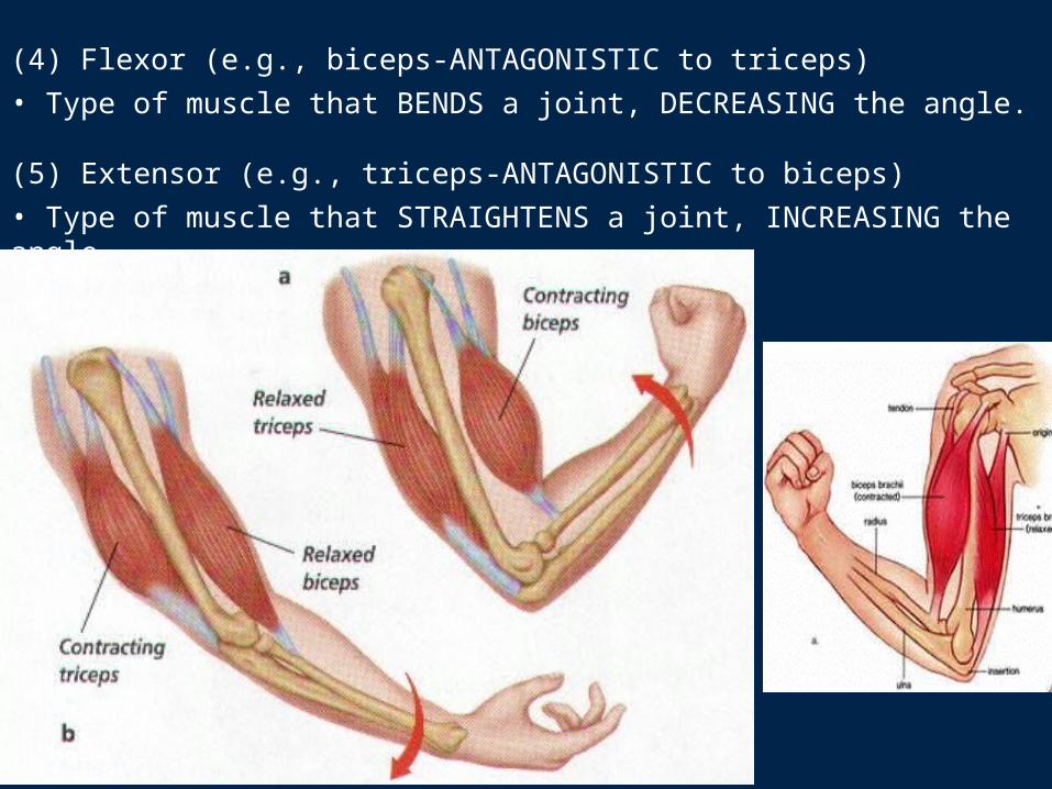
(4) Flexor (e.g., biceps-ANTAGONISTIC to triceps)• Type of muscle that BENDS a joint, DECREASING the angle.
(5) Extensor (e.g., triceps-ANTAGONISTIC to biceps)• Type of muscle that STRAIGHTENS a joint, INCREASING the angle.
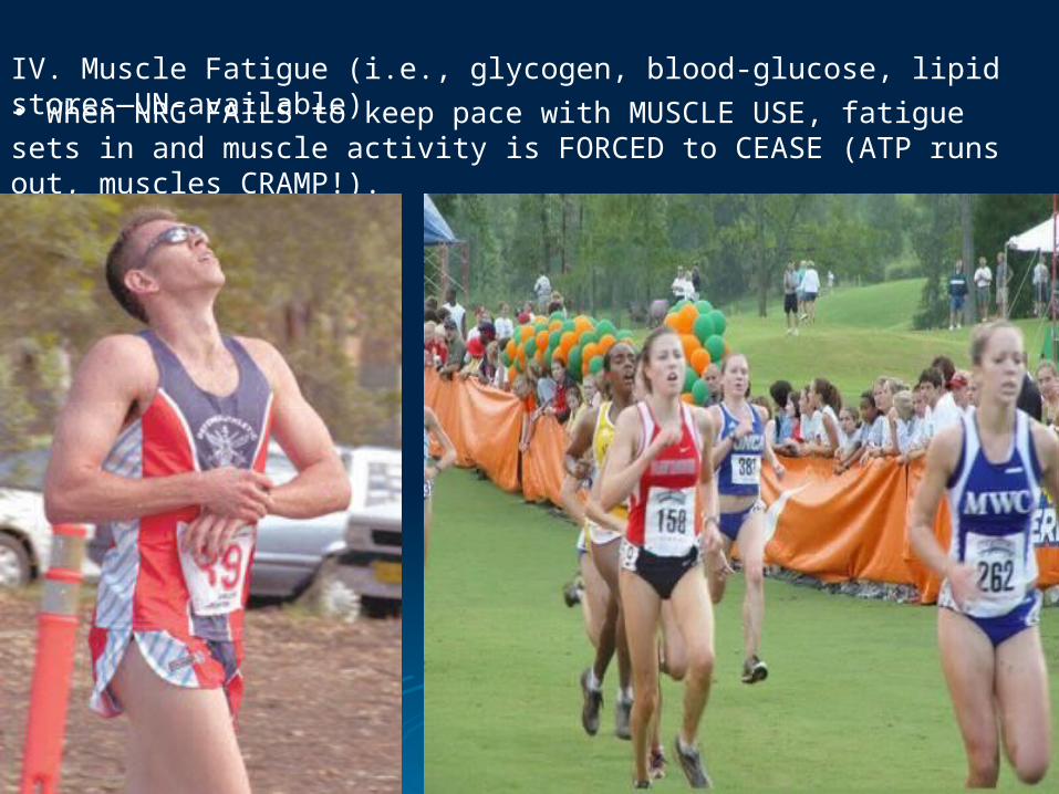
IV. Muscle Fatigue (i.e., glycogen, blood-glucose, lipid stores—UN-available)• When NRG FAILS to keep pace with MUSCLE USE, fatigue sets in and muscle activity is FORCED to CEASE (ATP runs out, muscles CRAMP!).
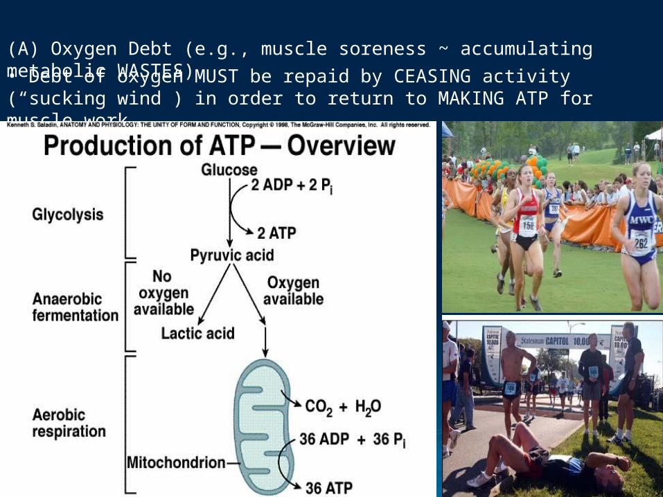
(A) Oxygen Debt (e.g., muscle soreness ~ accumulating metabolic WASTES)• Debt of oxygen MUST be repaid by CEASING activity (“sucking wind”) in order to return to MAKING ATP for muscle work.

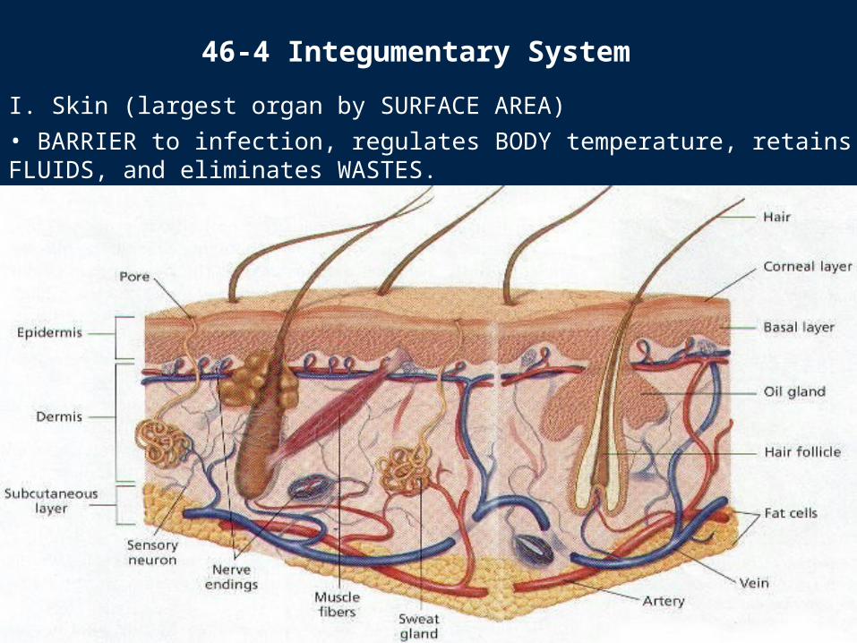
I. Skin (largest organ by SURFACE AREA)• BARRIER to infection, regulates BODY temperature, retains FLUIDS, and eliminates WASTES.
46-4 Integumentary System
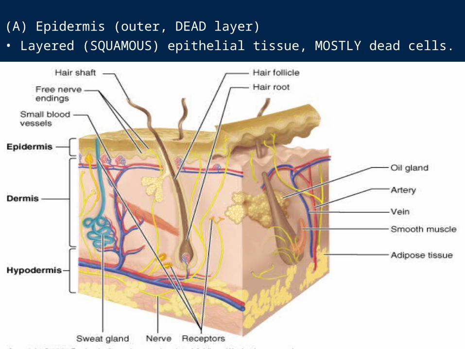
(A) Epidermis (outer, DEAD layer)• Layered (SQUAMOUS) epithelial tissue, MOSTLY dead cells.
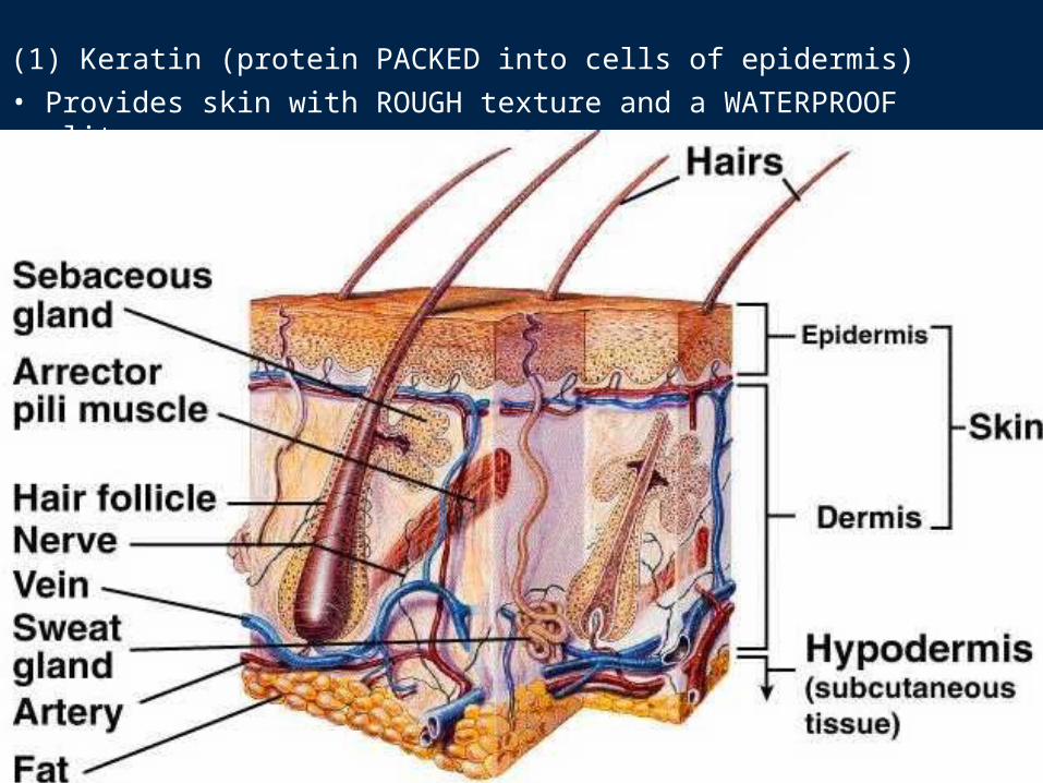
(1) Keratin (protein PACKED into cells of epidermis)• Provides skin with ROUGH texture and a WATERPROOF quality.
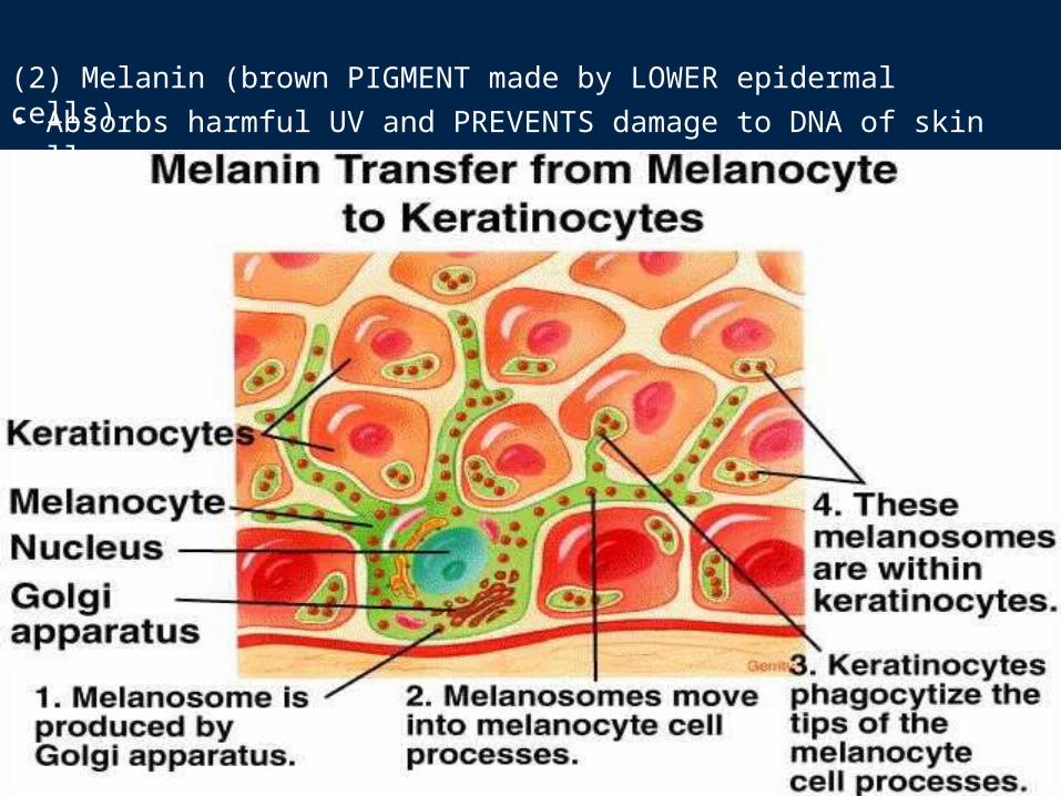
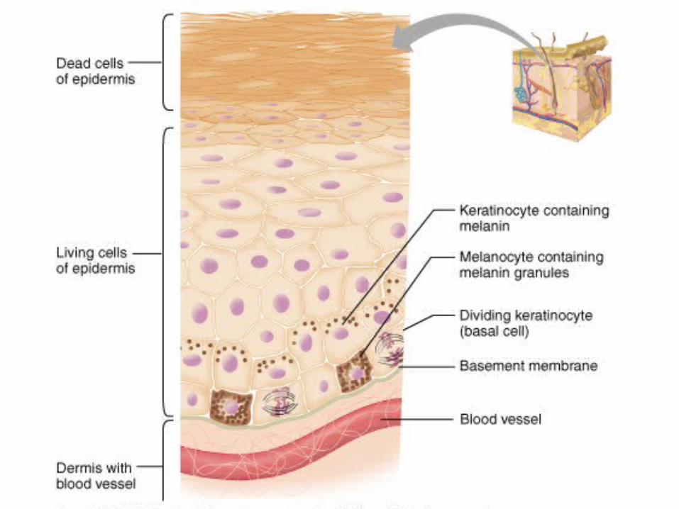
(2) Melanin (brown PIGMENT made by LOWER epidermal cells)• Absorbs harmful UV and PREVENTS damage to DNA of skin cells.
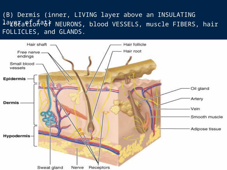
(B) Dermis (inner, LIVING layer above an INSULATING layer of fat)• Location of NEURONS, blood VESSELS, muscle FIBERS, hair FOLLICLES, and GLANDS.
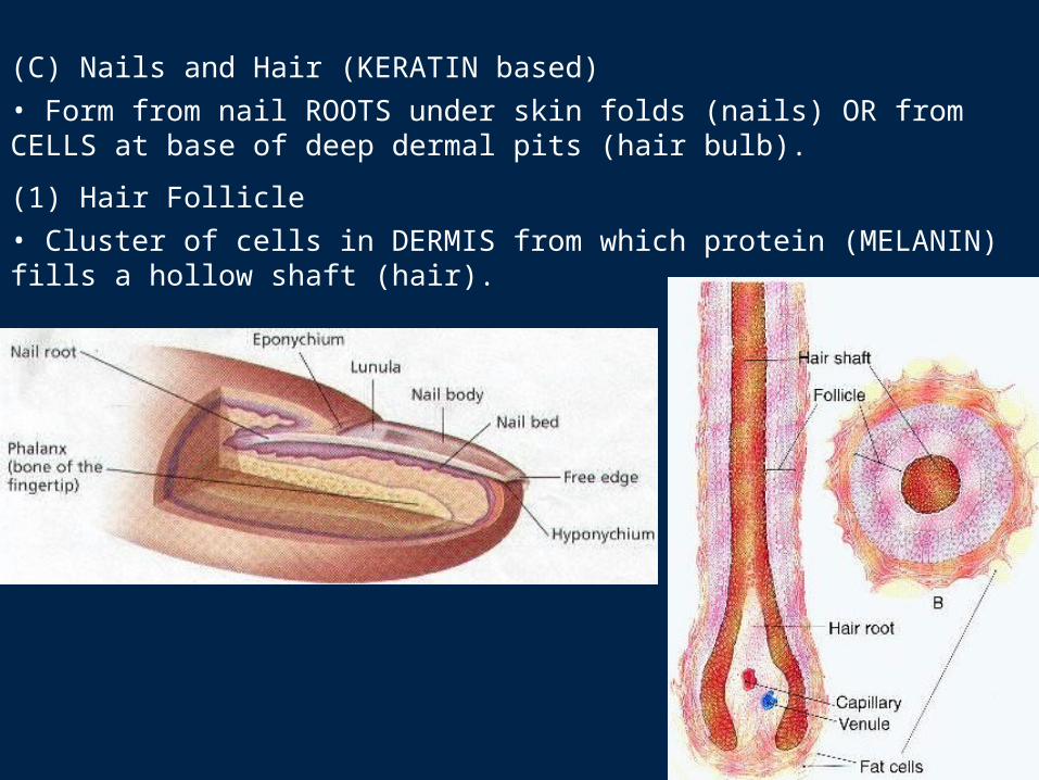
(C) Nails and Hair (KERATIN based)• Form from nail ROOTS under skin folds (nails) OR from CELLS at base of deep dermal pits (hair bulb).
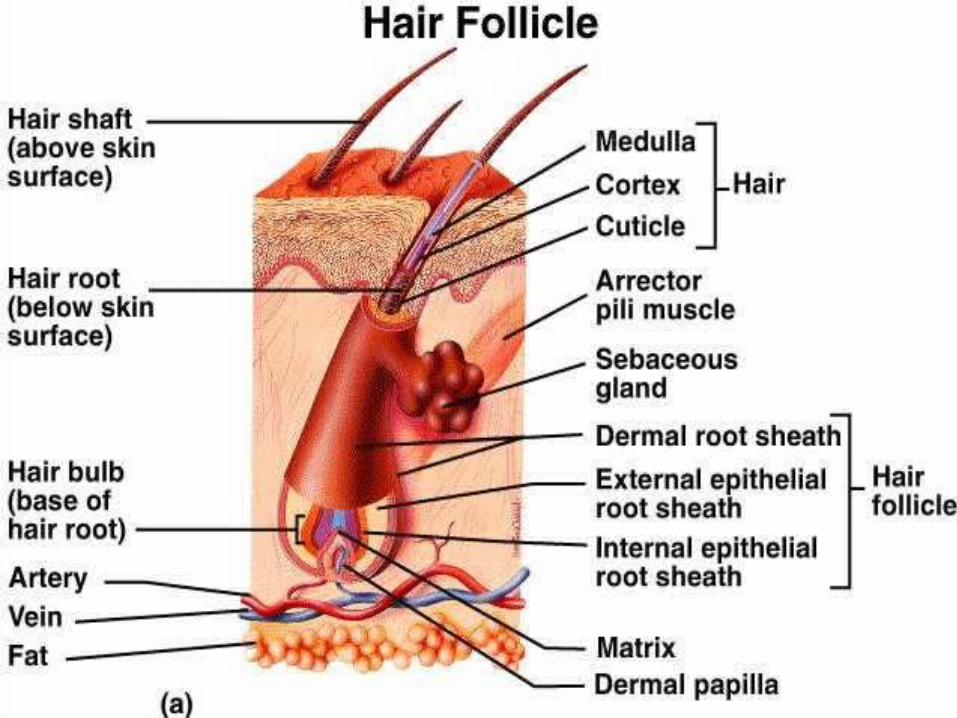
(1) Hair Follicle• Cluster of cells in DERMIS from which protein (MELANIN) fills a hollow shaft (hair).
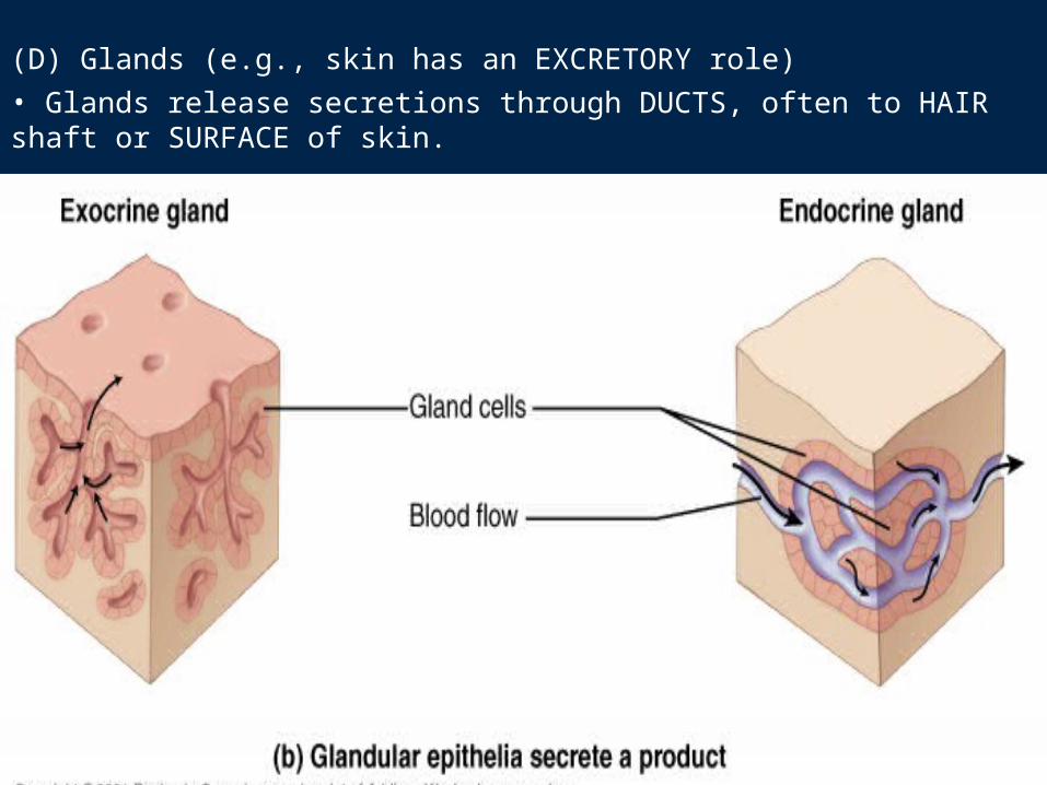
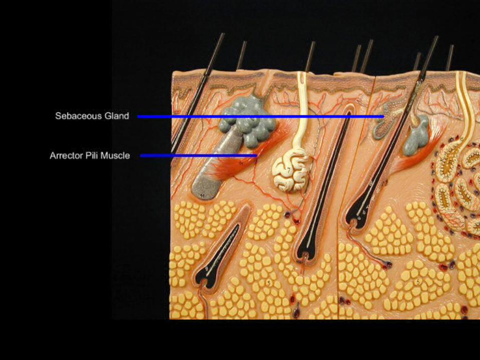
(D) Glands (e.g., skin has an EXCRETORY role)• Glands release secretions through DUCTS, often to HAIR shaft or SURFACE of skin.
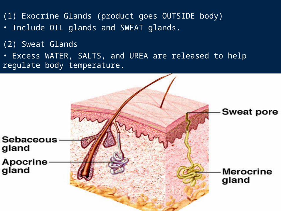
(1) Exocrine Glands (product goes OUTSIDE body)• Include OIL glands and SWEAT glands.
(2) Sweat Glands• Excess WATER, SALTS, and UREA are released to help regulate body temperature.
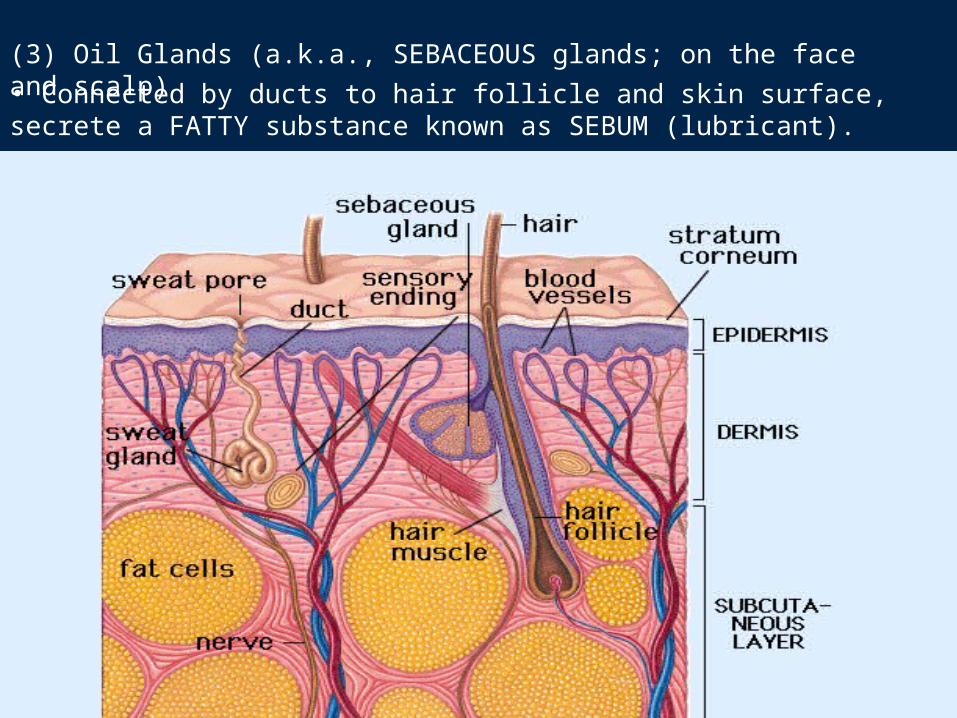
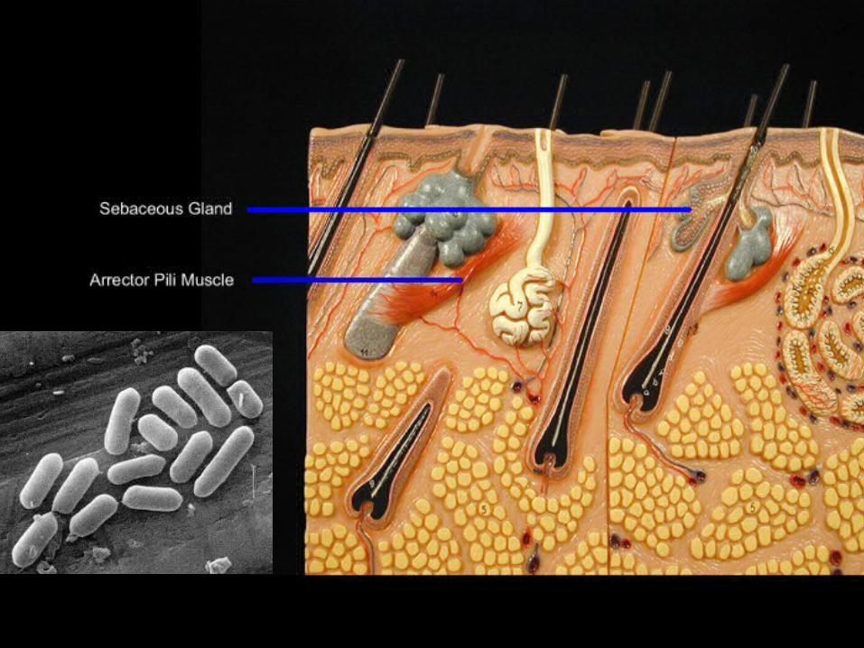
(3) Oil Glands (a.k.a., SEBACEOUS glands; on the face and scalp)• Connected by ducts to hair follicle and skin surface, secrete a FATTY substance known as SEBUM (lubricant).
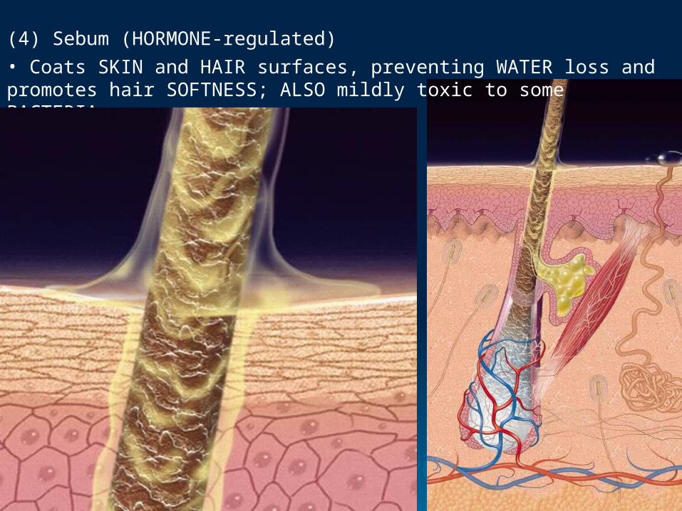
(4) Sebum (HORMONE-regulated)• Coats SKIN and HAIR surfaces, preventing WATER loss and promotes hair SOFTNESS; ALSO mildly toxic to some BACTERIA.
Extra Slides AND Answers for Critical Thinking Questions
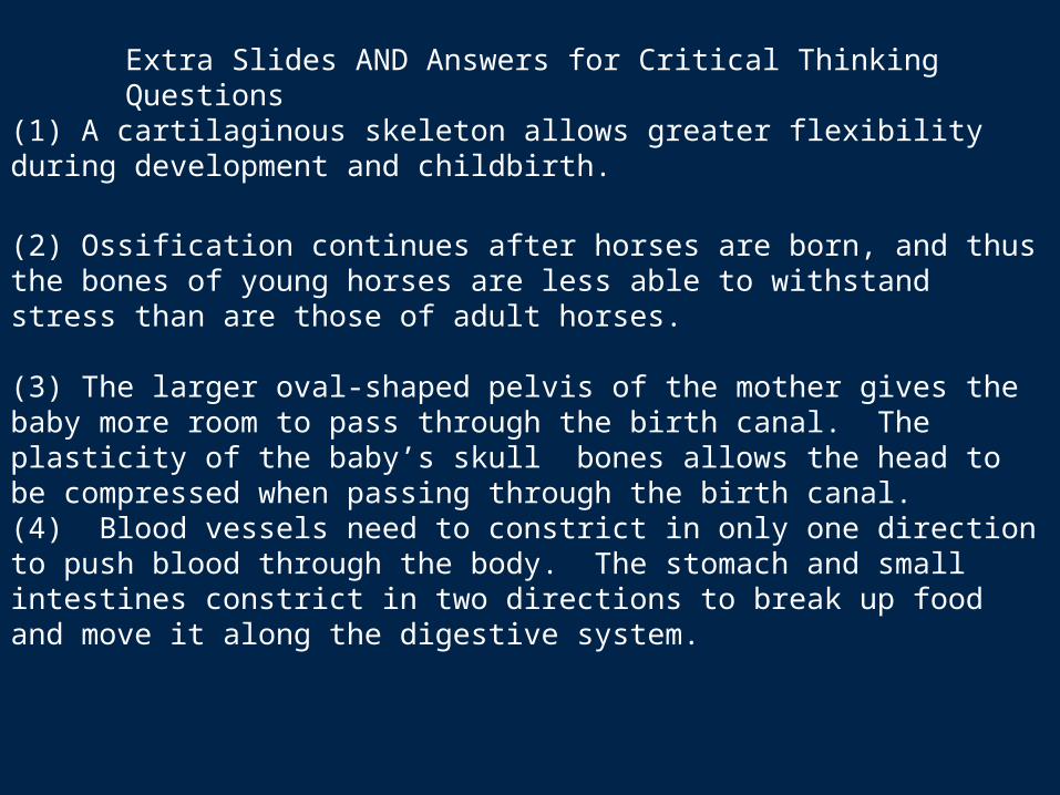
(1) A cartilaginous skeleton allows greater flexibility during development and childbirth.
(2) Ossification continues after horses are born, and thus the bones of young horses are less able to withstand stress than are those of adult horses.
(3) The larger oval-shaped pelvis of the mother gives the baby more room to pass through the birth canal. The plasticity of the baby’s skull bones allows the head to be compressed when passing through the birth canal.(4) Blood vessels need to constrict in only one direction to push blood through the body. The stomach and small intestines constrict in two directions to break up food and move it along the digestive system.
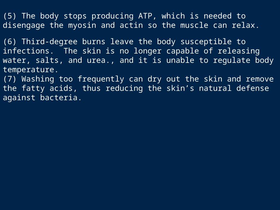
(5) The body stops producing ATP, which is needed to disengage the myosin and actin so the muscle can relax.
(6) Third-degree burns leave the body susceptible to infections. The skin is no longer capable of releasing water, salts, and urea., and it is unable to regulate body temperature.
(7) Washing too frequently can dry out the skin and remove the fatty acids, thus reducing the skin’s natural defense against bacteria.