Embed Size (px)
Citation preview

Chapter 3
Microbes and the Fossil Record: Selected
Topics in Paleomicrobiology
Alexandru M.F. Tomescu, Ashley A. Klymiuk, Kelly K.S. Matsunaga,
Alexander C. Bippus, and Glenn W.K. Shelton
Abstract The study of microbial fossils involves a broad array of disciplines and
covers a vast diversity of topics, of which we review a select few, summarizing the
state of the art. Microbes are found as body fossils preserved in different modes and
have also produced recognizable structures in the rock record (microbialites,
microborings). Study of the microbial fossil record and controversies arising from
it have provided the impetus for the assembly and refining of powerful sets of
criteria for recognition of bona fide microbial fossils. Different types of fossil
evidence concur in demonstrating that microbial life was present in the Archean,
close to 3.5 billion years ago. Early eukaryotes also fall within the microbial realm
and criteria developed for their recognition date the oldest unequivocal evidence
close to 2.0 billion years ago (Paleoproterozoic), but Archean microfossils >3
billion years old are strong contenders for earliest eukaryotes. In another dimension
of their contribution to the fossil record, microbes play ubiquitous roles in fossil
preservation, from facilitating authigenic mineralization to replicating soft tissue
with extracellular polymeric substances, forming biofilms that inhibit decay of
biological material, or stabilizing sediment interfaces. Finally, studies of the micro-
bial fossil record are relevant to profound, perennial questions that have puzzled
humanity and science—they provide the only direct window onto the beginnings
and early evolution of life; and the methods and criteria developed for recognizing
ancient, inconspicuous traces of life have yielded an approach directly applicable to
the search for traces of life on other worlds.
A.M.F. Tomescu (*) • K.K.S. Matsunaga • A.C. Bippus • G.W.K. Shelton
Department of Biological Sciences, Humboldt State University, Arcata, CA 95521, USA
e-mail: [email protected]
A.A. Klymiuk
Department of Ecology and Evolutionary Biology, University of Kansas, Lawrence, KS 66047,
USA
© Springer International Publishing Switzerland 2016
C.J. Hurst (ed.), Their World: A Diversity of Microbial Environments, Advances inEnvironmental Microbiology 1, DOI 10.1007/978-3-319-28071-4_3
69

3.1 Microbial Fossils: A Vast Field of Study
Knowledge of the microbial fossil record has expanded tremendously in more than
50 years since early discoveries (e.g., Tyler and Barghoorn 1954; Barghoorn and
Tyler 1965), both in depth—geologic time—and breadth—types of organisms,
modes of preservation, and types of fossil evidence. Along with the new discoveries
of fossil microbes and microbially induced structures, and keeping pace with
technological advances in analytical tools, the paleontological community devel-
oped and expanded the set of methods used to study these fossils and refined the
types of questions addressed, as well as the criteria applied to them. At the same
time, the community of scientists itself broadened its scope and expanded its ranks
to include paleobiology, geobiology, geochemistry, taphonomy, and other areas of
research in its sphere of investigation. As a result of this explosive growth,
paleomicrobiology is today just as vast an area of science as its “neo” counterpart
and could itself be the subject of a multivolume book. That is why for this chapter,
we had to select only some of the topics of major interest in paleomicrobiology
which we review to summarize the current state of the art.
One of the topics is the recognition of microbial fossils and the criteria used for it
(Sect. 3.3). These provide the foundation of all work involving microbial body
fossils and are especially relevant to the search for the earliest traces of life. The
development of these criteria over time, by discovery and critical scrutiny of
increasingly older Precambrian microbial fossils, provides a telling example of
the workings of science, in general, and paleobiology, in particular, as an objective
empirical approach to questions about nature. As a logical follow-up on the criteria
for recognition of microbial body fossils, we discuss microbially induced sedimen-
tary structures and other traces of microbial activity (microbially induced struc-
tures), their classification, and criteria of recognition. These provide a powerful
complement to the study of the microbial fossil record, even in the absence of body
fossils, and are active and growing fields of inquiry. This section is prefaced by a
review of microbial fossil preservation (Sect. 3.2), which provides a broader
context for the different aspects comprising the recognition of the fossils. The
next topic involves the earliest records of life and a review of the Archean fossil
record (Sect. 3.4). Aside from pushing back in deep time the history of life on Earth,
these fossil discoveries and the controversies they engendered were crucial in
shaping both the methods and the theoretical bases for the study of microbial
fossils. As a part of this topic, we summarize a few of the now-classic debates
which animated (or are still animating) the scientific community and provided
much of the impetus for the development of a powerful set of criteria for microbial
fossil recognition. Another topic covers the rise of early eukaryotes as reflected by
the microbial fossil record, with a discussion of the criteria used to recognize them
and a survey of the earliest types (Sect. 3.5). Next, we review the role of microbially
mediated processes in the various fossilization pathways of other organisms—
microbial-associated mineralization, plant, animal, and trace fossil preservation
(Sect. 3.6)—and the fossil record of symbioses that involve microbial participants
70 A.M.F. Tomescu et al.

(Sect. 3.7). The chapter ends with a discussion of future directions of investigation
in the study of microbial fossils and of the role of paleomicrobiology in the study
of life.
Throughout the chapter, we focus mainly on the record of body fossils, with
some detours into geochemistry and sedimentology for discussions of biogenicity,
microbially induced structures, and fossilization processes. The survey of the
prokaryote and eukaryotic fossil record is limited to early occurrences—Archean
for the former (4.0–2.5 Ga ¼ billion years) and Paleoproterozoic and
Mesoproterozoic (2.5–1.0 Ga) for the latter. However, the discussions of the roles
of microbes in fossilization and of the fossil record of symbioses draw on examples
from throughout the geologic time scale.
3.2 Microbial Fossil Preservation
Traces of microbial life occur as (1) body fossils, which can be preserved in several
modes, (2) structures (micro- and macroscopic) generated by microbial presence
and activities, and (3) chemical compounds present in the rock record as a result of
microbial metabolism (chemical biosignatures) (Fig. 3.1). This chapter deals
mostly with body fossils and, to a somewhat lesser extent, with microbially induced
structures. While chemical biosignatures can offer very useful insights into early
life on Earth and investigations of biogenicity of candidate microfossils (Brasier
and Wacey 2012), in this chapter, the impressive body of work produced by
geochemists [e.g., Knoll et al. (2012) and references therein] is touched upon
only lightly.
3.2.1 Body Fossils
The modes of preservation of microbial body fossils parallel those described for
plant fossils (e.g. Schopf 1975; Stewart and Rothwell 1993) and include perminer-alization (also known as petrifaction), coalified compression, authigenic or
duripartic preservation, and cellular replacement with minerals. In perminera-
lization, minerals (usually calcium carbonate, silica, iron sulfide) precipitate from
solutions inside and around cells, so the organisms end up incorporated in a mineral
matrix and preserved three dimensionally, sometimes in exquisite detail. The
quality of cellular preservation depends on the extent of decomposition of the
organisms preceding the permineralization phase. Many early prokaryotes are
preserved as permineralizations. In filamentous types, such as cyanobacteria,
permineralized specimens often preserve mainly the external cellular envelopes
(sheaths), whereas cells and their contents are altered to various degrees or not
preserved at all (e.g., Eoschizothrix; Seong-Joo and Golubic 1998) (Fig. 3.1a, b).
This is consistent with the results of chemical, structural (Helm et al. 2000), and
3 Microbes and the Fossil Record: Selected Topics in Paleomicrobiology 71
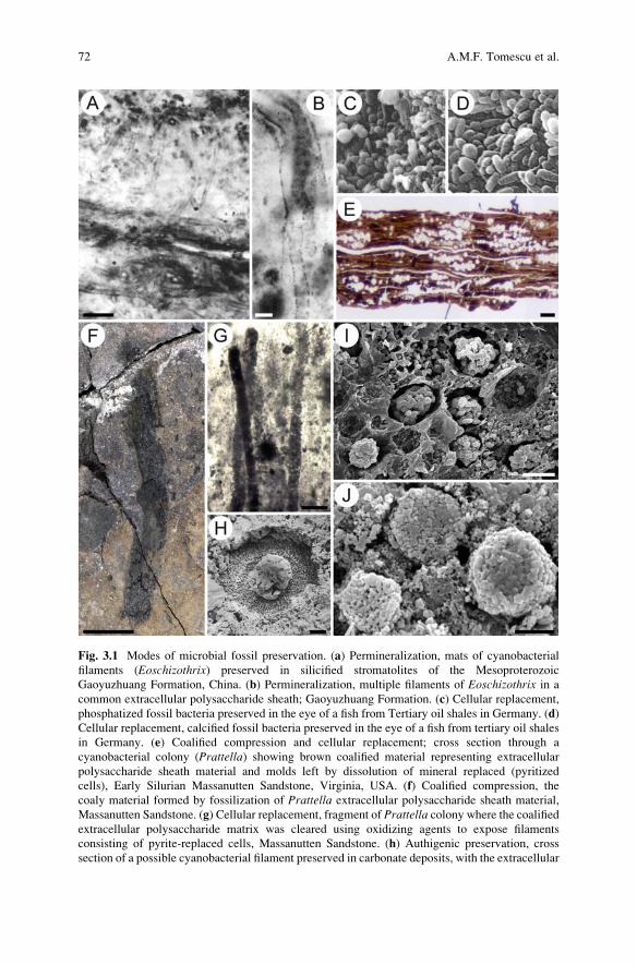
Fig. 3.1 Modes of microbial fossil preservation. (a) Permineralization, mats of cyanobacterial
filaments (Eoschizothrix) preserved in silicified stromatolites of the Mesoproterozoic
Gaoyuzhuang Formation, China. (b) Permineralization, multiple filaments of Eoschizothrix in a
common extracellular polysaccharide sheath; Gaoyuzhuang Formation. (c) Cellular replacement,
phosphatized fossil bacteria preserved in the eye of a fish from Tertiary oil shales in Germany. (d)
Cellular replacement, calcified fossil bacteria preserved in the eye of a fish from tertiary oil shales
in Germany. (e) Coalified compression and cellular replacement; cross section through a
cyanobacterial colony (Prattella) showing brown coalified material representing extracellular
polysaccharide sheath material and molds left by dissolution of mineral replaced (pyritized
cells), Early Silurian Massanutten Sandstone, Virginia, USA. (f) Coalified compression, the
coaly material formed by fossilization of Prattella extracellular polysaccharide sheath material,
Massanutten Sandstone. (g) Cellular replacement, fragment of Prattella colony where the coalifiedextracellular polysaccharide matrix was cleared using oxidizing agents to expose filaments
consisting of pyrite-replaced cells, Massanutten Sandstone. (h) Authigenic preservation, cross
section of a possible cyanobacterial filament preserved in carbonate deposits, with the extracellular
72 A.M.F. Tomescu et al.

experimental taphonomic (Bartley 1996) studies, which have stressed the higher
resistance to degradation of extracellular polymeric substances (i.e., sheath and
slime) in contrast that of the cell contents.
Coalified compressions are formed when the layers of sediment that incorporate
the organisms are subjected to lithostatic pressure during rock forming processes
(diagenesis). The pressure and temperature associated with burial in the Earth’scrust induce changes in the geometry and chemistry of cells along a gradient of
coalification of the carbonaceous material. For unicellular microfossils, a high
degree of coalification can lead to complete obliteration of diagnostic features, to
the point of rendering them unrecognizable as biogenic objects. However, lesser
degrees of coalification can preserve diagnostic features even down to the ultra-
structure level, as in the case of some early unicellular eukaryotes (Javaux
et al. 2001, 2004) (Figs. 3.9e, f, 3.10e, 3.11b), while also increasing the preservation
potential of fossils by rendering their organic compounds more chemically inert.
Sometimes, microbial colonies can form compressions, as in the case of
cyanobacterial colonies whose copious extracellular sheath material is coalified
(Tomescu et al. 2006, 2008) (Fig. 3.1e, f).
Authigenic preservation refers to removal of the organic material previously
enclosed in rock (by oxidation, decomposition) and its replacement with secondary
material (precipitated minerals or sediment) that forms casts, whereas duripartic
preservation involves the precipitation of minerals due to metabolic processes of
the organisms that are fossilized. Duripartic mineral precipitation can occur in the
cell walls, in the extracellular sheaths of colonies, or around the organisms, forming
molds that can preserve cell-level structural details. Various minerals are known to
form on microbial cell surfaces as a consequence of interactions between the
microbial metabolism and the chemistry of its environment (reviewed by Southam
and Donald 1999). Many fossil cyanobacteria are preserved as calcium carbonate
(micrite) rinds that coated the organisms (e.g.,Girvanella; Golubic and Knoll 1993)(Fig. 3.1h), which corresponds to a combination of authigenic and duripartic
preservation. Cellular replacement is a cell-to-cell process in which diagenetic
minerals precipitate inside individual cells, replacing their content. Different by
its discrete nature from both authigenic and duripartic preservation which involve
wholesale processes, cellular replacement is nevertheless more akin to authigenic
preservation. Pyrite is widespread in cellular replacement (e.g., Munnecke
et al. 2001; Kremer and Kazmierczak 2005) (Fig. 3.1i, j), but the list of minerals
⁄�
Fig. 3.1 (continued) sheath preserved as micrite and the filament lumen filled with sparry calcite.
(i) Prattella SEM of framboidal pyrite aggregates replacing individual cyanobacterial cells of
Prattella and occupying molds in the coalified extracellular polysaccharide matrix, Massanutten
Sandstone. (j) Cellular replacement, coccoid cells of a cyanobacterial mat replaced by framboidal
pyrite aggregates, Silurian, Poland. Scale bars: (a) 50 μm, (b) 10 μm, (c) the bacterial cells are
0.5–1.5 μm, (d) the bacterial cells are 0.5–1 μm, (e) 20 μm, (f) 1 cm, (g) 50 μm, (h) 10 μm, (i) 5 μm,
(j) 5 μm. Credits—images used with permission from (a) John Wiley & Sons (Seong-Joo and
Golubic 1998). (c), (d) Blackwell Science Ltd. (Liebig 2001). (h) Blackwell Science Ltd. (Golubic
and Knoll 1993). (j) Society for Sedimentary Geology (Kremer and Kazmierczak 2005)
3 Microbes and the Fossil Record: Selected Topics in Paleomicrobiology 73

is broad and includes apatite, calcite, siderite, hematite, and silica (Liebig 2001;
Noffke et al. 2013b) (Fig. 3.1c, d).
In some instances, modes of preservation are combined, as in the case of the
cyanobacterium Prattella (Tomescu et al. 2006, 2009) in which the macroscopic
colonies of multitrichomous filaments are enveloped in high amounts of extracel-
lular slime which forms coalified compressions, but within these compressions
individual cyanobacterial cells are preserved by cellular replacement with
framboidal pyrite aggregates (Fig. 3.1e, f, g, i).
3.2.2 Microbially Induced Structures
Microbially induced structures are structures produced by interactions between
microbes and the sediment or rock. The different types of microbially induced
structures encompass a wide spectrum of morphologies and range in size from the
submillimeter scale up several orders of magnitude; some can form associations
and layers with sizes ranging up to the kilometer scale. Microbially induced
structures are known in large numbers from Proterozoic and Phanerozoic rocks,
but their Archean record is less extensive (Awramik and Grey 2005; Noffke
et al. 2003, 2006, 2013a). Two broad categories of microbially induced structures
can be distinguished: microbialites and microborings.
3.2.2.1 Microbialites
Microbialites (Burne and Moore 1987) are constructional organosedimentary struc-
tures formed as a result of interactions between microbial mats and sediment.
Depending on whether their formation involves the precipitation of minerals or
not, microbialites are further divided into stromatolites (Kalkowsky 1908) and
microbially induced sedimentary structures (MISS), respectively (Noffke and
Awramik 2013).
Microbially Induced Sedimentary Structures
Because they do not involve mineral precipitation, MISS do not form thick or tall
buildups like the stromatolites; instead, they have a more flattened,
two-dimensional form. Microbial mats form at the interface between their substrate
(usually the sediment surface) and water or air, and MISS formed as a result of
interactions between the microbial mat and sediment (stabilizing, trapping) mark
the transient location of such surfaces. Modern analogues of MISS found in tidal
siliciclastic and terrestrial environments, which host a wide range of types with
different morphologies (Noffke et al. 1996; Beraldi-Campesi and Garcia-Pichel
74 A.M.F. Tomescu et al.

2011), allow us to understand the processes that lead to the formation of MISS
encountered in the fossil record (Schieber et al. 2007).
The types of interactions between mats and sediment that lead to both MISS and
stromatolite formation are binding and biostabilization, baffling and trapping, and
growth (Noffke and Awramik 2013). Binding of microbial populations by cooper-
ative secretion of extracellular polymeric substances leads to formation of a micro-
bial mat. Once established, the mat alters the structure and physical properties of the
sediment, stabilizing it (biostabilization) and, thus, reducing the effects of erosion
(e.g., Fang et al. 2014). Sudden changes of fluid dynamic patterns at the interface
between the sediment and water (or air) that increase the risk of bacterial mat
erosion trigger increased or renewed biostabilizing activity in the microbial mat.
Baffling and trapping occur when microbial mats baffle the water current around
them, causing suspended particles to sediment and become trapped in the mat.
Growth is the lateral and vertical expansion of the microbial mat by production of
more extracellular polymeric substances or more cells, and sediment may become
trapped within the mat while growth and associated binding are taking place.
MISS are ultimately formed when the surfaces hosting microbial mats are buried
in sediment and lithified; then, the finer-grained layer trapped and bound by the
microbial mat serves to separate two sedimentary beds and assist the preservation of
any surface structure (Noffke 2009; Noffke and Awramik 2013). Thus, in the
sedimentary record, MISS are recovered as structures on bedding planes, associated
with recognizable microscale sedimentary patterns immediately beneath the bed-
ding plane (laminae of organic matter concentration, grain size sorting, heavy
mineral concentrations, etc., microtextures—Noffke 2009) (Figs. 3.2a–c, 3.3a,
3.11f). The wide morphological variety of MISS is the result of different sedimen-
tary environments, substrates (sediment), types of interaction, and taphonomic
processes. Because these interactions and processes have not changed significantly
over 3.5 billion years, the morphologies of MISS in the fossil record are directly
comparable with microbial mat-induced sedimentary structures documented in
modern environments (Noffke et al. 2013b). Although identified as such relatively
recently, MISS have received a lot of attention as traces of early life and a
considerable body of literature on their genesis, recognition, classification, and
geologic record has accumulated (e.g., Noffke 2009, 2010; Noffke and Awramik
2013; Noffke et al. 2013b).
Stromatolites
We use the term stromatolites here to designate any sedimentary structure formed
as a result of microbial mats baffling, biostabilizing, and trapping sediment, in
association with mineral precipitation. In other words, stromatolites can be
regarded as MISS cemented by mineral precipitation. On the centimeter-to-meter
scale, these structures exhibit varied morphologies, including wavy laminations,
domes, and branched or unbranched columns (Awramik and Grey 2005)
(Figs. 3.3b–d, 3.6b, 3.7 h, 3.8f, g), some of which have received specific names—
3 Microbes and the Fossil Record: Selected Topics in Paleomicrobiology 75

e.g., dendrolites and thrombolites (Aitken 1967; Kennard and James 1986; Shapiro
2000). However, here we group all these different morphologies under the umbrella
term stromatolite as they all share a laminated organization (the precipitated
mineral is often calcium carbonate) and origin involving the activity of microbial
mats (Shapiro 2000; Awramik and Grey 2005). Reitner et al. (2011) provide a
comprehensive review of stromatolite research.
The most widely accepted mechanism for stromatolite formation involves sed-
iments that are trapped and bound within microbial mats by the same basic
mechanisms that form MISS and carbonate or some other type of mineral precip-
itation that cements the layer thus formed (Noffke and Awramik 2013). Over time,
the microbial mat grows above the cemented portion forming a new layer which is,
in turn, cemented, leading to upward growth of the stromatolite. The repetition of
recolonization of the upper stromatolite surface by the microbial mat and subse-
quent cementation with precipitated minerals produces the characteristic laminated
structure of stromatolites. Laboratory studies suggest that the extracellular poly-
meric substances produced by the microbial mats play an important role in carbon-
ate precipitation (Altermann et al. 2006; Dupraz et al. 2009) and that heterotrophic
members of microbial mat communities may initiate mineral precipitation (Noffke
and Awramik 2013). If the precipitating minerals permineralize the microbial cells
that form the mats, body fossils can be associated with the stromatolites. Although
such occurrences tend to be rare (Wacey 2009), they have been documented both in
the fossil record (e.g., Schopf and Blacic 1971; Schopf and Sovietov 1976; Knoll
and Golubic 1979) and in modern stromatolites (Kremer et al. 2012a).
Structures described from shallow, hypersaline marine environments, such as
Shark Bay in Western Australia (e.g., Awramik and Riding 1988), are often
presented as good modern analogues for ancient stromatolites (Fig. 3.2f). This is
because these structures are formed by microbial mats that trap and bind sediment,
producing morphologies similar to those of stromatolites from the geologic record
(Awramik and Grey 2005; Awramik 2006). However, most modern structures
proposed as stromatolites are primarily formed by binding or trapping of sediments,
without significant precipitation, whereas mineral (usually carbonate) precipitation
is a prominent feature of ancient stromatolites (Kazmierczak and Kempe 2006;
Noffke and Awramik 2013). To date, the only modern stromatolite analogues
formed by both microbial mat activity and carbonate precipitation are those
described from caldera lakes in Tonga (south Pacific) by Kazmierczak and
Kempe (2006) and Kremer et al. (2012a) (Fig. 3.2d, e). Kazmierczak and Kempe
(2006) propose that the highly alkaline chemistry of the caldera lakes is similar to
the chemistry of Precambrian seas that hosted the wealth of stromatolites
documented in the geologic record and that the rarity of this particular type of
conditions in modern marine environments should account for the infrequency of
modern carbonate-precipitating stromatolites and the presence of only comparable
but nonprecipitating structures in modern benthic environments.
76 A.M.F. Tomescu et al.

Fig. 3.2 Microbially induced structures. (a) Wrinkle structures representing buried microbial
mats, Archean Mozaan Group, Pongola Supergroup, South Africa. (b) Wrinkle structures
representing in situ preserved, thin microbial mats, Neoproterozoic Nama Group, Namibia. (c)
Landscape-scale preservation of MISS on a hill side; tidal flat morphology resulting from partial
erosion of a mat-stabilized sedimentary surface—the raised flat-topped areas are ancient microbial
mats and the ripple marked depressions represent areas where the mats were eroded, Cretaceous
Dakota Sandstone, Colorado, USA. (d) Stromatolites from caldera lakes on Niuafo‘ou Island
(Tonga Archipelago, south Pacific), recently recognized as the closest modern analogues of
Precambrian Stromatolites. (e) Vertical sections through Niuafo‘ou Island stromatolites showing
the variety of internal structures. (f) Stromatolites such as these, from Shark Bay (Western
Australia), were recognized early as modern analogues of Precambrian stromatolites. Scale bars:
(a) 2 cm, (b) 5 cm, (d) hammer for scale 28 cm, (f) measuring pole painted in 10 cm intervals.
Credits—images used with permission from (a) Geological Society of America (Noffke
et al. 2003). (b) Elsevier Science Publishers (Noffke 2009). (c) Society for Sedimentary Geology
(Noffke and Chafetz 2012). (d) and (e) Springer-Verlag (Kazmierczak and Kempe 2006). (f)
Geological Society of America (Noffke and Awramik 2013)
3 Microbes and the Fossil Record: Selected Topics in Paleomicrobiology 77

3.2.2.2 Microborings
Microborings are microscopic, tubular, usually branched cavities that record the
activity of euendolithic microbes. Although the differences are sometimes blurred,
only some of the rock-inhabiting (endolithic) microbes, the euendoliths, actively
bore into rock, whereas others occupy preexisting fissures and pore spaces of the
rock (chasmoendoliths and cryptoendoliths, respectively) (Golubic et al. 1981;
McLoughlin et al. 2007). In the rock record, microborings are found in both
sedimentary (Campbell 1982; McLoughlin et al. 2007) (Fig. 3.8h) and volcanic
rocks (Furnes et al. 2004, 2007; McLoughlin et al. 2012) (Figs. 3.4d–f, 3.7d, g,
3.8i). Living analogues have also been discovered in a wide range of environments,
including near-surface sedimentary rocks (Knoll et al. 1986) and volcanic glass
(Thorseth et al. 1991; Fisk et al. 1998).
Modern euendoliths employ several metabolic strategies, including
photoautotrophy in near-surface environments and chemolithoautotrophy in deeper
endolithic environments (McLoughlin et al. 2007). In carbonate rocks, endoliths
Fig. 3.3 Microbially induced structures. (a) Fine carbonaceous laminations with one layer folded
over itself and overlain by a deposit of microbial mat-like fragments, Archean Kromberg Forma-
tion, Onverwacht Group, South Africa. (b) Vertical section through stromatolite deposit,
Neoproterozoic Shisanlitai Formation, China. (c) Stromatolite in the Archean Tumbiana Forma-
tion, Fortescue Group, Western Australia. (d) Complex lamination at several scales in a thin
section through Tumbiana Formation stromatolite. Scale bars: (a) 500 μm, (b) 2 cm, (c) 5 cm, (d)
5 mm. Credits—images used with permission from (a) Elsevier Science Publishers (Walsh 1992).
(b) Geological Society of America (Noffke and Awramik 2013). (c) Elsevier Science Publishers
(Lepot et al. 2009b). (d) Springer Science + Business Media B.V. (Wacey 2009)
78 A.M.F. Tomescu et al.

Fig. 3.4 (a) Chains of non-syngenetic endolithic fossilized coccoidal cells (arrows) penetrating acrack around a magnetite grain in Archean rocks of the Isua Greenstone Belt, Greenland. (b)
Indigenous and syngenetic heterotrophic bacteria preserved in fossil extracellular polysaccharide
sheaths of a cyanobacterial colony (Prattella); the bacterial cells are exposed in fresh breaks of thecarbonaceous material (as seen here in SEM) which demonstrate that they did not penetrate
through fissures at a later time; the bacterial cells also exhibit plastic deformation characteristic
of soft, organic bodies (toward bottom right), corroborating hypotheses of biogenicity, Early
Silurian Massanutten Sandstone, Virginia, USA. (c) Ambient inclusion trail with terminal pyrite
grain and jagged tube edges, Archean Apex chert, Warrawoona Group, Western Australia. (d), (e),
and (f) Microbial bioerosion structures (microborings) in volcanic glass of the Troodos ophiolite
(Cretaceous, Cyprus), (d) Tubulohyalichnus spiralis, (e) Tubulohyalichnus annularis with
unevenly spaced annulations, (f) Tubulohyalichnus annularis with uniformly spaced annulations
and a terminal swelling. Scale bars: (a) 50 μm, (b) 1 μm, (c) 10 μm, (d) 10 μm, (e) 20 μm, (f)
20 μm. Credits—images used with permission from (a) Elsevier Science Publishers (Westall and
Folk 2003). (c) Geological Society of London (Wacey et al. 2008). (d), (e), and (f) Geological
Society of London (McLoughlin et al. 2009
3 Microbes and the Fossil Record: Selected Topics in Paleomicrobiology 79

have been shown to dissolve the host rock by producing organic acids or
bioalkalization (McLoughlin et al. 2007) and exant euendoliths that live within
volcanic glass or other siliceous rocks dissolve the host rock using similar
pH-altering mechanisms (Callot et al. 1987; Thorseth et al. 1995; Staudigel
et al. 1998, 2008; Budel et al. 2004).
3.3 Recognizing Microbial Fossils
As their biogenicity receives support from both chemical and morphological lines
of evidence, microbially induced structures represent more reliable indicators of
prehistoric life than exclusively chemical biosignatures. Nevertheless, the most
robust line of evidence in documenting the presence of microbial life, especially
in the search for the earliest records of it in the Archean, is represented by body
fossils (Ueno et al. 2001a). Yet when objects are recognized as candidate microbial
fossils in the rock record, their identification as actual fossilized microbes
(biogenicity) can be hampered by a series of factors (Buick 1990; Schopf and
Walter 1983). The small size of microbes renders them easily degradable during
diagenesis and, hence, unrecognizable. A wide variety of non-biogenic objects are
known that mimic biogenic morphologies (mineral dendrites, crystallites, spher-
oids, filaments; Schopf and Walter 1983; Westall 1999; Brasier et al. 2006).
Because of their simple morphology, fossil microbes are difficult to tell apart,
unequivocally, from abiogenic microbial-looking objects, and we are missing a
lot of the data needed to predict what kinds of such abiogenic objects may have
been produced by diagenesis in the host rocks (Buick 1990). Furthermore, it has
been argued that early microbial life may have looked and lived differently than
modern microbes (Buick 1990), but exactly because we are looking for the earliest
forms of life, we have no reference base, so we don’t know what types of fossil to
expect (Schopf and Walter 1983); for example, Archean microbes may not be
directly comparable to modern counterparts whose morphologies and metabolisms
may have been shaped by adaptation to living in a world filled with complex
eukaryotes which were absent in the Archean (Brasier and Wacey 2012). Because
of all these reasons, the literature on microfossils includes various terms conveying
different degrees of certainty about the biogenicity (or absence thereof) of fossil-
like objects: dubiofossils (fossil-like objects of uncertain origin; Hofmann 1972),
pseudofossils (fossil-like objects undoubtedly produced by abiogenic processes;
Hofmann 1972), bacteriomorphs (abiotic structures morphologically similar to
bacteria; Westall 1999), biomorphs (abiogenic structures that mimic biological
structures; Lepot et al. 2009a).
80 A.M.F. Tomescu et al.

3.3.1 Recognizing Microbial Body Fossils
The need to recognize bona fide microbial fossils and distinguish them from
abiogenic fossil-like objects on empirical bases was fueled by discoveries of
putative microbial fossils in Precambrian rocks. Questions on the biogenicity of
such fossils coming from progressively older rocks have been approached from two
epistemologically opposite directions. One of these relies on inductive lines of
reasoning focusing on demonstration of biogenicity by application of a set of
criteria, whereas the other emphasizes falsification of non-biogenicity in a suite
of contexts that range from geologic to metabolic. The former approach (traditionalapproach hereafter) was perfected in time by trial and error, whereas the latter
(contextual approach hereafter) is a more recent development that stems from work
on some of the oldest putative traces of life for which the simple application of the
traditional set of criteria does not produce sufficient resolution and unequivocal
conclusions (Brasier et al. 2006; Brasier and Wacey 2012). However, in theory, if
they are applied rigorously and given enough relevant data, the two approaches to
demonstrating biogenicity should ultimately lead to similar conclusions.
3.3.1.1 The Traditional Approach
The now-classic set of criteria for biogenicity used in the traditional approach was
distilled over many years (e.g., Cloud 1973; Cloud and Hagen 1965; Knoll and
Barghoorn 1977; Cloud and Morrison 1979; Schopf and Walter 1983; Buick 1990;
Walsh 1992; Golubic and Knoll 1993; Horodyski and Knauth 1994; Morris
et al. 1999; Schopf 1999; Southam and Donald 1999; Westall 1999; Schopf
et al. 2010) and broadened based on the accumulation of knowledge brought
about by successive discoveries of putative fossil microbiota. Each new discovery
presented scientists with its own type of putative fossils, set of geologic conditions
leading to fossilization (taphonomy), and modes of preservation. Each claim for the
oldest record of fossils in a given category at a given moment was thoroughly
scrutinized by the community (Brasier and Wacey 2012) which resulted in rejection
or acceptance of the new record—see, for example, Barghoorn and Tyler’s (1965)reevaluation of the initial inferences of Tyler and Barghoorn (1954) or Knoll and
Barghoorn’s (1975) rejection of the presence of eukaryotes in the 800 Ma (million
years) Bitter Springs Formation (Australia); numerous other examples are summa-
rized by Schopf and Walter (1983), and some are discussed below for the Akilia,
Isua, Apex Chert, and Martian meteorite ALH84001 controversies and debates. Of
these grew an increasingly more comprehensive, objective, and stringent set of
criteria which is in use today (with some differences between authors). In general,
application of these criteria involves addressing two fundamental types of ques-
tions: (1) Is the putative fossil indigenous to, and formed at the same time with, the
host rock (indigenousness and syngenicity), as opposed to a modern contaminant or
material introduced in the rock at a later time after rock formation? (2) Is the nature
3 Microbes and the Fossil Record: Selected Topics in Paleomicrobiology 81
of the putative fossil demonstrably biological (biogenicity) (Table 3.1)? Only if it
passes these two tests is a candidate fossil confirmed as a bona fide microbial body
fossil.
Indigenousness
To demonstrate indigenousness of the putative fossils, one has to demonstrate that
they are embedded in the prehistoric rock matrix. Contamination by modern biota
within the rock can arise by percolation through cracks and microfissures or during
sampling, but it can also be comprised of modern endolithic organisms inhabiting
pores and fissures beneath the rock surface (e.g., recent endoliths inhabiting the
cracks and fissures of the 3.7 Ga rocks at Isua, Greenland, along with carbonaceous
remains washed into cracks by rainwater; Westall and Folk 2003) (Fig. 3.4a).
Because of these, many authors recommend use of fresh samples from beneath
the weathering front of outcrops and use of petrographic thin sections to ascertain
microscopically that the putative fossils do not occur on fissures (Schopf andWalter
1983; Buick 1990; Morris et al. 1999). Scanning electron microscopy can also
provide evidence for indigenousness when fresh breaks in the rock are analyzed and
they reveal breaking of the putative fossils in the same plane, which also indicates
syngenicity (Fig. 3.4b); alternatively, putative fossils found exclusively on surfaces
with dissolution features are suspect of representing contamination (Morris
et al. 1999). If the microfossils are extracted by dissolution of the rock, special
care must be taken to avoid any modern contaminants in the facilities and on
equipment (Buick 1990) and to exclude from analysis the outermost layers of
rock samples that may introduce contaminants acquired during sampling (Redecker
et al. 2000).
Syngenicity
Demonstrating the syngenicity (also referred to as syngeneity) of candidate fossilsinvolves proving that they were placed in the rock matrix upon its formation and not
later (Schopf and Walter 1983; Buick 1990). For this, the age of the rock and the
processes which led to its formation need to be well understood. Fossils need to be
fully enclosed in the host rock as identifiable in petrographic thin sections, and
when broken, they should fracture in a manner consistent with the way the ground-
mass of the host rock breaks around them (Morris et al. 1999). If the candidate
fossils form only very localized assemblages or are consistently associated with
discontinuities in the rock structures, their syngenicity is questionable as they may
have been transported and emplaced along veins or other secondary diagenetic
structures. Additionally, one expects to see overall consistency between the chem-
istry of syngenetic fossils and host rock, therefore presence in the candidate fossils
of elements or compounds that are absent in the groundmass of the host rock
supports non-syngenicity (Morris et al. 1999). In one example, Javaux
82 A.M.F. Tomescu et al.
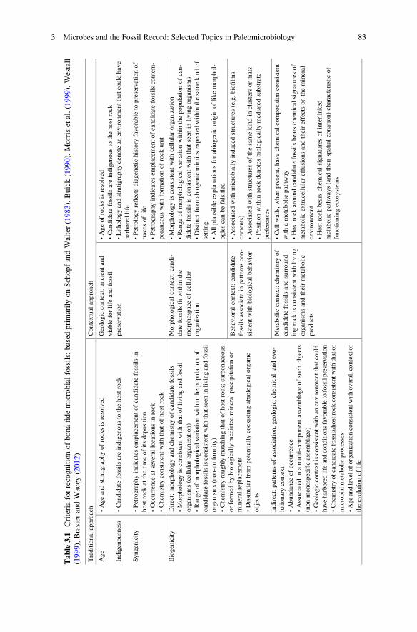
Table
3.1
Criteriaforrecognitionofbonafidemicrobialfossils;based
primarilyonSchopfandWalter(1983),Buick(1990),Morriset
al.(1999),Westall
(1999),Brasier
andWacey
(2012)
Traditional
approach
Contextual
approach
Age
Indigenousness
Syngenicity
•Ageandstratigraphyofrocksisresolved
•Candidatefossilsareindigenousto
thehostrock
•Petrographyindicates
emplacementofcandidatefossilsin
hostrock
atthetimeofitsdeposition
•Occurrence
atseveral
locationsin
rock
•Chem
istryconsistentwiththat
ofhostrock
Geologic
context:ancientand
viable
forlife
andfossil
preservation
•Ageofrocksisresolved
•Candidatefossilsareindigenousto
thehostrock
•Lithologyandstratigraphydenotean
environmentthatcouldhave
harboredlife
•Petrologyreflectsdiagenetic
history
favorable
topreservationof
traces
oflife
•Petrographyindicates
emplacementofcandidatefossilscontem-
poraneouswithform
ationofrock
unit
Biogenicity
Direct:morphologyandchem
istryofcandidatefossils
•Morphologyisconsistentwiththatoflivingandfossil
organisms(cellularorganization)
•Rangeofmorphological
variationwithin
thepopulationof
candidatefossilsisconsistentwiththatseen
inlivingandfossil
organisms(non-uniform
ity)
•Chem
istryroughly
matchingthatofhostrock;carbonaceous
orform
edbybiologically
mediatedmineral
precipitationor
mineral
replacement
•Dissimilar
from
potentially
coexistingabiological
organic
objects
Indirect:patternsofassociation,geologic,chem
ical,andevo-
lutionarycontext
•Abundance
ofoccurrence
•Associated
inamulti-componentassemblageofsuch
objects
(non-m
onospecificassemblage)
•Geologiccontextisconsistentwithan
environmentthatcould
haveharboredlifeandconditionsfavorabletofossilpreservation
•Chem
istryofcandidatefossils/hostrock
consistentwiththatof
microbialmetabolicprocesses
•Ageandleveloforganizationconsistentwithoverallcontextof
theevolutionoflife
Morphological
context:candi-
datefossilsfitwithin
the
morphospaceofcellular
organization
•Morphologyisconsistentwithcellularorganization
•Rangeofmorphological
variationwithin
thepopulationofcan-
didatefossilsisconsistentwiththat
seen
inlivingorganisms
•Distinctfrom
abiogenicmim
icsexpectedwithin
thesamekindof
setting
•Allplausible
explanationsforabiogenic
origin
oflikemorphol-
ogiescanbefalsified
Behavioralcontext:candidate
fossilsassociatein
patternscon-
sistentwithbiological
behavior
•Associated
withmicrobiallyinducedstructures(e.g.biofilm
s,
cements)
•Associated
withstructuresofthesamekindin
clustersormats
•Positionwithin
rock
denotesbiologically
mediatedsubstrate
preferences
Metaboliccontext:chem
istryof
candidatefossilsandsurround-
ingrock
isconsistentwithliving
organismsandtheirmetabolic
products
•Cellwalls,when
present,havechem
ical
compositionconsistent
withametabolicpathway
•Hostrock
aroundcandidatefossilsbears
chem
ical
signaturesof
metabolicextracellulareffusionsandtheireffectsonthemineral
environment
•Hostrock
bears
chem
ical
signaturesofinterlinked
metabolicpathways(andtheirspatialzonation)characteristic
of
functioningecosystem
s
3 Microbes and the Fossil Record: Selected Topics in Paleomicrobiology 83

et al. (2010) demonstrated syngenicity of 3.2 Ga microfossils from the Moodies
Group (South Africa) (Fig. 3.10a) by showing that the organic matter comprising the
microfossils had undergone the same degree of metamorphism as dispersed organic
matter in the host rock, based on Raman spectrometry. Recently, Olcott Marshall
et al. (2014) demonstrated that the carbonaceous material in the 3.46 Ga Apex Chert
(Warrawoona Group, Australia) represents four generations of material with different
thermal alteration histories and associated with different episodes of matrix formation,
indicating that at least some of the four generations (if not all) are not syngenetic.
Biogenicity
The biogenicity of candidate fossils is demonstrated both by direct assessment of
the objects themselves—morphology and chemistry—and indirectly, based on their
broader taphonomic, geologic, chemical, and evolutionary-biostratigraphic context.
The shapes and sizes of candidate fossils have to be consistent with those of known
fossil and living organisms (Schopf and Walter 1983). Morphologies indicative of
biogenicity include cells exhibiting phases of division (Knoll and Barghoorn 1977)
(Fig. 3.7c, f) or plastic deformation characteristic of soft, organic bodies (Tomescu
et al. 2008) (Fig. 3.4b). Morphological requirements for confirmation of biogenicity
include sizes within the range of known microbes (>0.01 μm3) and hollow objects
(coated in carbonaceous material), i.e., walls or sheaths of cells or cell colonies
(filaments) with or without internal divisions (Buick 1990).
Ideally, the candidate fossils show cellular elaboration, but this criterion is the
source of much debate (Buick 1990) as abiogenic objects can mimic some features
of cellular organization. Several authors recommend special caution in the inter-
pretation of spheroids comparable to coccoid prokaryotes, even when these exhibit
morphologies comparable to dividing cells, as such morphologies can be formed by
abiogenic processes (Westall 1999; Brasier and Wacey 2012). In such cases,
independent lines of evidence are required to corroborate biogenicity. Furthermore,
even more complex filamentous morphologies comparable to Precambrian micro-
fossils can be generated abiotically, as shown by Garcia-Ruiz et al.’s (2003)
experiments on precipitates formed by metallic salts in silica gels; however, the
structures thus formed are not hollow. Abiogenic structures mimicking microbial
filaments are also formed when local dissolution of the rock matrix allows for
displacement of crystals representing mineral inclusions which leave trails (Knoll
and Barghoorn 1974) (Fig. 3.4c); when carbonaceous inclusions from the rock are
also included in the trails, these can be easily mistaken for microbial filaments
(Lepot et al. 2009a). Only careful study of the microstructure and distribution of
carbonaceous matter, along with the fact that the “filaments” have mineral crystals
at their ends, reveals the abiogenic nature of such biomorphs (Brasier et al. 2002;
Lepot et al. 2009a).
The chemistry of candidate fossils can help in assessment of their biogenicity,
which is supported by the presence of cell walls or internal structures consisting of
kerogen (geologically transformed organic matter; see Sect. 3.6.2.1) and by chem-
ical compositions that roughly match that of the rock groundmass but show elevated
84 A.M.F. Tomescu et al.
carbon content (Buick 1990; Morris et al. 1999). Stable carbon isotope ratios (δ13C)of carbonaceous material in candidate fossils have been used extensively in dis-
cussions of biogenicity (Westall 1999), and 13C-depleted values are thought to
indicate biological fractionation of carbon and, thus, biogenicity (e.g., Ueno
et al. 2001b). However, a survey of the modern biota reveals that biogenic δ13Cvalues can vary at least as broadly as �41‰ to �3‰ PDB (Pee Dee Belemnite, a
standard used for reporting carbon isotopic compositions and based on the Creta-
ceous marine fossil cephalopod Belemnitella americana), overlapping toward the
top of this range with inorganic carbon (Buick 2001; Schidlowski 2000; Fletcher
et al. 2004). Therefore, caution should be applied in drawing generalizations based
on δ13C values (Buick 2001), which should at best be used to support biogenicity
only in conjunction with other independent sources of evidence.
The requirement for presence of organic carbon compounds (kerogen) excludes
most traces of microbial life comprised exclusively of inorganic material, such as
some microbially induced structures for which biogenicity criteria are discussed
below. A particular case of inorganic structures of biogenic origin are the
magnetosomes, ferromagnetic magnetite particles that are biomineralization prod-
ucts of magnetotactic bacteria. Magnetosomes are produced inside the bacterial
cells, and when the latter are degraded, their magnetosomes form chains that mark
the location of former filaments, but very similar magnetite grains can also have a
fully abiogenic origin. The equivocal nature of such magnetite grains fuelled a
significant part of the Martian meteorite ALH84001 debate (McKay et al. 1996;
Thomas-Keprta et al. 2001; Golden et al. 2004; see below Sect. 3.4.4). More
recently, Gehring et al. (2011) were able to identify dispersed magnetite particles
in Holocene lake sediments as magnetosomes using two-frequency ferromagnetic
resonance spectroscopy, thus opening the way to detection of this group of bacteria
based on acellular but biogenic body fossils.
Whether organic carbon is present or not, another set of morphological criteria
address biogenicity in terms of assemblage-level features. Candidate fossils
co-occurring with more clearly discernable microfossils (e.g., spheroids
co-occurring with rod-shaped fossils or a fossilized biofilm) are more likely to
have a biogenic origin (Westall 1999). While some morphological variation is to be
expected in assemblages of bona fide microbial fossils, the fossils have to be
consistent in morphology and size throughout the assemblages (Figs. 3.7c, f and
3.8a, b, d), and significant disparities in the size of morphologically similar objects
within an assemblage indicate abiogenic origin (Buick 1990; Westall 1999). Also at
the scale of the entire candidate fossil assemblage, occurrence in abundance
throughout the rock volume (a criterion for indigenousness and syngenicity as
well) and the presence of multiple morphological types, thus non-monospecific
assemblages (Fig. 3.7a), support biogenicity (Schopf and Walter 1983).
In a broader perspective, beyond the realm of morphology, the geology of the
host rock has to reflect both genesis in an environment favorable to the presence of
life and a subsequent geologic history conducive to fossil preservation (Schopf and
Walter 1983; Buick 1990). Microbial body fossils are not thought to preserve in
metamorphic rocks formed beyond low-grade metamorphism conditions. Microbe-
like objects found in medium- to high-grade metamorphic rocks or in igneous rocks
3 Microbes and the Fossil Record: Selected Topics in Paleomicrobiology 85
are either abiogenic, or if they are bona fide fossils, they represent nonindigenous
microbes (contaminants) or non-syngenetic microbial fossils. The chemistry of the
host rock can also be used to support inferences of biogenicity of candidate fossils
when it roughly matches that of the putative fossils, and it is characterized by
significant levels of elements and minerals formed by the direct or indirect activities
of microbes (e.g., pyrite produced by sulfate-reducing bacteria or magnetite, as
discussed above) (Morris et al. 1999; Westall 1999). Finally, the level of biological
complexity and organization of candidate fossils has to be consistent with the age of
the host rock and the overall context of the known history of life on Earth (Schopf
and Walter 1983). In this context, candidate fossils that appear out of context are
likely to be abiogenic, nonindigenous, or non-syngenetic.
3.3.1.2 The Contextual Approach
The search for the oldest traces of life adds another set of challenges (as discussed
by Brasier et al. 2006) to those encountered in documenting the microbial fossil
record in younger rocks. First, whereas Proterozoic (<2500 Ma old) rocks have
yielded a rich microfossil record, older rocks (especially pre-Neoarchean;
>2800 Ma old) have produced very rare candidate fossils that the traditional
approach to biogenicity can resolve unequivocally. Second, the environments of
early Earth were very different from those we are familiar with or that we can even
imagine today, and the potential life forms they hosted were very likely more
similar to those of modern environments we are just exploring today (e.g., deep
intraterrestrial endoliths, hyperthermophiles, anaerobes) than to anything else.
Third, there is currently increasing recognition that a variety of abiogenic self-
organizing structures generated by natural processes can mimic the morphological
complexity of bona fide traces of life.
In most cases, the situations generated by these constraints reside beyond the
sphere of resolution of the traditional inductive approach to biogenicity. Further-
more, the initial recognition of putative microfossils is based on intuition and
experience; however, intuition and experience are double-edged swords, as they
can easily lead one down the path of simply seeking evidence in support of a
preferred interpretation, without consideration of alternative explanations. Such
considerations, along with the challenges of identifying traces of life deeper and
deeper in the rock record, have led some workers (e.g., Brasier et al. 2002, 2006) to
reject the traditional approach and adopt a falsificationist approach wherein micro-
bial structures are not accepted as biogenic until the alternative null hypothesis of
abiogenicity is falsified. This approach emphasizes the integrative use of a com-
prehensive set of methods and an outlook based on asking open-ended questions
about types of processes (biogenic and abiogenic) and geologic settings, and
whether they could have produced the candidate fossil structures, rather than on
comparisons with known fossil or modern organisms and structures (Brasier
et al. 2006). In other words, instead of proving the biogenicity of structures, this
approach strives to demonstrate that they cannot be abiogenic. For this, questions
86 A.M.F. Tomescu et al.

are asked to assess the level of support for biogenicity in a hierarchy of contexts:
(1) geologic, (2) morphological, (3) behavioral-taphonomic, and (4) metabolic
(Table 3.1).
The method of the contextual approach has been formalized by Brasier
et al. (2006), Wacey (2009), and Brasier and Wacey (2012). Not surprisingly,
some of the criteria applied in the contextual approach necessarily overlap with
those of the traditional approach. However, due to the degree of generality of
questions asked in applying it, the applicability of this approach extends beyond
body fossils, to microbially induced structures. The approach has been used to
reject the biogenicity of putative prokaryote fossils of the 3.46 Ga old Apex Chert in
Australia (Brasier et al. 2002) (Fig. 3.5a) and to demonstrate the biogenicity of
prokaryote fossils in the 3.4 Ga old Strelley Pool Formation in Australia (Wacey
et al. 2011a) (Fig. 3.8a, b) and in an extensive critical analysis of all claims for early
Archean life (Wacey 2009). Below we summarize the elements of the contextual
approach as set forth by Brasier and Wacey (2012)—see also Table 3.1.
1. In a geologic context, questions are aimed at establishing the age of the rock
hosting the candidate fossils, as well as the indigenousness and syngenicity of
the latter, much in the same way that these are addressed traditionally. Further-
more, regional stratigraphy and petrology are mapped and sampled at the
kilometer -to-meter scale in order to gain an understanding of whether the past
local environments reflected by the rock record could have harbored life and
whether the rock sequence reflects a diagenetic and post-diagenetic history
favorable to fossil preservation. Detailed mapping of petrography and geochem-
istry at the cm-to-nm scale are then used to document spatial and temporal
Fig. 3.5 Debates and controversies. (a) Putative microbial filament (Primaevifilum amoenum)from the Archean Apex Chert (Warrawoona Group, Western Australia). (b) and (c) Limonite-
stained inclusions or cavities in Archean rocks of the Isua Greenstone Belt (Greenland), initially
interpreted as microbial fossils (Isuasphaera isua). (d) Structures from the ALH 84001 Martian
meteorite initially interpreted as bacterial magnetosomes. Scale bars: (a), (b), and (c) 10 μm; (d)
10 nm. Credits—images used with permission from (a) Elsevier Science Publishers (Schopf
et al. 2007). (b) and (c) Springer-Verlag (Pflug 1978b). (d) Wikimedia Commons; file:
ALH84001_structures.jpg; author: NASA
3 Microbes and the Fossil Record: Selected Topics in Paleomicrobiology 87

relationships between the candidate fossils and their emplacement in the rock, on
the one hand, and the host rock and its history, on the other hand. These allow for
more in-depth assessment of the suitability of the host rocks for fossil preserva-
tion and for reconstruction of a detailed time line of the events of putative fossil
formation and rock genesis, which allows for assessment of indigenousness and
syngenicity. Inability to document in detail all of these aspects of the geologic
context leaves the door open for alternative untestable (because unknown)
hypotheses of nonindigenousness, non-syngenicity, or non-biogenicity.
2. The morphological context provides information important in assessing the
biogenicity of candidate fossils regarded as members of a population of similar
objects. The level of support for biogenicity is tested by documenting the
morphospace occupied by the population (ranges of morphological and size
variation) and asking whether it fits within the morphospace of cellular organi-
zation in terms of both shape and size and range of variation. For example,
ranges that are too broad may indicate abiogenic structures whose variability is
not constrained by genetics or habitat. Importantly, biogenicity is assessed at the
same time in terms of the potentiality for the documented morphospace to be
occupied by abiogenic objects expected within the geological context under
consideration—for this the morphology of candidate fossils has to be considered
in concert with their chemistry. All plausible explanations for abiogenic origin
of objects with morphologies similar to that of the candidate fossils have to be
falsified to demonstrate biogenicity. In this context, Brasier and Wacey (2012)
emphasize the need for greater emphasis on improved mathematical modeling of
morphospace occupation by different populations of objects, in order to produce
more powerful tests for distinguishing biological populations from amalgam-
ations of abiogenic structures (e.g., Boal and Ng 2010).
3. Because living organisms have behaviors which may be reflected in the taphon-
omy of their fossils, support for biogenicity has to be tested in a behavioral-taphonomic context. Patterns of association documented within assemblages of
candidate fossils are assessed for the presence of features characteristic of
biological behaviors. These include populations of candidate fossils assembled
in clusters or mats reflecting colonial associations (Figs. 3.6a, 3.7a, b, 3.8d), or
populations associated with microbially induced structures and textures
(biofilm-like textures, biogenic or organomineral cements), as well as assem-
blages of candidate fossils positioned in response to substrate preferences
(Fig. 3.8b). Just like with the morphological context, care must be taken to
falsify abiogenic explanations for these types of association patterns: abiotic
mineral growth can also form clusters, sometimes at the contact between
contrasting lithologies which may be interpreted as a substrate surface colonized
by a microbial mat; and inferences of biogenicity need to be corroborated by
chemical data.
4. The metabolism of living organisms influences their environment and this may
be reflected in the chemistry of candidate fossils and their host rock, which offers
a metabolic context for testing hypotheses of biogenicity. Candidate fossils that
comprise a carbonaceous fraction are tested for 12C-enriched stable carbon
88 A.M.F. Tomescu et al.

isotope ratios characteristic of biogenic carbonaceous material (kerogen). An
enrichment in chemical elements comprising major building blocks of living
matter (hydrogen, oxygen, nitrogen, sulfur, phosphorus) relative to the host rock
matrix is also to be expected in biogenic objects. Due to the small size of
microfossils, their chemistry is compared to that of surrounding mineral grains.
Additionally, bona fide body fossils will be associated with chemical signatures
generated by their metabolic extracellular effusions in the host rock (discussed in
some detail below—see Sect. 3.6.2 Authigenic mineralization). In such cases, if
microbial communities are preserved in situ, the host rock can record the
chemical signatures of interlinked metabolic pathways (e.g., carbon fixation
and carbon respiration; Brasier and Wacey 2012) and their spatial zonation
characteristic of functioning microbial ecosystems.
3.3.2 Recognizing Microbially Induced Structures
The recognition of diverse structures in the geologic record as traces of microbial
life follows the same paradigms as that of microbial body fossils. Some authors
apply sets of criteria in a traditional inductive approach, while others favor a
context-based falsificationist approach, and the criteria used vary somewhat
among authors. The methods used for assessing the criteria also vary somewhat
for different types of microbially induced structures (MISS, stromatolites,
microborings) because of differences in the types of microorganisms that generated
the structures and their mode of formation. However, the fundamental requirements
for recognizing diverse structures in the geologic record as traces of microbial life
are the same as for microbial body fossils. Biogenic-like morphologies are what
initially recommends them as candidate microbially induced structures, and the
Fig. 3.6 Archean Dresser Formation (Warrawoona Group, Western Australia). (a) Putative
filamentous microfossils; some exhibit helical geometries, others are interwoven, branched, or
radiate from kerogen clots. (b) Putative stromatolite. Scale bars: (a) 50 μm, (b) lens cap diameter
ca. 6 cm. Credits—images used with permission from (a) and (b) Springer Science + Business
Media B.V. (Wacey 2009)
3 Microbes and the Fossil Record: Selected Topics in Paleomicrobiology 89
geologic context needs to reflect conditions suitable for life and syngenicity (except
for microborings which can be emplaced subsequent to the formation of their host
rock) (Schopf and Walter 1983; Buick 1990; McLoughlin et al. 2007). Aside from
morphology, geochemical analyses are called upon in the assessment of
biogenicity, to test for evidence for metabolic activity (Brasier et al. 2006;
McLoughlin et al. 2007; Brasier and Wacey 2012). Furthermore, in a contextual
approach (as outlined above), the null hypothesis of abiotic origin must be rejected
for any candidate microbially induced structure (Brasier et al. 2006; Brasier and
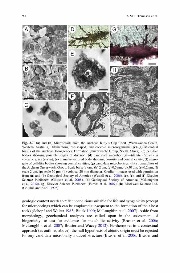
Fig. 3.7 (a) and (b) Microfossils from the Archean Kitty’s Gap Chert (Warrawoona Group,
Western Australia), filamentous, rod-shaped, and coccoid microorganisms. (c)–(g) Microbial
fossils of the Archean Hooggenoeg Formation (Onverwacht Group, South Africa), (c) cell-like
bodies showing possible stages of division, (d) candidate microborings—titanite (brown) in
volcanic glass (green), (e) granular-textured body showing porosity and central cavity, (f) aggre-
gate of cell-like bodies showing central cavities, (g) candidate microborings. (h) Stromatolites of
the Archean Onverwacht Group. Scale bars: (a) and (b) 2 μm, (c) 0.5 μm, (d) 50 μm, (e) 0.2 μm, (f)
scale 2 μm, (g) scale 50 μm, (h) coin ca. 20 mm diameter. Credits—images used with permission
from (a) and (b) Geological Society of America (Westall et al. 2006). (c), (e), and (f) Elsevier
Science Publishers (Glikson et al. 2008). (d) Geological Society of America (McLoughlin
et al. 2012). (g) Elsevier Science Publishers (Furnes et al. 2007). (h) Blackwell Science Ltd.
(Golubic and Knoll 1993)
90 A.M.F. Tomescu et al.

Wacey 2012). Several authors have proposed different recognition criteria and
ways in which these may be satisfied for specific types of structures.
3.3.2.1 Microbialites
Most microbialites lack microfossils and determining their biogenicity requires
attention to the geologic context, overall morphology, microstructure, and chemis-
try. Modern analogues of stromatolites are very rare (Kazmierczak and Kempe
2006) (Fig. 3.2d, f) and little is known about their formation from direct observa-
tions. As a result, definitions of stromatolites and the criteria used to recognize them
as biogenic structures are widely different between authors and are still debated. In
contrast to stromatolites, which have been recognized and studied for at least a
century (Awramik and Grey 2005), the study of microbially induced sedimentary
Fig. 3.8 Microbial fossils of the Archean Kelly Group (Western Australia); (a)–(h) Strelley Pool
Formation, (i) Euro Basalt. (a) Cluster of cells, some showing cell wall folding and invagination.
(b) Cells attached to quartz grain exhibiting preferred alignment parallel to the surface of the
quartz grain. (c) Carbonaceous threads of rodlike objects representing putative microbial fila-
ments. (d) Colony of loosely clustered hollow spheroidal microfossils. (e) Pair of linearly arranged
lenticular carbonaceous microfossils. (f) and (g) Stromatolites. (h) Microbial etch pits
(microborings) in pyrite. (i) Segmented microborings in volcanic glass. Scale bars: (a) and (b)
20 μm, (c) 20 μm, (d) 20 μm, (e) 10 μm, (f) 4 cm, (g) 3 cm, (h) 3 μm, (i) 25 μm. Credits—images
used with permission from (a) and (b) Nature Publishing Group (Wacey et al. 2011a). (c), (d), and
(e) Elsevier Science Publishers (Sugitani et al. 2013). (f) and (g) Springer Science + Business
Media B.V. (Wacey 2009). (h) Elsevier Science Publishers (Wacey et al. 2011b). (i) Elsevier
Science Publishers (Furnes et al. 2007)
3 Microbes and the Fossil Record: Selected Topics in Paleomicrobiology 91

structures as fossil traces of life, and especially in the search for the earliest traces of
life, is a relatively recent development. In part because of its recent rise, but also
due to the wealth of modern sedimentary environments that host microbial mats
providing as many modern analogues, the study of MISS is firmly grounded in an
empirical comparative approach that was pioneered by Noffke and Krumbein
(Noffke et al. 1996; Noffke et al. 2001).
Microbially Induced Sedimentary Structures
Noffke (2009) provided the most recent treatment of the lines of evidence used for
recognizing MISS, summarizing them in a set of six criteria of biogenicity which
we discuss below. These criteria are based on numerous studies of both modern and
fossil microbial mats and MISS, but emphasize MISS formed by photoautotrophic
mat-building microbes in aqueous environments.
1. Broader geologic context: MISS occur in sedimentary rocks that have experi-
enced, at most, low-grade metamorphism (lower greenschist facies). Higher-
grade metamorphism is unfavorable not only to the preservation of body fossils
but also to MISS preservation—the often complicated diagenesis of metamor-
phic rocks can render syngenicity difficult to assess, metamorphic textures and
structures can overprint the sedimentary structures, and, overall, biosignatures
are altered and difficult (or impossible) to recognize.
2. Sequence stratigraphy: MISS are associated with regression-transgression turn-
ing points (the term transgression indicates a relative rise in sea level, while
regression indicates a relative drop in sea level). More specifically, it is the
transgressive phases that succeed those turning points that witness expansion of
tidal flat and shallow shelf areas favorable to microbial mat formation along the
passive continental margins, due to sea level rise.
3. Depositional environment: MISS occur in rocks formed in environments favor-
able to the establishment and preservation of microbial mats. Although modern
photoautrophic mat-building microbes are often found in environments charac-
terized by fine-grained sand substrates and low current velocities (10–25 cm/s),
it is conceivable that chemoautotrophic microbes could build mats beyond the
photic zone and on substrates suitable to their metabolic needs.
4. Hydraulic regime: the types of MISS and their spatial and stratigraphic distri-
bution are consistent with the hydraulic regime implied by sedimentology, as
specific types of modern MISS have been shown to characterize environments
with different hydraulic regimes (e.g., shallow shelf, lagoon, different tidal
zones).
5. Morphology: constrained by the same biology throughout the ages, microbial
mats have maintained the same functional morphology traits, therefore geome-
tries and dimensions of fossil MISS match those of modern MISS.
6. Microtexture: microscopic textures and structures related to, caused by, or
representing microbial mats are found associated with the macroscopic MISS.
92 A.M.F. Tomescu et al.

These include (often poorly) fossilized or mineral-replaced microbial filaments,
wavy laminae concentrating organic matter and associated with sharp microstra-
tigraphic geochemical gradients (Noffke et al. 2013b), finer-grained clasts or
heavy minerals, upward-fining microsequences, and minute pores left by gases
accumulating under the microbial mat.
Stromatolites
The realization, early in the history of stromatolite studies, that some laminated
precipitation structures represent microbially induced structures led to a period of
somewhat undiscerning application of the label of biogenicity to any finely lami-
nated structure in the rock record, which resulted in a plethora of reports of
stromatolites (and therefore microbial fossils) from rocks of all ages [e.g., the
3.4 Ga Buck Reef Chert of South Africa, listed as a stromatolite by Schopf
(2006) but not treated as one by Tice and Lowe (2004)]. The subsequent realization
that not all laminated structures are biogenic (e.g., Lowe 1994) led to disagreement
over what defines a stromatolite (Hofmann et al. 1999; Awramik and Grey 2005).
Because of that, distilling a set of criteria of biogenicity for stromatolites is much
more difficult than for MISS (e.g., Riding 2011). Whereas some authors define
stromatolites as laminated structures that are formed by microbial activities and
mineral precipitation (Awramik and Margulis 1974; Buick et al. 1981), others use
the term stromatolite for laminated, lithified sedimentary structures, regardless of
the involvement of microbes in their formation (e.g., Semikhatov et al. 1979;
Antcliffe and McLoughlin 2009). Consequently, the disagreement also extends
over whether such laminated structures could represent good evidence for ancient
life, and a number of different sets of criteria for stromatolite biogenicity have been
proposed.
Buick et al. (1981) assembled probably the most stringent set of criteria that
focuses on morphological lines of evidence:
1. The candidate structures (termed stromatoloids; Buick et al. 1981) occur in
sedimentary or metamorphosed sedimentary rocks;
2. The structures are synsedimentary with the host rock; because most stromato-
lites are built by photosynthetic microbial communities that tend to be thicker in
positions receiving the most light and therefore raised with respect to the rest of
the substrate;
3. Most structures in a stromatolite have a convex-upward morphology (e.g.,
Figs. 3.2d–f, 3.3b–d, 3.6b, 3.7h, 3.8f, g) and
4. Individual laminae are thicker over the upward-facing convex surfaces (e.g.,
Figs. 3.3b, c, 3.7h, 2.8g);
5. Laminations are wavy and wrinkled or have several orders of curvature (e.g.,
Figs. 3.2e, 3.3c, d, 3.7 h) because bedding irregularities are amplified by the
phototropic tendencies of the microbial communities listed above;
6. Microbial body or trace fossils are present in the structures;
3 Microbes and the Fossil Record: Selected Topics in Paleomicrobiology 93

7. Changes in the microfossil assemblages are associated with changes in the
morphology of sedimentary structures, indicating a relationship between the
microbial communities and the formation of the structure; and
8. Microfossils are preserved in situ and in positions that indicate accretion activ-
ities—binding, trapping, or precipitation.
It is worth noting that, based on strict morphological criteria and including
requirements that microfossils be present and proven to contribute to sedimentary
accretion, this set of criteria diagnoses as abiogenic most structures accepted as
stromatolites by other systems of evaluation.
In his criteria for stromatolite biogenicity, Walter (1983) was concerned with
syngenicity—orientation of laminated structures with respect to bedding planes
indicating formation at the same time with adjacent layers and occurrence in rocks
where the laminated structures can only be explained as primary sedimentary
features—and biogenicity as reflected in morphology, microbial origin, and chem-
istry. Hofmann et al. (1999) used careful studies of morphology, sedimentology,
and local microstratigraphic relationships in their assessment of the biogenicity of
3.45 Ga conical laminated structures from the Warrawoona Group (Australia).
Their approach was aimed at rejecting hypotheses of abiogenic origin and their
arguments were later regarded as a set of criteria for biogenicity (Awramik and
Grey 2005). Hofmann et al. (1999) rejected regional deformational processes as a
potential explanation, based on the geographically broad extent of the structures,
combined with their narrowly constrained stratigraphic and lithologic circum-
stances. They also used the geometry and relative position of laminae to reject
sideways compression (folding), downright directed slumping, and strictly chemi-
cal precipitation as explanations for different morphological aspects of the struc-
tures, supporting their formation by upward accretion. Furthermore, these authors
used the steep angles of the structures to reject simple sedimentation as a formation
mechanism, supporting the presence biological binding. Finally, Hofmann
et al. (1999) invoked the wide acceptance of other independent lines of evidence
(at the time; some of these were later contested), such as body fossils and chemical
biosignatures, for the presence of life in coeval rocks, as making biogenic causes
even more plausible.
The contextual approach (see above; Brasier et al. 2002; Brasier andWacey 2012)
is applicable, with some changes, to MISS and stromatolites (Wacey 2009). The fact
that stromatolite-like morphologies have been proven to form by abiogenic processes
(Grotzinger and Rothman 1996; McLoughlin et al. 2008) implies that morphology
alone is not enough to determine biogenicity and that microstructural and geochem-
ical analyses are necessary to reject the null hypothesis of abiogenic formation for
any given stromatolite. It is interesting to note that Awramik and Grey (2005) have
expressed doubt about whether any abiogenically formed structures can convincingly
resemble stromatolites. On a more general level, these authors criticized the contex-
tual approach for setting lofty standards for paleontology. They point out that
alternative modes of genesis are thoroughly considered by workers adopting a
traditional inductive approach to stromatolite biogenicity, even if the associated
94 A.M.F. Tomescu et al.

deliberations are not included in the final publication. Instead of what they call the
binary character of the contextual approach, Awramik and Grey (2005) advocate an
approach that emphasizes morphology (without excluding other types of data) aimed
at finding evidence for the contribution of biogenic processes to the formation of
candidate structures and placement of that evidence on a scale of credibility (unequiv-
ocal-compelling-presumptive-permissive-suggestive evidence).
Most of the disputes on stromatolite biogenicity and the criteria to assess it are
certainly rooted in the absence of microbial body fossils from many candidate
stromatolites (Hofmann et al. 1999). Addressing this topic, Kremer et al. (2012a)
studied calcification and silicification processes in cyanobacterial mats that form
the best modern analogues of fossil stromatolites, in Tonga (south Pacific;
Kazmierczak and Kempe 2006) (Fig. 3.2d, e). They showed that morphological
preservation of cyanobacteria by primary mineralization depends on two main
factors: the type of mineral phase and the time of mineralization. Variations in
the two factors produce a wide spectrum of modes of morphological preservation
which encompass several stages of degradation that were documented for both
filamentous and coccoidal cyanobacteria. These observations led Kremer
et al. (2012a) to suggest that Archean life may have been more abundant than
previously thought based on meager findings, but difficult to recognize because of
the alteration of original microbial body fossils or sedimentary structures due to
recrystallization and mineral replacement.
3.3.2.2 Microborings
In contrast to stromatolites, microborings have abundant modern counterparts.
However, the abundance and ubiquity of modern microborings (included by some
authors in the broader category of bioalteration textures; Furnes et al. 2007) is onlystarting to become appreciated and studied. At a general level, the criteria used to
establish that microborings are ancient and biogenic are the same as those applied to
other microbially induced structures and to body fossils. However, due to their
relatively simple morphology and unique mode of formation of microborings, the
specific application of these criteria is somewhat different than for other fossil
biosignatures. In their treatment of microborings, McLoughlin et al. (2007) and
Furnes et al. (2007) propose an approach that includes evaluation in three con-
texts—geologic, morphological, and geochemical-metabolic—aimed at determin-
ing the age of structures and at falsifying an abiogenic origin.
1. Age: since microbes producing microborings are at work today just as they were
in the Proterozoic, it is imperative that microborings reported from ancient rocks
and presumably of ancient age be demonstrated to have originated early during
diagenesis of the host rocks. Age is established based on the relationships of the
microboring with surrounding features of the rock. Early diagenetic
microborings will crosscut early stage fractures in the host rock and will be
crosscut by later (younger) metamorphic mineral growths (McLoughlin
3 Microbes and the Fossil Record: Selected Topics in Paleomicrobiology 95
et al. 2007). Furthermore, ancient microborings will show the same level of
metamorphosis as their host rock (Furnes et al. 2007), and in the case of
microborings in volcanic rock filled with titanite (Fig. 3.7d, g), the absolute
age of the titanite can be directly determined by U-Pb dating (e.g., the
microborings of the 3.35 Ga Euro Basalt, Pilbara Craton, Australia; Banerjee
et al. 2007) (Fig. 3.8i).
2. Geologic context: the regional and local scale context of the microborings has to
be consistent with the presence of life. This is becoming an increasingly less
stringent requirement as modern and fossil biogenic microborings are being
discovered in (unforeseen) settings that broaden our ideas about the range of
environments where we can expect to find traces of life (e.g., subseafloor oceanic
crust; Furnes et al. 2001; McLoughlin et al. 2012). The geologic setting of
microborings is expected to be consistent with behavioral aspects of microbial
biology—microborings should demonstrate substrate preferences in the host
rock for metabolically useful compounds, or if their origin is cyanobacterial,
the stratigraphy and sedimentology of the host rock should be consistent with a
shallow, photic depositional environment (McLoughlin et al. 2007).
3. Morphological context: the morphology of microborings has to be consistent
with biogenic origin. Since microborings have a relatively simple morphology,
consisting only of branched tubes, it is difficult to establish a set of morpholog-
ical criteria that applies to all microborings and excludes all abiogenic mimics.
Nevertheless, a list of features that would lend support to a biogenic origin of
such tubes would include μm-scale size and branching or changes in direction as
they encounter other such structures (Furnes et al. 2007). These features, con-
sidered singly, cannot provide compelling evidence for biogenicity, and mor-
phology alone can rarely be used to determine the biogenicity of putative
microborings, without supporting geochemical evidence (McLoughlin
et al. 2007).
4. Geochemical-metabolic context: geochemical evidence provides the strongest
support for the biogenicity of most microborings. Euendoliths producing
microborings are chemolithoautotrophic prokaryotes, so microborings preferen-
tially occupying areas of the host rock rich in compounds that are metabolically
important for bacteria, such as metal inclusions, provide evidence for biological
processing (Brasier et al. 2006). Finer scale geochemical evidence from both the
microtubes and their filling can provide even stronger support for biogenicity.
For example, depletion of Mg, Ca, Fe, Na in the rock matrix around putative
microborings indicates metabolic processing by euendoliths (Alt and Mata
2000). Fine (<1 μm thick) linings of C, N, and P in both recent and prehistoric
microtubes have been interpreted as cellular remains (Giovannoni et al. 1996;
Furnes and Muehlenbachs 2003), so similar linings of biologically important
elements may be used as evidence for a biogenic origin of putative microborings.
Additionally, analyses of in situ carbon within the microborings can also provide
insights into their potential biogenicity (Furnes et al. 2004; McLoughlin
et al. 2012).
96 A.M.F. Tomescu et al.

5. Null hypothesis of abiogenicity: in most cases, the alternative explanation of
micron-sized tubular structures resides in ambient inclusion trails (AIT) (Lepotet al. 2009a); therefore it is essential that putative microborings are shown to not
belong in this category of structures. Although the exact conditions that lead to
formation of AIT are not well understood (McLoughlin et al. 2007), they have
been suggested to form by the movement of a crystal (e.g., pyrite, garnet)
through the crystalline silica matrix of the rock. The movement is thought to
be the result of pressure-solution processes and potentially driven by the thermal
decomposition of organics into gases (Tyler and Barghoorn 1963; Knoll and
Barghoorn 1974; Wacey et al. 2008; Lepot et al. 2009a).
While relatively few morphological characters can be used in support of the
biogenicity of putative microborings [although see McLoughlin et al.’s (2009)
formal taxonomy of microborings as trace fossils (Fig. 3.4d–f)], many more
features can be used to identify tubular structures as AIT of abiogenic origin.
McLoughlin et al. (2007) provided a list of characters present in AIT that can be
used to distinguish them from bona fide microborings: (1) presence of a mineral
grain at the end of the tube (Fig. 3.4c); (2) longitudinal striations caused by the
facets of the mineral grain as it was driven through the rock; (3) angular cross-
sectional geometry and twisted paths, particularly toward the end of the tubes (due
to the increasing resistance of the host rock); and (4) a tendency to crosscut other
tubes or branch, with sudden changes in diameter at branching points—branches
with different diameters are caused when the mineral grain forming the AIT splits
and the resulting fragments continue to form one AIT each.
Despite the abiogenic mode of AIT formation, an interesting aspect of these
structures that can be relevant to early microbial life is the nature of the organic
material that drives the movement of the trail-forming crystals. Wacey et al.’s(2008) study of the 3.4 Ga Strelley Pool sandstone (Australia) provides a good
example of biogenicity assessment for tubular trace fossils and AIT recognition
using the criteria outlined above. Furthermore, using nanoSIMS (secondary ion
mass spectrometry) analyses, these authors were able to confirm the role of
decomposing organic matter in AIT formation (proposed by Knoll and Barghoorn
1974) and to demonstrate features consistent with biogenic origin for the organic
material associated with the trails.
3.4 Earth’s Oldest Fossils and the Archean Fossil Record
3.4.1 An Archean-Proterozoic Disparity
Archean evidence for life includes all types of fossils discussed thus far: microbial
body fossils (microfossils), microbially induced structures (stromatolites, MISS),
and chemical biosignatures. This section focuses primarily on microfossils, with
microbially induced structures addressed only in the summary of the oldest
3 Microbes and the Fossil Record: Selected Topics in Paleomicrobiology 97
evidence for life (Sect. 3.4.5). A sweeping glance at the Precambrian microfossil
record reveals a marked change in the quality and number of fossils from the
Proterozoic to the Archean (Schopf et al. 2010). The Proterozoic rock record
hosts numerous localities with well-preserved, unequivocal microfossils (Sergeev
2009). The high diversity of microfossil types and morphologies seen among these
localities suggests that prokaryotic life arose and started diversifying much earlier,
in the Archean (Altermann and Schopf 1995; Sergeev 2009). However, despite
decades of research aimed at finding signals of life in the Archean, microfossils
from this time period are comparatively rare and often highly controversial (Schopf
and Walter 1983; Altermann and Schopf 1995; Wacey 2009). A historical focus on
the Proterozoic by researchers may have been at the origin of this disparity in the
early stages of the discipline. Nevertheless, continued studies are now indicating
that this discrepancy in the quality and abundance of microfossils between the
Archean and the Proterozoic is predominantly an artifact of the geologic record and
not the result of lack of study or differences in the early biosphere (Schopf and
Walter 1983; Schopf et al. 2010; Wacey 2012). This can be attributed to a series of
factors that include (1) relatively low levels of cratonic sedimentation (i.e., on
continental landmasses) during the Archean; (2) comparatively intense metamor-
phism and deformation of Archean sedimentary rocks, which are predominantly
found today in metamorphic greenstone belts (Schopf and Walter 1983; Buick
1990) that consist of metamorphosed basalt with minor sedimentary rock inter-
layers; and (3) the fact that the silica forming most Archean cherts that preserve
candidate fossils precipitated from relatively hot, acidic, and concentrated hydro-
thermal fluids, whereas Proterozoic cherts formed from cooler, more neutral, and
dilute surficial fluids (Buick 1990). Such processes degrade the remains of organ-
isms and biological compounds, complicating efforts to identify them in Archean
rocks. Thus, although Archean rocks can be found in many areas, those that are well
exposed, have undergone relatively little metamorphism, are of clear sedimentary
origin, and have yielded bona fide body fossils thus far are found in only two places
on Earth: the Kaapvaal Craton in South Africa and the Pilbara Craton of Western
Australia (Wacey 2009; Hickman and Van Kranendonk 2012), which may have
been part of the same landmass in the Archean (Zegers et al. 1998) and possibly in
close vicinity to each other. It is from these two regions that nearly all Archean
microfossils known to date originate, including the very earliest evidence of life.
3.4.2 Brief History of Discovery
The first bona fide Precambrian microfossils were reported in 1907 from ca. 1 Ga
old Torridonian sedimentary phosphates in Scotland (Peach et al. 1907; Wacey
2009), but it was not until Tyler and Barghoorn (1954) first published descriptions
of microfossils from the Gunflint iron formation (Canada) that sustained work went
into documenting microbial diversity in the Precambrian (Schopf andWalter 1983).
Dated to 1.9 Ga, the Gunflint iron formation microfossils provided the first solid
98 A.M.F. Tomescu et al.

evidence of Proterozoic life older than 1 billion years. Much older microfossils
were reported from Archean rocks of South Africa during the 1960s and from
Australia as early as the late 1970s (Knoll and Barghoorn 1977; Dunlop et al. 1978;
Awramik et al. 1983). However, nearly all of these claims were questioned or
contested in some capacity (Nisbet 1980; Schopf and Packer 1987; Buick 1984). It
was not until microfossils from the 3.46 Ga Apex Basalt (Australia) (Fig. 3.5a) were
described that an assemblage of early Archean (Paleoarchean) microfossils was
widely accepted as evidence of early Archean life (Schopf and Packer 1987; Schopf
1993). For over a decade, these fossils were embraced as the best and earliest
evidence for life on Earth (Brasier et al. 2004). In 2002 Brasier et al. presented
evidence refuting all the lines of evidence on which the biogenicity claims for the
Apex microfossils were based, sparking a debate that continues to the present day
(e.g., Schopf and Kudryavtsev 2012; Pinti et al. 2013). Despite their later conten-
tiousness, the initial wide acceptance of these fossils established an enduring
paradigm that shapes our conception of the timing of the emergence of life. This
idea of an early Archean origin for life has been dubbed the “Early Eden Hypoth-
esis” (Brasier et al. 2004). Regardless of the biogenicity of the Apex microfossils, a
look at the Archean microfossil record known to date reveals numerous lines of
evidence which, even in the absence of a smoking gun, point strongly toward the
emergence of life around 3.5 billion years ago (see below).
3.4.3 Salient Patterns in the Archean Fossil Record
Several major patterns emerge from a review of the Archean record of life. In terms
of the host rock, Archean microfossils occur in sedimentary rocks, most commonly
cherts and sandstones, formed in marine environments and metamorphosed to
greenschist facies. Most of these fossils are found in cherts (Table 3.2) and a
small proportion originate from sandstones such as those from the Strelley Pool
Formation in Australia and Moodies Group in South Africa (Noffke et al. 2006;
Wacey 2009). Despite their rarity in the Archean microfossil record, sandstones
record valuable data that are difficult to extract from other types of rock, including
successive depositional and diagenetic stages or microbially induced structures
(e.g., Noffke et al. 2006, 2013a; Wacey et al. 2011b). Sandstones may also provide
some protection to the fossil structures from mechanical stress and strain during
metamorphism (Wacey 2009). Although early studies focused on rocks originating
from environments thought to be most conducive to early life at the time, such as
shallow marine environments, more recent work has focused on searching for
biosignatures in what were previously considered unlikely contexts, such as in
hydrothermal deposits and pillow basalts (Rasmussen 2000; Furnes et al. 2004;
Duck et al. 2007; Furnes et al. 2007; Wacey 2009; McLoughlin et al. 2012).
Geographically, as previously mentioned, Archean microfossils occur exclu-
sively in rocks of the Kaapvaal Craton in South Africa and in the Pilbara Craton
of Western Australia. However, within each of these regions, fossil localities have
3 Microbes and the Fossil Record: Selected Topics in Paleomicrobiology 99

Table 3.2 Archean microfossil record, with an emphasis on rock units older than 3 Ga and
microbial body fossils, MISS microbially induced sedimentary structures
Age (Ma) Rock unit Location Fossil evidence References
3480 Dresser Formation
(Warrawoona
Group, Pilbara
Supergroup)
Australia Microfossilsa—fila-
ments spiral, tubular
branched and
unbranched, some
septate; spheroids
MISS
?Stromatolites
Buick et al. (1981),
Ueno et al. (2001a,
b), Awramik and
Grey (2005), Van
Kranendonk (2006),
Schopf (2006),
Wacey (2009),
Noffke
et al. (2013a)
?3470b ?Mount Ada Basalt
(Warrawoona
Group, Pilbara
Supergroup)
Australia Microfossils—tubu-
lar sheaths; fila-
ments unbranched,
some septate
Awramik
et al. (1983)
3466 Kitty’s Gap Chert
in Panorama For-
mation
(Warrawoona
Group, Pilbara
Supergroup)
Australia Microfossils—colo-
nies of primarily
coccoid cells, some
chain-like; some fil-
aments and rare
rod-shaped cells
Westall et al. (2006)
3465 Apex Chert in
Apex Basalt
(Warrawoona
Group, Pilbara
Supergroup)
Australia Microfossilsc—fila-
ments unbranched
septate
Schopf and Packer
(1987), Schopf
(1993, 2006)
3450 Hooggenoeg For-
mation
(Onverwacht
Group, Swaziland
Supergroup)
South Africa Microfossils—nar-
row filaments; clus-
ters of spherical,
subspherical struc-
tures, cell walls;
rod-shaped cells
Microborings in pil-
low lavas
Walsh (1992),
Walsh and Westall
(2003), Westall
et al. (2001, 2006),
Furnes et al. (2004),
Glikson
et al. (2008), Wacey
(2009), Fliegel
et al. (2010),
McLoughlin
et al. (2012)
3426–3350 Strelley Pool For-
mation (Kelly
Group, Pilbara
Supergroup)
Australia Microfossils—
threadlike and hol-
low tubular fila-
ments, filmlike
structures, hollow
spheroids, lenticular
microfossils (ellip-
soids)
Stromatolites
Microborings
Hofmann
et al. (1999), Brasier
et al. (2006),
Allwood
et al. (2007), Wacey
et al. (2006, 2011a,
b), Sugitani
et al. (2010, 2013)
(continued)
100 A.M.F. Tomescu et al.

Table 3.2 (continued)
Age (Ma) Rock unit Location Fossil evidence References
3416–3334 Kromberg Forma-
tion (Onverwacht
Group, Swaziland
Supergroup)
South Africa Microfossils—
spheroids, ellip-
soids, rod-shaped
cells, spindle-
shaped microfossils
Microborings in pil-
low lavas
Walsh (1992),
Furnes et al. (2007),
Wacey (2009)
3416 Buck Reef Chert
(Onverwacht
Group, Swaziland
Supergroup)
South Africa Microfossils—occa-
sional filaments in
laminations
MISS
Tice and Lowe
(2004), Wacey
(2009)
3350 Euro Basalt (Kelly
Group, Pilbara
Supergroup)
Australia Microborings Banerjee
et al (2007), Wacey
(2009)
3260 Swartkoppie For-
mation
(Onverwacht
Group, Swaziland
Supergroup)
South Africa Microfossils—coc-
coid cells
Schopf (2006)
3245 Sheba Formation
(Fig Tree Group,
Swaziland
Supergroup)
South Africa Microfossils—coc-
coid cells
Schopf (2006)
3240–3235 Kangaroo Caves
Formation (Sulphur
Springs Group,
Pilbara
Supergroup)
Australia Microfossils—
unbranched fila-
ments and bundles
of tubes, spheroids
(small, 50–100 nm
diameter)
Rasmussen (2000),
Duck et al. (2007),
Wacey (2009)
3200 Dixon Island For-
mation (West
Pilbara
Superterrane)
Australia Microfossils—fila-
ments, spheroids,
microbial mat
remnants
Kiyokawa
et al. (2006), Schopf
et al. (2006), Wacey
(2009)
3200 Moodies Group
(Swaziland
Supergroup)
South Africa Microfossils—
spheroids
MISS
Noffke et al. (2006),Wacey (2009),
Javaux et al. (2010)
3000 Cleaverville For-
mation (Gorge
Creek Group, De
Grey Supergroup)
Australia Possible microfos-
sils—spheroids
Wacey (2009)
3190–2970 Farrell Quartzite
(Gorge Creek
Group, De Grey
Supergroup)
Australia Microfossils—
threadlike, filmlike,
spheroidal, lenticu-
lar, or spindle-like
Sugitani
et al. (2007), Wacey
(2009)
2750 Hardey Formation
(Fortescue Group,
Mount Bruce
Supergroup)
Australia MISS Rasmussen
et al. (2009)
(continued)
3 Microbes and the Fossil Record: Selected Topics in Paleomicrobiology 101
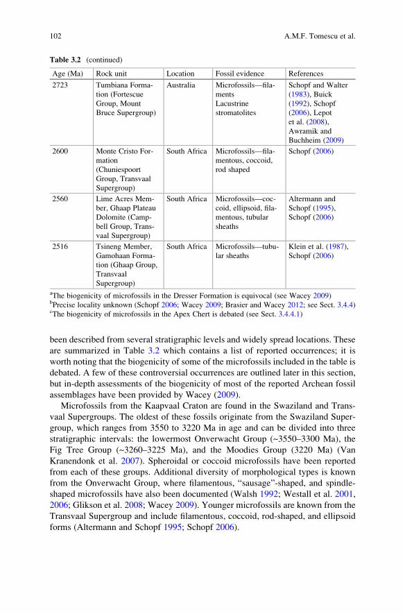
been described from several stratigraphic levels and widely spread locations. These
are summarized in Table 3.2 which contains a list of reported occurrences; it is
worth noting that the biogenicity of some of the microfossils included in the table is
debated. A few of these controversial occurrences are outlined later in this section,
but in-depth assessments of the biogenicity of most of the reported Archean fossil
assemblages have been provided by Wacey (2009).
Microfossils from the Kaapvaal Craton are found in the Swaziland and Trans-
vaal Supergroups. The oldest of these fossils originate from the Swaziland Super-
group, which ranges from 3550 to 3220 Ma in age and can be divided into three
stratigraphic intervals: the lowermost Onverwacht Group (~3550–3300 Ma), the
Fig Tree Group (~3260–3225 Ma), and the Moodies Group (3220 Ma) (Van
Kranendonk et al. 2007). Spheroidal or coccoid microfossils have been reported
from each of these groups. Additional diversity of morphological types is known
from the Onverwacht Group, where filamentous, “sausage”-shaped, and spindle-
shaped microfossils have also been documented (Walsh 1992; Westall et al. 2001,
2006; Glikson et al. 2008; Wacey 2009). Younger microfossils are known from the
Transvaal Supergroup and include filamentous, coccoid, rod-shaped, and ellipsoid
forms (Altermann and Schopf 1995; Schopf 2006).
Table 3.2 (continued)
Age (Ma) Rock unit Location Fossil evidence References
2723 Tumbiana Forma-
tion (Fortescue
Group, Mount
Bruce Supergroup)
Australia Microfossils—fila-
ments
Lacustrine
stromatolites
Schopf and Walter
(1983), Buick
(1992), Schopf
(2006), Lepot
et al. (2008),
Awramik and
Buchheim (2009)
2600 Monte Cristo For-
mation
(Chuniespoort
Group, Transvaal
Supergroup)
South Africa Microfossils—fila-
mentous, coccoid,
rod shaped
Schopf (2006)
2560 Lime Acres Mem-
ber, Ghaap Plateau
Dolomite (Camp-
bell Group, Trans-
vaal Supergroup)
South Africa Microfossils—coc-
coid, ellipsoid, fila-
mentous, tubular
sheaths
Altermann and
Schopf (1995),
Schopf (2006)
2516 Tsineng Member,
Gamohaan Forma-
tion (Ghaap Group,
Transvaal
Supergroup)
South Africa Microfossils—tubu-
lar sheaths
Klein et al. (1987),
Schopf (2006)
aThe biogenicity of microfossils in the Dresser Formation is equivocal (see Wacey 2009)bPrecise locality unknown (Schopf 2006; Wacey 2009; Brasier and Wacey 2012; see Sect. 3.4.4)cThe biogenicity of microfossils in the Apex Chert is debated (see Sect. 3.4.4.1)
102 A.M.F. Tomescu et al.
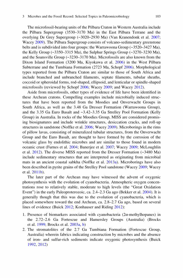
The microfossil-bearing units of the Pilbara Craton in Western Australia include
the Pilbara Supergroup (3530–3170 Ma) in the East Pilbara Terrane and the
overlying De Grey Supergroup (~3020–2930 Ma) (Van Kranendonk et al. 2007;
Wacey 2009). The Pilbara Supergroup consists of volcano-sedimentary greenstone
belts and is subdivided into four groups: the Warrawoona Group (~3520–3427 Ma),
the Kelly Group (~3350–3315 Ma), the Sulphur Springs Group (~3270–3230 Ma),
and the Soansville Group (~3230–3170 Ma). Microfossils are also known from the
Dixon Island Formation (3200 Ma, Kiyokawa et al. 2006) in the West Pilbara
Subterrane and the Tumbiana Formation (2723 Ma, Schopf 2006). Morphological
types reported from the Pilbara Craton are similar to those of South Africa and
include branched and unbranched filaments, septate filaments, tubular sheaths,
coccoid or spheroidal forms, rod-shaped, ellipsoid, and lenticular or spindle-shaped
microfossils (reviewed by Schopf 2006; Wacey 2009; and Wacey 2012).
Aside from microfossils, other types of evidence of life have been identified in
these Archean cratons. Compelling examples include microbially induced struc-
tures that have been reported from the Moodies and Onverwacht Groups in
South Africa, as well as the 3.48 Ga Dresser Formation (Warrawoona Group),
and the 3.35 Ga Euro Basalt and ~3.42–3.35 Ga Strelley Pool Formation (Kelly
Group) in Australia. In rocks of the Moodies Group, MISS are considered promis-
ing biosignatures and include wrinkle structures, desiccation cracks, and roll-up
structures in sandstone (Noffke et al. 2006; Wacey 2009). Microborings in the rims
of pillow lavas, consisting of mineralized tubular structures, from the Onverwacht
Group and the Euro Basalt, are thought to have formed by the corrosion of the
volcanic glass by endolithic microbes and are similar to those found in modern
oceanic crust (Furnes et al. 2004; Banerjee et al. 2007; Wacey 2009; McLoughlin
et al. 2012). The diverse MISS reported from the Dresser Formation (~3480 Ma)
include sedimentary structures that are interpreted as originating from microbial
mats in an ancient coastal sabkha (Noffke et al. 2013a). Microborings have also
been described in pyrite grains of the Strelley Pool sandstone (Wacey 2009; Wacey
et al. 2011b).
The later part of the Archean may have witnessed the advent of oxygenic
photosynthesis with the evolution of cyanobacteria. Atmospheric oxygen concen-
trations rose to relatively stable, moderate to high levels (the “Great Oxidation
Event”) in the early Paleoproterozoic, ca. 2.4–2.3 Ga ago (Bekker et al. 2004). It is
generally though that this was due to the evolution of cyanobacteria, which is
placed somewhere toward the end Archean, ca. 2.8–2.7 Ga ago, based on several
lines of evidence (Buick 2012; Konhauser and Riding 2012):
1. Presence of biomarkers associated with cyanobacteria (2α-methylhopanes) in
the 2.72–2.6 Ga Fortescue and Hamersley Groups (Australia) (Brocks
et al. 1999; Brocks et al. 2003a, b)
2. The stromatolites of the 2.7 Ga Tumbiana Formation (Fortescue Group,
Australia) wherein fabrics indicating construction by microbes and the absence
of iron- and sulfur-rich sediments indicate oxygenic photosynthesis (Buick
1992, 2012)
3 Microbes and the Fossil Record: Selected Topics in Paleomicrobiology 103
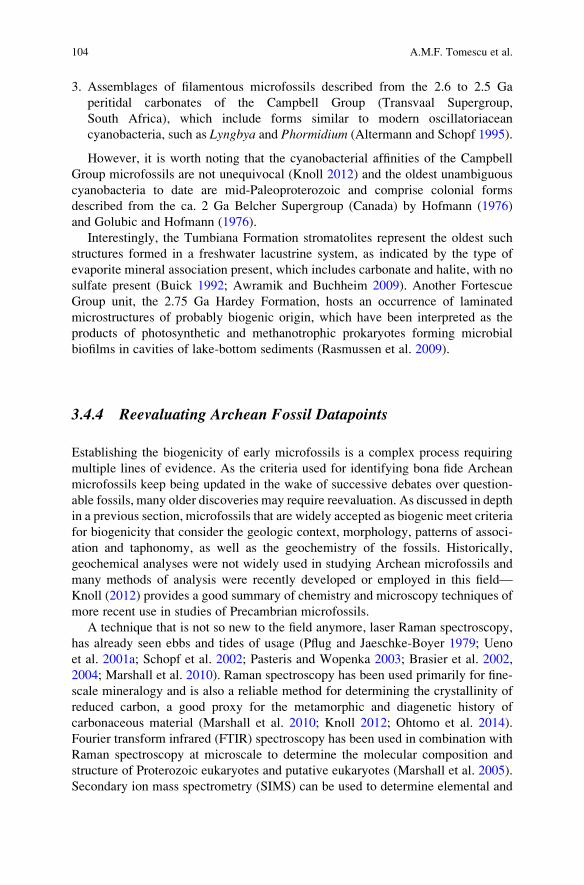
3. Assemblages of filamentous microfossils described from the 2.6 to 2.5 Ga
peritidal carbonates of the Campbell Group (Transvaal Supergroup,
South Africa), which include forms similar to modern oscillatoriacean
cyanobacteria, such as Lyngbya and Phormidium (Altermann and Schopf 1995).
However, it is worth noting that the cyanobacterial affinities of the Campbell
Group microfossils are not unequivocal (Knoll 2012) and the oldest unambiguous
cyanobacteria to date are mid-Paleoproterozoic and comprise colonial forms
described from the ca. 2 Ga Belcher Supergroup (Canada) by Hofmann (1976)
and Golubic and Hofmann (1976).
Interestingly, the Tumbiana Formation stromatolites represent the oldest such
structures formed in a freshwater lacustrine system, as indicated by the type of
evaporite mineral association present, which includes carbonate and halite, with no
sulfate present (Buick 1992; Awramik and Buchheim 2009). Another Fortescue
Group unit, the 2.75 Ga Hardey Formation, hosts an occurrence of laminated
microstructures of probably biogenic origin, which have been interpreted as the
products of photosynthetic and methanotrophic prokaryotes forming microbial
biofilms in cavities of lake-bottom sediments (Rasmussen et al. 2009).
3.4.4 Reevaluating Archean Fossil Datapoints
Establishing the biogenicity of early microfossils is a complex process requiring
multiple lines of evidence. As the criteria used for identifying bona fide Archean
microfossils keep being updated in the wake of successive debates over question-
able fossils, many older discoveries may require reevaluation. As discussed in depth
in a previous section, microfossils that are widely accepted as biogenic meet criteria
for biogenicity that consider the geologic context, morphology, patterns of associ-
ation and taphonomy, as well as the geochemistry of the fossils. Historically,
geochemical analyses were not widely used in studying Archean microfossils and
many methods of analysis were recently developed or employed in this field—
Knoll (2012) provides a good summary of chemistry and microscopy techniques of
more recent use in studies of Precambrian microfossils.
A technique that is not so new to the field anymore, laser Raman spectroscopy,
has already seen ebbs and tides of usage (Pflug and Jaeschke-Boyer 1979; Ueno
et al. 2001a; Schopf et al. 2002; Pasteris and Wopenka 2003; Brasier et al. 2002,
2004; Marshall et al. 2010). Raman spectroscopy has been used primarily for fine-
scale mineralogy and is also a reliable method for determining the crystallinity of
reduced carbon, a good proxy for the metamorphic and diagenetic history of
carbonaceous material (Marshall et al. 2010; Knoll 2012; Ohtomo et al. 2014).
Fourier transform infrared (FTIR) spectroscopy has been used in combination with
Raman spectroscopy at microscale to determine the molecular composition and
structure of Proterozoic eukaryotes and putative eukaryotes (Marshall et al. 2005).
Secondary ion mass spectrometry (SIMS) can be used to determine elemental and
104 A.M.F. Tomescu et al.

isotopic compositions and map them on samples at high resolution (House
et al. 2000; De Gregorio et al. 2009). The molecular composition of microfossils
has been assessed using laser pyrolysis gas chromatography—mass spectrometry
(Arouri et al. 2000). Furthermore, several types of electron microscopy (transmis-
sion electron microscopy [TEM], scanning-transmission electron microscopy
[STEM], and high-resolution transmission electron microscopy [HRTEM]) have
been employed to examine the morphology and nanostructure of carbon (Ohtomo
et al. 2014) and microfossils (Javaux et al. 2004, 2010). Fossil imaging is also
performed using confocal laser microscopy (Schopf et al. 2006; Lepot et al. 2008).
Synchrotron-based scanning-transmission X-ray microscopy (STXM) has been
used in combination with X-ray absorption near-edge structure spectroscopy
(XANES) for imaging, as well as elemental and molecular mapping (Boyce
et al. 2002; De Gregorio et al. 2009).
These techniques represent important sources of new information in the
reevaluation of older discoveries. This is the case for some of the fossils listed in
Table 3.2, including microfossils from the Kromberg Formation and some of the
microfossils reported from the Hooggenoeg Formation (Onverwacht Group)
(Wacey 2009), as well as those from the Tumbiana Formation (Buick 2001).
Moreover, in order to identify the oldest evidence of life, the age of the rock and
syngenicity of the microfossils must be firmly established, for which thorough
understanding of the geologic context of the locality is key. When microfossils
from the ~3470 Ma Mount Ada Basalt were reported in 1983, these were the oldest
convincing microfossils known at the time (Awramik et al. 1983). However, the
precise collection locality remains unknown and has never been resampled, which
casts reasonable doubts on the age of these fossils, as they could originate from the
much younger Fortescue Group (Schopf 2006; Wacey 2009; Brasier and Wacey
2012).
3.4.4.1 The Apex Chert Debate
Microfossils from cherts in the Apex Basalt (Warrawoona Group, Australia)
(Fig. 3.5a) were first reported by Schopf and Packer (1987) and later described by
Schopf (1993). Eleven taxa of filamentous prokaryotes were circumscribed based
on various aspects of their morphology. They were interpreted as bona fide Archean
microfossils based on their frequent presence in the rocks, the early Archean age of
the fossiliferous cherts, the occurrence of the fossils within clasts of the brecciated
chert and absence from the surrounding matrix, and their morphological complexity
and similarity to younger prokaryotes (Schopf 1993). Data from Laser-Raman
analysis was later presented in support of biogenicity (Schopf et al. 2002), showing
that the microfossils contained kerogen (geologically transformed organic matter).
The Apex fossils were widely accepted as the oldest and best-preserved microbial
fossils (Marshall et al. 2011) until Brasier et al. (2002) disputed their biogenicity,
sparking a debate that has made them the most controversial Archean microfossils.
3 Microbes and the Fossil Record: Selected Topics in Paleomicrobiology 105
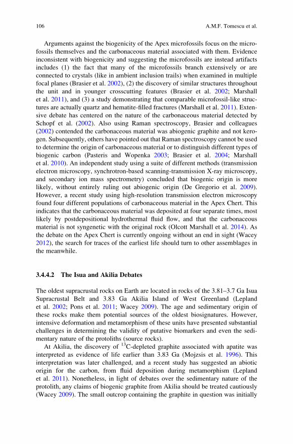
Arguments against the biogenicity of the Apex microfossils focus on the micro-
fossils themselves and the carbonaceous material associated with them. Evidence
inconsistent with biogenicity and suggesting the microfossils are instead artifacts
includes (1) the fact that many of the microfossils branch extensively or are
connected to crystals (like in ambient inclusion trails) when examined in multiple
focal planes (Brasier et al. 2002), (2) the discovery of similar structures throughout
the unit and in younger crosscutting features (Brasier et al. 2002; Marshall
et al. 2011), and (3) a study demonstrating that comparable microfossil-like struc-
tures are actually quartz and hematite-filled fractures (Marshall et al. 2011). Exten-
sive debate has centered on the nature of the carbonaceous material detected by
Schopf et al. (2002). Also using Raman spectroscopy, Brasier and colleagues
(2002) contended the carbonaceous material was abiogenic graphite and not kero-
gen. Subsequently, others have pointed out that Raman spectroscopy cannot be used
to determine the origin of carbonaceous material or to distinguish different types of
biogenic carbon (Pasteris and Wopenka 2003; Brasier et al. 2004; Marshall
et al. 2010). An independent study using a suite of different methods (transmission
electron microscopy, synchrotron-based scanning-transmission X-ray microscopy,
and secondary ion mass spectrometry) concluded that biogenic origin is more
likely, without entirely ruling out abiogenic origin (De Gregorio et al. 2009).
However, a recent study using high-resolution transmission electron microscopy
found four different populations of carbonaceous material in the Apex Chert. This
indicates that the carbonaceous material was deposited at four separate times, most
likely by postdepositional hydrothermal fluid flow, and that the carbonaceous
material is not syngenetic with the original rock (Olcott Marshall et al. 2014). As
the debate on the Apex Chert is currently ongoing without an end in sight (Wacey
2012), the search for traces of the earliest life should turn to other assemblages in
the meanwhile.
3.4.4.2 The Isua and Akilia Debates
The oldest supracrustal rocks on Earth are located in rocks of the 3.81–3.7 Ga Isua
Supracrustal Belt and 3.83 Ga Akilia Island of West Greenland (Lepland
et al. 2002; Pons et al. 2011; Wacey 2009). The age and sedimentary origin of
these rocks make them potential sources of the oldest biosignatures. However,
intensive deformation and metamorphism of these units have presented substantial
challenges in determining the validity of putative biomarkers and even the sedi-
mentary nature of the protoliths (source rocks).
At Akilia, the discovery of 13C-depleted graphite associated with apatite was
interpreted as evidence of life earlier than 3.83 Ga (Mojzsis et al. 1996). This
interpretation was later challenged, and a recent study has suggested an abiotic
origin for the carbon, from fluid deposition during metamorphism (Lepland
et al. 2011). Nonetheless, in light of debates over the sedimentary nature of the
protolith, any claims of biogenic graphite from Akilia should be treated cautiously
(Wacey 2009). The small outcrop containing the graphite in question was initially
106 A.M.F. Tomescu et al.
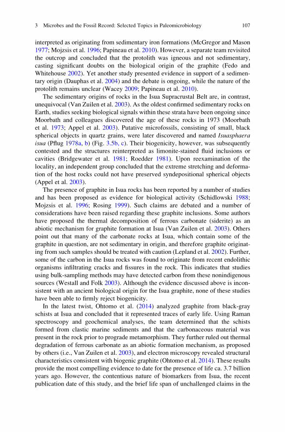
interpreted as originating from sedimentary iron formations (McGregor and Mason
1977; Mojzsis et al. 1996; Papineau et al. 2010). However, a separate team revisited
the outcrop and concluded that the protolith was igneous and not sedimentary,
casting significant doubts on the biological origin of the graphite (Fedo and
Whitehouse 2002). Yet another study presented evidence in support of a sedimen-
tary origin (Dauphas et al. 2004) and the debate is ongoing, while the nature of the
protolith remains unclear (Wacey 2009; Papineau et al. 2010).
The sedimentary origins of rocks in the Isua Supracrustal Belt are, in contrast,
unequivocal (Van Zuilen et al. 2003). As the oldest confirmed sedimentary rocks on
Earth, studies seeking biological signals within these strata have been ongoing since
Moorbath and colleagues discovered the age of these rocks in 1973 (Moorbath
et al. 1973; Appel et al. 2003). Putative microfossils, consisting of small, black
spherical objects in quartz grains, were later discovered and named Isuasphaeraisua (Pflug 1978a, b) (Fig. 3.5b, c). Their biogenicity, however, was subsequently
contested and the structures reinterpreted as limonite-stained fluid inclusions or
cavities (Bridgewater et al. 1981; Roedder 1981). Upon reexamination of the
locality, an independent group concluded that the extreme stretching and deforma-
tion of the host rocks could not have preserved syndepositional spherical objects
(Appel et al. 2003).
The presence of graphite in Isua rocks has been reported by a number of studies
and has been proposed as evidence for biological activity (Schidlowski 1988;
Mojzsis et al. 1996; Rosing 1999). Such claims are debated and a number of
considerations have been raised regarding these graphite inclusions. Some authors
have proposed the thermal decomposition of ferrous carbonate (siderite) as an
abiotic mechanism for graphite formation at Isua (Van Zuilen et al. 2003). Others
point out that many of the carbonate rocks at Isua, which contain some of the
graphite in question, are not sedimentary in origin, and therefore graphite originat-
ing from such samples should be treated with caution (Lepland et al. 2002). Further,
some of the carbon in the Isua rocks was found to originate from recent endolithic
organisms infiltrating cracks and fissures in the rock. This indicates that studies
using bulk-sampling methods may have detected carbon from these nonindigenous
sources (Westall and Folk 2003). Although the evidence discussed above is incon-
sistent with an ancient biological origin for the Isua graphite, none of these studies
have been able to firmly reject biogenicity.
In the latest twist, Ohtomo et al. (2014) analyzed graphite from black-gray
schists at Isua and concluded that it represented traces of early life. Using Raman
spectroscopy and geochemical analyses, the team determined that the schists
formed from clastic marine sediments and that the carbonaceous material was
present in the rock prior to prograde metamorphism. They further ruled out thermal
degradation of ferrous carbonate as an abiotic formation mechanism, as proposed
by others (i.e., Van Zuilen et al. 2003), and electron microscopy revealed structural
characteristics consistent with biogenic graphite (Ohtomo et al. 2014). These results
provide the most compelling evidence to date for the presence of life ca. 3.7 billion
years ago. However, the contentious nature of biomarkers from Isua, the recent
publication date of this study, and the brief life span of unchallenged claims in the
3 Microbes and the Fossil Record: Selected Topics in Paleomicrobiology 107

field of early biosignature research caution against immediate adoption of these
findings which await scientific consensus and independent corroboration.
3.4.4.3 The Alan Hills 84001 Martian Meteorite Controversy
Although only indirectly relevant to questions about early life on Earth, the Alan
Hills—ALH84001 Martian meteorite controversy is included here because of its
relevance to issues of biogenicity and their implications for astrobiology. In 1996,
McKay et al. argued for the presence of traces of Martian life in meteorite ALH
84001. The evidence they presented in support of their claim came from several
types of data: (1) polycyclic aromatic hydrocarbons interpreted as diagenetic
products of microorganisms and considered to be indigenous to the meteorite,
(2) chemistry and mineral composition and structures suggestive of (microbial)
biogenic products or biologic behavior, (3) magnetite particles similar to
magnetofossils (magnetosomes) left by magnetotactic bacteria (Fig. 3.5d), and
(4) submicron bacteriomorphs similar to nanobacteria (Folk 1993) and bacterial
fossils.
McKay et al.’s (1996) claim that these ALH 84001 features represented evidence
of ancient Martian life was met with a great deal of skepticism (Bradley et al. 1997
and others). Multiple research groups have verified the presence of polycyclic
aromatic hydrocarbons in ALH84001 (Clemett et al. 1998; Steele et al. 2012).
However, mechanisms for abiotic formation of polycyclic aromatic hydrocarbons
have been documented (Zolotov and Shock 2000; McCollum 2003; Treiman
2003a). Additionally, Steele et al. (2012) demonstrated that abiotically formed
organic compounds are present on multiple Martian meteorites (including
ALH84001) and that these compounds were not contaminants from Earth. These
findings are inconsistent with the original claims of biogenic polycyclic aromatic
hydrocarbons in ALH84001. The claim that the mineral composition—magnetite,
iron sulfide, and siderite—is indicative of life and its byproducts has also been
scrutinized. This suite of minerals is well known in low-temperature, aqueous
systems (Anders 1996; Golden et al. 2000) and has not been shown to be exclu-
sively indicative of biologic behavior (Treiman 2003b).
There is disagreement over how similar the magnetite grains in ALH84001 are
to the magnetosomes of Earth bacteria (such as the marine magnetotactic vibrio
Magnetovibrio blakemorei strain MV-1) (Clemett et al. 2002; Treiman 2003b).
While some authors point to close similarity (Thomas-Keprta et al. 2001), others
have documented differences in morphology between magnetosomes of MV-1 and
other bacterial strains, and the magnetite crystals in ALH84001 (Buseck et al. 2001;
Golden et al. 2004). Golden et al. (2004) demonstrated that the morphology of
ALH84001 magnetite crystals is replicated abiogenically and that the most com-
mon crystal morphology for biogenic magnetite is different from that in both
ALH84001 and abiogenic magnetite, concluding that rather than representing a
compelling biosignature, the morphology of the ALH84001 magnetite crystals is
consistent with an abiogenic origin. Finally, the submicron bacteriomorphs from
108 A.M.F. Tomescu et al.

ALH84001 are largely discounted as biogenic structures (Treiman 2003b) based on
the fact that at < 100 nm they are just below the size of the smallest free-living
prokaryotes (Gorbushina and Krumbein 2000) and that a number of abiotic mech-
anisms have been proposed for the formation of such structures, including mineral
precipitation from solution (Bradley et al. 1998; Kirkland et al. 1999; Vecht and
Ireland 2000; Grasby 2003).
Overall it is the inability to reject the null hypothesis of abiotic formation
(Brasier and Wacey 2012) that does not allow the features described in
ALH84001 to be considered biogenic (Treiman 2003b). Since the polycyclic
aromatic hydrocarbons have been shown to be abiotic (Steele et al. 2012), the
mineral composition of the carbonate globules is not a definitive indicator of
biological activity, and both the magnetite grains and nano-bacteriomorphs shaped
could have formed abiotically, it is unlikely that ALH84001 contains biogenic
material. However, it will be interesting to see the results of the application of
Gehring et al.’s (2011) method of distinguishing between bacterial magnetosomes
and abiogenic magnetite to the ALH84001 magnetite grains.
3.4.5 Oldest Traces of Life
In the context of the findings summarized above, it is clear that there is a large body
of evidence suggesting that life has existed since the Paleoarchean
(3600–3200 Ma), despite the questionable or equivocal status of some of these
fossils (Knoll 2012). Although not all stromatolite and other microbially induced
structure occurrences are included in Table 3.2, it is worth noting that the strati-
graphic extent of stromatolites, MISS, and microborings mirrors closely that of
microfossils (e.g., Hofmann 2000; Awramik and Grey 2005; Brasier et al. 2006;
Fliegel et al. 2010). While the oldest reported putative microfossils originate from
the ~3480 Ma Dresser Formation (Australia) (Fig. 3.6a), their biogenicity remains
equivocal. The most convincing of these microfossils are carbonaceous filaments
reported by Ueno et al. (2001a, b), but their biogenic interpretation based on
morphology and stable carbon isotopes awaits further verification (Wacey 2009).
Microbial presence in the Dresser Formation is also supported by putative stromat-
olites (Buick et al. 1981; Awramik and Grey 2005; Van Kranendonk 2006; Wacey
2009) (Fig. 3.6b). Irrespective of the verdict on the microfossils and stromatolites,
unequivocal MISS described by Noffke et al. (2013a) in the Dresser Formation
demonstrate presence of microbial life very close to 3.5 Ga ago. The Dresser
Formation MISS formed in shallow-water, low-energy evaporitic coastal environ-
ments (Buick and Dunlop 1990; Noffke et al. 2013a).
Convincing microfossils, only slightly younger than the Dresser Formation
fossils, are known from the ~3466 Ma Kitty’s Gap Chert (Warrawoona Group)
(Westall et al. 2006) (Fig. 3.7a, b); the ~3450 Ma Hooggenoeg Formation
(Onverwacht Group), particularly those reported by Glikson et al. (2008)
(Fig. 3.7c, e, f); and the ~3426–3350 Strelley Pool Formation (Kelly Group)
3 Microbes and the Fossil Record: Selected Topics in Paleomicrobiology 109

(Wacey et al. 2011a; Sugitani et al. 2013) (Fig. 3.8a–e). Dresser Formation micro-
fossils notwithstanding, the Kitty’s Gap Chert, and Hooggenoeg Formation host the
oldest unequivocal microbial body fossils known to date. Although slightly youn-
ger, the Strelley Pool Formation microfossils provide very good evidence for life
corroborated by independent studies, with good geochemical evidence supporting
their biogenicity, and the presence of other types of fossils within the formation,
including microborings and stromatolites (Hofmann et al. 1999; Brasier et al. 2006;
Allwood et al. 2007; Wacey et al. 2006, 2011a, b; Sugitani et al. 2013) (Fig. 3.8f–h).
Aside from the Strelley Pool Formation microborings, which are hosted in sedi-
mentary rocks and the oldest known to date in the fossil record, the oldest
microborings in volcanic glass have been reported from 3.34 Ga in the Hooggenoeg
Formation (Furnes et al. 2004; Fliegel et al. 2010; McLoughlin et al. 2012)
(Fig. 3.7d, g) and from the 3.42–3.31 Ga Euro Basalt of the Kelly Group (Banerjee
et al. 2007) (Fig. 3.8i).
3.5 Microbial Eukaryotes: Recognition and Early Fossil
Record
Irrespective of the detailed circumstances of their evolution, as proposed by several
competing hypotheses, eukaryotes arose from prokaryotic stock (Knoll and
Bambach 2000; Lang and Burger 2012). Whereas in the case of the earliest pro-
karyotes proof of biogenicity is one of the biggest hurdles, in early eukaryotes their
very eukaryotic affinities are challenging to ascertain. Because of their prokaryotic
origins, it is to be expected that early eukaryotes were unicellular organisms and
that they will be difficult to distinguish from prokaryotes. Indeed, the earliest bona
fide eukaryotes and even older putative eukaryotes are unicellular dispersed micro-
fossils (Javaux et al. 2010; Knoll 2014). From the perspective of the paleontologist,
most early eukaryotes fall in the category of acritarchs (e.g., Figs. 3.9a–d, 3.10a–d,
g, 3.11a, c), an artificial group of organic-walled unicellular microfossils of large
size (50 μm or more) and uncertain biological affinities which comprise the most
abundant and widely distributed record of Proterozoic protists (Knoll et al. 2006;
Buick 2010).
3.5.1 Recognizing Early Eukaryotes
Known from dispersed unicellular microfossils in Proterozoic rocks (and possibly
going as far back in time as the Archean; Javaux et al. 2010; Knoll et al. 2006; Knoll
2014), early eukaryotes have to pass the same tests of indigenousness and
syngenicity as their prokaryotic counterparts (e.g., Bengtson et al. 2009; Javaux
et al. 2010), in addition to satisfying eukaryote-specific criteria. Putative eukaryotes
110 A.M.F. Tomescu et al.

have generally been recognized based on the fact that they display complexity
unknown in prokaryotes (Schopf and Klein 1992; Javaux 2007). This is expressed
in the interrelated abilities to synthesize complex polymers and produce complex
structures (e.g., Javaux et al. 2004; Javaux 2007). The production of complex
polymers is reflected in the recalcitrant nature of cell walls or their ability to
withstand acid maceration, a feature often considered important in distinguishing
taxonomic affinities at the domain level (e.g., Javaux et al. 2004; Knoll et al. 2006;
Javaux 2007). In turn, recalcitrant cell walls preserve complex cell surface struc-
tures. Aside from these, large size (> ca. 50 μm) is often treated as indicative of
eukaryotic affinities (e.g., Schopf and Klein 1992). However, size is not diagnostic
by itself, as large bacteria and cyanobacterial sheaths are known (e.g., Waterbury
and Stanier 1978; Schulz et al. 1999), as well as eukaryotes smaller than 1 μm in
diameter (e.g., Courties et al. 1994).
The hallmark of complex cell structure, cellular organelles, has yet to be
unequivocally substantiated in the early fossil record. Early reports of acritarchs
Fig. 3.9 Recognizing early unicellular eukaryotes—acritarchs. (a) Satka favosa, showing surfaceornamentation (cell wall consisting of interlocking panels), Mesoproterozoic Roper Group,
Australia. (b) Tappania plana, showing asymmetrically distributed long processes (some of
which are branched) protruding from the cell wall and bulbous protrusions (arrow) potentially
indicative of vegetative reproduction by “budding,” Roper Group. (c) Tappania plana, showingpossible excystment structure at the apex of a necklike extension (at top left), Roper group. (d)Tappania plana, showing asymmetrically distributed long cell surface processes, Roper Group. (e)
Leiosphaeridia jacutica, showing complex cell wall structure: two electron-dense, homogeneous
layers that sandwich a thick central layer with electron-dense, porous texture, Roper Group. (f)
Leiosphaeridia crassa, showing complex cell wall organization consisting of as many as four
structurally distinct layers preserved at places (arrow), Roper Group. Scale bars: (a) 40 μm, (b)
35 μm, (c) 20 μm, (d) 35 μm, (e) 1 μm, (f) 1.6 μm. Credits—images used with permission from (a),
(b), and (d) Nature Publishing Group (Javaux et al. 2001). (c), (e), and (f) Blackwell Publishing
Ltd. (Javaux et al. 2004)
3 Microbes and the Fossil Record: Selected Topics in Paleomicrobiology 111
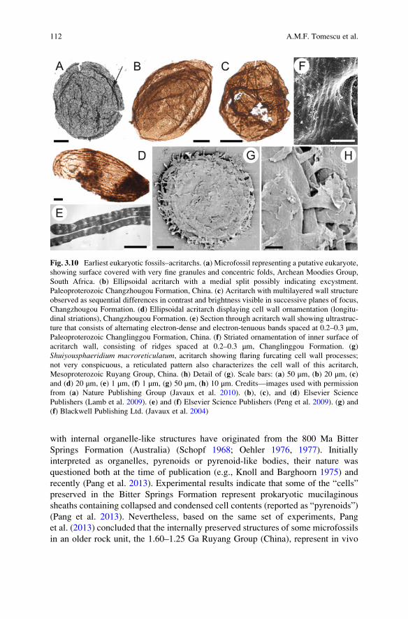
with internal organelle-like structures have originated from the 800 Ma Bitter
Springs Formation (Australia) (Schopf 1968; Oehler 1976, 1977). Initially
interpreted as organelles, pyrenoids or pyrenoid-like bodies, their nature was
questioned both at the time of publication (e.g., Knoll and Barghoorn 1975) and
recently (Pang et al. 2013). Experimental results indicate that some of the “cells”
preserved in the Bitter Springs Formation represent prokaryotic mucilaginous
sheaths containing collapsed and condensed cell contents (reported as “pyrenoids”)
(Pang et al. 2013). Nevertheless, based on the same set of experiments, Pang
et al. (2013) concluded that the internally preserved structures of some microfossils
in an older rock unit, the 1.60–1.25 Ga Ruyang Group (China), represent in vivo
Fig. 3.10 Earliest eukaryotic fossils–acritarchs. (a) Microfossil representing a putative eukaryote,
showing surface covered with very fine granules and concentric folds, Archean Moodies Group,
South Africa. (b) Ellipsoidal acritarch with a medial split possibly indicating excystment.
Paleoproterozoic Changzhougou Formation, China. (c) Acritarch with multilayered wall structure
observed as sequential differences in contrast and brightness visible in successive planes of focus,
Changzhougou Formation. (d) Ellipsoidal acritarch displaying cell wall ornamentation (longitu-
dinal striations), Changzhougou Formation. (e) Section through acritarch wall showing ultrastruc-
ture that consists of alternating electron-dense and electron-tenuous bands spaced at 0.2–0.3 μm,
Paleoproterozoic Changlinggou Formation, China. (f) Striated ornamentation of inner surface of
acritarch wall, consisting of ridges spaced at 0.2–0.3 μm, Changlinggou Formation. (g)
Shuiyousphaeridium macroreticulatum, acritarch showing flaring furcating cell wall processes;
not very conspicuous, a reticulated pattern also characterizes the cell wall of this acritarch,
Mesoproterozoic Ruyang Group, China. (h) Detail of (g). Scale bars: (a) 50 μm, (b) 20 μm, (c)
and (d) 20 μm, (e) 1 μm, (f) 1 μm, (g) 50 μm, (h) 10 μm. Credits—images used with permission
from (a) Nature Publishing Group (Javaux et al. 2010). (b), (c), and (d) Elsevier Science
Publishers (Lamb et al. 2009). (e) and (f) Elsevier Science Publishers (Peng et al. 2009). (g) and
(f) Blackwell Publishing Ltd. (Javaux et al. 2004)
112 A.M.F. Tomescu et al.
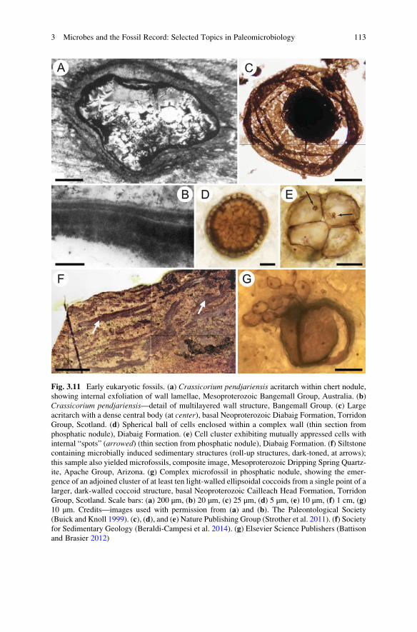
Fig. 3.11 Early eukaryotic fossils. (a) Crassicorium pendjariensis acritarch within chert nodule,
showing internal exfoliation of wall lamellae, Mesoproterozoic Bangemall Group, Australia. (b)
Crassicorium pendjariensis—detail of multilayered wall structure, Bangemall Group. (c) Large
acritarch with a dense central body (at center), basal Neoproterozoic Diabaig Formation, Torridon
Group, Scotland. (d) Spherical ball of cells enclosed within a complex wall (thin section from
phosphatic nodule), Diabaig Formation. (e) Cell cluster exhibiting mutually appressed cells with
internal “spots” (arrowed) (thin section from phosphatic nodule), Diabaig Formation. (f) Siltstone
containing microbially induced sedimentary structures (roll-up structures, dark-toned, at arrows);
this sample also yielded microfossils, composite image, Mesoproterozoic Dripping Spring Quartz-
ite, Apache Group, Arizona. (g) Complex microfossil in phosphatic nodule, showing the emer-
gence of an adjoined cluster of at least ten light-walled ellipsoidal coccoids from a single point of a
larger, dark-walled coccoid structure, basal Neoproterozoic Cailleach Head Formation, Torridon
Group, Scotland. Scale bars: (a) 200 μm, (b) 20 μm, (c) 25 μm, (d) 5 μm, (e) 10 μm, (f) 1 cm, (g)
10 μm. Credits—images used with permission from (a) and (b). The Paleontological Society
(Buick and Knoll 1999). (c), (d), and (e) Nature Publishing Group (Strother et al. 2011). (f) Society
for Sedimentary Geology (Beraldi-Campesi et al. 2014). (g) Elsevier Science Publishers (Battison
and Brasier 2012)
3 Microbes and the Fossil Record: Selected Topics in Paleomicrobiology 113
protoplasm condensation corresponding to an encystment stage common in some
algae, thus supporting their eukaryotic affinities.
3.5.1.1 Morphological Criteria
Dispersed unicellular microfossils are most compellingly recognized as eukaryotes
based on morphological criteria (Javaux et al. 2003) although geochemical data on
biomarkers have also been used, sometimes exclusively (e.g., Brocks et al. 1999), in
support of eukaryote presence. In terms of morphology, three categories of features
are considered diagnostic of eukaryotic nature, especially when co-occurring, in
studies of microfossils (Javaux et al. 2003): complex cell surface features,
excystment structures, and complex wall organization.
1. Complex cell surface features: these include ornamentation (Figs. 3.9a and
3.10a, d, f) and processes protruding from the cell surface (Figs. 3.9b, d and
3.10 g, h). The use of complex surface features to recognize eukaryotic cells
stems from Cavalier-Smith’s (2002) reasoning that production of such surface
features would require that the cell possesses an endomembrane system and
cytoskeleton, which are fundamental components of the eukaryotic cell. Javaux
et al. (2004) point out that while prokaryotic organisms can synthesize both cell
wall ornament and preservable structures, wall ornamentation rarely occurs on
the size scale observed in Precambrian candidate eukaryotes and is seldom found
on preservable structures.
Regular polygonal patterns occurring at the micron scale are seen on the
surfaces of Proterozoic eukaryotic microfossils (Javaux et al. 2003, 2004).
Although many prokaryotes possess oblique, square, or hexagonal crystalline
arrays on their surfaces, these shapes occur at the nanometer scale. Furthermore,
their easy removability by chemicals in culture studies suggests that they are
unlikely to preserve or to survive the acid maceration processes used to extract
dispersed carbonaceous microfossils from the rock (Javaux et al. 2003). Other
examples of surface ornamentation include striations and lineations (Fig. 3.10d),
chagrinate (covered with very fine granules) surfaces (Fig. 3.10a), cruciform
structures, or internal (inner wall surface) striations (Fig. 3.10f) (Javaux
et al. 2001, 2004; Butterfield 2009). Sometimes, larger bulbous protrusions
suggestive of vegetative reproduction through budding are present, as in
Tappania plana (Javaux et al. 2001, 2004) (Fig. 3.9b).
Cell surface processes documented in early eukaryotes have varied morphol-
ogies, from spiny or thin and unbranched to bulbous or branched (Javaux
et al. 2003). Some can be long (e.g., up to 60 μm in Tappania plana of the
1.50–1.45 Ga Roper Group, Australia; Javaux et al. 2001) (Fig. 3.9b, d), while
others are septate, branched (Fig. 3.9b and 3.10 g, h), and anastomosing (e.g., the
putative basal fungus “Tappania” from the 850 to 750 Ma Wynniatt Formation,
Canada; Butterfield 2009). Processes with irregular length, exhibiting hetero-
morphism within one cell or asymmetric distribution on the cell surface (Javaux
114 A.M.F. Tomescu et al.

et al. 2001; Knoll et al. 2006) (Fig. 3.9b, d), can only be accounted for by active
growth and remodeling of the cell which requires a dynamic cytoskeletal
architecture and regulatory networks that characterize eukaryotes (Javaux
et al. 2001, 2004).
2. Excystment structures: these are openings in the walls of microfossils that are
thought to be produced by liberation of internal vegetative cells from the wall of
a resting stage, another eukaryotic feature (Fig. 3.9c and 3.10b). Commonly
recognized in Phanerozoic microfossils, excystment structures are much more
equivocal in Precambrian fossils (Javaux et al. 2003). For instance, in the Roper
Group fossils, at least two taxa show possible excystment structures. Because
these structures are perforations or splits that run some length of the cell wall,
they are not regularly occurring (there are few occurrences in the population),
and the microfossils generally lack evidence of vegetative cells having been
present, interpretation as degradational ruptures or breaks in the microfossil cell
wall seems equally likely. More complex structures, such as those seen in
Tappania plana which possesses a slit at the apex of a necklike extension
(Javaux et al. 2003) (Fig. 3.9c), are more credible as eukaryotic excystment
structures.
3. Complex cell wall organization: the structural complexity of eukaryotic cell walls
can be preserved in microfossils and distinguished from acetolysis-resistant struc-
tures formed by bacteria (Javaux et al. 2003). Thus, ultrastructural features
(Figs. 3.9e, f, 3.10e, and 3.11b) can provide evidence for eukaryotic affinities,
even in microfossils in which morphology is not diagnostic (Javaux et al. 2004).
These authors undertook a careful TEM study of cell wall ultrastructure demon-
strating that while some eukaryotes (recognized based on morphological charac-
ters observed in light microscopy and SEM) share nondiagnostic wall
ultrastructures consisting of single, homogeneous, electron-dense layers of vari-
able thickness, in others cell wall organization is more complex (Fig. 3.9e, f). In
the latter microfossils, some of which do not display other features indicative of
eukaryotic affinities, cell walls exhibit at least four distinct types of structures
characterized by multiple layers of different density and organization (e.g., dense,
porous, laminar, fibrous) (Javaux et al. 2004) (Fig. 3.9f).
Discussing the multilayered structure of Leiosphaeridia crassa from the
Roper Group, Javaux et al. (2004) point out that such structures occur in the
acetolysis-resistant walls of many green algae. Several authors have discussed
similarities between the cell wall structure of different early eukaryotes and
those of extant chlorococcalean green algae (Arouri et al. 1999), prasinophyte
green algae (Loeblich 1970), or volvocalean green algae (Moczydlowska and
Willman 2009). Javaux et al. (2004) also discuss the possibility that acetolysis-
resistant walls structurally similar to those described in the early putative
eukaryotes are produced by prokaryotic organisms; this seems not to be the
case, as few bacteria make spores with comparable size, surface ornamentation,
and preservation potential. For example, myxobacteria (which are mostly ter-
restrial) produce spore-enclosing structures (sporangioles) up to 50 μm in diam-
eter, but these have smooth walls and unknown chemical composition and are
3 Microbes and the Fossil Record: Selected Topics in Paleomicrobiology 115

not known to preserve in sediments. Actinobacteria have spores up to 3 μm in
diameter that can be ornamented, but their surface ornaments are very small,
proteinaceous, and are unlikely to survive fossilization processes. In contrast, the
extracellular polysaccharide sheaths that envelop coccoidal cyanobacterial col-
onies can exceed 100 μm in size and are commonly fossilized (Bartley 1996);
however, these envelopes are smooth walled, and their ultrastructure consists of
fibrous layers distinct from those of eukaryotes (Javaux et al. 2004).
3.5.1.2 Geochemistry: Hydrocarbon Biomarkers
Steranes are molecular fossils representing the geologically stable derivatives of
sterols, which are eukaryote-specific compounds (Summons and Lincoln 2012). As
a result, steranes in the rock record have been used as biomarkers for eukaryote
presence. The oldest claims for eukaryotes are based exclusively on the presence of
steranes, unaccompanied by body fossils, in Archean rocks dated at 2.72–2.60 Ma
in Australia (Fortescue and Hamersley Groups; Brocks et al. 1999; Brocks
et al. 2003a, b) and at 2.67–2.46 Ga in South Africa (Transvaal Supergroup;
Waldbauer et al. 2009). However, when not backed by microfossil evidence, the
use of hydrocarbon biomarkers as evidence for eukaryotes is contentious for several
reasons (Buick 2010; Knoll 2014). One of the reasons is that sterol biosynthesis
requires molecular oxygen, which was not present in the Archean, except maybe for
rare, very localized “oxygen oases” produced by photosynthetic cyanobacteria in
microbial mats (Knoll 2014). Another reason is that although very rare, some
prokaryotic organisms are known to synthesize sterols; this reason is relatively
easy to dismiss as known prokaryotic sterols are simple and, thus, could not have
generated the complex steranes that have been reported from Archean rocks (Knoll
2014). The most contentious issues are those of indigenousness and syngenicity,
because hydrocarbons are known to migrate through rock over significant distances
and thicknesses from their layers of origin (geological contaminants) or can be
introduced in drill core samples by the drilling process (modern contaminants)
(Knoll 2014). Indeed, based on very different carbon isotope ratios measured in
different organic fractions of the same rocks, Rasmussen et al. (2008) showed that
the biomarkers reported by Brocks et al. (1999) are not indigenous. To date, the
oldest unequivocally dated steranes come from >2.2 Ga fluid inclusions trapped by
metamorphic processes in closed systems, and therefore contamination-proof, in
the Matinenda Formation (Canada; Dutkiewicz et al. 2006; George et al. 2008).
3.5.2 Oldest Evidence for Eukaryotes
As pointed out by Knoll (2014), when it comes to the oldest evidence for eukary-
otes, there is no clear-cut age limit—rather, the geological record presents us with
evidence that falls along a sliding scale of certainty that ranges from bona fide,
116 A.M.F. Tomescu et al.
confidently interpreted fossils down to around 1.8–2.0 Ga ago to increasingly more
equivocal fossils or chemical biosignatures in older rocks. At least ten rock units
around the world have yielded Paleoproterozoic and Mesoproterozoic acritarchs of
eukaryotic affinities to date (Table 3.3); the morphological diversity of acritarchs in
most of these units has been carefully summarized by Knoll et al. (2006). Some of
these units also host putative multicellular eukaryotes.
3.5.2.1 Acritarchs
The oldest acritarchs interpreted with reasonable confidence as eukaryotes have
been reported from the late Paleoproterozoic Changzhougou Formation and the
overlying Changlinggou Formation (Chang Cheng Group, China; Lamb et al. 2009;
Peng et al. 2009) dated between 1.80 and 1.65 Ga (Table 3.3) (Fig. 3.10b–f). They
include several distinct forms with complex cell wall structure (Fig. 3.10c, e), wall
ornamentation (Fig. 3.10d, f), or putative excystment structures (Fig. 3.10b), indi-
cating that eukaryotes had already started diversifying before the end of the
Paleoproterozoic (Lamb et al. 2009). The ca. 1.65 Ga Mallapunyah Formation
(Australia) has also yielded acritarchs with ornamented cell walls interpreted as
eukaryotes (Javaux et al. 2004). In the Mesoproterozoic, several rock units dated
between 1.6 and 1.1 Ga host diverse eukaryotic acritarch assemblages—the Ruyang
Group in China (Xiao et al. 1997; Yin 1997; Javaux et al. 2004) (Fig. 3.10g, h), the
Sarda and Avadh Formations in India (Knoll et al. 2006), and the Bangemall Group
in Australia (Buick and Knoll 1999) (Fig. 3.11a, b). Detailed studies of both the
morphology and the distribution of eukaryotic acritarchs in the Roper Group of
Australia (Fig. 3.9a–f) have revealed high morphological diversity and suggest
niche partition among early eukaryotes 1.50–1.45 Ga ago, at the beginning of the
Mesoproterozoic (Javaux et al. 2001, 2003, 2004). Another early Mesoproterozoic
unit containing diverse acritarchs is the Chamberlain Shale of Montana
(1.47–1.42 Ga; Horodyski 1980). Considerable eukaryotic acritarch diversity has
been reported from the middle and late Mesoproterozoic Thule Supergroup
(1.3–1.2 Ga; Greenland), which includes several fossiliferous subunits (Samuelsson
et al. 1999), and the Lakhanda Group (1.1–1.0 Ga; Siberia; Knoll et al. 2006).
The oldest evidence for eukaryotic life on land may be preserved in the
ca. 1.2 Ga Dripping Spring Quartzite (Apache Group, Arizona; Beraldi-Campesi
et al. 2014) (Fig. 3.11f). Here, diverse and abundant MISS preserved on
paleosurfaces that display desiccation features and bear strong morphological
resemblance to modern terrestrial biocrusts co-occur with eukaryote- and
prokaryote-like microfossils in river floodplain deposits. These have been
interpreted as evidence that microbial communities, including eukaryotic compo-
nents, were already adapted to live in dry habitats and formed biological soil crust-
like communities long before the advent of land plants (Beraldi-Campesi
et al. 2014). Previously, the ca. 1 Ga Torridon Group in Scotland was considered
to provide the oldest evidence for eukaryotic life on continents represented by
3 Microbes and the Fossil Record: Selected Topics in Paleomicrobiology 117
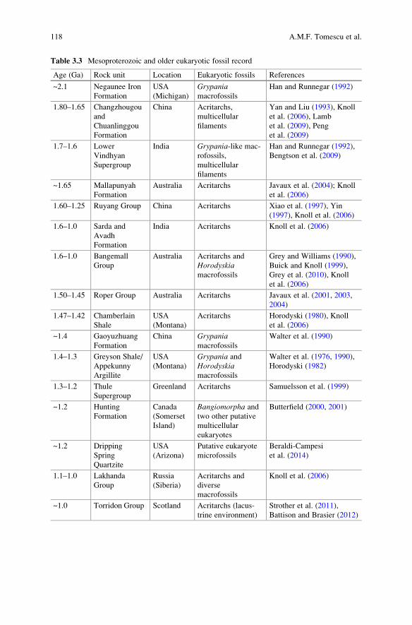
Table 3.3 Mesoproterozoic and older eukaryotic fossil record
Age (Ga) Rock unit Location Eukaryotic fossils References
~2.1 Negaunee Iron
Formation
USA
(Michigan)
Grypaniamacrofossils
Han and Runnegar (1992)
1.80–1.65 Changzhougou
and
Chuanlinggou
Formation
China Acritarchs,
multicellular
filaments
Yan and Liu (1993), Knoll
et al. (2006), Lamb
et al. (2009), Peng
et al. (2009)
1.7–1.6 Lower
Vindhyan
Supergroup
India Grypania-like mac-
rofossils,
multicellular
filaments
Han and Runnegar (1992),
Bengtson et al. (2009)
~1.65 Mallapunyah
Formation
Australia Acritarchs Javaux et al. (2004); Knoll
et al. (2006)
1.60–1.25 Ruyang Group China Acritarchs Xiao et al. (1997), Yin
(1997), Knoll et al. (2006)
1.6–1.0 Sarda and
Avadh
Formation
India Acritarchs Knoll et al. (2006)
1.6–1.0 Bangemall
Group
Australia Acritarchs and
Horodyskiamacrofossils
Grey and Williams (1990),
Buick and Knoll (1999),
Grey et al. (2010), Knoll
et al. (2006)
1.50–1.45 Roper Group Australia Acritarchs Javaux et al. (2001, 2003,
2004)
1.47–1.42 Chamberlain
Shale
USA
(Montana)
Acritarchs Horodyski (1980), Knoll
et al. (2006)
~1.4 Gaoyuzhuang
Formation
China Grypaniamacrofossils
Walter et al. (1990)
1.4–1.3 Greyson Shale/
Appekunny
Argillite
USA
(Montana)
Grypania and
Horodyskiamacrofossils
Walter et al. (1976, 1990),
Horodyski (1982)
1.3–1.2 Thule
Supergroup
Greenland Acritarchs Samuelsson et al. (1999)
~1.2 Hunting
Formation
Canada
(Somerset
Island)
Bangiomorpha and
two other putative
multicellular
eukaryotes
Butterfield (2000, 2001)
~1.2 Dripping
Spring
Quartzite
USA
(Arizona)
Putative eukaryote
microfossils
Beraldi-Campesi
et al. (2014)
1.1–1.0 Lakhanda
Group
Russia
(Siberia)
Acritarchs and
diverse
macrofossils
Knoll et al. (2006)
~1.0 Torridon Group Scotland Acritarchs (lacus-
trine environment)
Strother et al. (2011),
Battison and Brasier (2012)
118 A.M.F. Tomescu et al.

diverse acritarchs described from lacustrine deposits (Strother et al. 2011; Battison
and Brasier 2012) (Fig. 3.11c–e, g).
3.5.2.2 Macrofossils
Enigmatic macrofossils reported from the Paleoproterozoic and Mesoproterozoic
may also represent eukaryotic organisms. The most common of these are Grypaniaand Horodyskia. Their regular morphology and macroscopic size suggest a eukary-
otic origin (Knoll 2014). Grypania occurs as strap-shaped compressions of origi-
nally cylindrical organisms that form coils up to 24 mm across (Walter et al. 1976,
1990) and has been interpreted as a sessile algal eukaryote that was most likely
multinucleate (coenocytic or multicellular) (Han and Runnegar 1992). Horodyskiafossils consist of 1–4 mm spheroidal (or sometimes conical, ovoid or rectangular)
bodies connected by thin threads to form uniseriate structures (Yochelson and
Fedonkin 2000). Eukaryotic affinity of Horodyskia is considered probable, but
not beyond debate (Knoll et al. 2006). The oldest Grypania are known from
mid-Paleoproterozoic rocks older than the earliest eukaryotic acritarchs (2.1 Ga;
Negaunee Iron Formation, Michigan; Han and Runnegar 1992). The genus is also
known from several younger rock units which include the ca. 1.4 Ga Gaoyuzhuang
Formation in China (Walter et al. 1990), the 1.4–1.3 Ga Greyson Shale (Appekunny
Argillite) of Montana (Walter et al. 1976, 1990; Horodyski 1982), and possibly
from the 1.7–1.6 Ga lower Vindhyan Supergroup in India (Rohtas Formation,
where it has been described as Katnia; Tandon and Kumar 1977; Han and Runnegar
1992). The oldest Horodyskia fossils have been reported from the Mesoproterozoic
Bangemall Group in Australia (Grey and Williams 1990) but the genus is also
known from the Greyson Shale of Montana (Horodyski 1982).
Tubular objects 100–180 μm in diameter with walls replaced by apatite are
preserved in the lower Vindhyan Supergroup of India (Tirohan Dolomite; Bengtson
et al. 2009). These fossils date from around the Paleoproterozoic-Mesoproterozoic
boundary (1.7–1.6 Ga) and display regular annulation (shallow transverse grooves)
on the outer surface corresponding to internal septa. Bengtson et al. (2009) interpret
these fossils as filamentous algae (multicellular eukaryotes). Similar tubular or
filamentous fossils are described by Yan and Liu (1993) from the late Paleopro-
terozoic in the 1.8–1.65 Ga Chang Cheng Group (China) and by Butterfield (2001)
from the mid-Mesoproterozoic in the 1.2 Ga Hunting Formation (Somerset Island,
arctic Canada). These fossils are associated with two other multicellular types: flat,
layered units with internal differentiation and a stratified cellular structure (com-
pared to phylloid algae; Butterfield 2001) and Bangiomorpha, the first unequivocaloccurrence of complex multicellularity in the fossil record, the oldest reported
occurrence of sexual reproduction, and the oldest record for an extant phylum
(Rhodophyta, the red algae) and an extant family (Bangiaceae) (Butterfield
2000). Finally, in the late Mesoproterozoic, the Lakhanda Group hosts, along
with diverse acritarchs, larger eukaryotic fossils compared to xanthophyte algae,
3 Microbes and the Fossil Record: Selected Topics in Paleomicrobiology 119
fungi, and metazoans (Knoll et al. 2006; Hermann and Podkovyrov 2006; German
and Podkovyrov 2009).
3.5.2.3 Eukaryotes in the Archean?
Some microfossils older than the Chang Cheng Group biota have been discussed as
potential eukaryotes. The most prominent case is that of the spheroidal microfossils
described as Eosphaera by Barghoorn and Tyler (1965) from a diverse biota in the
Gunflint Iron Formation of Ontario (ca. 1.88 Ga; Schneider et al. 2002). Eosphaerafossils are small (up to 15 μm) and comprised of two concentric spherical envelopes
enclosing up to 15 small spheroidal tubercles in the circular space between the two
envelopes. Although Eosphaera has been compared to volvocalean green algae
(Kazmierczak 1979), currently most authors agree that the Gunflint microfossil
assemblage is entirely prokaryotic (e.g., Awramik and Barghoorn 1977) and that
Eosphaera does not preserve sufficient detail in support of eukaryotic affinities
(Knoll 1992). However, other lines of evidence indicate that eukaryotic life might
have originated before the late Paleoproterozoic and even as early as the Archean
(older than 2.5 Ga).
The shales of the Francevillian B Formation of Gabon, dated at 2.1 Ga, have
yielded pyritized macrofossils consisting of elongated to isodiametric flattened
specimens up to 12 cm in size, with a thicker central area and thinner radially
patterned, undulate, and lobed margins (El Albani et al. 2010). The morphology of
the fossils has been interpreted as reflecting growth that requires cell-to-cell
signaling and coordinated growth responses indicative of well-integrated colonial
organization or multicellular life in the early Paleoproterozoic, an interpretation
bolstered by the detection of steranes in the same layers (El Albani et al. 2010).
Both the Gabonese macrofossils and Grypania—if the latter indeed represents a
multicellular (or multinucleate) eukaryote—with its oldest occurrence at 2.1 Ga
(Han and Runnegar 1992), predate the oldest unicellular eukaryotes (acritarchs) of
the Chang Cheng Group (Lamb et al. 2009; Peng et al. 2009) by at least 300 million
years. Assuming that unicellular eukaryotes evolved before multicellular forms,
this implies that the prototypical unicellular eukaryote evolved prior to 2.1 Ga and
we should expect to find eukaryotic acritarchs older than that age. Javaux
et al. (2010) have reported organic-walled microfossils from shallow-marine
deposits in the 3.2 Ga Moodies Group of South Africa. These microfossils are
older than the oldest claims for sterane biomarkers (2.7–2.5 Ga; Brocks et al. 1999;
Waldbauer et al. 2009). They are the oldest and largest Archean organic-walled
microfossils reported to date (Javaux et al. 2010) and co-occur with microbially
induced sedimentary structures (Noffke et al. 2006). The fossils display features
consistent with eukaryotic affinities, such as good organic preservation, which is
indicative of cell walls containing recalcitrant polymers and large sizes
(ca. 30–300 μm) (Fig. 3.10a). These microfossils could, in principle, be eukaryotic,
but their lack of cell surface ornamentation and their simple cell wall structure also
make them easily comparable to the extracellular envelopes of some bacteria, thus
120 A.M.F. Tomescu et al.

precluding unequivocal interpretation as eukaryotes (Knoll 2014). It will be inter-
esting to see if geochemical studies of the Moodies Group (not published to date)
ascertain the presence of sterane biomarkers.
3.5.3 Salient Patterns in the Early Eukaryotic Fossil Record
The oldest bona fide unicellular eukaryotes are known from 1.80 to 1.65 Ga
(mid-Paleoproterozoic) rocks, but the fossil record provides strong evidence that
organisms capable of producing macroscopic bodies by growth processes that
required coordinated responses, whether in a coenocyte or an aggregation of
cells, had evolved by 2.1 Ga. This mode of development is not known in pro-
karyotes but is consistent with coenocytic or multicellular organization in eukary-
otes, in which multicellularity has been achieved independently and to different
extents in different clades (Niklas and Newman 2013; Niklas et al. 2013). Since
complex intracellular organization evolved in eukaryotes well before the appear-
ance of metazoans (Knoll et al. 2006), the earliest unicellular eukaryotes should be
sought after in rocks older than 2.1 Ga (early Paleoproterozoic and Archean),
ideally using approaches that combine morphological and ultrastructural charac-
terization with geochemical studies.
Precambrian eukaryotes have been reported predominantly from rocks deposited
in marine environments, and only starting with the ca. 1.2 Ga Dripping Spring
Quartzite (Beraldi-Campesi et al. 2014) do we see eukaryotes on continents.
Considering the living environments of the early marine acritarchs, Knoll
et al. (2006) echo the views of Butterfied (2005a, b) that not all acritarchs represent
reproductive cysts of planktonic algae—some must be the remains of benthic
heterotrophic organisms. Javaux et al. (2001) documented, in their study of the
Roper Group acritarchs, an onshore-offshore pattern in fossil distribution between
depositional environments ranging from marginal marine to basinal. Overall, their
data show seaward decrease in abundance and decline in diversity, as well as
changing dominance among different species. These are interpreted as reflecting
the effects of natural selection by physical habitat variables on species distributions
by ca. 1.5 Ga, contributing to the rise of biological diversity (Javaux et al. 2001).
Late Proterozoic and Mesoproterozoic rocks contain abundant eukaryotic fos-
sils, but morphological diversity (disparity; Wills 2001) of these fossil assemblages
maintains low to moderate levels (summarized by Knoll et al. 2006). Among these,
microfossils of large size and with complex wall structure and surface ornamenta-
tion are frequent in rocks up to ca. 1.6 Ga, but in older rocks, the cell surface
ornamentation and ultrastructure are less distinctive, leading some to posit some
residual uncertainty concerning taxonomic assignments at the domain level (Knoll
2014). A significant leap in the level of eukaryote disparity becomes apparent
around 800 Ma, marking the end of the long interval between the evolution of the
first eukaryotes and their taxonomic radiation in the second half of the
Neoproterozoic (Knoll et al. 2006).
3 Microbes and the Fossil Record: Selected Topics in Paleomicrobiology 121
The morphologies of Proterozoic eukaryotes are varied, from simple
unornamented cells, morphologically complex ornamented unicells, to three-
dimensionally complex multicellular organisms displaying cellular differentiation,
but for many of these fossils, phylogenetic placement is difficult because the dearth
and generality of characters preserved do not support unequivocal taxonomic
affinities (Knoll et al. 2006). For example, Knoll et al. (2006) point out that
filamentous fossils document the early evolution of a molecular capacity for simple
multicellularity which was subsequently exploited by multiple clades. Branching of
structures, thought to require a more sophisticated intracellular organization (Knoll
et al. 2006), cell-to-cell signaling, and polarity of cell development and division, is
first seen in the bifurcating processes of unicellular Tappania of the Roper Group
(Javaux et al. 2001) and in ca. 1 Ga filamentous fossils (Knoll et al. 2006). By
1.2 Ga, multicellularity and polarity of cell divisions had led to the evolution of
complex body plans—e.g., Bangiomorpha (Butterfield 2000).
3.6 Roles of Microbes in Taphonomic Pathways
Another dimension of the microbial contribution to the fossil record has to do with
the role of microbes in processes leading to fossilization (fossil preservation).
Taphonomy, a field of paleontological investigation pioneered by Ivan Efremov
(1940), is concerned with elucidating decay, disarticulation, transport, and burial
pathways that result in fossilization—or not. Taphonomic studies document
biostratinomy (physical and chemical changes incurred after death) and diagenesis
(physical and chemical changes incurred after burial). Most investigations of
taphonomy are biased toward those fossilization pathways leading to Konservat-
Lagerstatten, deposits in which fossilized organisms exhibit exceptional preserva-
tion (Schiffbauer and Laflamme 2012). Sometimes these even include the preser-
vation of soft tissues which may have resulted from carcass burial in an anoxic
environment. Microbes play substantial roles in the formation of most Konservat-
Lagerstatten, where they act as agents of degradation, as well as, ironically,
preservation. Much of what is known about the roles of microbes in taphonomic
pathways derives from studies of Konservat-Lagerstatten.
Another important body of evidence contributing to understanding of microbial
roles in taphonomic pathways results from experimental investigations of taphon-
omy in living organisms and modern environments; these experiments are some-
times referred to as “actualistic” (not to be confused with the philosophic position,
although the association is enlightening). By comparing the outcomes of actualistic
experiments with observations from the fossil record, paleontologists have gained a
strong appreciation for the necessity and ubiquity of microbial contributions to
exceptional preservation. First and foremost, microbes facilitate authigenic miner-
alization, which is a special type of “cast and mold” formation, acting as nucleation
sites and by altering local geochemical gradients. Microbes can also entirely
replicate soft tissue by consuming original tissue and replacing it with extracellular
122 A.M.F. Tomescu et al.

polymeric substances (EPS). Finally, microbial mats can form “death masks” over
specimens, which inhibit decay and disarticulation in addition to promoting min-
eralization, as well as stabilizing sediment interfaces, which has important impli-
cations for studies of fossil trackways. It is important to note that many of the roles
that microbes play in taphonomic pathways occur concurrently (e.g., Darroch
et al. 2012; Cosmidis et al. 2013; Iniesto et al. 2013). As such, although we have
broken the following discussion into several major categories of microbe-mineral-
fossil interactions, it should be understood that these are artificial and intergrading.
3.6.1 Cautionary Tales
As microbial contribution to exceptional preservation is most often implicated in
preservation of ephemeral structures and soft tissue, it is useful to briefly review
what is presently known about the organic structures that can, and (thus far) cannot,
be found in the fossil record. This is particularly germane as many of the debates
surrounding these structures invoke or include fossil or living bacteria. For instance,
a putative dinosaurian heart has been refuted as such, but the cemented sediment
does bear evidence of microbiogenic texture (Cleland et al 2011). Structures
preserved within the body cavities of three early Cretaceous birds from the Jehol
biota have been interpreted as ovarian follicles (Zheng et al. 2013), but this is
unlikely, as gonads are among the first visceral organs to suffer autolytic and
bacterial decay (Mayr and Manegold 2013). Finally, the sensational report of
nonmineralized soft tissue in a tyrannosaurid (Schweitzer et al. 2005) was later
reinterpreted as evidence that many fossil specimens are colonized by microbes
after fossilization and exhumation (Kaye et al. 2008), in addition to during
biostratinomy. Schweitzer et al. (2005) originally reported transparent hollow
tubules obtained from acid digestion of dinosaur bone. The tubules were interpreted
as blood vessels and contained spherical microstructures that were interpreted as
nuclei from endothelial cells (Schweitzer et al. 2005). In their actualistic
reinvestigation, Kaye et al. (2008) acid-digested a variety of fossil bones and
recovered structures very similar to those observed by Schweitzer et al. (2005).
SEM-EDX analyses indicated that the microspheres were likely to have originally
been framboidal pyrite, diagenetically altered to iron oxides (Kaye et al. 2008),
consistent with an earlier hypothesis put forth by Martill and Unwin (1997). Fourier
transform infrared (FTIR) spectroscopy indicated that as opposed to reflecting
diagenetically altered collagen, the material was comparable to modern biofilms;
their lack of antiquity was confirmed by carbon dating. Kaye et al. (2008) suggested
that recent biofilms had lined voids in fossil bones, thus producing the hollow
structures morphologically similar to blood vessels and osteocytes.
Unlike the aforementioned putative soft tissues, some reports of original bio-
polymers in dinosaurian tissue are more compelling, having been assessed with
FTIR spectroscopy. These include collagen peptides (San Antonio et al. 2011) and
amide groups (Manning et al. 2009) reported from Cretaceous bones and in
3 Microbes and the Fossil Record: Selected Topics in Paleomicrobiology 123
embryonic Jurassic bone, which presently constitutes the oldest evidence of in situ
preservation of complex organics in vertebrate remains (Reis et al. 2013). Other
biopolymers known to be highly resistant with good fossilization potential include
chitin, cellulose, sporopollenin (Briggs 1999; Wolfe et al. 2012), and the com-
pounds found in melanosomes (Liu and Simon 2003; Wilson et al. 2007; Vinther
et al. 2008, 2010), the latter of which have been previously interpreted as fossilized
bacteria (Wuttke 1983a). Not only can melanosomes be used to reconstruct color
patterning (Li et al. 2010a; Knight et al. 2011), their presence is tightly correlated
with the preservation potential of integument (Zhang et al. 2010). Maganins, which
are peptides found in dermal secretions of some amphibians, also contribute to
preservation of integument (McNamara et al. 2009). Maganins can permeate and
lyse microbial cell membranes, and because they persist post mortem, they inhibit
microbial decay to the extent that Rana skin remains intact more than 50 days,
during which time all contents of the body cavity entirely decay (Wuttke 1983b).
Finally, reports of nuclei in the fossil record (e.g., Baxter 1950; Millay and
Eggert 1974; Brack-Hanes and Vaughn 1978; Martill 1990; Schweitzer et al. 2007)
have been problematic. Actualistic experiments have demonstrated pseudonucleic
structures (Westall et al. 1995), but intracellular mineralization of legitimate nuclei
remains elusive, with a sole exception to date—the recently published nuclei and
chromosomes preserved in the root cells of a Jurassic (ca. 187 Ma) Osmunda fern
rhizome from Scania (Sweden; Bomfleur et al. 2014). In the Miocene Clarkia flora,
where plant fossils are preserved as compressions and ultrastructural TEM inves-
tigations have substantiated reports of nuclei, they are preserved at much lower
abundance than chloroplasts or mitochondria (Niklas 1983), which typically
autosenesce before nuclear disorganization occurs (Woolhouse 1984). This seem-
ing discrepancy may result from partitioning of protoplast into nucleate and enu-
cleate microprotoplasts via osmotic shock (Niklas et al. 1985). By isolating plastids
from nuclei, nuclear-controlled autosenescence (Yoshida 1962) could have been
circumvented (Niklas 1983). While this model would account for some reports of
fossil nuclei in the absence of plastids, it is possible that other reports of fossil
nuclei in plants represent condensed cellular contents (Niklas 1983). For instance,
osmotic stress can cause protoplast shrinkage away from the cell walls within
10 min (Munns 2002). The shrunken protoplast, which may be as little as 1/3 or
less of the cell volume, remains centrally positioned by attachment of plasma
membrane strands (Fig. 5, Munns 2002). Plasma shrinkage has also been invoked
(Pang et al. 2013) in refuting putative nuclei in Ediacaran Doushantuo Formation
(ca. 555 Ma, China) specimens, which have become the focus of taxonomic
disputes (e.g., Huldtgren et al. 2011). Because nuclei are composed of water,
proteins, and nucleic acids, they are easily degraded in comparison to lipids
(Eglinton et al. 1991); in animal fossils at least, all lipid bilayers could be expected
to fossilize, and the lack of mitochondria in the Doushantuo specimens suggests the
nucleus-like bodies are not what they seem (Pang et al. 2013).
124 A.M.F. Tomescu et al.

3.6.2 Authigenic Mineralization
Authigenic minerals are defined simply as those minerals which are formed in situ
and, contra Schopf et al. (2011), are not implicitly biogenic, although many
biologically mediated pathways induce the precipitation of iron, carbonate, phos-
phate, sulfur, silicate, and clay minerals (e.g., Konhauser 1998; Bazylinski and
Frankel 2003; Briggs 2003a, b; Martin et al. 2004; Peckmann and Goedert 2005;
Konhauser et al. 2011; Darroch et al. 2012). Carbonaceous polymers alone may be
sufficient to precipitate many minerals (McNamara et al. 2009; Schopf et al. 2011;
Roberts et al. 2013)—this process has been termed organomineralization (Trichet
and Defarge 1995) and is considered abiogenic, although the polymers may be
derived from living tissue. Biomineralization, on the other hand, encompasses both
active and passive precipitations of minerals and is defined as occurring in the
presence of living cells (Trichet and Defarge 1995).
Active biomineralization occurs when the precipitation of minerals is under
direct control of the cell (biologically controlled biomineralization; Konhauser
and Riding 2012), as in the case of magnetite crystals formed within magnetotactic,
microaerophilic bacteria (Bazylinski 1996). Passively induced precipitation (bio-
logically induced biomineralization; Konhauser and Riding 2012) can occur
through metabolic processes that alter local chemistry permitting stoichiometric
precipitation (Lovley and Phillips 1986; Lovley et al. 1987; Roh et al. 2003; Straub
et al. 2004) and may also occur through mineral nucleation on EPS or microbial
surfaces (e.g., Ferris et al. 1987; Thompson and Ferris 1990; Fortin et al. 1997;
Leveille et al. 2000; Van Lith et al. 2003). EPS are chemically complex bacterial
exudates that contain mucus, sugars, extracellular DNAs, and proteins (Wingender
et al. 2012) and are anionic, resulting in metal ion sorption (Beveridge 1989).
Bacterial cells are also characterized by the electric double layer immediately
external to the cell walls, which results from proton shuffling to establish a proton
motive force, forming a highly localized electrochemical gradient (Poortinga
et al. 2002).
3.6.2.1 Organomineralization
Biomineralization has been generally recognized as a key factor in exceptional
preservation (Briggs et al. 1993; Briggs and Kear 1993; Briggs and Wilby 1996;
Martin et al. 2004; Darroch et al. 2012), but it is important to emphasize that
fossilization may readily result from organomineralization as well. For instance,
sulfurization, a common diagenetic phenomenon (Sinninghe Damste and de Leeuw
1990), can preserve soft tissue as sulfur-rich organic residue (McNamara
et al. 2009, 2010). Similarly, diagenetic (post-burial) processes often reduce recal-
citrant biomolecules like chitin, cellulose, and lignin into geologically stable
macromolecules, termed kerogen (Briggs 2003a, b); kerogen is composed of
interlinked polycyclic aromatic hydrocarbons, and organomineralization could
3 Microbes and the Fossil Record: Selected Topics in Paleomicrobiology 125
proceed via hydrogen bonding between metal ions and peripheral hydrogen atoms
(Schopf et al. 2011).
The precipitation of dolomite (Ca(Mg)CO3) and disordered high-Mg carbonates
can also occur via organomineralization (Roberts et al. 2013), and Klymiuk
et al. (2013a) have suggested that some kerogenous plant fossils preserved in
calcium carbonate concretions are at least partially mineralized with disordered
high-Mg carbonates. The lack of an actualistic explanation for the precipitation,
under ambient temperatures, of the geologically significant amounts of dolomite
found in the rock record has often been referred to as “the dolomite problem,” as it
was only known to occur under a very narrow set of conditions, in rarely occurring
environment—hypersaline sabkhas, in association with sulfate-reducing bacteria
(Wright and Wacey 2004). Roberts et al. (2013) achieved abiotic nucleation of
high-Mig carbonates on carboxylated polystyrene, and they suggest that Mg2+ ions
are complexed and dewatered by surface-bound carboxyl groups. Because the
model does not require metabolic activity, any type of organic matter—including
microbial cell walls, which are rich in carboxyl groups—could serve as nucleation
sites for dolomite precipitation (Roberts et al. 2013).
Fossils of 63 adult frogs, constituents of the late Miocene Libros biota of
northeast Spain, provide evidence that tissue composition itself can control precip-
itation of authigenic phosphates (McNamara et al. 2009). In some of the specimens,
the presence of distinctive microfabrics (Wilby and Briggs 1997; Briggs 2003a, b;
Chin et al. 2003; Briggs et al. 2005) indicates that phosphate nucleated directly
upon soft tissue without bacterial intermediates (McNamara et al. 2009). Bacterial
contributions are often implicated in phosphatization, as calcium carbonate
(CaCO3) precipitation is thermodynamically favored over calcium phosphate
(CaPO4) at pH below 6.38 (Briggs 2003a, b), and bacterial decay leads to localized
acidification. Phosphatization of the Libros frogs, however, was probably not
significantly enhanced by diffusion of acidic microbial metabolites from internal
biofilms, but was instead facilitated primarily by release of SO42� during degrada-
tion of glycosaminoglycans (GAGs), which are abundant in extant adult Rana skin
where they are chemically bound to collagen fibers (McNamara et al. 2009).
Collagen contains abundant hydroxyl groups, to which Ca2+ readily adsorbs;
calcium phosphate precipitates are extremely localized to dermal collagen fibers
in the fossil frogs and absent elsewhere in the specimens (McNamara et al. 2009).
The Libros frogs also serve as an opportunity to make an important distinction:
mineral precipitation was itself not bacterially mediated, but the decay of collagen,
which increases hydroxyproline and therefore the abundance of hydroxyl nucle-
ation sites (Lawson and Czernuszka 1998), was almost certainly facilitated by
bacterial collagenases (McNamara et al. 2009). The distinction as to whether
mineralization is abiogenic or biogenically mediated is one that explicitly concerns
how the mineral is precipitated and does not preclude subsidiary microbial inter-
actions. Because decaying organic matter comprises heterogeneous microenviron-
ments (Briggs and Wilby 1996), it is probable that most taphonomic pathways
include both organomineralization and biomineralization.
126 A.M.F. Tomescu et al.

3.6.2.2 Biomineralization
Burgess Shale-Type Preservation
Burgess Shale-type preservation is ubiquitous in Ediacaran and Cambrian deposits
(Gabbott et al. 2004; Xiao et al. 2002; Zhu et al. 2008; Anderson et al. 2011; Cai
et al. 2012). Factors thought to be crucial in the formation of Burgess Shale-type
deposits include dysoxia (low oxygen content) or anoxia (Gaines et al. 2012; Cai
et al. 2012), the lack of biogenic reworking of sediment (bioturbation) (Orr
et al. 2003), and inhibition of autolysis. Many authors have hypothesized that
early diagenetic mineralization is an intrinsic component of Burgess Shale-type
taphonomic pathways. Butterfield (1995) suggested that authigenic clay minerals
inhibit autolytic decay, whereas Orr et al. (1998) suggested that they adsorb to
organic matter and create templates. Gabbott et al. (2004) considered pyritization
more relevant for the Chengjiang biota, where it was precipitated on organic
remains during bacterial sulfate and iron reduction. Finally, Petrovich (2001) has
suggested that Fe2+ ions adsorbed to organic tissues, promoting nucleation of iron-
rich clays. Still others (Gaines et al. 2008; Page et al. 2008; Butterfield et al. 2007)
have maintained that clays are metamorphic in nature and play no role in early
diagenesis.
Analyses of 53 fossils from 11 Burgess Shale-type deposits using scanning
electron microscopy—energy-dispersive X-ray spectroscopy (SEM-EDX)—indi-
cate that all share a principal taphonomic pathway (Gaines et al. 2008). According
to Gaines et al. (2008), conservation of organic tissues (i.e., as kerogen), and not
early diagenetic authigenic mineralization, is the primary mechanism responsible
for preservation. Fossils from all sites display carbonaceous preservation, ranging
from continuous robust through degraded films, but all are enriched with C and
depleted of Al, Si, and O with respect to the surrounding matrix (Gaines
et al. 2008). This principal pathway may be occasionally augmented by authigenic
mineral replication of some tissues (Gaines et al. 2008), including pyrite precipi-
tated in gut tracts of trilobites (Lin 2007), and calcium phosphate replication of
some soft tissues (Butterfield 2002, 2007). More recent analyses, however, have
examined unmetamorphosed fossils preserved in the Doushantuo Formation, which
also exhibit Burgess Shale-type preservation (Anderson et al. 2011). All of the
Doushantuo fossils examined are closely associated with clay and pyrite minerals,
which appears to confirm the role of these two components in early diagenesis
(Anderson et al. 2011).
The presence of authigenic clay minerals can be explained in several ways:
reducing conditions at the sediment-water interface could be deflocculating alumi-
nosilicate grains (Gaines et al. 2005); clay minerals could be attracted and adsorbed
onto organic surfaces (Gabbott 1998), a process which is facilitated by sulfate
reduction and reduced pH (Martin et al. 2004); clay minerals could be
authigenically precipitated from pore waters, in which localized zones of reduced
pH resulted in dissolution of metals from resident sediments (Gabbott et al. 2001);
3 Microbes and the Fossil Record: Selected Topics in Paleomicrobiology 127

or clay minerals could conceivably be bound to organic surfaces by bacterial
biofilms (Konhauser et al. 1998; Toporski et al. 2002). By invoking reducing
conditions and low pH, these models find at least a subsidiary role for sulfate-
reducing bacteria, and actualistic studies of decay of lobster eggs indicate that
quartz and clay mineral attachment to decaying tissue is enhanced in the presence of
sulfate-reducing bacteria, precipitating in proximity to decaying tissue, while iron
sulfides precipitate in surrounding sediments (Martin et al. 2004).
Phosphate Mineralization of Soft Tissue
In order to preserve details of soft tissue, mineralization must necessarily outpace
decay (Allison 1988a); thus phosphatization, the replication of soft tissues by
calcium phosphate (apatite, Ca3PO4), represents a biostratinomic or early diage-
netic taphonomic pathway and a discrete temporal window for preservation. Phos-
phatization of soft tissues is relatively common in vertebrate fossils (Martill 1987,
1988, 1989, 1991; Martill and Unwin 1989; Kellner 1996a, b; Briggs et al. 1997;
Frey et al. 2003), and, with only few exceptions (e.g., McNamara et al. 2009), it is
generally conceded that bacteria have a vital role in soft tissue mineralization by
Ca3PO4. Anaerobic decay generates steep geochemical gradients favoring Ca3PO4
precipitation over CaCO3 (Briggs and Kear 1993, 1994; Hof and Briggs 1997;
Sagemann et al. 1999; Briggs 2003a, b), and carbonate precipitation is inhibited at
pH < 6.38 (Briggs 2003a, b) or when PO43� concentrations are high (Sagemann
et al. 1999). Chemical microenvironments in the vicinity of decaying carcasses
geochemically differ from surrounding sediments in terms of ion concentration, pH,
and Eh (oxidation potential) (Sagemann et al. 1999), and these differences become
more extreme toward the interior of the carcass (Briggs and Wilby 1996). If PO43�
ions are sourced from the decaying tissue itself, mineral precipitation will be highly
localized, whereas if PO43� is derived from external sediments, or from microbial
consortia engaged in active phosphate precipitation (Wilby et al. 1996; Cosmidis
et al. 2013), phosphatization is likely to be extensive (Wilby and Briggs 1997).
Invertebrates tend to be less prone to widespread soft tissue phosphatization. In
arthropods, muscle, hepatopancreas, gills, nerve ganglia (Briggs and Kear 1993,
1994; Hof and Briggs 1997; Sagemann et al. 1999), and midgut glands (Butterfield
2002) can be selectively mineralized. Actualistic experiments produce only partial
phosphatization (Briggs and Kear 1994; Hof and Briggs 1997; Sagemann
et al. 1999). For shrimp carcasses inoculated with sulfate-reducing bacteria and
sulfide-oxidizing and fermenting bacteria, decay was most intense and phosphati-
zation most extensive under anaerobic sulfate reduction (Sagemann et al. 1999).
While this implies a prominent role for sulfate-reducing bacteria in invertebrate soft
tissue phosphatization, Briggs et al. (2005) report organomineralization in a spec-
imen of Mesolimulus, a horseshoe crab known from the Jurassic Solnhofen and
Nusplingen biotas. Muscle fibers with distinct banding have been preserved by
apatite precipitation directly onto fibers, although structures interpreted as
cyanobacterial body fossils have also been observed (Briggs et al. 2005). The
128 A.M.F. Tomescu et al.
preserved midgut of a trilobite (Lerosey-Aubril et al. 2012) was probably also
organomineralized; the surrounding sediment is low in phosphate, and although
microbial mats can concentrate phosphates (Wilby et al. 1996), none are present
within the surrounding sediments. Instead, Lerosey-Aubril et al. (2012) suggest that
epithelial cells of the midgut probably contained mineral calcium phosphate con-
cretions, a form of mineral storage in preparation for ecdysis (molting).
Microbial activity can contribute phosphates to a microenvironment through
active and passive mineralization. Calcium phosphate (apatite) is actively precip-
itated intra- and extracellularly by a number of bacteria, including Bacterionemamatruchotii, Chromohalobacter marismortui, Escherichia coli, Providenciarettgeri, Ramlibacter tataouinensis, and Serratia sp. (reviewed in Cosmidis
et al. 2013). Microbial metabolic activities alone may, however, be sufficient to
generate amounts of free phosphate high enough to passively precipitate large
quantities of laminar calcium phosphate (Arning et al. 2009). Lipid biomarkers
associated with sulfate-reducing bacteria and abundance of the giant sulfur bacteria
Thiomargarita namibiensis are correlated with PO43� concentrations in modern
phosphate-rich sediments (Schulz and Schulz 2005; Arning et al. 2008). Phospha-
tized fossils have not been assessed for biomarkers, but coccoid, spiral, and
bacillus-like structures interpreted as bacterial body fossils are occasionally
observed in zones of soft-tissue fossilization in vertebrates and invertebrates
(Briggs et al. 1997; Toporski et al. 2002; Briggs et al. 2005; Skawina 2010; Pinheiro
et al. 2012). While morphology cannot be used to circumscribe phylogenetic
affinities of these putative fossil bacteria, actualistic experiments in freshwater
decay of invertebrate tissue (Skawina 2010) reveal a succession of bacterial
morphotypes that are generally correlative with the stage of decay: cocci predom-
inate over bacilli when pH >7 (i.e., at the beginning and end of decay; Briggs and
Kear 1994; Hof and Briggs 1997; Skawina 2010), whereas bacilli are more prev-
alent when pH is reduced <7. These observations thus provide a tentative link
between bacillus-type structures and sulfate-reducing agents of anaerobic decay.
Iron Minerals and Pyritization
Although some iron oxides can be precipitated abiotically in steep geochemical
gradients, like those resulting from collagen decay (Kremer et al. 2012b), precip-
itation of many iron oxides, iron carbonates, and iron sulfide (pyrite) is bacterially
mediated (Ferris et al. 1987; Beveridge 1989; Ferris 1993; Konhauser 1998;
Bazylinski and Frankel 2003; Chatellier et al. 2004; Konhauser et al. 2011). For
instance, direct precipitation of iron oxides onto bacterial EPS releases a proton into
the extracellular microenvironment; by acidifying their immediate surroundings,
aerobic iron-oxidizing bacteria can enhance the proton motive force, thus increas-
ing the energy-generating potential of a cell (Chan et al. 2004). Alternately,
authigenic pyrite formation is frequently a result of bacterial metabolisms
employing sulfate reduction. Sulfate-reducing bacteria utilize sulfate as a terminal
electron acceptor under anaerobic conditions; H2S results as a metabolic by-product
3 Microbes and the Fossil Record: Selected Topics in Paleomicrobiology 129

and reacts with dissolved iron, precipitating iron sulfide (Frankel and Bazylinski
2003). In some cases, the cellular membrane itself can serve as an anionic matrix,
immobilizing Fe2+, which then reacts with metabolic H2S, autolithifying the bac-
terial cell in the process (Ferris et al. 1988). Despite the ubiquity of these processes
in modern environments, there are relatively few reports of pyritized bacterial body
fossils (Southam et al. 2001; Schieber and Riciputi 2005; Cosmidis et al. 2013; and
possibly those reported by Tomescu et al. 2008).
Bacteriogenic pyrite is generally depleted in 34S (Canfield and Thamdrup 1994),
and when crystals do not nucleate directly upon cell surfaces, they are framboidal,
or lacking distinct crystal faces (Popa et al. 2004). Although the presence of
framboidal pyrite is not in itself positively indicative of sulfate-reducing metabo-
lisms (Butler and Rickard 2000; P�osfai and Dunin-Borkowski 2006), there is a
strong association between bacterial EPS and the formation of pyrite framboids
(Maclean et al. 2008). Framboidal pyrite commonly occurs below the reduction-
oxidation transition zone in subaqueous microbial mats (Popa et al. 2004). Focused
ion beam SEM-EDX provides a novel view of the interior of low-temperature
framboidal pyrite aggregates formed in microbial mats: organic matrix occurs
between individual pyrite crystals, suggesting that nanocrystals nucleate directly
within the organic matrix of bacterial EPS (MacLean et al. 2008). Extensive
precipitation of pyrite, whether on cell surfaces or within EPS, can result in high-
fidelity preservation of fossil morphology (Grimes et al. 2001, 2002; Gabbott
et al. 2004; Brock et al. 2006; Darroch et al. 2012; Wang et al. 2012). In some
cases, pyrite framboids form inside bacterial cells, replacing them, as is the case
with the cyanobacteria reported by Tomescu et al. (2006, 2009) from the Early
Silurian Massanutten Sandstone. Pyrite often completely replaces organics, as in
the Jehol biota, where insects were initially preserved in pyrite that was later
weathered to iron oxides (Wang et al. 2012), and cellularly preserved Devonian,
Mississippian, and Eocene fossil plants that have been replaced with pyrite (e.g.,
Allison 1988b; Rothwell et al. 1989; Tomescu et al. 2001).
An actualistic experiment in organomineralization of a celery petiole (Grimes
et al. 2001) has yielded important insights into authigenic pyrite formation in plant
tissues. Grimes et al. (2001) demonstrated that pyrite readily precipitates on inner
plant cell wall surfaces, within cell walls, and in the middle lamella between cells.
Pyrite initially nucleates on the inner walls of parenchyma cells, before penetrating
inward (Grimes et al. 2001). Inward penetration occurs through successive nucle-
ation upon previously precipitated crystals, rather than by continued crystal growth
(Grimes et al. 2001). Pyrite precipitates only between fibrils of cellulose and not on
lignified surfaces; minerals do not replace the plant tissue but rather nucleate within
fluid-filled spaces between cellulose fibrils (Grimes et al. 2001). Thus, pyritized
plant tissues probably represent middle lamella regions, as opposed to replaced
cellulose, and if present, lignin will be coalified (Grimes et al. 2001, 2002).
Taphonomic pathways hypothesized for the London Clay (one of the more
diverse Eocene floras in Europe; Collinson et al. 2010), which invoke pyritic
replacement of plant tissues in the presence of sulfate-reducing bacteria, have
also been experimentally investigated (Grimes et al. 2001; Brock et al. 2006). In
130 A.M.F. Tomescu et al.
both studies, the experimental system rapidly became driven by anaerobic respira-
tion, with diffuse precipitation of iron sulfides into sediment surrounding Platanustwigs. Local pH also declined, but as decay tapered off—at 12 and 5 weeks (Grimes
et al. 2001; Brock et al. 2006 respectively)—pH increased, reflecting a metabolic
shift to sulfide oxidation (Brock et al. 2006). Although pyrite precipitated on plant
surfaces, few of the twigs exhibited internal sulfide minerals (Grimes et al. 2001;
Brock et al. 2006); these results may have been due to inherent heterogeneity of the
system (Brock et al. 2006) or hydrophobic moieties in lignin molecules (Jung and
Deetz 1993; Grimes et al. 2001).
Doushantuo fossils preserved in small cm-sized chert nodules represent a more
specialized pyritization pathway. The chert nodules, which contain microbial mat
fragments at their core, are surrounded by silica cortex and inner pyrite rim, with an
exterior rim of late diagenetic blocky calcite (Xiao et al. 2010). Because pyrite
crystals are immersed in groundmass silica, which exhibits no concentric growth
zones, the two minerals were probably syngenetic and swiftly precipitated (Xiao
et al. 2010). Formation of pyrite-silica rims was likely facilitated by local pH
changes related to bacterial sulfate reduction (Xiao et al. 2010), consistent with
diffusion-precipitation models that posit a spherical precipitation front formed at
the boundary between diffusing H2S and a surrounding Fe2+ reservoir (Raiswell
et al. 1993; Coleman and Raiswell 1995). Pervasive marine anoxia and substan-
tially higher levels of dissolved iron during the Ediacaran are thought to have
encouraged proliferation of sulfate-reducing bacteria (Canfield et al. 2008; Li
et al. 2010b). Sulfate reduction generates alkalinity (HCO3�), thereby promoting
CaCO3 precipitation, but because sulfate reduction and pyrite precipitation have a
net increase of protons, pH declines (Xiao et al. 2010). Within a narrow window
(pH 9.0–10.0), the solubilities of carbonate and silica behave inversely, and silica
precipitates at the same time CaCO3 enters solution; therefore, proton generation by
sulfate-reducing bacteria led to silicification, while H2S generated during metabo-
lism led to pyrite precipitation in the Doushantuo nodules (Xiao et al. 2010).
Compression-Impression Leaf Fossilization
The presence of biofilms may be integral to preservation of leaves as compression-
impression fossils. Two taphonomic pathways invoking microbially mediated
preservation have been proposed for compression-impression plant macrofossils.
The exceptional preservation of plant and insect biota at the Eocene Florissant
locality is probably a special case and not widely replicated in the paleobotanical
record: fossilization is thought to have been facilitated by extensive diatom blooms
in response to increased levels of dissolved silica (derived from periodic volcanic
ashfall) within a lacustrine system (O’Brien et al. 2002). When exposed along
bedding planes, it is apparent that the fossils are encased within diatomaceous
laminae; O’Brien et al. (2002) suggest that diatom biofilms were established on
3 Microbes and the Fossil Record: Selected Topics in Paleomicrobiology 131
floating plant and insect debris, with subsequent sinking and incorporation into the
sediment.
The second taphonomic pathway implicating biofilms was first proposed by
Spicer (1977) who recognized that iron was often precipitated on submerged
surfaces of leaves prior to burial, and leaf compression fossils are often encrusted
with iron oxides (Dunn et al. 1997). Leaf surfaces, however, are covered by
hydrophobic cuticles that inhibit metal binding. Degradation of cuticle followed
by authigenic mineralization of cellulose could account for this seeming oxymoron,
but although exceptional instances of cellulose preservation have been demon-
strated (Wolfe et al. 2012), cellulose typically degrades rapidly in comparison to
cuticle (Spicer 1981). Dissimilatory iron-reducing bacteria have also been invoked
to explain this oxymoron (Spicer 1977), but they are restricted to aerobic environ-
ments with abundant Fe3+ (Dunn et al. 1997). By contrast, leaf surfaces host a
variety of microbes capable of forming biofilms (Morris et al. 1997). The anionic
nature of bacterial EPS facilitates metal binding (Beveridge 1989), and experiments
conducted by Dunn et al. (1997) show that mineral precipitation does not occur in
the absence of biofilm formation.
3.6.3 Doushantuo “Embryos”
The Doushantuo biota, preserved in Ediacaran marine deposits, has been recently
subject to intense debates, centering on the presence of microscopic multiloculate
structures that have been interpreted as having bilateral symmetry and thus
representing bilaterian embryos in varying stages of development (Xiao
et al. 1998; Huldtgren et al 2011; Yin et al. 2014). A recent analysis using
backscattered electron imaging, electron probe microanalysis, and synchrotron
X-ray tomography microscopy (SRXTM) compared the so-called embryos to
preserved cells of other Doushantuo fossils with uncontested affinities to distin-
guish between crystal structures of mineralized phases preserving (or replacing)
original structure and those attributable to later diagenetic effects, including void
filling (Cunningham et al. 2012a). Supposed nuclei exhibit crystal textures charac-
teristic of void filling, and cells purported to represent later developmental stages
are instead diagenetic artifacts (Cunningham et al. 2012a). In light of these ana-
lyses, the Doushantuo specimens are unlikely to actually represent fossilized
embryos (trace fossil evidence, however, suggests that bilaterians may have
evolved by this time; see Pecoits et al. 2012). Nevertheless, these enigmatic
specimens triggered an intense period of research into taphonomic pathways that
could replicate the structures.
Under abiotic conditions, invertebrate egg surfaces could be mineralized in
solutions of calcium carbonate and calcium phosphate in as little as 1–2 weeks
(Hippler et al. 2012) to 1 month (Martin et al. 2005). Sediments could also bind to
the egg surfaces, similarly replicating exterior morphology (Martin et al. 2005).
Although eggs may not exhibit substantial decay for up to a year, neither
132 A.M.F. Tomescu et al.

experiment was able to induce internal mineralization (Martin et al. 2005; Hippler
et al. 2012).
Unlike abiotic mineralization, bacterial pseudomorphing does replicate internal
structure. A bacterial pseudomorph can be formed by establishment of a surface
biofilm and then invasion of bacteria into the interior, where they consume cyto-
plasm, replacing it with EPS and bacterial biomass (Raff et al. 2008, 2013). Local
biofilms are initially formed at structural boundaries (Stoodley et al. 2002) and then
act as scaffolds, conjoining to form the full pseudomorphs (Raff et al. 2013).
Localized surface heterogeneities promote generation of very small biofilms
conformed to the local shape, and therefore a fully pseudomorphed structure is
composed of numerous local biofilms (Raff et al. 2013). If autolysis of embryos is
blocked by anoxia or reducing conditions (Raff et al. 2006), they are reliably
pseudomorphed, in aerobic and anaerobic conditions, by natural seawater bacterial
populations dominated by gammaproteobacteria (Raff et al. 2008, 2013). Further-
more, Raff et al. (2013) were able to identify the single species that could each
replicate one of three taphonomic pathways observed in embryos exposed to natural
seawater populations (Raff et al. 2013).
Because pseudomorphs initiate as minute biofilms, microbial flora may be
heterogeneous across a specimen. Single-taxon experiments demonstrate that dif-
ferent colonizers will yield different taphonomic outcomes. Pseudoalteromonastunicata produced high-fidelity pseudomorphs that replicated both external and
internal structure within 2–3 days, a timeline comparable to pseudomorph genera-
tion in natural seawater. Vibrio harveyi generated pseudomorphs replicating exter-
nal surfaces only, while Pseudoalteromonas luteoviolacea resulted in complete
degradation within a few days (Raff et al. 2013). Finally, although Pseudoal-teromonas atlantica is known to form surface biofilms, it did not interact with the
embryos, suggesting that not all biofilm formers are competent pseudomorphers
(Raff et al. 2013). Species identity may not be foremost in determining taphonomic
outcomes (Raff et al. 2013), but rather may depend on the suite of genetic capabil-
ities (Burke et al. 2011).
Competition experiments (Raff et al. 2013) illustrated that the products of an
initial pseudomorphing strain could be obliterated by tissue-destructive strains, but
once formed, bacterial pseudomorphs of Artemia embryos and nauplius larvae were
stable for up to 19 months. These experiments demonstrated that if Pseudoal-teromonas luteoviolacea comprised more than 5 % of a mixed population,
pseudomorphing was inhibited. This suggests that preservation of soft tissue may
depend upon favorable competitive outcomes between closely related species.
Hypotheses regarding the taxonomic affinities of the Doushantuo “embryos”
have also been tested using actualistic experiments. Bailey et al. (2007) suggested
that the Doushantuo fossils could represent fossilized giant sulfur bacteria similar to
Thiomargarita, in which a large central vacuole accounts for 98 % of the cell
volume. Thiomargarita reproduces by reductive cell division (Kalanetra
et al. 2005) and thus provides a morphological analogue to “cleaving cells” seen
in the putative embryos. During laboratory decay of Thiomargarita, however, cellmembranes decayed in advance of mucus sheaths, which may remain stable for
3 Microbes and the Fossil Record: Selected Topics in Paleomicrobiology 133
months or years prior to degradation (Cunningham et al. 2012b). Furthermore,
when Thiomargarita cells were inoculated with pseudomorphing bacteria of the
type used in experiments by Raff et al. (2008), the cells collapsed into their central
vacuole (Cunningham et al. 2012b). Despite similar morphologies, it is thus
unlikely that the Doushantuo specimens represent giant sulfur bacteria.
3.6.4 Microbial Mats
Microbial mats, which are composed of biofilms with microbial cells spatially
organized in EPS (Stoodley et al. 2002), are vertically stratified communities
defined by light penetration and vertical redox gradients generated by microbial
metabolic activities (Wierzchos et al. 1996). The upper surfaces are dominated by
photosynthetic cyanobacteria, with aerobic heterotrophic microbes in the oxidized
upper layer (Visscher and Stolz 2005). These overlie a deeper anoxic layer char-
acterized by anoxygenic photosynthetic bacteria, fermenters, and chemolithoau-
totrophic sulfur bacteria; lowermost layers contain dissimilatory sulfate and sulfur-
reducing bacteria and methanogens (Dupraz and Visscher 2005; Visscher and Stolz
2005). Transitions between these zones may occur within millimeters. Oxygenation
and carbon dynamics within mats are also subject to temporal shifts: in daylight,
photosynthesis results in supersaturated O2 concentrations and high carbon produc-
tion. After dark, microbes employing aerobic respiratory pathways rapidly consume
the accumulated carbon and render much of the mat anoxic; thereafter, respiration
switches to sulfate reduction, and sulfides accumulate, peaking near dawn (Canfield
and Des Marais 1991; Visscher et al. 1991; Canfield et al. 2004). Microbial mats
induce precipitation of both iron sulfides and carbonates, and because they grow
continually, mats also trap sedimentary particles and organic remains (Krumbein
1979; Visscher and Stolz 2005).
3.6.4.1 Death Masks
First proposed by Gehling (1999), the microbial death mask model of fossil
preservation invokes anaerobic sulfate-reducing metabolisms, which induce for-
mation of pyritic shrouds that drape the decaying carcass and preserve features as
mineralized casts (Gehling 1999; Gehling et al. 2005; Callow and Brasier 2009).
Pyrite precipitation through death masks formed beneath active microbial mats
constitutes the leading hypothesis for Ediacaran-type preservation (Gehling 1999;
Gehling et al. 2005; Darroch et al. 2012). In Ediacaran-type deposits, framboidal
pyrite is found in direct association with some three-dimensionally preserved
fossils (Laflamme et al. 2011), and beds commonly contain oxidized weathering
products of pyrite (goethite and limonite) as well as sedimentary textures charac-
teristic of preserved microbial mats (Gehling 1999; Laflamme et al. 2012). It has
also been suggested that some Ediacaran morphologies may even be taphomorphs,
134 A.M.F. Tomescu et al.
preservational variants of structures produced during postmortem microbial decay
(Liu et al. 2011).
Microbial mats were probably extensive across Proterozoic seafloors, where
they substantially contributed to sediment lithification (Gehling et al. 2005; Droser
et al. 2006). Although the rise of metazoan grazers diminished the extent to which
microbial mats influenced sedimentary structure on global scale (Orr et al. 2003),
they remained important agents in the formation of Konservat-Lagerstatten (Gall
et al. 1985; Gall 1990; Seilacher et al. 1985; Briggs 2003a, b; Fregenal-Martınez
and Buscalioni 2010). Mats contribute to fossilization through biostratinomic and
early diagenetic processes. The former include envelopment of carcasses in which
the microbial mat can replicate the body surface on the underside of the mat and
protect the body from scavengers and disarticulation. Anoxic conditions within the
mat can also inhibit microbial decay, and mineral precipitation can stabilize the
specimen (Gall 1990; Wilby et al. 1996; Gehling 1999; Briggs 2003a, b; Briggs
et al. 2005; Martill et al. 2008; Iniesto et al. 2013). Although iron sulfide precip-
itation is usually invoked in death mask preservation, microbial mats also precip-
itate carbonates and phosphates (Reid et al. 2000; Decho and Kawaguchi 2003;
Dupraz et al. 2009; Cosmidis et al. 2013).
In the first experiment to test death mask hypotheses, Darroch et al. (2012)
followed the taphonomic pathways of lepidopteran larvae placed on top of micro-
bial mats, marine sand, and sterilized sand, which were allowed to decay over a
6-week period. By the end of the experiment, only specimens from microbial mat
arrays consistently exhibited Ediacaran-type epirelief structure that replicated mor-
phology. Iron sulfides precipitated within a day, reaching their maximum extent
within 2 weeks and reentering solution by the end of the experiment (Darroch
et al. 2012), likely due to a shift to sulfide-oxidizing metabolism. These results
suggest that the temporal window in which Ediacaran-type preservation can occur
is short. The formation of abundant iron sulfides, despite negative stoichiometric
bias due to the use of low-sulfate freshwater, provides substantial support for the
importance of sulfate-reducing bacteria in Ediacaran-type preservation. However,
there is little evidence for iron sulfides precipitating as finely grained cements
capable of replicating morphology (sensu Gehling 1999). Instead, sediments and
some clay minerals appear to have been stabilized by microbial EPS (Darroch
et al. 2012), which may play a critical role in early cementation of grains and
preservation of morphological detail (Briggs and Kear 1994; Wilby et al. 1996;
Briggs et al. 2005; Laflamme et al. 2011).
The progression of decay in vertebrates has also been assessed in the context of
microbial mats. Neon tetra carcasses were maintained on microbial mats and
control sediment over a 27-month period (Iniesto et al. 2013). Control fish exhibited
advanced decay by 15 days, and by day 50 the entire specimen was readily
disarticulated. By comparison, structural integrity of mat fish was stable between
7 and 30 days; organized scales and tegument persisted at least 3 months in the mat
fish, whereas control fish were almost entirely decomposed to a few fragmentary
skeletal remains by the end of three months (Iniesto et al. 2013). The proliferation
of the microbial mat over decaying fish had significant implications for
3 Microbes and the Fossil Record: Selected Topics in Paleomicrobiology 135
preservation: by day 30, carcasses had been almost entirely covered by
cyanobacterial filaments, which thickened over time such that by day 270, fish
carcasses were immersed in the mat to the depth of transition zone between oxic
and anoxic layers, with full incorporation into the anoxic layer by day 540 (Iniesto
et al. 2013). SEM-EDX analyses also demonstrated that despite Ca- and
Mg-enriched water chemistry, the major mineral precipitated in experimental
mats was calcium carbonate, with spherules appearing in localized patches by
day 7 and a thin film of calcium carbonate covering the whole carcass by day 15.
Iniesto et al. (2013) also examined several decayed microbial mat carcasses using
magnetic resonance imaging, which revealed that internal skeletal organization and
soft organs were readily apparent even at day 270, indicating that immersion in the
mat inhibited decomposition. This experiment illustrated that microbial mats
directly contribute to preservation of fish in two major ways: formation of a
cyanobacterial sarcophagus which prevents disarticulation and inhibits decay and
early biostratinomic precipitation of a calcium-rich film (Iniesto et al. 2013).
3.6.4.2 Trackways
Microbial mats have long been understood as integral to preservation of vertebrate
trackways and footprints (Thulborn 1990; Conti et al. 2005; Marty et al. 2009).
However, there has been only one actualistic examination of track preservation in
modern microbial mats (Marty et al. 2009), despite the fact that debate in vertebrate
ichnology (study of trace fossils) often hinges on whether a track is a primary
imprint, an underprint, or an overprint. Primary imprints, discernable by the
presence of skin or claw impressions, are relatively rare in the fossil record. By
contrast, underprints, where the act of impression distorts underlying sediments
which then lithify, are thought to have been readily incorporated into the fossil
record (Lockley 1991). Overtracks, on the other hand, are formed when tracks are
stabilized by microbial mats and are then infilled with sediments; as this may
happen multiple times, with each new lamina stabilized by a successive mat, a
number of smaller-perimeter and less-detailed surfaces will develop (Thulborn
1990). Tracks preserved in this fashion could break along any fissile plane, and
the exposed surface might therefore represent a later infill and not the original
impression surface. Observations of footprints along modern tidal flats suggest that
most of the vertebrate fossil track record comprises modified true tracks and
overtracks (Marty et al. 2009). Furthermore, footprints are generally only produced
during wet periods; when dry, microbial mats are resistant to pressures, and even
large tracemakers will leave no impression. Because microbial mats are densely
consolidated once dried, they rarely disintegrate even under heavy rainfall, and
surfaces which retain impressions are therefore those which are subject to repeated
inundations, rarely drying (Marty et al. 2009). This suggests that tracks represent
narrow taphonomic windows—those on the same bedding plane are likely to have
been emplaced within hours to a few days of each other. Understanding essential
factors leading to fossilization of tracks has important implications for
136 A.M.F. Tomescu et al.
biomechanical modeling and behavioral interpretations (e.g., Currie and Sarjeant
1979; Lockley 1986).
3.6.5 Microbial Interactions with Bone
Microbial degradation of buried bones is not necessarily immediate (Hedges 2002),
especially in waterlogged and humid sites (Jans et al. 2004). Because most envi-
ronments are not geochemically stable with respect to the apatite (calcium phos-
phate) they contain, bones are not in thermodynamic equilibrium with soil solution,
and biological attack generally precedes demineralization (Collins et al. 2002).
Under normal environmental parameters, collagen is recalcitrant (Collins
et al. 2002). Deterioration of collagen, which is enhanced by a variety of microbial
collagenases (McDermid et al. 1988; Harrington 1996), follows demineralization
(Collins et al. 2002). Although the low permeability and high mineral content of
bone can initially inhibit substantial decomposition prior to demineralization (Child
1995), microbial “bone taint” can begin as early as 6 h postmortem (Roberts and
Mead 1985), owing to proliferation of gut and muscle flora, which initially prop-
agate along foramina housing vessels and nerves (Child 1995). Soft tissue decay
reduces pH, accelerating demineralization that continues as bones are incorporated
into sediment (Child 1995). In addition to bacteria, common soil fungi, including
Mucor, are also capable of producing microscopic focal destructions (Child 1995).
Mycelial proliferation may facilitate bacterial proliferation within bone because
fungal hyphae readily cross voids in cancellous bone (Daniel and Chin 2010).
Fungal proliferation in bone is often inhibited by accumulating NH4+ and nitrogen
which result from autolysis, microbial degradation, and acid hydrolysis of collagen
(Child 1995).
Microbial decay is the ultimate fate of most bone incorporated into sediment,
and several studies have demonstrated that biofilm establishment is imperative for
preservation. Carbonates can substitute in bone lattice for PO43� (Timlin
et al. 2000). Carpenter (2005) demonstrated that carbonate minerals develop on
bone when it is exposed to calcium carbonate solutions in the presence of bacteria,
whereas CaCO3 did not precipitate in sterile solution. Daniel and Chin (2010) built
on this study, defining the temporal window in which precipitation commences.
Cubes of bone which were not surface-sterilized prior to suspension in a sand
matrix exhibited extensive mineral deposition, particularly within interior cancel-
lous bone. Samples that had been washed, but not sterilized, showed a similar
pattern, although interior deposition was lower. By contrast, sterilized samples
showed little deposition that was limited to the exterior surfaces (Daniel and Chin
2010). These studies confirm the importance of biofilms that both induce mineral
precipitation and trap sediment around bone and also indicate that early diagenetic
mineralization is more likely to initiate in cancellous bone than compact bone
(Daniel and Chin).
3 Microbes and the Fossil Record: Selected Topics in Paleomicrobiology 137

Bones deposited in marine settings are subject to colonization by anaerobic-
oxidizing archaeans and sulfate-reducing bacteria (Shapiro and Spangler 2009).
High-sulfide concentrations occur in the proximity of skeletons as a result of
oxidation of lipids by sulfate-reducing bacteria, and carbonates precipitate readily
(Allison et al. 1991; Shapiro and Spangler 2009). Epifluorescence bacterial counts
show that highest concentrations of bacteria occur at bone surfaces, with no
statistical difference between bone buried in sediment or exposed at the sediment–
water interface (Deming et al. 1997). Rich mats of microbial biofilms cover up to
50 % of the bone surface, and destruction of the outer edges of bones is facilitated
by bacterial boring (Allison et al. 1991). Features consistent with microboring and
bacteriogenic precipitation of carbonates and iron sulfides have been observed in
fossil whale falls from the Eocene through Pleistocene and in some Cretaceous
plesiosaurs (Amano and Little 2005; Shapiro and Spangler 2009). Aragonite with
botryoidal fabrics, associated with methane oxidation (Campbell et al. 2002), also
occurs in some specimens (Shapiro and Spangler 2009).
3.7 Microbial Symbioses in the Fossil Record
Symbiotic associations of different trophic consequences (mutualistic, parasitic,
etc.) involving microbes continue to be discovered in high numbers in all modern
ecosystems, and they are probably even more widely spread than we could imagine.
Therefore, it is not surprising that their fossil counterparts are also being discovered
at high rates in the rock record. As a result, many symbiotic associations known
from the modern biota have been reported from the fossil record. Some of these
associations fit into well-circumscribed categories, such as the lichen symbioses
and mycorrhizal associations, which are very briefly addressed here because of the
imminent publication of a book that will provide an exhaustive treatment of the
fungal fossil record (Taylor et al. 2014). Other associations described in the fossil
record belong to more loosely circumscribed types, such as the endophytic syn-
drome or the bacterial-cyanobacterial mat consortia (the oldest record of which has
been described in ca. 440 Ma Silurian cyanobacterial colonies; Tomescu
et al. 2008).
3.7.1 Lichen Symbioses
Lichens are symbioses between a phylogenetically heterogeneous assemblage of
mycobionts—predominantly ascomycete fungi (Gargas et al. 1995; Grube and
Winka 2002; Prieto and Wedin 2013)—and photobionts, the majority of which
are chlorophyte algae (Honegger 2009). A smaller number of mycobionts, perhaps
10 %, form symbioses with cyanobacteria (Honegger 2009). The lichenized habit
has been independently gained and lost multiple times (Lutzoni et al. 2001, 2004;
138 A.M.F. Tomescu et al.

Nelsen et al. 2009), and few characters used in traditional morphological classifi-
cation are phylogenetically informative (e.g., Stenroos and DePriest 1998; Grube
and Kroken 2000). Thus, although the fossil record of lichens [comprehensively
reviewed by Rikkinen and Poinar (2008) and Matsunaga et al. (2013)] is stratigra-
phically extensive, the phylogenetic affinities of most fossil lichen symbionts
cannot be determined with confidence. Several Paleogene specimens preserved in
amber have been attributed to living lineages (e.g., Peterson 2000; Poinar et al.
2000; Rikkinen 2003; Rikkinen and Poinar 2002, 2008), but few fossils exhibit the
key reproductive features that would allow identification of the mycobiont.
The oldest record of a putative lichen is a ca. 585 Ma phosphatized algal mat
from the Doushantuo Formation containing several 0.5–0.9 μm wide filaments that
are interpreted as coenocytic hyphae with terminal sporulation, thought to be
comparable to those of the glomeromycete Geosiphon (Yuan et al. 2005), which
forms an endosymbiotic association with Nostoc cyanobacteria (Gehrig et al. 1996;Kluge et al. 2003). Yuan et al. (2005) interpreted these Ediacaran hyphae as
evidence of a presymbiotic syndrome defined by facultative use of algal products
by a marine fungus. The fungal affinities of the filaments are, however, rather
suspect due to their small diameter: hyphae ofGeosiphon are significantly larger, asare those of Mucoromycota (Schussler and Kluge 2001; Deacon 2006). New
techniques, including Raman spectroscopy or time-of-flight mass spectrometry,
and confocal laser scanning microscopy may provide more insight into the nature
of these earliest lichen-like specimens. No other evidence exists for lichen symbi-
oses prior to the Devonian: suggestions that some Ediacaran fossils conventionally
considered metazoans instead represent lichens (Retallack 1994, 2013) have been
refuted (Antcliffe and Hancy 2013a, b; Xiao et al. 2013).
Other contenders for the earliest lichens include those described from early
Devonian of Scotland and Wales. Honneger et al. (2013a, b) have described two
fossil lichens preserved in siltstone from the Welsh borderlands; the specimens
consist of septate hyphae of probable ascomycete affinity, which hosted
cyanobacteria and unicellular chlorophyte algae. As in some extant lichens
(Cardinale et al. 2006; Grube and Berg 2009), the latter also appears to have been
colonized by non-photosynthetic bacteria, including actinomycetes (Honegger
et al. 2013a). More important to our understanding of the fossil record of lichen
symbioses, both Welsh lichens are dorsiventrally organized with internal stratifi-
cation consistent with modern lichens. This stands in contrast to another Devonian
structure, the enigmatic Winfrenatia reticulata of Scotland’s Rhynie Chert.
Winfrenatia was described as a morphologically primitive crustose cyanolichen
(Taylor et al. 1995a, 1997). It lacks internal stratification, and cyanobacteria are
thought to have been housed in depressions pocking the surface of the thallus
(Taylor et al. 1995a, 1997).
As described, the architecture of Winfrenatia is unlike that of any living lichen
(Taylor et al. 1997; Honegger et al. 2013b). Additional specimens of Winfrenatia,described by Karatygin et al. (2009), suggest that much of the thallus is composed
of sheaths of extracellular polymeric substances of filamentous biofilm-forming
cyanobacteria. While some modern lichens contain multiple photobiont species,
3 Microbes and the Fossil Record: Selected Topics in Paleomicrobiology 139

several authors have suggested that Winfrenatia instead represents opportunistic
fungal parasitism of cyanobacterial colonies (Poinar et al. 2000; Karatygin
et al. 2009). While this interpretation accounts for multiple cyanobacteria, and
the lack of internal stratification, the three-dimensional complexity of the speci-
mens remains problematic. We suggest instead that Winfrenatia represents a
microbial mat with intrinsic fungal biota. Cantrell and Duval-Perez (2012) have
isolated 43 species of fungi from a hypersaline microbial mat, including Aspergil-lus, Cladosporium, and Acremonium species. Microbial mats are composed of
aggregated biofilms, which are themselves highly structured multispecies commu-
nities with complex internal architecture: most cells are arranged in sessile
microcolonies surrounded by EPS and separated by minute water channels
(Costerton et al. 1995; Stoodley et al. 2002). Such channelization would have
aided percolation of silica-rich water of the hydrothermal pools in which the Rhynie
Chert was deposited (Rice et al. 2002). Furthermore, microbial mats have complex
three-dimensional morphology resulting from desiccation, gas production, and
water flow (Gerdes et al. 1993), accounting for the pocket-like depressions evident
in Winfrenatia.
3.7.2 Mycorrhizal Symbioses
Of the microbes that colonize root tissue, mycorrhizal fungi in particular are
thought to have been integral to the evolution of land plants and their successful
exploitation of terrestrial soils (Pirozynski and Malloch 1975; Humphreys
et al. 2010; Wang et al. 2010; Bidartondo et al. 2011). Three highly conserved
genes (DMI1, DMI3, and IPD3) found in all major land plant lineages are necessary
for mycorrhizal formation, suggesting that this symbiotic relationship evolved with
the common ancestor of liverworts and vascular plants (Wang et al. 2010).
Arbuscular mycorrhizal fungi are known from at least the Ordovician (Redecker
et al. 2000) and were highly diverse by the Devonian (e.g., Remy et al. 1994; Taylor
et al. 1995b; Taylor and Taylor 2000; Dotzler et al. 2006, 2009; Garcia Massini
2007; Krings et al. 2012; Strullu-Derrien et al. 2014). Despite the ephemeral nature
of absorptive arbuscules, they have been observed in numerous fossil plants,
including the Triassic Antarcticycas (Phipps and Taylor 1996) and the seed fern
Glossopteris (Harper et al. 2013), as well as the Eocene coniferMetasequoia milleri(Stockey et al. 2001). The fossil record for ectomycorrhizae, on the other hand, is
exceedingly sparse: a Suillus- or Rhizopogon-like fungus is known from the Eocene
Princeton Chert, where it formed an ectomycorrhizal association with the extinct
pine, Pinus arnoldii (LePage et al. 1997; Klymiuk et al. 2011).
140 A.M.F. Tomescu et al.
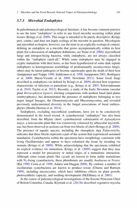
3.7.3 Microbial Endophytes
In paleobotanical and paleomycological literature, it has become common practice
to use the term “endophyte” to refer to any fossil microbe occurring within plant
tissues (Krings et al. 2009). This usage is intended to be purely descriptive (Krings
pers. comm.) and does not imply ecology of the microbe in question. Mycologists
and microbial ecologists, however, use the term in an explicitly ecological context,
defining an endophyte as a microbe that grows asymptomatically within its host
plant (for a discussion of endophyte definitions, see Stone et al. 2000); mycorrhizal
fungi and nitrogen-fixing bacteria, while endophytic, are not always classified
within the “endophyte catch-all.” While some endophytes may be engaged in
cryptic mutualism with their hosts, as has been hypothesized of some dark septate
endophytes (a heterogeneous assemblage of predominantly ascomycetous fungi),
others may be latent pathogens or become saprotrophs upon the death of their host
(Jumpponen and Trappe 1998; Saikkonen et al. 1998; Jumpponen 2001; Rodriguez
et al. 2009; Macia‐Vicente et al. 2009; Newsham 2011). Some fossil fungi
described as endophytes (as defined by Krings et al. 2009) elicited host responses
characteristic of infection or parasitism (e.g., Krings et al. 2007; Schwendemann
et al. 2010; Taylor et al. 2012). Recently, a study of the Early Devonian vascular
plant Horneophyton lignieri, eliciting comparisons with modern basal land plants
(embryophytes), has demonstrated the presence of endophytes belonging to two
major fungal lineages, the Glomeromycota and Mucoromycotina, and revealed
previously undocumented diversity in the fungal associations of basal embryo-
phytes (Strullu-Derrien et al. 2014).
Endophytes, excluding mycorrhizal symbionts, have yet to be conclusively
demonstrated in the fossil record. A cyanobacterial “endophyte” has also been
described, from the Rhynie chert: cyanobacterial colonization of Aglaophytonmajor, a nonvascular plant that was extensively colonized by arbuscular mycorrhi-
zae, has been observed in sections cut from two blocks of chert (Krings et al. 2009).
The presence of aquatic species, including the charophyte alga Palaeonitella,indicates that these blocks represent a part of the system that experienced sustained
inundation. Cyanobacteria within the specimens have morphology consistent with
living Oscillatoriales and appear to have colonized the tissue by invading via
stomata (Krings et al. 2009). While acknowledging that the specimens exhibited
no explicit evidence for mutualism, Krings et al. (2009) suggest that they may
represent a model for precursory or initial stages of a mutualistic interaction.
Although some extant plants like cycads are known to form stable mutualisms
with N2-fixing cyanobacteria, these photobionts are usually Anabaena or Nostoc(Rai 1990; Costa et al. 1999; Adams and Duggan 2008). By contrast, a number of
oscillatorian cyanobacteria are known to produce toxins (Chorus and Bartram
1999), including microcystins, which have inhibitory effects on plant growth,
photosynthetic capacity, and seedling development (McElhiney et al. 2001).
In the course of paleomycological investigations of the Eocene Princeton Chert
of British Columbia, Canada, Klymiuk et al. (2013b) described vegetative mycelia
3 Microbes and the Fossil Record: Selected Topics in Paleomicrobiology 141
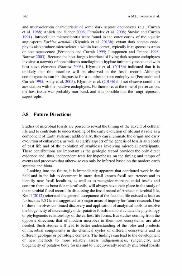
and microsclerotia characteristic of some dark septate endophytes (e.g., Currah
et al. 1988; Ahlich and Sieber 2006; Fernandez et al. 2008; Stoyke and Currah
1991). Intracellular microsclerotia were found in the outer cortex of the aquatic
angiosperm Eorhiza arnoldii (Klymiuk et al. 2013b); extant dark septate endo-
phytes also produce microsclerotia within host cortex, typically in response to stress
or host senescence (Fernando and Currah 1995; Jumpponen and Trappe 1998;
Barrow 2003). Because the host-fungus interface of living dark septate endophytes
involves a network of nonchitinous mucilaginous hyphae intimately associated with
host sieve elements (Barrow 2003), Klymiuk et al. (2013b) indicated that it is
unlikely that this interface will be observed in the fossil record. Although
conidiogenesis can be diagnostic for a number of root endophytes (Fernando and
Currah 1995; Addy et al. 2005), Klymiuk et al. (2013b) did not observe conidia in
association with the putative endophytes. Furthermore, at the time of preservation,
the host tissue was probably moribund, and it is possible that the fungi represent
saprotrophs.
3.8 Future Directions
Studies of microbial fossils are poised to reveal the timing of the advent of cellular
life and to contribute to understanding of the early evolution of life and its role as a
component of Earth systems; additionally, they can illuminate the origin and early
evolution of eukaryotes, as well as clarify aspects of the genesis of fossils as records
of past life and of the evolution of symbioses involving microbial participants.
These contributions are important as the geologic record provides the only direct
evidence and, thus, independent tests for hypotheses on the timing and tempo of
events and processes that otherwise can only be inferred based on the modern earth
systems and biota.
Looking into the future, it is immediately apparent that continued work in the
field and in the lab to document in more detail known fossil occurrences and to
identify new fossil localities, as well as to recognize more potential fossils and
confirm them as bona fide microfossils, will always have their place in the study of
the microbial fossil record. In discussing the fossil record of Archean microbial life,
Knoll (2012) reiterated the general acceptance of the fact that life existed at least as
far back as 3.5 Ga and suggested two major areas of inquiry for future research. One
of these involves continued discovery and application of analytical tools to resolve
the biogenicity of increasingly older putative fossils and to elucidate the physiology
or phylogenetic relationships of the earliest life forms. But studies coming from the
opposite direction, that of modern microbes in their host ecosystems, are also
needed. Such studies will lead to better understanding of the roles and products
of microbial components in the chemical cycles of different ecosystems and in
different geologic or petrologic contexts. The findings can lead to the development
of new methods to more reliably assess indigenousness, syngenicity, and
biogenicity of putative body fossils and to unequivocally identify microbial fossils
142 A.M.F. Tomescu et al.
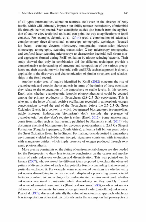
of all types (stromatolites, alteration textures, etc.) even in the absence of body
fossils, which will ultimately improve our ability to trace the trajectory of microbial
life through the rock record. Such actualistic studies also benefit from the applica-
tion of cutting-edge analytical tools and can point the way to applications in fossil
contexts. For example, Schmid et al. (2014) used a combination of advanced
complementary three-dimensional microscopy tomography techniques (focused
ion beam—scanning electron microscopy tomography, transmission electron
microscopy tomography, scanning-transmission X-ray microscopy tomography,
and confocal laser scanning microscopy) to characterize bacterial cell-(iron) min-
eral aggregates formed during Fe(II) oxidation by nitrate-reducing bacteria. Their
study showed that only in combination did the different techniques provide a
comprehensive understanding of structure and composition of the various precip-
itates and their association with bacterial cells and EPS; such an approach is directly
applicable to the discovery and characterization of similar structures and relation-
ships in the fossil record.
Another major area of inquiry identified by Knoll (2012) concerns the rise of
cyanobacteria and aerobic photosynthesis in terms of the timing of these events, as
they relate to the oxygenation of the atmosphere to stable levels. In this context,
Knoll asks whether cyanobacteria (aerobic photosynthesizers) could be counted
among the primary producers in Neoarchean (2.8–2.5 Ga) ecosystems. This is
relevant to the issue of small positive oscillations recorded in atmospheric oxygen
concentrations toward the end of the Neoarchean, before the 2.5–2.3 Ga Great
Oxidation Event, in a context in which documented biosignatures (stromatolites,
stable isotopes, hydrocarbon biomarkers) don’t exclude the presence of
cyanobacteria, but they don’t require it either (Knoll 2012). Some answers may
come from studies such as that recently published by Planavsky et al. (2014) who
document chemical biosignatures for oxygenic photosynthesis in 2.95 Ga Sinqeni
Formation (Pongola Supergroup, South Africa), at least a half billion years before
the Great Oxidation Event. In the Sinqeni Formation, rocks deposited in a nearshore
environment yielded molybdenum isotopic signatures consistent with interaction
with manganese oxides, which imply presence of oxygen produced through oxy-
genic photosynthesis.
More precise constraints on the dating of environmental changes are also needed
for the Proterozoic, to draw less tentative conclusions on the causes and mecha-
nisms of early eukaryote evolution and diversification. This was pointed out by
Javaux (2007), who reviewed the different ideas proposed to explain the observed
pattern of diversification of early eukaryote-like fossils, concluding that no event in
particular explained it. For example, some unanswered questions are whether early
eukaryotes diversifying in the marine realm displaced a preexisting cyanobacterial
biota or evolved in an ecologically undersaturated environment and whether
eukaryotes remained in minority while diversifying or they quickly formed
eukaryote-dominated communities (Knoll and Awramik 1983), or when eukaryotes
did invade the continents. In terms of recognition of early (unicellular) eukaryotes,
Peat et al. (1978) discussed critically the value of an actualistic approach which can
bias interpretations of ancient microfossils under the assumption that prokaryotes in
3 Microbes and the Fossil Record: Selected Topics in Paleomicrobiology 143

the Precambrian were morphologically similar to modern prokaryotes and
suggested that it is not out of the realm of possibility to discover disproportionately
large (eukaryote-like) prokaryote microfossils in Precambrian rocks.
Like in the case of prokaryote evolution, answers to questions concerning early
eukaryotes will come both from continued studies of the fossil record and from
better understanding of their taphonomy and modes of preservation based on
actualistic studies. Early on, Golubic and Barghoorn (1977) emphasized the need
for ultrastructural studies of diagenetic alteration in the cell walls of extant micro-
organisms, for application in the microbial fossil record. Conversely yet
convergently, Javaux et al. (2003) pointed out that taphonomic studies [like those
conducted by Knoll and Barghoorn (1975) or Bartley (1996) on prokaryotes] are
needed to elucidate the probability of preservation of intracellular—components
such as pyrenoids, starch, and cytoplasm—which, contrary to conventional
wisdom, is far from vanishingly small (e.g., Bomfleur et al. 2014). By improving
understanding of fossils and their mode of formation, such studies will lead to more
detailed and accurate interpretations of the fossils’ implications for the taxonomy,
ecology, and evolution of ancient microbes.
Microbially induced sedimentary structures have entered the sphere of interest
of Precambrian biological evolution studies relatively recently, benefiting fully
from well-developed and articulated concepts of sedimentology, an outlook empha-
sizing actualistic studies, and the breadth of modern environments in which they are
formed. Recently, Wilmeth et al. (2014) documented domal sand structures of
putative microbial origin in the 1.09 Ga Copper Harbor Conglomerate of Michigan,
expanding the fossil record of continental microbialites to siliciclastic fluvial
environments.
In contrast to MISS, stromatolites, although recognized and studied for more
than a century, lack extensive modern analogues and have ranked among the more
contentious Precambrian fossil biosignatures. It is therefore exciting to note a
resurgence of actualistic studies addressing stromatolite structures and the micro-
bial communities that build them from several perspectives. Kremer et al.’s (2012a)studies of microbial taphonomy and fossilization potential in modern stromatolites
in Tonga are such a study (see “Stromatolites” in sect. 3.2.2.1). Related to this, a
study by Knoll et al. (2013) documented in detail both the composition of micro-
fossil assemblages and the sedimentary structures (petrofabrics) in
Mesoproterozoic carbonate platform microbialites of the Angmaat/Society Cliffs
Formation (Baffin and Bylot Islands). Their study revealed covariation of micro-
fossil assemblages with petrofabrics, supporting hypotheses that link stromatolite
microstructure to the composition and diversity of microbial mat communities.
Mirroring Knoll et al.’s (2013) work in modern microbialites, Russell
et al. (2014) studied the microbial communities building microbialites in Pavilion
Lake (Canada). Using molecular analyses, Russell et al. (2014) documented diverse
communities including phototrophs (cyanobacteria) as well as heterotrophs and
photoheterotrophs. They also showed that the microbialite-building communities
are more diverse than the non-lithifying microbial mats in the lake and that
microbial community composition does not correlate with depth-related changes
144 A.M.F. Tomescu et al.

in microbialite morphology, suggesting that microbialite structure may not be under
strict control of microbial community composition. Finally, in a study of extracel-
lular polymeric substances and functional gene diversity within biofilm communi-
ties of modern oolitic sands (analogous in genesis to stratiform stromatolites) from
Great Bahama Bank, Diaz et al. (2014) suggest that carbonate precipitation in
marine oolitic biofilms is spatially and temporally controlled by a consortium of
microbes with diverse physiologies (photosynthesizers, heterotrophs, denitrifiers,
sulfate reducers, and ammonifiers) and point to a role of EPS-mediated microbial
calcium carbonate precipitation in the formation of the microlaminated oolitic
structures.
Long time considered ill positioned to make independent contributions beyond
merely documenting historical confirmation of events and processes, paleontology
(including Precambrian paleobiology; Schopf 2009) witnessed in the 1970s–1980s
the “paleobiological revolution” that reinstated it at the “high table” of evolutionary
biology (Sepkoski and Ruse 2009). Building on this newfound identity and adding
to it a developmental anatomy - comparative morphology twist, today paleontology
is poised to make meaningful contributions to the field of evolutionary-
developmental biology by documenting in fossils and tracing through time the
anatomical and morphological fingerprints of genetic pathways that regulate devel-
opment (e.g., Rothwell et al. 2014). In this context, a recent study by Flood
et al. (2014) inspires some exciting ideas. These authors used comparative geno-
mics to study phylogenetically distant bacteria that induce formation of wrinkle
structures (a type of MISS) in modern sediments. Their results suggest that hori-
zontally transferred genes may code for phenotypic traits that underlie similar
biostabilizing influences of these organisms on sediments. On the one hand, this
implies that the ecological utility of some phenotypic traits such as the construction
of mats and biofilms, along with the lateral mobility of genes in the microbial
world, render inferences of phylogenetic relationships from gross morphological
features preserved in the rock record uncertain (Flood et al. 2014). On the other
hand, this study expands the range of phenotypic traits that can be used as mor-
phological fingerprints for shared genetic pathways, to the realm of micro- and
macroscale sedimentary structures.
Deep in the rock record, the fossil evidence may not look spectacular by most
standards—tiny microfossils, wrinkles on a rock face, readings of chemical com-
position on a computer screen, or a spectrometer curve. Yet the studies of the
microbial fossil record can have tremendous outcomes, as they can bring key
contributions to addressing two of the most profound and perennial questions that
have puzzled humanity and science. On the one hand, tracing the microbial fossil
record is our only direct way of catching a glimpse of the beginnings and early
evolution of life, and this chapter has attempted to provide an introduction and
overview of the paradigms that underpin such studies. On the other hand, the
methods and ideas developed as a result of studies of the microbial fossil record
for recognizing ancient and inconspicuous traces of life, as well as the knowledge
and experience accumulated in the process, have crystallized in an approach that is
directly applicable to the search for traces of life on other planets (e.g., Brasier and
3 Microbes and the Fossil Record: Selected Topics in Paleomicrobiology 145
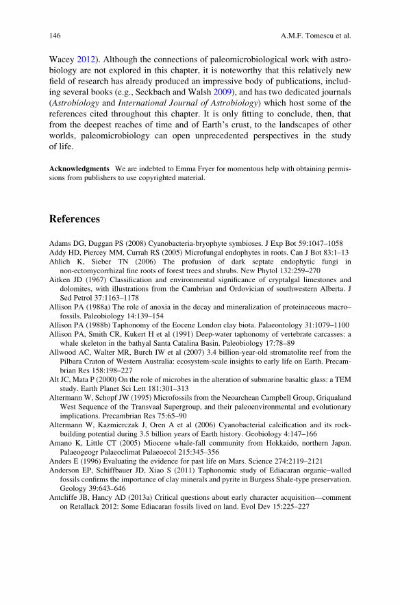
Wacey 2012). Although the connections of paleomicrobiological work with astro-
biology are not explored in this chapter, it is noteworthy that this relatively new
field of research has already produced an impressive body of publications, includ-
ing several books (e.g., Seckbach and Walsh 2009), and has two dedicated journals
(Astrobiology and International Journal of Astrobiology) which host some of the
references cited throughout this chapter. It is only fitting to conclude, then, that
from the deepest reaches of time and of Earth’s crust, to the landscapes of other
worlds, paleomicrobiology can open unprecedented perspectives in the study
of life.
Acknowledgments We are indebted to Emma Fryer for momentous help with obtaining permis-
sions from publishers to use copyrighted material.
References
Adams DG, Duggan PS (2008) Cyanobacteria-bryophyte symbioses. J Exp Bot 59:1047–1058
Addy HD, Piercey MM, Currah RS (2005) Microfungal endophytes in roots. Can J Bot 83:1–13
Ahlich K, Sieber TN (2006) The profusion of dark septate endophytic fungi in
non-ectomycorrhizal fine roots of forest trees and shrubs. New Phytol 132:259–270
Aitken JD (1967) Classification and environmental significance of cryptalgal limestones and
dolomites, with illustrations from the Cambrian and Ordovician of southwestern Alberta. J
Sed Petrol 37:1163–1178
Allison PA (1988a) The role of anoxia in the decay and mineralization of proteinaceous macro–
fossils. Paleobiology 14:139–154
Allison PA (1988b) Taphonomy of the Eocene London clay biota. Palaeontology 31:1079–1100
Allison PA, Smith CR, Kukert H et al (1991) Deep-water taphonomy of vertebrate carcasses: a
whale skeleton in the bathyal Santa Catalina Basin. Paleobiology 17:78–89
Allwood AC, Walter MR, Burch IW et al (2007) 3.4 billion-year-old stromatolite reef from the
Pilbara Craton of Western Australia: ecosystem-scale insights to early life on Earth. Precam-
brian Res 158:198–227
Alt JC, Mata P (2000) On the role of microbes in the alteration of submarine basaltic glass: a TEM
study. Earth Planet Sci Lett 181:301–313
Altermann W, Schopf JW (1995) Microfossils from the Neoarchean Campbell Group, Griqualand
West Sequence of the Transvaal Supergroup, and their paleoenvironmental and evolutionary
implications. Precambrian Res 75:65–90
Altermann W, Kazmierczak J, Oren A et al (2006) Cyanobacterial calcification and its rock-
building potential during 3.5 billion years of Earth history. Geobiology 4:147–166
Amano K, Little CT (2005) Miocene whale-fall community from Hokkaido, northern Japan.
Palaeogeogr Palaeoclimat Palaeoecol 215:345–356
Anders E (1996) Evaluating the evidence for past life on Mars. Science 274:2119–2121
Anderson EP, Schiffbauer JD, Xiao S (2011) Taphonomic study of Ediacaran organic–walled
fossils confirms the importance of clay minerals and pyrite in Burgess Shale-type preservation.
Geology 39:643–646
Antcliffe JB, Hancy AD (2013a) Critical questions about early character acquisition—comment
on Retallack 2012: Some Ediacaran fossils lived on land. Evol Dev 15:225–227
146 A.M.F. Tomescu et al.

Antcliffe JB, Hancy AD (2013b) Reply to Retallack (2013): Ediacaran characters. Evol Dev
15:389–392
Antcliffe JB, McLoughlin N (2009) Deciphering fossil evidence for the origin of life and the origin
of animals: common challenges in different worlds. In: Seckbach J, Walsh M (eds) From
fossils to astrobiology. Springer, Dordrecht, pp 211–229
Appel PWU, Moorbath S, Myers JS (2003) Isuasphaera isua (Pflug) revisited. Precambrian Res
126:309–312
Arning ET, Birgel D, Brunner B et al (2009) Bacterial formation of phosphatic laminites off Peru.
Geobiology 7:295–307
Arning ET, Birgel D, Schulz-Vogt HN et al (2008) Lipid biomarker patterns of phosphogenic
sediments from upwelling regions. Geomicrobiol J 25:69–82
Arouri K, Greenwood PF, Walter MR (1999) A possible chlorophycean affinity of some
Neoproterozoic acritarchs. Org Geochem 30:1323–1337
Arouri KR, Greenwood PF, Walter MR (2000) Biological affinities of Neoproterozoic acritarchs
from Australia: microscopic and chemical characterisation. Org Geochem 31:75–89
Awramik SM (2006) Respect for stromatolites. Nature 441:700–701
Awramik SM, Buchheim HP (2009) A giant, Late Archean lake system: the Meentheena Member
(Tumbiana Formation; Fortescue Group), Western Australia. Precambrian Res 174:215–240
Awramik SM, Barghoorn ES (1977) The Gunflint microbiota. Precambrian Res 5:121–142
Awramik SM, Grey K (2005) Stromatolites: biogenicity, biosignatures, and bioconfusion. In:
Gladstone GR, Hoover RB, Levin GV et al. (eds) Astrobiology and Planetary Missions. SPIE
Proceedings 5906:1–9
Awramik SM, Margulis L (1974) Stromatolite Newslett 2:5
Awramik SM, Riding R (1988) Role of algal eukaryotes in subtidal columnar stromatolite
formation. Proc Nat Acad Sci USA 85:1327–1329
Awramik SM, Schopf JW, Walter MR (1983) Filamentous fossil bacteria from the Archean of
Western Australia. Precambrian Res 20:357–374
Bailey JV, Joye SB, Kalanetra KM et al (2007) Palaeontology: undressing and redressing
Ediacaran embryos (Reply). Nature 446:E10–E11
Banerjee NR, Simonetti A, Furnes H et al (2007) Direct dating of Archean microbial ichnofossils.
Geology 35:487–490
Barghoorn ES, Tyler SA (1965) Microorganisms from the Gunflint chert. Science 147:563–577
Barrow JR (2003) Atypical morphology of dark septate fungal root endophytes of Bouteloua in
arid southwestern USA rangelands. Mycorrhiza 13:239–247
Bartley JK (1996) Actualistic taphonomy of cyanobacteria: implications for the Precambrian fossil
record. Palaios 11:571–586
Battison L, Brasier MD (2012) Remarkably preserved prokaryote and eukaryote microfossils
within 1 Ga-old lake phosphates of the Torridon Group, NW Scotland. Precambrian Res
196–197:204–217
Baxter RW (1950) Peltastrobus reedae: a new sphenopsid cone from the Pennsylvanian of
Indiana. Bot Gaz 112:174–182
Bazylinski DA (1996) Controlled biomineralization of magnetic minerals by magnetotactic
bacteria. Chem Geol 132:191–198
Bazylinski DA, Frankel RB (2003) Biologically controlled mineralization in prokaryotes. Rev
Miner Geochem 54:217–247
Bekker A, Holland HD, Wang P-L et al (2004) Dating the rise of atmospheric oxygen. Nature
427:117–120
Bengtson S, Belivanova V, Rasmussen B et al (2009) The controversial “Cambrian” fossils of the
Vindhyan are real but more than a billion years older. Proc Nat Acad Sci USA 106:7729–7734
Beraldi-Campesi H, Garcia-Pichel F (2011) The biogenicity of modern terrestrial roll-up struc-
tures and its significance for ancient life on land. Geobiology 9:10–23
Beraldi-Campesi H, Farmer JD, Garcia-Pichel F (2014) Modern terrestrial sedimentary
biostructures and their fossil analogs in Mesoproterozoic subaerial deposits. Palaios 29:45–54
3 Microbes and the Fossil Record: Selected Topics in Paleomicrobiology 147
Beveridge TJ (1989) Role of cellular design in bacterial metal accumulation and mineralization.
Ann Rev Microbiol 43:147–171
Bidartondo MI, Read DJ, Trappe JM et al (2011) The dawn of symbiosis between plants and fungi.
Biol Lett 7:574–577
Boal D, Ng R (2010) Shape analysis of filamentous Precambrian microfossils and modern
cyanobacteria. Paleobiology 36:555–572
Bomfleur B, McLoughlin S, Vajda V (2014) Fossilized nuclei and chromosomes reveal 180million
years of genomic stasis in royal ferns. Science 343:1376–1377
Boyce CK, Hazen RM, Knoll AH (2002) Non-destructive, in situ, cellular-scale mapping of
elemental abundances including organic carbon in permineralized fossils. Proc Nat Acad Sci
USA 98:5970–5974
Brack-Hanes SD, Vaughn JC (1978) Evidence of Paleozoic chromosomes from lycopod
microgametophytes. Science 200:1383–1385
Bradley JP, Harvey RP, McSween HY Jr (1997) No ‘nannofossils’ in martian meteorite. Nature
390:454–455
Bradley JP, McSween HY Jr, Harvey RP (1998) Epitaxial growth of nanophase magnetite in
Martian meteorite ALH 84001: implications for biogenic mineralization. Meteorit Planet Sci
33:765–773
Brasier MD, Wacey D (2012) Fossils and astrobiology: new protocols for cell evolution in deep
time. Int J Astrobiol 11:217–228
Brasier MD, Green OR, Jephcoat AP et al (2002) Questioning the evidence for Earth’s oldest
fossils. Nature 417:76–81
Brasier M, Green O, Lindsay J et al (2004) Earths oldest (~3.5 Ga) fossils and the ‘early eden
hypothesis’: questioning the evidence. Origins Life Evol B 34:257–269
Brasier M, McLoughlin N, Green O et al (2006) A fresh look at the fossil evidence for Early
Archaean cellular life. Phil Trans R Soc B 361:887–902
Bridgewater D, Allaart JH, Schopf JW et al (1981) Microfossil-like objects from the Archean of
Greenland: a cautionary note. Nature 289:51–53
Briggs DE (1999) Molecular taphonomy of animal and plant cuticles: selective preservation and
diagenesis. Phil Trans R Soc Lond B354:7–17
Briggs DE (2003a) The role of decay and mineralization in the preservation of soft-bodied fossils.
Ann Rev Earth Planet Sci 31:275–301
Briggs DE (2003b) The role of biofilms in the fossilization of non-biomineralized tissues. In:
Krumbein WE, Paterson DM, Zavarzin GA (eds) Fossil and recent biofilms. Kluwer, Dor-
drecht, pp 281–290
Briggs DE, Kear AJ (1993) Fossilization of soft tissue in the laboratory. Science 259:439–1442
Briggs DE, Kear AJ (1994) Decay and mineralization of shrimps. Palaios 9:431–456
Briggs DE, Wilby PR (1996) The role of the calcium carbonate-calcium phosphate switch in the
mineralization of soft-bodied fossils. J Geol Soc 153:665–668
Briggs DEG, Kear AJ, Martill DM et al (1993) Phosphatization of soft-tissue in experiments and
fossils. J Geol Soc 150:1035–1038
Briggs DE, Moore RA, Shultz JW et al (2005) Mineralization of soft-part anatomy and invading
microbes in the horseshoe crab Mesolimulus from the Upper Jurassic Lagerstatte of
Nusplingen, Germany. Proc R Soc B: Biol Sci 272:627–632
Briggs DE, Wilby PR, Perez-Moreno BP et al (1997) The mineralization of dinosaur soft tissue in
the Lower Cretaceous of Las Hoyas, Spain. J Geol Soc 154:587–588
Brock F, Parkes RJ, Briggs DE (2006) Experimental pyrite formation associated with decay of
plant material. Palaios 21:499–506
Brocks JJ, Logan GA, Buick R et al (1999) Archean molecular fossils and the early rise of
eukaryotes. Science 285:1033–1036
Brocks JJ, Buick R, Logan GA et al (2003a) Composition and syngeneity of molecular fossils from
the 2.78 to 2.45 billion-year-old Mount Bruce Supergroup, Pilbara Craton, Western Australia.
Geochim Cosmochim Acta 67:4289–4319
148 A.M.F. Tomescu et al.

Brocks JJ, Buick R, Logan GA et al (2003b) A reconstruction of Archean biological diversity
based on molecular fossils from the 2.78 to 2.45 billion-year-old Mount Bruce Supergroup,
Hamersley Basin, Western Australia. Geochim Cosmochim Acta 67:4321–4335
Budel B, Weber B, Kuhl M et al (2004) Reshaping of sandstone surfaces by cryptoendolithic
cyanobacteria: bioalkalization causes chemical weathering in arid landscapes. Geobiology
2:261–268
Buick R (1984) Carbonaceous filaments from North Pole, Western Australia: are they fossil
bacteria in Archean stromatolites? Precambrian Res 24:157–172
Buick R (1990) Microfossil recognition in Archean rocks: an appraisal of spheroids and filaments
from the 3500 m.y. old chert-barite unit at North Pole, Western Australia. Palaios 5:441–459
Buick R (1992) The antiquity of oxygenic photosynthesis: evidence from stromatolites in
sulphate-deficient Archaean lakes. Science 255:74–77
Buick R (2001) Life in the Archean. In: Briggs DEG, Crowther PR (eds) Palaeobiology
II. Blackwell Science, Oxford, pp 13–21
Buick R (2010) Ancient acritarchs. Nature 463:885–886
Buick R (2012) Geobiology of the Archean Eon. In: Knoll AH, Canfield DE, Konhauser KO (eds)
Fundamentals of geobiology. Wiley-Blackwell, Chichester, pp 351–370
Buick R, Dunlop J (1990) Evaporitic sediments of Early Archaean age from the Warrawoona
Group, North Pole, Western Australia. Sedimentology 37:247–277
Buick R, Knoll AH (1999) Acritarchs and microfossils from the Mesoproterozoic Bangemall
Group, Northwestern Australia. J Paleontol 73:744–764
Buick R, Dunlop JSR, Groves DI (1981) Stromatolite recognition in ancient rocks: an appraisal of
irregularly laminated structures in an Early Archaean chert-barite unit from North Pole,
Western Australia. Alcheringa 5:161–181
Burke C, Steinberg P, Rusch D et al (2011) Bacterial community assembly based on functional
genes rather than species. Proc Nat Acad Sci USA 108:14288–14293
Burne RV, Moore L (1987) Microbialites: organosedimentary deposits of benthic microbial
communities. Palaios 2:241–254
Buseck PR, Dunin-Borkowski RE, Devouard B et al (2001) Magnetite morphology and life on
Mars. Proc Nat Acad Sci USA 98:13490–13495
Butler IB, Rickard D (2000) Framboidal pyrite formation via the oxidation of iron (II) monosulfide
by hydrogen sulphide. Geochim Cosmochim Acta 64:2665–2672
Butterfield NJ (1995) Secular distribution of Burgess‐Shale‐type preservation. Lethaia 28:1–13Butterfield NJ (2000) Bangiomorpha pubescens n. gen., n. sp.: implications for the evolution of
sex, multicellularity and the Mesoproterozoic/Neoproterozoic radiation of eukaryotes. Paleo-
biology 26:386–404
Butterfield NJ (2001) Paleobiology of the Late Mesoproterozoic (ca. 1200 Ma) hunting formation,
Somerset Island, arctic Canada. Precambrian Res 111:235–256
Butterfield NJ (2002) Leanchoilia guts and the interpretation of three-dimensional structures in
Burgess Shale-type fossils. Paleobiology 28:155–171
Butterfield NJ (2005a) Probable Proterozoic fungi. Paleobiology 31:165–182
Butterfield NJ (2005b) Reconstructing a complex early Neoproterozoic eukaryote, Wynniatt
Formation, arctic Canada. Lethaia 38:155–169
Butterfield NJ, Balthasar U, Wilson LA (2007) Fossil diagenesis in the Burgess Shale.
Palaeontology 50:537–543
Butterfield NJ (2009) Modes of pre-Ediacaran multicellularity. Precambrian Res 173:201–2011
Cai Y, Schiffbauer JD, Hua H et al (2012) Preservational modes in the Ediacaran Gaojiashan
Lagerstatte: Pyritization, aluminosilicification, and carbonaceous compression. Palaeogeogr
Palaeoclimat Palaeoecol 326:109–117
Callot G, Maurette M, Pottier L et al (1987) Biogenic etching of microfractures in amorphous and
crystalline silicates. Nature 328:147–149
3 Microbes and the Fossil Record: Selected Topics in Paleomicrobiology 149

Callow RH, Brasier MD (2009) Remarkable preservation of microbial mats in Neoproterozoic
siliciclastic settings: implications for Ediacaran taphonomic models. Earth Sci Rev
96:207–219
Campbell SE (1982) Precambrian endoliths discovered. Nature 299:429–431
Campbell KA, Farmer JD, Des Marais D (2002) Ancient hydrocarbon seeps from the Mesozoic
convergent margin of California: carbonate geochemistry, fluids and palaeoenvironments.
Geofluids 2:63–94
Canfield DE, Des Marais DJ (1991) Aerobic sulfate reduction in microbial mats. Science
251:1471–1473
Canfield DE, Thamdrup B (1994) The production of 34S-depleted sulfide during bacterial dispro-
portionation of elemental sulfur. Science 266:1973–1975
Canfield DE, Poulton SW, Knoll AH et al (2008) Ferruginous conditions dominated later
Neoproterozoic deep-water chemistry. Science 321:949–952
Canfield DE, Sørensen KB, Oren A (2004) Biogeochemistry of a gypsum‐encrusted microbial
ecosystem. Geobiology 2:133–150
Cantrell SA, Duval-Perez L (2012) Microbial mats: an ecological niche for fungi. Front Microbiol
3:1–7
Cardinale M, Puglia AM, Grube M (2006) Molecular analysis of lichen‐associated bacterial
communities. FEMS Microbiol Ecol 57:484–495
Carpenter K (2005) Experimental investigation of the role of bacteria in bone fossilization. N Jb
Geol Palaont Mh 11:83–94
Cavalier-Smith T (2002) The neomuran origin of archaebacteria: the negibacteria root of the
universal tree and bacteria megaclassification. Int J Syst Microbiol 52:7–76
Chan CS, De Stasio G, Welch SA et al (2004) Microbial polysaccharides template assembly of
nanocrystal fibers. Science 303:1656–1658
Chatellier X, West MM, Rose J et al (2004) Characterization of iron-oxides formed by oxidation of
ferrous ions in the presence of various bacterial species and inorganic ligands. Geomicrobiol J
21:99–112
Child AM (1995) Towards and understanding of the microbial decomposition of archaeological
bone in the burial environment. J Archaeol Sci 22:165–174
Chin K, Eberth DA, Schweitzer MH et al (2003) Remarkable preservation of undigested muscle
tissue within a Late Cretaceous tyrannosaurid coprolite from Alberta, Canada. Palaios
18:286–294
Chorus I, Bartram J (1999) Toxic cyanobacteria in water: a guide to their public health conse-
quences, monitoring and management. Routledge, London
Cleland TP, Stoskopf MK, Schweitzer MH (2011) Histological, chemical, and morphological
reexamination of the “heart” of a small Late Cretaceous Thescelosaurus. Naturwissenschaften98:203–211
Clemett SJ, Dulay MT, Gilette JS et al (1998) Evidence for the extraterrestrial origin of polycyclic
aromatic hydrocarbons (PAHs) in the Martian meteorite ALH 84001. Faraday Discuss
109:417–436
Clemett SJ, Thomas-Keprta KL, Shimmin J et al (2002) Crystal morphology of MV-1 magnetite.
Am Mineral 87:1727–1730
Cloud P (1973) Pseudofossils: a plea for caution. Geology 1:123–127
Cloud PE Jr, Hagen H (1965) Electron microscopy of the Gunflint microflora. Proc Nat Acad Sci
USA 54:1–8
Cloud P, Morrison K (1979) On microbial contaminants, micropseudofossils, and the oldest
records of life. Precambrian Res 9:81–91
Coleman ML, Raiswell R (1995) Source of carbonate and origin of zonation in pyritiferous
carbonate concretions; evaluation of a dynamic model. Am J Sci 295:282–308
Collins MJ, Nielsen-Marsh CM, Hiller J (2002) The survival of organic matter in bone: a review.
Archaeometry 44:383–394
150 A.M.F. Tomescu et al.
Collinson ME, Manchester SR, Wilde V et al (2010) Fruit and seed floras from exceptionally
preserved biotas in the European Paleogene. Bull Geosci 85:155–162
Conti MA, Morsilli M, Nicosia U et al (2005) Jurassic dinosaur footprints from southern Italy:
footprints as indicators of constraints in paleogeographic interpretation. Palaios 20:534–550
Cosmidis J, Benzerara K, Menguy N et al (2013) Microscopy evidence of bacterial microfossils in
phosphorite crusts of the Peruvian shelf: Implications for phosphogenesis mechanisms. Chem
Geol 359:10–22
Costerton JW, Lewandowski Z, Caldwell DE et al (1995) Microbial biofilms. Ann Rev Microbiol
49:711–745
Costa JL, Paulsrud P, Lindblad P (1999) Cyanobiont diversity within coralloid roots of selected
cycad species. FEMS Microb Ecol 28:85–91
Courties C, Vaquer A, Troussellier M et al (1994) Smallest eukaryotic organism. Nature 370:255
Cunningham JA, Thomas CW, Bengtson S et al (2012a) Distinguishing geology from biology in
the Ediacaran Doushantuo biota relaxes constraints on the timing of the origin of bilaterians.
Proc R Soc B: Biol Sci 279:2369–2376
Cunningham JA, Thomas CW, Bengtson S et al (2012b) Experimental taphonomy of giant sulphur
bacteria: implications for the interpretation of the embryo-like Ediacaran Doushantuo fossils.
Proc R Soc B: Biol Sci 279:1857–1864
Currah RS, Hambleton S, Smreciu A (1988) Mycorrhizae and mycorrhizal fungi of Calypsobulbosa. Am J Bot 75:739–752
Currie PJ, Sarjeant WA (1979) Lower Cretaceous dinosaur footprints from the Peace River
Canyon, British Columbia, Canada. Palaeogeogr Palaeoclim Palaeoecol 28:103–115
Daniel JC, Chin K (2010) The role of bacterially mediated precipitation in the permineralization of
bone. Palaios 25:507–516
Darroch SA, Laflamme M, Schiffbauer JD et al (2012) Experimental formation of a microbial
death mask. Palaios 27:293–303
Dauphas N, Van Zuilen M, Wadhwa M et al (2004) Clues from Fe isotope variations on the origin
of early Archean BIFs from Greenland. Science 306:2077–2080
De Gregorio BT, Sharp TG, Flynn GJ et al (2009) Biogenic origin for Earth’s oldest putative
microfossils. Geology 37:631–634
Deacon JW (2006) Fungal biology. Wiley-Blackwell, Oxford
Decho AW, Kawaguchi T (2003) Extracellular polymers (EPS) and calcification within modern
marine stromatolites. In: Krumbein WE, Paterson DM, Zavarzin GA (eds) Fossil and recent
biofilms. Springer, Dordrecht, pp 227–240
Deming JW, Reysenbach AL, Macko SA et al (1997) Evidence for the microbial basis of a
chemoautotrophic invertebrate community at a whale fall on the deep seafloor: bone‐coloniz-ing bacteria and invertebrate endosymbionts. Microsc Res Tech 37:162–170
Diaz MR, van Norstrand JD, Eberli GP et al (2014) Functional gene diversity of oolitic sands from
Great Bahama Bank. Geobiology 12:231–49. doi:10.1111/gbi.12079
Dotzler N, Krings M, Taylor TN et al (2006) Germination shields in Scutellospora(Glomeromycota: Diversisporales, Gigasporaceae) from the 400 million-year-old Rhynie
chert. Mycol Progr 5:178–184
Dotzler N, Walker C, Krings M et al (2009) Acaulosporoid glomeromycotan spores with a
germination shield from the 400-million-year-old Rhynie chert. Mycol Progr 8:9–18
Droser ML, Gehling JG, Jensen SR (2006) Assemblage palaeoecology of the Ediacara biota: the
unabridged edition? Palaeogeogr Palaeoclim Palaeoecol 232:131–147
Duck LJ, Glikson M, Golding SD et al (2007) Microbial remains and other carbonaceous forms
from the 3.24 Ga Sulphur Springs black smoker deposit, Western Australia. Precambrian Res
154:205–220
Dunlop JSR, Milne VA, Groves DI et al (1978) A new microfossils assemblage from the Archean
of Western Australia. Nature 274:676–678
Dunn KA, McLean RJC, Upchurch GR et al (1997) Enhancement of leaf fossilization potential by
bacterial biofilms. Geology 25:1119–1122
3 Microbes and the Fossil Record: Selected Topics in Paleomicrobiology 151
Dupraz C, Visscher PT (2005) Microbial lithification in marine stromatolites and hypersaline
mats. Trends Microbiol 13:429–438
Dupraz C, Reid RP, Braissant O et al (2009) Processes of carbonate precipitation in modern
microbial mats. Earth Sci Rev 96:141–162
Dutkiewicz A, Volk H, George SC et al (2006) Biomarkers from Huronian oil-bearing fluid
inclusions: an uncontaminated record of life before the Great Oxidation Event. Geology
34:437–440
Efremov IA (1940) Taphonomy: a new branch of paleontology. Pan-Am Geol 74:81–93
Eglinton G, Logan GA, Ambler RP et al (1991) Molecular preservation [and discussion]. Phil
Trans R Soc Lond B 333:315–328
El Albani A, Bengtson S, Canfield DE et al (2010) Large colonial organisms with coordinated
growth in oxygenated environments 2.1 Gyr ago. Nature 466:100–104
Fang HW, Shang QQ, Chen MH et al (2014) Changes in the critical erosion velocity for sediment
colonized by biofilm. Sedimentology 61:648–659
Fedo CM, Whitehouse MJ (2002) Metasomatic origin of quartz-pyroxene rock, Akilia, Greenland,
and implications for Earth’s earliest life. Science 296:1448–1452Fernando AA, Currah RS (1995) Leptodontidium orchidicola (Mycelium radicis atrovirens com-
plex): aspects of its conidiogenesis and ecology. Mycotaxon 54:287–294
Fernandez N, Messuti MI, Fontenla S (2008) Arbuscular mycorrhizas and dark septate fungi in
Lycopodium paniculatum (Lycopodiaceae) and Equisetum bogotense (Equisetaceae) in a
Valdivian temperate forest of Patagonia, Argentina. Am Fern J 98:117–127
Ferris FG (1993) Microbial biomineralization in natural environments. Earth Sci 47:233–250
Ferris FG, Fyfe WS, Beveridge TJ (1987) Bacteria as nucleation sites for authigenic minerals in a
metal-contaminated lake sediment. Chem Geol 63:225–232
Ferris FG, Fyfe WS, Beveridge TJ (1988) Metallic ion binding by Bacillus subtilis: implications
for the fossilization of microorganisms. Geology 16:149–152
Fisk MR, Giovannoni SJ, Thorseth IH (1998) Alteration of oceanic volcanic glass: textural
evidence of microbial activity. Science 281:978–979
Fletcher BJ, Beerling DJ, Chaloner WG (2004) Stable carbon isotopes and the metabolism of the
terrestrial Devonian organism Spongiophyton. Geobiology 2:107–119
Fliegel D, Kosler J, McLoughlin N et al (2010) In-situ dating of the Earth’s oldest trace fossil at3.34 Ga. Earth Planet Sci Lett 299:290–298
Flood BE, Bailey JV, Biddle JF (2014) Horizontal gene transfer and the rock record: comparative
genomics of phylogenetically distant bacteria that induce wrinkle structure formation in
modern sediments. Geobiology 12:119–132
Folk RL (1993) SEM imaging of bacteria and nannobacteria in carbonate sediments and rocks. J
Sed Petrol 63:990–999
Fortin D, Ferris FG, Beveridge TJ (1997) Surface-mediated mineral development by bacteria. In:
Banfield JF, Nealson KH (eds) Geomicrobiology: interactions between microbes and minerals:
Reviews in Mineralogy 35. Mineralogical Society of America, Washington, pp 161–180
Frankel RB, Bazylinski DA (2003) Biologically induced mineralization by bacteria. Rev Mineral
Geochem 54:95–114
Fregenal-Martınez MA, Buscalioni AD (2010) A holistic approach to the palaeoecology of Las
Hoyas Konservat-Lagerstatte (La Huerguina Formation, Lower Cretaceous, Iberian Ranges,
Spain). J Iberian Geol 36:297–326
Frey E, Martill DM, Buchy MC (2003) A new species of tapejarid pterosaur with soft–tissue head
crest. Geol Soc Lond Spec Publ 217:65–72
Furnes H, Muehlenbachs K (2003) Bioalteration recorded in ophiolitic pillow lavas. Geol Soc
Lond Spec Publ 218:415–426
Furnes H, Staudigel H, Thorseth IH et al (2001) Bioalteration of basaltic glass in the oceanic crust.
Geochem Geophys Geosyst 2:1049. doi:10.1029/2000GC000150
Furnes H, Banerjee NR, Muehlenbachs K et al (2004) Early life recorded in Archean pillow lavas.
Science 304:578–581
152 A.M.F. Tomescu et al.

Furnes H, Banerjee NR, Staudigel H et al (2007) Comparing petrographic signatures of
bioalteration in recent to Mesoarchean pillow lavas: tracing subsurface life in oceanic igneous
rocks. Precambrian Res 158:156–176
Gabbott SE (1998) Taphonomy of the Ordovician Soom Shale Lagerstatte: an example of soft
tissue preservation in clay minerals. Palaeontology 41:631–668
Gabbott SE, Norry MJ, Aldridge RJ et al (2001) Preservation of fossils in clay minerals; a unique
example from the Upper Ordovician Soom Shale, South Africa. Proc Yorkshire Geol Polytech
Soc 53:237–244
Gabbott SE, Xian-Guang H, Norry MJ et al (2004) Preservation of Early Cambrian animals of the
Chengjiang biota. Geology 32:901–904
Gaines RR, Briggs DE, Yuanlong Z (2008) Cambrian Burgess Shale-type deposits share a
common mode of fossilization. Geology 36:755–758
Gaines RR, Kennedy MJ, Droser ML (2005) A new hypothesis for organic preservation of Burgess
Shale taxa in the middle Cambrian Wheeler Formation, House Range, Utah. Palaeogeogr
Palaeoclim Palaeoecol 220:193–205
Gall JC, Bernier P, Gaillard C et al (1985) Influence du developpement d’un voile algaire sur la
sedimentation et la taphonomie des calcaires lithographiques. Exemple du gisement de Cerin
(Kimmeridgien superieur, Jura meridional francais). Mem Phys Chim Sci l’univers, Sci de la
Terre 301:547–552
Gall JC (1990) Les voiles microbiens. Leur contribution �a la fossilisation des organismes au corps
mou. Lethaia 23:21–28
Gaines RR, Hammarlund EU, Hou X, Qi C, Gabbott SE, Zhao Y, Peng J, Canfield DE (2012)
Mechanism for Burgess Shale-type preservation. Proc Natl Acad Sci USA 109:5180–5184
Garcia Massini JL (2007) A glomalean fungus from the Permian of Antarctica. Int J Plant Sci
168:673–678
Garcia-Ruiz JM, Hyde ST, Carnerup AM et al (2003) Self-assembled silica-carbonate structures
and detection of ancient microfossils. Science 302:1194–1197
Gargas A, DePriest PT, Grube M et al (1995) Multiple origins of lichen symbioses in fungi
suggested by SSU rDNA phylogeny. Science 268:1492–1495
Gehling JG (1999) Microbial mats in terminal Proterozoic siliciclastics; Ediacaran death masks.
Palaios 14:40–57
Gehling JG, Droser ML, Jensen SR et al (2005) Ediacara organisms: relating form to function. In:
Evolving form and function: fossils and development, Symposium Proceedings, Peabody
Museum of Natural History, Yale University, New Haven, p 43–67
Gehrig H, Schussler A, Kluge M (1996) Geosiphon pyriforme, a fungus forming endocytobiosis
with Nostoc (Cyanobacteria), is an ancestral member of the glomales: evidence by SSU rRNA
Analysis. J Mol Evol 4:371–81
Gehring AU, Kind J, Charilaou M et al (2011) The detection of magnetotactic bacteria and
magnetofossils by means of magnetic anisotropy. Earth Planet Sci Lett 309:113–117
George SC, Volk H, Dutkiewicz A et al (2008) Preservation of hydrocarbons and biomarkers in oil
trapped inside fluid inclusions for >2 billion years. Geochim Cosmochim Acta 72:844–870
Gerdes G, Claes M, Dunajtschik-Piewak K et al (1993) Contribution of microbial mats to
sedimentary surface structures. Facies 29:61–74
German TN, Podkovyrov VN (2009) New insights into the nature of the Late Riphean
Eosolenides. Precambrian Res 173:154–162
Giovannoni SJ, Fisk MR, Mullins TD et al (1996) Genetic evidence for endolithic microbial life
colonizing basaltic glass/seawater interfaces. In: Alt JC, Kinoshita H, Stokking LB et al (eds)
Proceedings of the Ocean Drilling Program. Sci Results 148:207–214
Glikson M, Duck LJ, Golding SD et al (2008) Microbial remains in some earliest Earth rocks:
comparison with a potential modern analogue. Precambrian Res 164:187–200
Golden DC, Ming DW, Schwandt CS et al (2000) An experimental study on kinetically-driven
precipitation of Ca-Mg-Fe carbonates from solution: implications for the low temperature
3 Microbes and the Fossil Record: Selected Topics in Paleomicrobiology 153

formation of carbonates in Martian meteorite Allan Hills 84001. Meteorit Planet Sci
35:457–465
Golden DC, Ming DW, Morris RV et al (2004) Evidence for exclusively inorganic formation of
magnetite in Martian meteorite ALH84001. Am Mineral 89:681–695
Golubic S, Barghoorn ES (1977) Interpretation of microbial fossils with special reference to the
Precambrian. In: Flugel E (ed) Fossil algae: recent results and developments. Springer, Berlin,
pp 1–14
Golubic S, Hofmann HJ (1976) Comparison of modern and mid-Precambrian Entophysalidaceae
(Cyanophyta) in stromatolitic algal mats: cell division and degradation. J Paleontol
50:1074–1092
Golubic S, Knoll AH (1993) Prokaryotes. In: Lipps JH (ed) Fossil prokaryotes and protists.
Blackwell, Boston, pp 51–76
Golubic S, Friedmann I, Schneider J (1981) The lithobiontic ecological niche, with special
reference to microorganisms. J Sed Petrol 51:475–478
Gorbushina AA, Krumbein WE (2000) Subaerial microbial mats and their effects on soil and rock.
In: Riding RE, Awramik SM (eds) Microbial sediments. Springer, Berlin, pp 161–170
Grasby SE (2003) Naturally precipitating vaterite (μ-CaCO3) spheres: unusual carbonates formed
in an extreme environment. Geochim Cosmochim Acta 67:1659–1666
Grey K, Williams IR (1990) Problematic bedding-plane markings from the Middle Proterozoic
Manganese Supergroup, Bangemall Basin, Western Australia. Precambrian Res 46:307–327
Grey K, Yochelson EL, Fedonkin MA et al (2010) Horodyskia williamsii new species, a
Mesoproterozoic macrofossil from Western Australia. Precambrian Res 180:1–17
Grimes ST, Brock F, Rickard D et al (2001) Understanding fossilization: experimental pyritization
of plants. Geology 29:123–126
Grimes ST, Davies KL, Butler IB et al (2002) Fossil plants from the Eocene London Clay: the use
of pyrite textures to determine the mechanism of pyritization. J Geol Soc 159:493–501
Grotzinger JP, Rothman DH (1996) An abiotic model for stomatolite morphogenesis. Nature
383:423–425
Grube M, Berg G (2009) Microbial consortia of bacteria and fungi with focus on the lichen
symbiosis. Fungal Biol Rev 23:72–85
Grube M, Kroken S (2000) Molecular approaches and the concept of species and species
complexes in lichenized fungi. Mycol Res 104:1284–1294
Grube M, Winka K (2002) Progress in understanding the evolution and classification of lichenized
ascomycetes. Mycologist 16:67–76
Han T-M, Runnegar B (1992) Megascopic eukaryotic algae from the 2.1 billion-tear-old Negaunee
Iron Formation, Michigan. Science 257:232–235
Harper CJ, Taylor TN, Krings M et al (2013) Mycorrhizal symbiosis in the Paleozoic seed fern
Glossopteris from Antarctica. Rev Palaeobot Palynol 192:22–31
Harrington DJ (1996) Bacterial collagenases and collagen-degrading enzymes and their potential
role in human disease. Infect Immun 64:1885–1891
Hedges RE (2002) Bone diagenesis: an overview of processes. Archaeometry 44:319–328
Helm RF, Huang Z, Edwards D et al (2000) Structural characterization of the released polysac-
charide of desiccation-tolerant Nostoc commune DRH-1. J Bacteriol 182:974–982Hermann TN, Podkovyrov VN (2006) Fungal remains from the Late Riphean. Paleontol J
40:207–214
Hickman AH, Van Kranendonk MJ (2012) Early Earth evolution: evidence from the 3.5–1.8 Ga
geologic history of the Pilbara region of Western Australia. Episodes 35:283–297
Hippler D, Hu N, Steiner M et al (2012) Experimental mineralization of crustacean eggs: new
implications for the fossilization of Precambrian-Cambrian embryos. Biogeosciences
9:1765–1775
Hof CH, Briggs DE (1997) Decay and mineralization of mantis shrimps (Stomatopoda; Crusta-
cea); a key to their fossil record. Palaios 12:420–438
154 A.M.F. Tomescu et al.

Hofmann HJ (1972) Precambrian remains in Canada: fossils, dubiofossils and pseudofossils. In:
Proceedings of the 24th International Geological Congress, Section 1, p 20–30
Hofmann HJ (1976) Precambrian microflora, Belcher Islands, Canada—significance and system-
atics. J Paleo 50:1040–1073
Hofmann HJ (2000) Archean stromatolites as microbial archives. In: Riding RE, Awramik SM
(eds) Microbial sediments. Springer, Berlin, pp 315–327
Hofmann HJ, Grey K, Hickman AH et al (1999) Origin of 3.45 Ga coniform stromatolites in
Warrawoona Group, Western Australia. Geol Soc Am Bull 111:1256–1262
Honegger R (2009) Lichen-forming fungi and their photobionts. In: Deising H (ed) The Mycota,
vol 5, Plant relationships. Springer, Berlin, pp 305–333
Honegger R, Axe L, Edwards D (2013a) Bacterial epibionts and endolichenic actinobacteria and
fungi in the Lower Devonian lichen Chlorolichenomycites salopensis. Fungal Biol
117:512–518
Honegger R, Edwards D, Axe L (2013b) The earliest records of internally stratified cyanobacterial
and algal lichens from the Lower Devonian of theWelsh Borderland. New Phytol 197:264–275
Horodyski RJ (1980) Middle Proterozoic shale-facies microbiota from the Lower Belt Supergroup,
Little Belt Mountains, Montana. J Paleo 54:649–663
Horodyski RJ (1982) Problematic bedding-plane markings from the Middle Proterozoic
Appekunny Argillite, Belt Supergroup, Northwestern Montana. J Paleo 56:882–889
Horodyski RJ, Knauth LP (1994) Life on land in the Precambrian. Science 263:494–498
House CH, Schopf JW, McKeegan KD et al (2000) Carbon isotopic composition of individual
Precambrian microfossils. Geology 28:707–710
Huldtgren T, Cunningham JA, Yin C et al (2011) Fossilized nuclei and germination structures
identify Ediacaran “animal embryos” as encysting protists. Science 334:1696–1699
Humphreys CP, Franks PJ, Rees M et al (2010) Mutualistic mycorrhiza-like symbiosis in the most
ancient group of land plants. Nat Commun 1:103
Iniesto M, Lopez-Archilla AI, Fregenal-Martınez M et al (2013) Involvement of microbial mats in
delayed decay: an experimental essay on fish preservation. Palaios 28:56–66
Jans MME, Nielsen-Marsh CM, Smith CI et al (2004) Characterisation of microbial attack on
archaeological bone. J Archaeol Sci 31:87–95
Javaux EJ (2007) Patterns of diversification in early eukaryotes. Carnets de Geologie/Notebooks
on Geology 2007(01):38–42
Javaux EJ, Knoll AH, Walter MR (2001) Morphological and ecological complexity in early
eukaryotic ecosystems. Nature 412:66–69
Javaux EJ, Knoll AH, Walter MR (2003) Recognizing and interpreting the fossils of early
eukaryotes. Orig Life Evol Biosph 33:75–94
Javaux EJ, Knoll AH, Walter MR (2004) TEM evidence for eukaryotic diversity in
mid-Proterozoic oceans. Geobiology 2:121–132
Javaux EJ, Marshall CP, Bekker A (2010) Organic-walled microfossils in 3.2-billion-year-old
shallow-marine siliciclastic deposits. Nature 463:934–938
Jumpponen A (2001) Dark septate endophytes–are they mycorrhizal? Mycorrhiza 11:207–211
Jumpponen A, Trappe JM (1998) Dark septate endophytes: a review of facultative biotrophic root‐colonizing fungi. New Phytol 140:295–310
Jung HG, Deetz DA (1993) Cell wall lignification and degradability. In: Jung HG, Buxton DR,
Hatfield RD et al (eds) Forage cell wall structure and digestibility. American Society of
Agronomy, Crop Science Society of America, Soil Science Society of America, p 315–346
Kalanetra KM, Joye SB, Sunser NR et al (2005) Novel vacuolate sulfur bacteria from the Gulf of
Mexico reproduce by reductive division in three dimensions. Environ Microbiol 7:1451–1460
Kalkowsky E (1908) Oolith und Stromatolith im norddeutschen Buntsandstein. Z Dtsch Geol Ges
60:68–125
Karatygin IV, Snigirevskaya NS, Vikulin SV (2009) The most ancient terrestrial lichen
Winfrenatia reticulata: a new find and new interpretation. Paleontol J 43:107–114
3 Microbes and the Fossil Record: Selected Topics in Paleomicrobiology 155

Kaye TG, Gaugler G, Sawlowicz Z (2008) Dinosaurian soft tissues interpreted as bacterial
biofilms. PLoS One 3, e2808
Kazmierczak J (1979) The eukaryotic nature of Eosphaera-like ferriferous structures from the
Precambrian Gunflint Iron Formation, Canada: a comparative study. Precambrian Res 9:1–22
Kazmierczak J, Kempe S (2006) Genuine modern analogues of Precambrian stromatolites from
caldera lakes of Niuafo’ou Island, Tonga. Naturwissenschaften 93:119–126
Kellner AW (1996a) Reinterpretation of a remarkably well preserved pterosaur soft tissue from the
Early Cretaceous of Brazil. J Vert Paleo 16:718–722
Kellner AWA (1996b) Fossilized theropod soft tissue. Nature 379:32
Kennard JM, James NP (1986) Thrombolites and stromatolites; two distinct types of microbial
structures. Palaios 1:492–503
Kirkland BL, Lynch FL, Rahnis MA et al (1999) Alternative origins for nannobacteria-like objects
in calcite. Geology 27:347–350
Kiyokawa S, Ito T, Ikehara M et al (2006) Middle Archean volcano-hydrothermal sequence:
bacterial microfossil-bearing 3.2 Ga Dixon Island Formation, coastal Pilbara terrane, Australia.
Geol Soc Am Bull 118:3–22
Klein C, Beukes NJ, Schopf JW (1987) Filamentous microfossils in the early Proterozoic Trans-
vaal Super group: their morphology, significance, and paleoenviron mental setting. Precam-
brian Res 36:81–94
Kluge M, Mollenhauer D, Wolf E et al (2003) The Nostoc-Geosiphon endocytobiosis. In: Rai AN,Bergman B, Rasmussen U (eds) Cyanobacteria in symbiosis. Kluwer, Dordrecht, pp 19–30
Klymiuk AA, Stockey RA, Rothwell GW (2011) The first organismal concept for an extinct
species of Pinaceae: Pinus arnoldii Miller. Int J Plant Sci 172:294–313
Klymiuk AA, Harper CJ, Moore DM et al (2013a) Reinvestigating Carboniferous “actinomy-
cetes”: authigenic formation of biomimetic carbonates provides insight into early diagenesis of
permineralized plants. Palaios 28:80–92
Klymiuk AA, Taylor TN, Taylor EL et al (2013b) Paleomycology of the Princeton Chert II. Dark-
septate fungi in the aquatic angiosperm Eorhiza arnoldii indicate a diverse assemblage of root-
colonizing fungi during the Eocene. Mycologia 105:1100–1109
Knight TK, Bingham PS, Lewis RD, Savrda CE (2011) Feathers of the Ingersoll shale, Eutaw
Formation (Upper Cretaceous), eastern Alabama: the largest collection of feathers from North
American Mesozoic rocks. Palaios 26:364–376
Knoll AH (1992) The early evolution of eukaryotes: a geological perspective. Science
256:622–627
Knoll AH (2012) The fossil record of microbial life. In: Knoll AH, Canfield DE, Konhauser KO
(eds) Fundamentals of geobiology. Wiley-Blackwell, Chichester, pp 297–314
Knoll AH (2014) Paleobiological perspectives on early eukaryotic evolution. Cold Spring Harb
Perspect Biol 6:a016121
Knoll AH, Awramik SM (1983) Ancient microbial ecosystems. In: Krumbein WE (ed) Microbial
geochemistry. Blackwell, Oxford, pp 287–315
Knoll AH, Bambach RK (2000) Directionality in the history of life: diffusion fom the left wall or
repeated scaling on the right? In: Erwin DH, Wing SL (eds) Deep time. Paleobiology’sperspective. The Paleontological Society. Supplement to Palaios 26:1–14
Knoll AH, Barghoorn ES (1974) Ambient pyrite in Precambrian chert: new evidence and a theory.
Proc Nat Acad Sci USA 71:2329–2331
Knoll AH, Barghoorn ES (1975) Precambrian eukaryotic organisms: a reassessment of the
evidence. Science 190:52–54
Knoll AH, Barghoorn ES (1977) Microfossils showing cell division from the Swaziland System of
South Africa. Science 198:396–398
Knoll AH, Golubic S (1979) Anatomy and taphonomy of a Precambrian algal stromatolite.
Precambrian Res 10:115–151
Knoll AH, Golubic S, Green J et al (1986) Organically preserved microbial endoliths from the late
Proterozoic of East Greenland. Nature 321:856
156 A.M.F. Tomescu et al.
Knoll AH, Javaux EJ, Hewitt D et al (2006) Eukaryotic organisms in Proterozoic oceans. Phil
Trans R Soc Lond B 361:1023–1038
Knoll AH, Canfield DE, Konhauser KO (eds) (2012) Fundamentals of geobiology. Wiley-
Blackwell, Chichester
Knoll AH, Worndle S, Kah LC (2013) Covariance of microfossil assemblages and microbialite
textures across an upper Mesoproterozoic carbonate platform. Palaios 28:453–470
Konhauser KO (1998) Diversity of bacterial iron mineralization. Earth Sci Rev 43:91–121
Konhauser KO, Riding R (2012) Bacterial biomineralization. In: Knoll AH, Canfield DE,
Konhauser KO (eds) Fundamentals of geobiology. Wiley-Blackwell, Chichester, pp 105–130
Konhauser KO, Fisher QJ, Fyfe WS et al (1998) Authigenic mineralization and detrital clay
binding by freshwater biofilms: the Brahmani River, India. Geomicrobiol J 15:209–222
Konhauser KO, Kappler A, Roden EE (2011) Iron in microbial metabolisms. Elements 7:89–93
Kremer B, Kazmierczak J (2005) Cyanobacterial mats from Silurian black radiolarian cherts:
phototrophic life at the edge of darkness? J Sediment Res 75:897–906
Kremer B, Kazmierczak J, Lukomska-Kowalczyk M et al (2012a) Calcification and silicification:
fossilization potential of cyanobacteria from stromatolites of Niuafo’ou’s caldera lakes
(Tonga) and implications for the early fossil record. Astrobiology 12:535–548
Kremer B, Owocki K, Kr�olikowska A et al (2012b) Mineral microbial structures in a bone of the
Late Cretaceous dinosaur Saurolophus angustirostris from the Gobi Desert, Mongolia—a
Raman spectroscopy study. Palaeogeogr Palaeoclim Palaeoecol 358:51–61
Krings M, Taylor TN, Hass H et al (2007) Fungal endophytes in a 400-million-yr-old land plant:
infection pathways, spatial distribution, and host responses. New Phytol 174:648–657
Krings M, Hass H, Kerp H et al (2009) Endophytic cyanobacteria in a 400-million-yr-old land
plant: a scenario for the origin of a symbiosis? Rev Palaeobot Palynol 153:62–69
Krings M, Taylor TN, Dotzler N (2012) Fungal endophytes as a driving force in land plant
evolution: evidence from the fossil record. In: Southworth D (ed) Biocomplexity of plant-
fungal interactions. Wiley, New York, pp 5–28
Krumbein WE (1979) Calcification by bacteria and algae. In: Trudinger PA, Swaine DJ (eds)
Biogeochemical cycling of mineral-forming element. Elsevier, Amsterdam, pp 47–68
Laflamme M, Schiffbauer JD, Narbonne GM et al (2011) Microbial biofilms and the preservation
of the Ediacara biota. Lethaia 44:203–213
Laflamme M, Schiffbauer JD, Narbonne GM (2012) Deep-water microbially induced sedimentary
structures (MISS) in deep time: the Ediacaran fossil Ivesheadia. In: Noffke NK, Chafetz H
(eds) Microbial mats in siliciclastic depositional systems through time. SEPM Special Publi-
cation 101:111–123
Lamb DM, Awramik SM, Chapman DJ et al (2009) Evidence for eukaryotic diversification in the
~1800 million-year-old Changzhougou Formation, North China. Precambrian Res 173:93–104
Lang BF, Burger G (2012) Mitochondrial and eukaryotic origins: a critical review. In: Marechal-
Drouard L (ed) Mitochondrial genome evolution. Elsevier—Academic, Amsterdam, pp 1–20
Lawson AC, Czernuszka JT (1998) Collagen-calcium phosphate composites. J Eng Med
212:413–425
LePage B, Currah R, Stockey R et al (1997) Fossil ectomycorrhizae from the Middle Eocene. Am J
Bot 84:410–410
Lepland A, Arrhenius G, Cornell D (2002) Apatite in early Archean Isua supracrustal rocks,
southern West Greenland: its origin, association with graphite and potential as a biomarker.
Precambrian Res 118:221–241
Lepland A, Van Zuilen MA, Philippot P (2011) Fluid-deposited graphite and its geobiological
implications in early Archean gneiss from Akilia, Greenland. Geobiology 9:2–9
Lepot K, Benzerara K, Brown GE Jr et al (2008) Microbially influenced formation of 2,724-
million-year-old stromatolites. Nat Geosci 1:118–121
Lepot K, Philippot P, Benzerara K et al (2009a) Garnet-filled trails associated with carbonaceous
matter mimicking microbial filaments in Archean basalt. Geobiology 7:393–402
3 Microbes and the Fossil Record: Selected Topics in Paleomicrobiology 157
Lepot K, Benzerara K, Rividi N et al (2009b) Organic matter heterogeneities in 2.72 Ga stromat-
olites: alteration versus preservation by sulfur incorporation. Geochim Cosmochim Acta
73:6579–6599
Lerosey-Aubril R, Hegna TA, Kier C et al (2012) Controls on gut phosphatisation: the trilobites
from the Weeks Formation Lagerstatte (Cambrian; Utah). PLoS One 7, e32934
Leveille RJ, Fyfe WS, Longstaffe FJ (2000) Geomicrobiology of carbonate-silicate microbialites
from Hawaiian basaltic sea caves. Chem Geol 169:339–355
Li Q, Gao KQ, Vinther J et al (2010a) Plumage color patterns of an extinct dinosaur. Science
327:1369–1372
Li C, Love GD, Lyons TW et al (2010b) A stratified redox model for the Ediacaran Ocean. Science
328:80–83
Liebig K (2001) Bacteria. In: Briggs DEG, Crowther PR (eds) Palaeobiology II. Blackwell,
Oxford, pp 253–256
Lin JP (2007) Preservation of the gastrointestinal system in Olenoides (Trilobita) from the Kaili
Biota (Cambrian) of Guizhou, China. Mem Assoc Australasian Palaeontol 33:179
Liu Y, Simon JD (2003) Isolation and biophysical studies of natural eumelanins: applications of
imaging technologies and ultrafast spectroscopy. Pigment Cell Res 16:606–618
Liu AG, Mcilroy D, Antcliffe JB, Brasier MD (2011) Effaced preservation in the Ediacara biota
and its implications for the early macrofossil record. Palaeontology 54:607–630
Lockley MG (1986) The paleobiological and paleoenvironmental importance of dinosaur foot-
prints. Palaios 1:37–47
Lockley MG (1991) Tracking dinosaurs: a new look at an ancient world. Cambridge University
Press, Cambridge
Loeblich TR (1970) Morphology, ultrastructure and distribution of Palaeozoic acritarchs. In:
Proceedings of the North American Palaeontological Convention G, p 705–788
Lovley DR, Stolz JF, Nord GL et al (1987) Anaerobic production of magnetite by a dissimilatory
iron-reducing microorganism. Nature 330:252–254
Lovley DR, Phillips EJP (1986) Organic matter mineralization with reduction of ferric iron in
anaerobic sediments: a review. Appl Environ Microbiol 51:683–689
Lowe DR (1994) Abiological origin of described stromatolites older than 3.2 Ga. Geology
22:387–390
Lutzoni F, Pagel M, Reeb V (2001) Major fungal lineages are derived from lichen symbiotic
ancestors. Nature 411:937–940
Lutzoni F, Kauff F, Cox CJ et al (2004) Assembling the fungal tree of life: progress, classification,
and evolution of subcellular traits. Am J Bot 91:1446–1480
Macia‐Vicente JG, Rosso LC, Ciancio A et al (2009) Colonisation of barley roots by endophytic
Fusarium equiseti and Pochonia chlamydosporia: effects on plant growth and disease. Ann
Appl Biol 155:391–401
MacLean LC, Tyliszczak T, Gilbert PU, Zhou D, Pray TJ, Onstott TC, Southam G (2008) A high‐resolution chemical and structural study of framboidal pyrite formed within a low‐temperature
bacterial biofilm. Geobiology 6:471–480
Manning PL, Morris PM, McMahon A et al (2009) Mineralized soft–tissue structure and chemistry
in a mummified hadrosaur from the Hell Creek Formation, North Dakota (USA). Proc R Soc B:
Biol Sci 276:3429–3437
Marshall CP, Javaux EJ, Knoll AH et al (2005) Combined micro-Fourier transform infrared
(FTIR) spectroscopy and micro-Raman spectroscopy of Proterozoic acritarchs: a new approach
to palaeobiology. Precambrian Res 138:208–224
Marshall CP, Edwards HGM, Jehlicka J (2010) Understanding the application of Raman spec-
troscopy to the detection of traces of life. Astrobiology 10:229–243
Marshall CP, Emry JR, Olcott Marshall A (2011) Haematite pseudomicrofossils present in the 3.5-
billion-year-old Apex Chert. Nat Geosci 4:240–243
Martill DM (1987) Prokaryote mats replacing soft tissues in Mesozoic marine reptiles. Modern
Geol 11:265–269
158 A.M.F. Tomescu et al.
Martill DM (1988) Preservation of fish in the Cretaceous Santana Formation of Brazil.
Palaeontology 31:1–18
Martill DM (1989) The Medusa effect: instantaneous fossilization. Geol Today 5:201–205
Martill DM (1991) Organically preserved dinosaur skin: taphonomic and biological implications.
Modern Geol 16:61–68
Martill DM (1990) Macromolecular resolution of fossilized muscle tissues from an elopomorph
fish. Nature 346:171–172
Martill DM, Unwin DM (1989) Exceptionally well preserved pterosaur wing membrane from the
Cretaceous of Brazil. Nature 340:138–140
Martill DM, Unwin DM (1997) Small spheres in fossil bones: blood corpuscles or diagenetic
products? Palaeontology 40:619–624
Martill DM, Brito PM, Washington-Evans J (2008) Mass mortality of fishes in the Santana
Formation (Lower Cretaceous, ?Albian) of northeast Brazil. Cretceous Res 29:649–658
Martin D, Briggs DE, Parkes RJ (2004) Experimental attachment of sediment particles to inver-
tebrate eggs and the preservation of soft-bodied fossils. J Geol Soc 161:735–738
Martin D, Briggs DE, Parkes RJ (2005) Decay and mineralization of invertebrate eggs. Palaios
20:562–572
Marty D, Strasser A, Meyer CA (2009) Formation and taphonomy of human footprints in
microbial mats of present-day tidal-flat environments: implications for the study of fossil
footprints. Ichnos 16:127–142
Matsunaga KK, Stockey RA, Tomescu AM (2013) Honeggeriella complexa gen. et sp. nov., a
heteromerous lichen from the Lower Cretaceous of Vancouver Island (British Columbia,
Canada). Am J Bot 100:450–459
Mayr G, Manegold A (2013) Can ovarian follicles fossilize? Nature 499, E1
McCollum TM (2003) Formation of meteorite hydrocarbons from thermal decomposition of
siderite (FeCO3). Geochim Cosmochim Acta 67:311–317
McDermid AS, McKee AS, Marsh PD (1988) Effect of environmental pH on enzyme activity and
growth of Bacteroides gingivalis W50. Infect Immun 56:1096–1100
McElhiney J, Lawton LA, Leifert C (2001) Investigations into the inhibitory effects of
microcystins on plant growth, and the toxicity of plant tissues following exposure. Toxicon
39:1411–1420
McGregor VR, Mason B (1977) Petrogenesis and geochemistry of metabasaltic and
metasedimentary enclaves in the Amitsoq gneisses, West Greenland. Am Mineral 62:887–904
McKay DS, Gibson EK Jr, Thomas-Keprta KL et al (1996) Search for past life on Mars: possible
relic biogenic activity in Martian meteorite ALH 84001. Science 273:924–930
McLoughlin N, Wacey D, Brasier MD et al (2007) On biogenicity criteria for endolithic
microborings on early Earth and beyond. Astrobiology 7:10–26
McLoughlin N, Wilson LA, Brasier MD (2008) Growth of synthetic stromatolites and wrinkle
structures in the absence of microbes—implications for the early fossil record. Geobiology
6:95–105
McLoughlin N, Furnes H, Banerjee NR et al (2009) Ichnotaxonomy of microbial trace fossils in
volcanic glass. J Geol Soc 166:159–169
McLoughlin N, Grosch EG, Kilburn MR et al (2012) Sulfur isotope evidence for Paleoarchean
subseafloor biosphere, Barberton, South Africa. Geology 40:1031–1034
McNamara M, Orr PJ, Kearns SL et al (2010) Organic preservation of fossil musculature with
ultracellular detail. Proc R Soc B: Biol Sci 277:423–427
McNamara ME, Orr PJ, Kearns SL et al (2009) Soft-tissue preservation in Miocene frogs from
Libros, Spain: insights into the genesis of decay microenvironments. Palaios 24:104–117
Millay MA, Eggert DA (1974) Microgametophyte development in the Paleozoic seed fern family
Callistophytaceae. Am J Bot 60:1067–1075
Moczydlowska M, Willman S (2009) Ultrastructure of cell walls in ancient microfossils as a proxy
to their biological affinities. Precambrian Res 173:27–38
3 Microbes and the Fossil Record: Selected Topics in Paleomicrobiology 159

Mojzsis SJ, Arrhenius G, McKeegan KD, Harrison TM, Nutman AP, Friend CRL (1996) Evidence
for life on Earth before 3,800 million years ago. Nature 384:55–59
Moorbath S, O’Nions RK, Pankhurst RJ (1973) Early Archaean age for the Isua iron formation,
West Greenland. Nature 240:138–139
Morris CE, Monier J, Jacques M (1997) Methods for observing microbial biofilms directly on leaf
surfaces and recovering them for isolation of culturable microorganisms. Appl Environ
Microbiol 63:1570–1576
Morris PA, Wentworth SJ, Allen CC et al (1999) Methods for determining biogenicity in Archean
and other ancient rocks. In: Lunar and Planetary Science Conference XXX, http://www.lpi.
usra.edu/meetings/LPSC99/pdf/1952.pdf
Munns R (2002) Comparative physiology of salt and water stress. Plant Cell Environ 25:239–250
Munnecke A, Servais T, Vachard D (2001) Halysis Hoeg, 1932—a problematic Cyanophyceae:
new evidence from the Silurian of Gotland (Sweden). N Jb Geol Palaont Mh 7:21–42
Nelsen MP, Lucking R, Grube M et al (2009) Unravelling the phylogenetic relationships of
lichenised fungi in Dothideomyceta. Stud Mycol 64:135–144
Newsham KK (2011) A meta‐analysis of plant responses to dark septate root endophytes. New
Phytol 190:783–793
Niklas KJ (1983) Organelle preservation and protoplast partitioning in fossil angiosperm leaf
tissues. Am J Bot 70:543–548
Niklas KJ, Newman SA (2013) The origins of multicellular organisms. Evol Dev 15:41–52
Niklas KJ, Brown RM Jr, Santos R (1985) Ultrastructural states of preservation in Clarkia
angiosperm leaf tissues: implications on modes of fossilization. In: Smiley CJ (ed) Late
Cenozoic History of the Pacific Northwest: interdisciplinary studies of the Clarkia beds of
Northern Idaho. Pacific Division of the AAAS, California Academy of Sciences, San
Francisco, p 143–159
Niklas KJ, Cobb ED, Crawford DR (2013) The evo-devo of multinucleate cells, tissues, and
organisms, and an alternative route to multicellularity. Evol Dev 15:466–474
Nisbet EG (1980) Archean stromatolites and the search for the earliest life. Nature 284:395–396
Noffke N (2009) The criteria for the biogeneicity of microbially induced sedimentary structures
(MISS) in Archean and younger, sandy deposits. Earth-Sci Rev 96:173–180
Noffke N (2010) Microbial mats in sandy deposits from the Archean Era to today. Springer,
New York
Noffke N, Awramik SM (2013) Stromatolites and MISS—differences between relatives. GSA
Today 23:4–9
Noffke N, Chafetz H (eds) (2012) Microbial mats in siliciclastic depositional systems through
time. SEPM Special Publicartion no. 101. Society for Sedimentary Geology, Tulsa
Noffke N, Gerdes G, Klenke T et al (1996) Microbially induced sedimentary structures—examples
from modern sediments of siliciclastic tidal flats. Zb Geol Palaont 1:307–316
Noffke N, Gerdes G, Klenke T et al (2001) Microbially induced sedimentary structures—a new
category within the classification of primary sedimentary structures. J Sediment Res
71:649–656
Noffke N, Hazen R, Nhleko N (2003) Earth’s earliest microbial mats in a siliciclastic marine
environment (2.9 Ga Mozaan Group, South Africa). Geology 31:673–676
Noffke N, Eriksson KA, Hazen RM et al (2006) A new window into Early Archean life: microbial
mats in Earths oldest siliciclastic tidal deposits (3.2 Ga Moodies Group, South Africa).
Geology 34:253–256
Noffke N, Christian D, Wacey D et al (2013a) Microbially induced sedimentary structures
recording an ancient ecosystem in the ca. 3.48 billion-year-old Dresser Formation, Pilbara,
Western Australia. Astrobiology 13:1103–1124
Noffke N, Decho AW, Stoodley P (2013b) Slime through time: the fossil record of prokaryote
evolution. Palaios 28:1–5
160 A.M.F. Tomescu et al.
O’Brien NR, Meyer HW, Reilly K et al (2002) Microbial taphonomic processes in the fossilization
of insects and plants in the late Eocene Florissant Formation, Colorado. Rocky Mtn Geol
37:1–11
Oehler DZ (1976) Transmission electron microscopy of organic microfossils from the late
Precambrian Bitter Springs Formation, Australia: techniques and survey of preserved ultra-
structure. J Paleo 50:90–106
Oehler DZ (1977) Pyrenoid-like structures in late Precambrian algae from the Bitter Springs
Formation of Australia. J Paleontol 51:885–901
Ohtomo Y, Kakegawa T, Ishida A et al (2014) Evidence for biogenic graphite in early Archean
Isua metasedimentary rocks. Nat Geosci 7:25–28
Olcott Marshall A, Jehlicka J, Rouzaud JN et al (2014) Multiple generations of carbonaceous
material deposited in Apex chert by basin-scale pervasive hydrothermal fluid flow. Gondwana
Res 25:284–289
Orr PJ, Benton MJ, Briggs DE (2003) Post-Cambrian closure of the deep-water slope-basin
taphonomic window. Geology 31:769–772
Orr PJ, Briggs DE, Kearns SL (1998) Cambrian Burgess Shale animals replicated in clay minerals.
Science 281:1173–1175
Page A, Gabbott SE, Wilby PR et al (2008) Ubiquitous Burgess Shale-style “clay templates” in
low-grade metamorphic mudrocks. Geology 36:855–858
Pang K, Tang Q, Schiffbauer JD et al (2013) The nature and origin of nucleus‐like intracellular
inclusions in Paleoproterozoic eukaryote microfossils. Geobiology 11:499–510
Papineau D, De Gregorio BT, Cody GD et al (2010) Ancient graphite in the Eoarchean quartz-
pyroxene rocks from Akilia in southern West Greenland I: petrographic and spectroscopic
characterization. Geochim Cosmochim Ac 74:5862–5883
Pasteris JD, Wopenka B (2003) Necessary, but not sufficient: Raman identification of disordered
carbon as a signature of ancient life. Astrobiology 3:727–738
Peach BN, Horne J, Gunn W et al (1907) The Geological Structure of the Northwest Highlands of
Scotland. Memoirs of the Geological Survey of Great Britain 1907:1–668
Peat CJ, Muir MD, Plumb KA et al (1978) Proterozoic microfossils from the Roper Group,
Northern Territory, Australia. BMR J Aus Geol Geophys 3:1–17
Peckmann J, Goedert JL (2005) Geobiology of ancient and modern methane-seeps. Palaeogeogr
Palaeoclim Palaeoecol 227:1–5
Pecoits E, Konhauser KO, Aubet NR, Heaman LM, Veroslavsky G, Stern RA, Gingras MK (2012)
Bilaterian burrows and grazing behavior at >585 million years ago. Science 336:1693–1696
Peng Y, Bao H, Yuan X (2009) New morphological observations for Paleoproterozoic acritarchs
from the Chuanlinggou Formation, North China. Precambrian Res 168:223–232
Peterson EB (2000) An overlooked fossil lichen (Lobariaceae). Lichenologist 32:298–300
Petrovich R (2001) Mechanisms of fossilization of the soft-bodied and lightly armored faunas of
the Burgess Shale and of some other classical localities. Am J Sci 301:683–726
Pflug HD (1978a) Frueheste bisher bekannte Lebewesen Isuasphaera isua n. gen. n. spec. aus der
Isua-Serie von Groenland (ca. 3800 Mio. J.). Oberhess naturwiss Z 44:131–145
Pflug HD (1978b) Yeast-like microfossils detected in oldest sediments of the Earth. Naturwis-
senschaften 65:611–615
Pflug HD, Jaeschke-Boyer H (1979) Combined structural and chemical analysis of 3,800-Myr-old
microfossils. Nature 280:483–486
Pinheiro FL, Horn BL, Schultz CL et al (2012) Fossilized bacteria in a Cretaceous pterosaur
headcrest. Lethaia 45:495–499
Pinti D, Mineau R, Clement V (2013) Comment on “Biogenicity of Earth’s oldest fossils: a
resolution of the controversy” by J. William Schopf and and Anatoliy B. Kudryavtsev,
Gondwana Research 22 (2012), 761–771. Gondwana Res 23:1652–1653
Pirozynski KA, Malloch DW (1975) The origin of land plants: a matter of mycotrophism.
BioSystems 6:153–164
3 Microbes and the Fossil Record: Selected Topics in Paleomicrobiology 161

Planavsky NJ, Asael D, Hofmann A et al (2014) Evidence for oxygenic photosynthesis half a
billion years before the Great Oxidation Event. Nat Geosci 7:283–286
Phipps CJ, Taylor TN (1996) Mixed arbuscular mycorrhizae from the Triassic of Antarctica.
Mycologia 88:707–714
Poinar GO, Peterson EB, Platt JL (2000) Fossil Parmelia in new world amber. Lichenologist
32:263–269
Pons ML, Quitte G, Fujii T et al (2011) Early Archean serpentine mud volcanoes at Isua,
Greenland, as a niche for early life. Proc Nat Acad Sci USA 108:17639–17643
Poortinga AT, Bos R, Norde W et al (2002) Electric double layer interactions in bacterial adhesion
to surfaces. Surf Sci Rep 47:1–32
Popa R, Kinkle BK, Badescu A (2004) Pyrite framboids as biomarkers for iron-sulfur systems.
Geomicrobiol J 21:193–206
P�osfai M, Dunin-Borkowski RE (2006) Sulfides in biosystems. Rev Miner Geochem 61:679–714
Prieto M, Wedin M (2013) Dating the diversification of the major lineages of Ascomycota (Fungi).
PLoS One 8, e65576
Raff EC, Villinski JT, Turner FR, Donoghue PC, Raff RA (2006) Experimental taphonomy shows
the feasibility of fossil embryos. Proc Natl Acad Sci USA 103:5846–5851
Raff EC, Andrews ME, Turner FR et al (2013) Contingent interactions among biofilm‐forming
bacteria determine preservation or decay in the first steps toward fossilization of marine
embryos. Evol Dev 15:243–256
Raff EC, Schollaert KL, Nelson DE et al (2008) Embryo fossilization is a biological process
mediated by microbial biofilms. Proc Nat Acad Sci USA 105:19360–19365
Rai AN (1990) Handbook of symbiotic cyanobacteria. CRC, Boca Raton, FL
Raiswell R, Whaler K, Dean S et al (1993) A simple three-dimensional model of diffusion-with-
precipitation applied to localised pyrite formation in framboids, fossils and detrital iron
minerals. Mar Geol 113:89–100
Rasmussen B (2000) Filamentous microfossils in a 3,235-million-year-old volcanogenic massive
sulfide deposit. Nature 405:676–679
Rasmussen B, Fletcher IR, Brocks JJ et al (2008) Reassessing the first appearance of eukaryotes
and cyanobacteria. Nature 455:1101–1104
Rasmussen B, Blake TS, Fletcher IR et al (2009) Evidence for microbial life in synsedimentary
cavities from 2.75 Ga terrestrial environments. Geology 37:423–426
Redecker D, Kodner R, Graham LE (2000) Glomalean fungi from the Ordovician. Science
289:1920–1921
Reid RP, Visscher PT, Decho AW et al (2000) The role of microbes in accretion, lamination and
early lithification of modern marine stromatolites. Nature 406:989–992
Reis RR, Huang TD, Roberts EM et al (2013) Embryology of Early Jurassic dinosaur from China
with evidence of preserved organic remains. Nature 496:210–214
Reitner J, Queric N-V, Arp G (eds) (2011) Advances in stromatolite geobiology. Springer, Berlin
Remy W, Taylor TN, Hass H et al (1994) Four hundred-million-year-old vesicular arbuscular
mycorrhizae. Proc Nat Acad Sci USA 91:11841–11843
Retallack GJ (2013) Ediacaran life on land. Nature 493:89–92
Retallack GJ (1994) Were the Ediacaran fossils lichens? Paleobiology 20:523–544
Rice CM, Trewin NH, Anderson LI (2002) Geological setting of the Early Devonian Rhynie
cherts, Aberdeenshire, Scotland: an early terrestrial hot spring system. J Geol Soc 159:203–214
Riding R (2011) The nature of stromatolites: 3,500 million years of history and one century of
research. In: Reitner J, Queric N-V, Arp G (eds) Advances in stromatolite geobiology.
Springer, Berlin, pp 29–74
Rikkinen J (2003) Calicioid lichens from European Tertiary amber. Mycologia 95:1032–1036
Rikkinen J, Poinar GO (2008) A new species of Phyllopsora (Lecanorales, lichen-forming
Ascomycota) from Dominican amber, with remarks on the fossil history of lichens. J Exp
Bot 59:1007–1011
162 A.M.F. Tomescu et al.
Rikkinen J, Poinar GO Jr (2002) Fossilised Anzia (Lecanorales) lichen-forming Ascomycota from
European Tertiary amber. Mycol Res 106:984–990
Roberts JA, Kenward PA, Fowle DA et al (2013) Surface chemistry allows for abiotic precipitation
of dolomite at low temperature. Proc Nat Acad Sci USA 110:14540–14545
Roberts TA, Mead GC (1985) Involvement of intestinal anaerobes in the spoilage of red meats,
poultry and fish. Soc Appl Bacteriol 13:333–349
Rodriguez RJ, White JF Jr, Arnold AE et al (2009) Fungal endophytes: diversity and functional
roles. New Phytol 182:314–330
Roedder E (1981) Are the 3,800-Myr-old Isua objects microfossils, limonite-stained fluid inclu-
sions, or neither? Nature 293:459–462
Roh Y, Zhang C-L, Vali H et al (2003) Biogeochemical and environmental factors in Fe
biomineralization: magnetite and siderite formation. Clays Clay Min 51:83–95
Rosing MT (1999) 13C-depleted carbon microparticles in >3700-Ma sea-floor sedimentary rocks
from West Greenland. Science 283:674–676
Rothwell GW, Scheckler SE, Gillespie WH (1989) Elkinsia gen. nov., a late Devonian gymno-
sperm with cupulate ovules. Bot Gaz 158:170–189
Rothwell GW, Wyatt SE, Tomescu AMF (2014) Plant evolution at the interface of paleontology
and developmental biology: an organism-centered paradigm. Am J Bot 101:899–913
Russell JA, Brady AL, Cardman Z et al (2014) Prokaryote populations of extant microbialites
along a depth gradient in Pavilion Lake, British Columbia, Canada. Geobiology 12:250.
doi:10.1111/gbi.12082
Sagemann J, Bale SJ, Briggs DE et al (1999) Controls on the formation of authigenic minerals in
association with decaying organic matter: an experimental approach. Geochim Cosmochim
Acta 63:1083–1095
Saikkonen K, Faeth SH, Helander M et al (1998) Fungal endophytes: a continuum of interactions
with host plants. Annu Rev Ecol Syst 29:319–343
Samuelsson J, Dawes PR, Vidal G (1999) Organic-walled microfossils from the Proterozoic Thule
Supergroup, Northwest Greenland. Precambrian Res 96:1–23
San Antonio JD, Schweitzer MH, Jensen ST et al (2011) Dinosaur peptides suggest mechanisms of
protein survival. PLoS One 6, e20381
Schidlowski M (1988) A 3,800-million-year isotopic record of life from carbon in sedimentary
rocks. Nature 333:313–318
Schidlowski M (2000) Carbon isotopes and microbial sediments. In: Riding RE, Awramik SM
(eds) Microbial sediments. Springer, Berlin, pp 84–95
Schieber J, Riciputi L (2005) Pyrite and marcasite coated grains in the Ordovician Winnipeg
Formation, Canada: an intertwined record of surface conditions, stratigraphic condensation,
geochemical “reworking”, and microbial activity. J Sediment Res 75:907–920
Schieber J, Bose PK, Eriksson PG et al (2007) Atlas of microbial mat features preserved within the
siliciclastic rock record. Atlases in Geosciences. Elsevier, Amsterdam
Schiffbauer JD, Laflamme M (2012) Lagerstatten through time: a collection of exceptional
preservational pathways from the terminal Neoproterozoic through today. Palaios 27:275–278
Schmid G, Zeitvogel F, Hao L et al (2014) 3-D analysis of bacterial cell-(iron)mineral aggregates
formed during Fe(II) oxidation by the nitrate-reducing Acidovorax sp. strain BoFeN1 using
complementary microsopy tomography approaches. Geobiology 12:340–361
Schneider DA, Bickford ME, Cannon WF et al (2002) Age of volcanic rocks and syndepositional
iron formations, Marquette Range Supergroup: implications for the tectonic setting of
Paleoproterozoic iron formations of the Lake Superior region. Can J Earth Sci 39:999–1012
Schopf JM (1975) Modes of fossil preservation. Rev Palaeobot Palynol 20:27–53
Schopf JW (1968) Microflora of the Bitter Springs Formation, Late Precambrian, Central
Australia. J Paleo 42:651–6881999
Schopf JW (1993) Microfossils of the Early Archean apex chert: new evidence of the antiquity of
life. Science 260:640–646
3 Microbes and the Fossil Record: Selected Topics in Paleomicrobiology 163
Schopf JW (1999) Fossils and pseudofossils: lessons from the hunt for early life on Earth. In: Size
limits of very small microorganisms Conference. National Academy Press, Washington, DC,
pp. 88–93
Schopf JW (2006) Fossil evidence of Archean life. Phil Trans R Soc B 361:869–885
Schopf JW (2009) Emergence of Precambrian paleobiology: a new field of science. In:
Sepkoski D, Ruse M (eds) The paleobiological revolution. Chicago University Press, Chicago,
pp 89–110
Schopf JW, Blacic JM (1971) New microorganisms from the Bitter Springs Formation (Late
Precambrian) of the north-central Amadeus Basin, Australia. J Paleo 45:925–961
Schopf JW, Klein C (1992) Times of origin and earliest evidence of major biologic groups. In:
Schopf JW, Klein C (eds) The Proterozoic biosphere: a multidisciplinary study. Cambridge
University Press, New York, pp 587–593
Schopf JW, Kudryavtsev AB (2012) Biogenicity of Earth’s earliest fossils: a resolution of the
controversy. Gondwana Res 22:761–771
Schopf JW, Packer BM (1987) Early Archean (3.3- billion to 3.5-billion-year-old) microfossils
from Warrawoona Group, Australia. Science 237:70–73
Schopf JW, Sovietov YK (1976) Microfossils in Conophyton from the Soviet Union and their
bearing on Precambrian biostratigraphy. Science 193:143–146
Schopf JW, Walter MR (1983) Archean microfossils: new evidence of ancient microbes. In:
Schopf JW (ed) Earth’s earliest biosphere. Princeton University Press, New Jersey, pp 214–239
Schopf JW, Kudryavtsev AB, Agresti DG et al (2002) Laser-Raman imagery of Earth’s earliestfossils. Nature 416:73–76
Schopf JW, Tripathi AB, Kudryavtsev AB (2006) Three-dimensional confocal optical imagery of
Precambrian microscopic organisms. Astrobiology 6:1–16
Schopf JW, Kudryavtsev AB, Czaja AD et al (2007) Evidence of Archean life: stromatolites and
microfossils. Precambrian Res 158:141–155
Schopf JW, Kudryavtsev AB, Sugitani K et al (2010) Precambrian microbe-like pseudofossils: a
promising solution to the problem. Precambrian Res 179:191–205
Schopf JW, Kudryavtsev AB, Tripathi AB et al (2011) Three-dimensional morphological (CLSM)
and chemical (Raman) imagery of cellularly mineralized fossils. In: Allison PA, Bottjer D
(eds) Taphonomy. Springer, Netherlands, pp 457–486
Schulz HN, Schulz HD (2005) Large sulfur bacteria and the formation of phosphorite. Science
307:416–418
Schulz HN, Brinkhoff T, Ferdelman TG et al (1999) Dense populations of a giant sulfur bacterium
in Namibian shelf sediments. Science 284:493–495
Schussler A, Kluge M (2001) Geosiphon pyriforme, an endocytosymbiosis between fungus and
cyanobacteria, and its meaning as a model system for arbuscular mycorrhizal research. In:
Esser K, Lemke PA, Hock B (eds) Fungal associations. Springer, Berlin, pp 151–161
Schweitzer MH,Wittmeyer JL, Horner JR et al (2005) Soft-tissue vessels and cellular preservation
in Tyrannosaurus rex. Science 307:1952–1955Schweitzer MH, Wittmeyer JL, Horner JR (2007) Soft tissue and cellular preservation in verte-
brate skeletal elements from the Cretaceous to the present. Proc R Soc B: Biol Sci 274:183–197
Schwendemann AB, Taylor TN, Taylor EL, Krings M (2010) Organization, anatomy, and fungal
endophytes of a Triassic conifer embryo. Am J Bot 97:1873–1883
Seckbach J, Walsh M (eds) (2009) From fossils to astrobiology. Springer, Dordrecht
Seilacher A, Reif WE, Westphal F et al (1985) Sedimentological, ecological and temporal patterns
of fossil Lagerstatten [and Discussion]. Phil Trans R Soc Lond B 311:5–24
Semikhatov SM, Gebelein CD, Cloud P et al (1979) Stromatolite morphogenesis—progress and
problems. Can J Earth Sci 16:992–1015
Seong-Joo L, Golubic S (1998) Multi-trichomous cyanobacterial microfossils from the
Mesoproterozoic Gaoyuzhuang Formation, China: paleoecological and taxonomic implica-
tions. Lethaia 31:169–184
164 A.M.F. Tomescu et al.

Sepkoski D, Ruse M (2009) Paleontology at the high table. In: Sepkoski D, Ruse M (eds) The
paleobiological revolution. Chicago University Press, Chicago, pp 1–11
Sergeev VN (2009) The distribution of microfossil assemblages in Proterozoic rocks. Precambrian
Res 173:212–222
Shapiro RS (2000) A comment on the systematic confusion of thrombolites. Palaios 15:166–169
Shapiro RS, Spangler E (2009) Bacterial fossil record in whale-falls: petrographic evidence of
microbial sulfate reduction. Palaeogeogr Palaeoclim Palaeocol 274:196–203
Sinninghe Damste JS, de Leeuw JW (1990) Analysis, structure and geochemical significance of
organically-bound sulphur in the geosphere: state of the art and future research. Org Geochem
16:1077–1101
Skawina A (2010) Experimental decay of gills in freshwater bivalves as a key to understanding
their preservation in Upper Triassic lacustrine deposits. Palaios 25:215–220
Southam G, Donald R (1999) A structural comparison of bacterial microfossils vs. ‘nanobacteria’and nanofossils. Earth-Sci Rev 48:251–264
Southam G, Donald R, R€ostad A et al (2001) Pyrite discs in coal: evidence for fossilized bacterial
colonies. Geology 29:47–50
Spicer RA (1977) The pre-depositional formation of some leaf impressions. Palaeontology
20:907–912
Spicer RA (1981) The sorting and deposition of allochthonous plant material in a modern
environment at Silwood Lake, Silwood Park, Berkshire, England. US Geol Surv Prof Paper
1143:1–77
Staudigel H, Yayanos A, Chastain R et al (1998) Biologically mediated dissolution of volcanic
glass in seawater. Earth Planet Sci Lett 164:233–244
Staudigel H, Furnes H, McLoughlin N et al (2008) 3.5 billion years of glass bioalteration: volcanic
rocks as basis for microbial life? Earth-Sci Rev 89:156–176
Steele A, McCubbin FM, Fries M et al (2012) A reduced organic Carbon component in Martian
basalts. Science 337:212–215
Stenroos SK, DePriest PT (1998) SSU rDNA phylogeny of cladoniiform lichens. Am J Bot
85:1548–1559
Stewart WN, Rothwell GW (1993) Paleobotany and the evolution of plants. Cambridge University
Press, New York
Stockey RA, Rothwell GW, Addy HD et al (2001) Mycorrhizal association of the extinct conifer
Metasequoia milleri. Mycol Res 105:202–205
Stone JK, Bacon CW, White JF Jr (2000) An overview of endophytic microbes: endophytism
defined. In: Bacon CW,White JF Jr (eds) Microbial endophytes. Marcel Dekker, New York, pp
3–29
Stoodley P, Sauer K, Davies DG et al (2002) Biofilms as complex differentiated communities.
Annu Rev Microbiol 56:187–209
Stoyke G, Currah RS (1991) Endophytic fungi from the mycorrhizae of alpine ericoid plants. Can J
Bot 69:347–352
Straub KL, Schunhuber WA, Bucholz-Cleven BEE (2004) Diversity of ferrous iron-oxidizing,
nitrate-reducing bacteria and their involvement in oxygen-independent iron cycling.
Geomicrobiol J 21:371–378
Strother PK, Battison L, Brasier MD et al (2011) Earth’s earliest non-marine eukaryotes. Nature
473:505–509
Strullu-Derrien C, Kenrick P, Pressel S et al (2014) Fungal associations in Horneophyton lignerifrom the Rhynie Chert (c. 407 million year old) closely resemble those in extant lower land
plants: novel insights into ancestral plant-fungus symbioses. New Phytol 203:964–979
Sugitani K, Grey K, Allwood A (2007) Diverse microstructures from Archaean chert from the
Mount Goldsworthy–Mount Grant area, Pilbara Craton, Western Australia: microfossils,
dubiofossils, or pseudofossils? Precambrian Res 158:228–262
3 Microbes and the Fossil Record: Selected Topics in Paleomicrobiology 165
Sugitani K, Lepot K, Nagaoka T et al (2010) Biogenicity of morphologically diverse carbonaceous
microstructures from the ca. 3400 Ma Strelley Pool Formation, in the Pilbara Craton, Western
Australia. Astrobiology 10:899–920
Sugitani K, Mimura K, Nagaoka T et al (2013) Microfossil assemblage from the 3400 Ma Strelley
Pool Formation in the Pilbara Craton, Western Australia: results form a new locality. Precam-
brian Res 226:59–74
Summons RE, Lincoln SA (2012) Biomarkers: informative molecules for studies in geobiology.
In: Knoll AH, Canfield DE, Konhauser KO (eds) Fundamentals of geobiology. Wiley-
Blackwell, Chichester, pp 269–296
Tandon KK, Kumar S (1977) Discovery of annelid and arthropod remains from Lower Vindhyan
rocks (Precambrian) of central India. Geophytology 7:126–130
Taylor TN, Taylor EL (2000) The Rhynie chert ecosystem: a model for understanding fungal
interactions. In: Bacon CW, White JF Jr (eds) Microbial endophytes. Marcel Dekker,
New York, pp 31–47
Taylor T, Hass H, Kerp H (1997) A cyanolichen from the Lower Devonian Rhynie chert. Am J Bot
84:992–992
Taylor TN, Hass H, Remy W et al (1995a) The oldest fossil lichen. Nature 378:244
Taylor TN, Remy W, Hass H et al (1995b) Fossil arbuscular mycorrhizae from the Early
Devonian. Mycologia 87:560–573
Taylor TN, Krings M, Dotzler N (2012) Fungal endophytes in Astromyelon-type (Sphenophyta,
Equisetales, Calamitaceae) roots from the Upper Pennsylvanian of France. Rev Palaeobot
Palynol 171:9–18
Taylor TN, Krings M, Taylor EL (2015) Fossil fungi. Academic, San Diego
Thomas-Keprta KL, Clemett SJ, Bazylinski DA et al (2001) Truncated hexa-octahedral magnetite
crystals in ALH84001: presumptive biosignatures. Proc Nat Acad Sci USA 98:2164–2169
Thompson JB, Ferris FG (1990) Cyanobacterial precipitation of gypsum, calcite, and magnesite
from natural alkaline lake water. Geology 18:995–998
Thulborn T (1990) Dinosaur tracks. Chapman and Hall, London
Tice MM, Lowe DR (2004) Photosynthetic microbial mats in the 3,416-Myr-old ocean. Nature
431:549–552
Timlin JA, Carden A, Morris MD et al (2000) Raman spectroscopic imaging markers for fatigue-
related microdamage in bovine bone. Anal Chem 72:2229–2236
Tomescu AMF, Rothwell GW, Mapes G (2001) Lyginopteris royalii sp. nov. from the Upper
Mississippian of North America. Rev Paleobot Palynol 116:159–173
Tomescu AMF, Rothwell GW, Honegger R (2006) Cyanobacterial macrophytes in an Early
Silurian (Llandovery) continental biota: Passage Creek, lower Massanutten Sandstone, Vir-
ginia, USA. Lethaia 39:329–338
Tomescu AMF, Honegger R, Rothwell GW (2008) Earliest fossil record of bacterial-
cyanobacterial mat consortia: the early Silurian Passage Creek biota (440 Ma, Virginia,
USA). Geobiology 6:120–124
Tomescu AMF, Rothwell GW, Honegger R (2009) A new genus and species of filamentous
microfossil of cyanobacterial affinity from Early Silurian fluvial environments (lower
Massanutten Sandstone, Virginia, USA). Bot J Linn Soc 160:284–289
Toporski JKW, Steele A, Westall F et al (2002) Morphologic and spectral investigation of
exceptionally well-preserved bacterial biofilms from the Oligocene Enspel formation, Ger-
many. Geochim Cosmochim Acta 66:1773–1791
Thorseth IH, Furnes H, Tumyr O (1991) A textural and chemical study of Icelandic palagonite of
varied composition and its bearing on the mechanisms of the glass-palagonite transformation.
Geochim Cosmochim Acta 55:731–749
Thorseth IH, Furnes H, Tumyr O (1995) Textural and chemical effects of bacterial activity on
basaltic glass: an experimental approach. Chem Geol 119:139–160
166 A.M.F. Tomescu et al.

Treiman AH (2003a) Submicron magnetite grains and carbon compounds in Martian meteorite
ALH84001: inorganic, abiotic formation by shock and thermal metamorphism. Astrobiology
3:369–392
Treiman AH (2003b) Traces of ancient Martian life in meteorite ALH84001: an outline of status in
late 2003. Lunar and Planetary Institute, Houston, http://planetaryprotection.nasa.gov/sum
mary/alh84001
Trichet J, Defarge C (1995) Non-biologically supported organomineralization. Bull Inst
Oceanograph Monaco 14:203–236
Tyler SA, Barghoorn ES (1954) Occurrence of structurally preserved plants in pre-Cambrian rocks
of the Canadian Shield. Science 119:606–608
Tyler SA, Barghoorn ES (1963) Ambient pyrite grains in Precambrian cherts. Am J Sci
261:424–432
Ueno Y, Isozaki Y, Yurimoto H et al (2001a) Carbon isotopic signatures of individual Archean
microfossils(?) from Western Australia. Int Geol Rev 43:196–212
Ueno Y, Maruyama S, Isozaki Y et al (2001b) Early Archean (ca. 3.5 Ga) microfossils and 13C-
depleted carbonaceous matter in the North Pole area, Western Australia: field occurrence and
geochemistry. In: Nakashima S, Maruyama S, Brack A et al (eds) Geochemistry and the origin
of life. Universal Academy Press, Tokyo, pp 203–236
Van Lith Y, Warthmann R, Vasconcelos C et al (2003) Microbial fossilization in carbonate
sediments: a result of the bacterial surface involvement in dolomite precipitation. Sedimentol-
ogy 50:237–245
Van Kranendonk MJ (2006) Volcanic degassing, hydrothermal circulation and the flourishing of
early life on Earth: a review of the evidence from c. 3490–3240Ma rocks of the Pilbara
Supergroup, Pilbara Craton, Western Australia. Earth-Sci Rev 74:197–240
Van Kranendonk MJ, Smithies RH, Hickman AH et al (2007) Review: secular tectonic evolution
of Archean continental crust: interplay between horizontal and vertical processes in the
formation of the Pilbara Craton, Australia. Terra Nova 19:1–38
van Zuilen MA, Lepland A, Teranes J et al (2003) Graphite and carbonates in the 3.8 Ga old Isua
Supracrustal Belt, southern West Greenland. Precambrian Res 126:331–348
Vecht A, Ireland TG (2000) The role of vaterite and aragonite in the formation of pseudo-biogenic
carbonate structures: implications for Martian exobiology. Geochim Cosmochim Acta
64:2719–2725
Vinther J, Briggs DE, Prum RO et al (2008) The colour of fossil feathers. Biol Lett 4:522–525
Vinther J, Briggs DE, Clarke J et al (2010) Structural coloration in a fossil feather. Biol Lett
6:128–131
Visscher PT, Beukema J, van Gemerden H (1991) In situ characterization of sediments: measure-
ments of oxygen and sulfide profiles with a novel combined needle electrode. Limnol Oceanogr
36:1476–1480
Visscher PT, Stolz JF (2005) Microbial mats as bioreactors: populations, processes, and products.
Palaeogeogr Palaeoclim Palaeoecol 219:87–100
Wacey D (2009) Early life on Earth: a practical guide. Springer, New York
Wacey D (2012) Earliest evidence for life on Earth: and Australia perspective. Aust J Earth Sci
59:153–166
Wacey D, McLoughlin N, Green OR et al (2006) The ~3.4 billion-year-old Strelley Pool Sand-
stone: a new window into early life on Earlt. Int J Astrobiol 5:333–342
Wacey D, Kilburn MR, McLoughlin N et al (2008) Use of NanoSIMS in the search for early life on
Earth: ambient inclusion trails in a c. 3400 Ma sandstone. J Geol Soc 165:43–53
Wacey D, Kilburn MR, Saunders M et al (2011a) Microfossils of sulphur-metabolizing cells in
3.4-billion-year-old rocks of Western Australia. Nat Geosci 4:698–702
Wacey D, Saunders M, Brasier MD et al (2011b) Earliest microbially mediated pyrite oxidation in
~3.4 billion-year-old sediments. Earth Planet Sci Let 301:393–402
3 Microbes and the Fossil Record: Selected Topics in Paleomicrobiology 167

Waldbauer JR, Sherman LS, Sumner DY et al (2009) Late Archean molecular fossils from the
Transvaal Supergroup record the antiquity of microbial diversity and aerobiosis. Precambrian
Res 169:28–47
Walsh MM (1992) Microfossils and possible microfossils from the Early Archean Onverwacht
Group, Barberton Mountain Land, South Africa. Precambrian Res 54:271–293
Walsh MM, Westall F (2003) Archean biofilms preserved in the Swaziland Supergroup,
South Africa. In: Krumbein WE, Paterson DM, Zavarzin GA (eds) Fossil and recent biofilms.
Kluwer, Dordrecht, pp 307–316
Walter MR (1983) Archean stromatolites: evidence of the Earth’s earliest benthos. In: Schopf JW
(ed) Earth’s earliest biosphere: its origin and evolution. Princeton University Press, Princeton,
pp 187–213
Walter MR, Oehler JH, Oehler DZ (1976) Megascopic algae 1300 million years old from the Belt
Supergroup, Montana: a reinterpretation of Walcott’sHelminthoidichnites. J Paleo 50:872–881Walter MR, Du RL, Horodyski RJ (1990) Coiled carbonaceous megafossils from the Middle
Proterozoic of Jixian (Tianjin) and Montana. Am J Sci 290-A:133–148
Wang B, Yeun LH, Xue JY et al (2010) Presence of three mycorrhizal genes in the common
ancestor of land plants suggests a key role of mycorrhizas in the colonization of land by plants.
New Phytol 186:514–525
Wang B, Zhao F, Zhang H et al (2012) Widespread pyritization of insects in the Early Cretaceous
Jehol Biota. Palaios 27:707–711
Waterbury JB, Stanier RY (1978) Patterns of growth and development in pleurocapsalean
cyanobacteria. Microbiol Rev 42:2–44
Westall F (1999) The nature of fossil bacteria: a guide to the search for extraterrestrial life. J
Geophys Res—Planets 104(E7):16437–16451
Westall F, Folk RL (2003) Exogenous carbonaceous microstructures in Early Archean cherts and
BIFs of the Isua Greenstone Belt: implications for the search for life in ancient rocks.
Precambrian Res 126:313–330
Westall F, Boni L, Guerzoni E (1995) The experimental silicification of microorganisms.
Palaeontology 38:495–528
Westall F, de Wit MJ, Dann J et al (2001) Early Archean fossil bacteria and biofilms in
hydrothermally-influenced sediments from the Barberton greenstone belt, South Africa. Pre-
cambrian Res 106:93–116
Westall F, de Vries ST, Nijman W et al (2006) The 3.446 Ga “Kitty’s Gap Chert”, an early
Archean microbial ecosystem. Geol S Am Special Paper 405:105–131
Wierzchos J, Berlanga M, Ascaso C et al (1996) Micromorphological characterization and
lithification of microbial mats from the Ebro Delta (Spain). Int Microbiol 9:289–295
Wilby PR, Briggs DE (1997) Taxonomic trends in the resolution of detail preserved in fossil
phosphatized soft tissues. Geobios 30:493–502
Wilby PR, Briggs DE, Bernier P et al (1996) Role of microbial mats in the fossilization of soft
tissues. Geology 24:787–790
Wills MA (2001) Disparity vs. diversity. In: Briggs DEG, Crowther PR (eds) Palaeobiology
II. Blackwell, Oxford, pp 495–500
Wilmeth DT, Dornbos SQ, Isbell JL (2014) Putative domal microbial structures in fluvial
siliciclastic facies of the Mesoproterozoic (1.09 Ga) Copper Harbor Conglomerate, Upper
Peninsula of Michigan, USA. Geobiology 12:99–108
Wilson AS, Dodson HI, Janaway RC, Pollard AM, Tobin DJ (2007) Selective biodegradation in
hair shafts derived from archaeological, forensic and experimental contexts. Br J Dermatol
157:450–457
Wolfe AP, Csank AZ, Reyes AV et al (2012) Pristine Early Eocene wood buried deeply in
kimberlite from northern Canada. PLoS One 7, e45537
Woolhouse HW (1984) The biochemistry and regulation of senescence in chloroplasts. Can J Bot
62:2934–2942
168 A.M.F. Tomescu et al.
Wingender J, Neu TR, Flemming HC (2012) Microbial extracellular polymeric substances:
characterization, structure and function. Springer Science & Business Media, Heidelberg, 258p
Wright DT, Wacey D (2004) Sedimentary dolomite: a reality check. Geol Soc Lond Special Pub
235:65–74
Wuttke M (1983a) Weichteilerhaltung durch lithifizierte Mikoorganismen bei mittel-eozanen
Vertebraten aus dem Olschiefer der ‘Grube Messel’ bei Darmstadt. Senckenbergiana Lethaea
64:503–527
Wuttke M (1983b) Aktuopalaontologische Studien uber den Zerfall von Wirbeltieren: Teil
1, Anura. Senckenbergiana Lethaea 64:529–560
Xiao S, Knoll AH, Kaufman AJ et al (1997) Neoproterozoic fossils in Mesoproterozoic rocks?
Chamostratigraphic resolution of a biostratigraphic conundrum from the North China Platform.
Precambrian Res 84:197–220
Xiao S, Zhang Y, Knoll AH (1998) Three-dimensional preservation of algae and animal embryos
in a Neoproterozoic phosphorite. Nature 391:553–558
Xiao S, Yuan X, Steiner M et al (2002) Macroscopic carbonaceous compressions in a terminal
Proterozoic shale: a systematic reassessment of the Miaohe biota, South China. J Paleo
76:347–376
Xiao S, Schiffbauer JD, McFadden KA et al (2010) Petrographic and SIMS pyrite sulfur isotope
analyses of Ediacaran chert nodules: implications for microbial processes in pyrite rim
formation, silicification, and exceptional fossil preservation. Earth Planet Sci Lett
297:481–495
Xiao S, Droser M, Gehling JG et al (2013) Affirming life aquatic for the Ediacara biota in China
and Australia. Geology 41:1095–1098
Yan Y, Liu Z (1993) Significance of eukaryotic organisms in the microfossil flora of Changcheng
system. Acta Micropalaeontol Sin 10:167–180
Yin L-M (1997) Acanthomorphic acritarchs from Meso-Neoproterozoic shales of the Ruyang
Group, Shanxi, China. Rev Palaeobot Palynol 98:15–25
Yin Z, Liu P, Li G et al (2014) Biological and taphonomic implications of Ediacaran fossil
embryos undergoing cytokinesis. Gondwana Res 25:1019–1026
Yochelson EL, Fedonkin MA (2000) A new tissue-grade organism 1.5 billion years old from
Montana. Proc Biol Soc Washington 113:843–847
Yoshida Y (1962) Nuclear control of chloroplast activity in Elodea leaf cells. Protoplasma
54:476–492
Yuan X, Xiao S, Taylor TN (2005) Lichen-like symbiosis 600 million years ago. Science
308:1017–1020
Zegers TE, de Wit MJ, Dann J, White SH (1998) Vaalbara, Earth’s oldest assembled continent? A
combined structural, geochronological, and palaeomagnetic test. Terra Nova 10:250–259
Zhang F, Kearns SL, Orr PJ et al (2010) Fossilized melanosomes and the colour of Cretaceous
dinosaurs and birds. Nature 463:1075–1078
Zheng X, O’Connor J, Huchzermeyer F et al (2013) Preservation of ovarian follicles reveals early
evolution of avian reproductive behaviour. Nature 495:507–511
Zhu M, Gehling JG, Xiao S et al (2008) Eight-armed Ediacara fossil preserved in contrasting
taphonomic windows from China and Australia. Geology 36:867–870
Zolotov MY, Shock EL (2000) An abiotic origin for hydrocarbons in the Allan Hills 84001
Martian meteorite through cooling of magmatic and impact-generated gases. Meteorit Planet
Sci 35:629–638
3 Microbes and the Fossil Record: Selected Topics in Paleomicrobiology 169


![Bioactive Powerpoint Microbes fighting microbes [Read-Only]](https://img.pdfslide.us/doc/110x75/625e85126147534db333a997/bioactive-powerpoint-microbes-fighting-microbes-read-only.jpg)