Embed Size (px)
Citation preview
1
Chapter 27 Hand Infections and Bites
Louis Carter
Many hand infections in Africa will present in an advanced stage and will require more than just systemic antibiotics and simple I and D. When the infection is advanced or occurs in an otherwise healthy person without injury, one must consider systemic underlying diseases as diabetes mellitus or HIV/AIDS. These infections will often need differentiation from fungal infections. Some regions of the world have unique conditions seen commonly in their region. Such is the case with felons and tenosynovitis that are frequently seen in dry, arid areas near a desert where there are many thorn bushes. Other areas where HIV/AIDS is common will see a high incidence of necrotizing fasciitis requiring radical surgical treatment. Many surgeons will not have access to sophisticated lab procedures, including reliable culture and sensitivities. Most infections seen will be bacterial. Common infections seen in the West and covered in major texts will be discussed briefly. Paronychia Simple paronychia is rarely seen early. When it is seen early, warm soaks, elevation, a cephalosporin antibiotic and with early I and D are usually adequate. When presentation is delayed, one may be dealing with destruction of the nail bed, osteomyelitis of the distal phalanx and/or a felon. The treatment for a felon will be discussed below. Destroyed nail bed will need debridement and later closure depending on viable tissues left behind. Osteomyelitis of the distal phalanx requires removal of the distal phalanx and later closure with available tissue. Often amputation will be required. With a severe paronychia, one must always suspect underlying osteomyelitis.
Fig 1 Fig 2
Severe paronychia treated by excision of nail and underlying nail bed
2
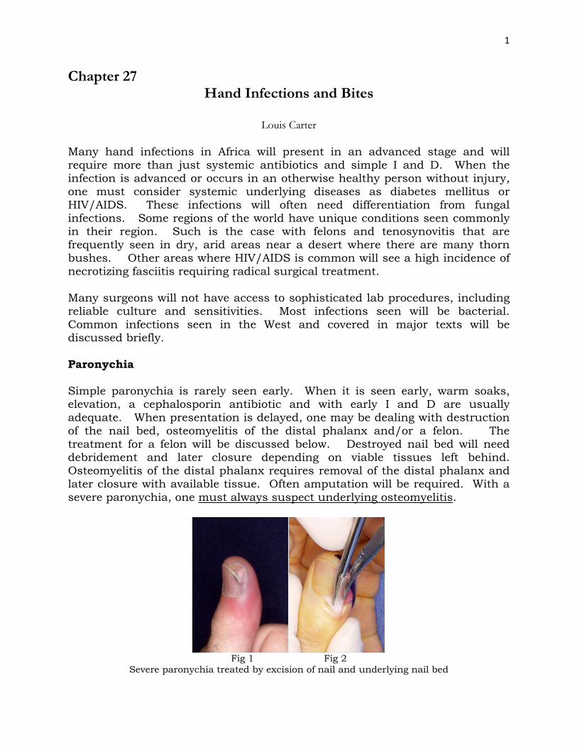
Felon This closed space infection within the septa of the palmar pad of the distal phalanx can be secondary to severe paronychia but is most often secondary to a puncture wound or bite wound into the pulp of the distal phalanx. Such puncture wounds of the hand and foot are commonly seen in desert and arid conditions where thorn bushes are common and the thorns can be several cm. long and very stiff, easily penetrating the sole of a shoe. Often the injury occurs at night when walking or working in the dark. A swollen painful distal phalanx is seen when the infection is confined to the distal phalanx. At this time simple I and D is all that is necessary. Many incisions have been described but the author has found one simple midline vertical incision, illustrated on the left below, on the volar aspect of the distal phalanx is sufficient. Once within the soft tissue, the septa medially and laterally may be divided and the purulence drained. Because the incision is midline, the major nerves and vessels are not damaged. This wound heals well with few long term complications. The incision on the lateral side of the distal phalanx in the drawing on the right below is advocated but not by the author.
Fig 3 Fir 4 Fig 5
These show the appropriate incisions to drain a felon. Some prefer the lateral incision while the author prefers the midline incision in figures 3 and 5 (Courtesy of Dr. Anthony Smith)
With a longstanding felon, osteomyelitis of the distal phalanx is common and the distal finger will require amputation. In addition, the infection may spread proximally and through the flexor tendon sheath with flexor tenosynovitis and later a possible deep space infection in the palm.
3
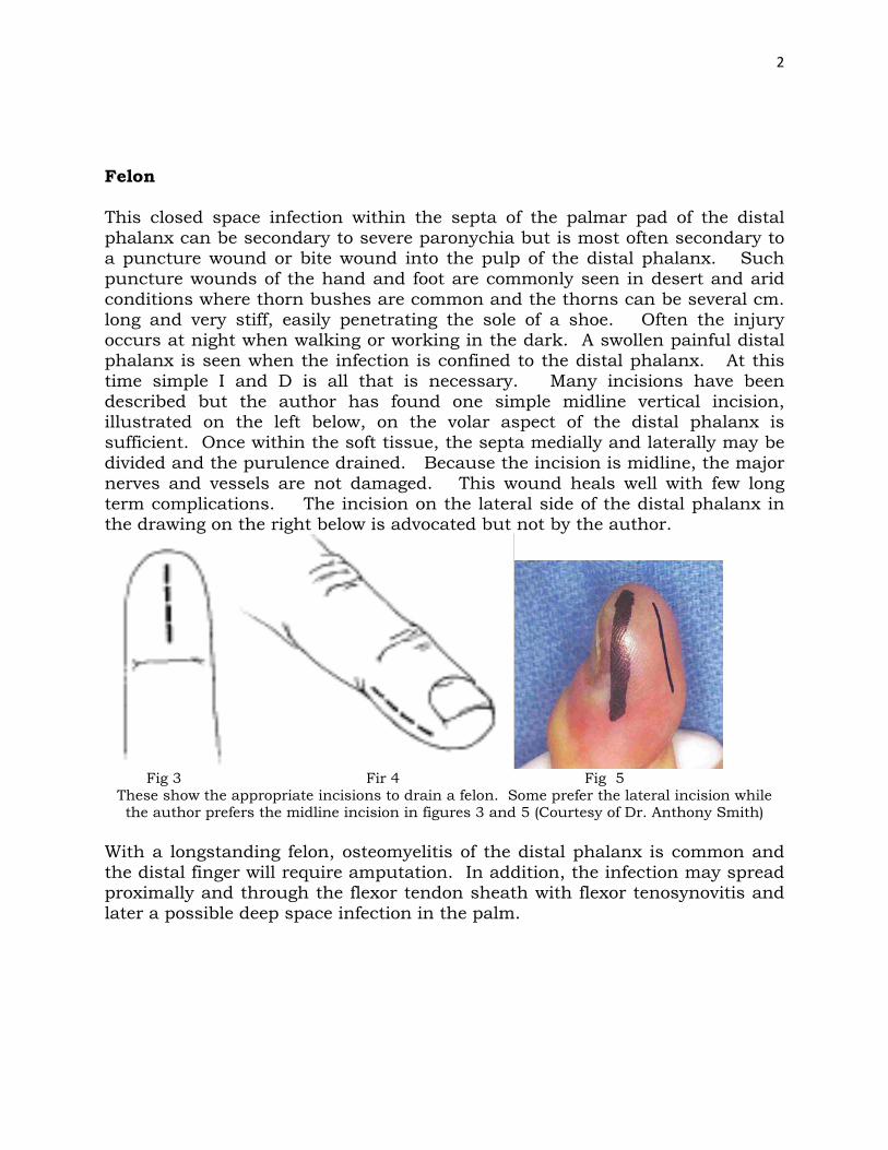
Fig 6 Fig 7 Fig 8 Fig 9
Felons and osteomyelitis secondary to thorn injuries Flexor Tenosynovitis
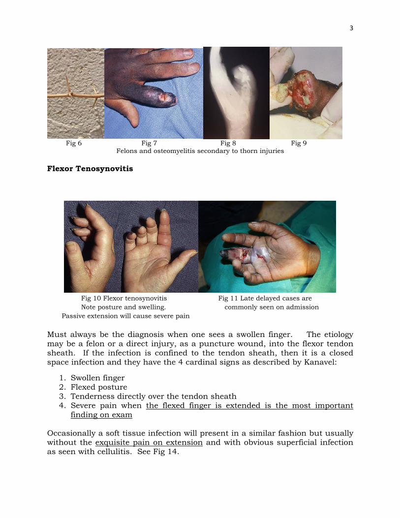
Fig 10 Flexor tenosynovitis Fig 11 Late delayed cases are Note posture and swelling. commonly seen on admission Passive extension will cause severe pain Must always be the diagnosis when one sees a swollen finger. The etiology may be a felon or a direct injury, as a puncture wound, into the flexor tendon sheath. If the infection is confined to the tendon sheath, then it is a closed space infection and they have the 4 cardinal signs as described by Kanavel:
1. Swollen finger 2. Flexed posture 3. Tenderness directly over the tendon sheath 4. Severe pain when the flexed finger is extended is the most important
finding on exam
Occasionally a soft tissue infection will present in a similar fashion but usually without the exquisite pain on extension and with obvious superficial infection as seen with cellulitis. See Fig 14.
4
Fig 12
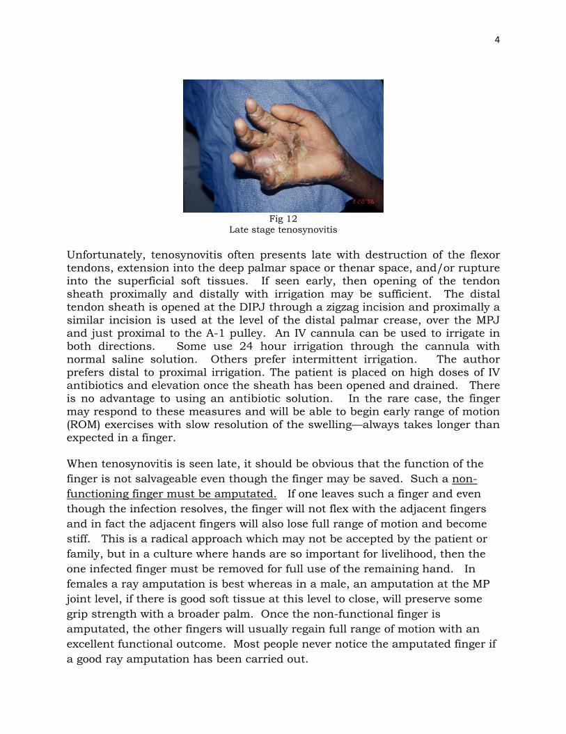
Late stage tenosynovitis Unfortunately, tenosynovitis often presents late with destruction of the flexor tendons, extension into the deep palmar space or thenar space, and/or rupture into the superficial soft tissues. If seen early, then opening of the tendon sheath proximally and distally with irrigation may be sufficient. The distal tendon sheath is opened at the DIPJ through a zigzag incision and proximally a similar incision is used at the level of the distal palmar crease, over the MPJ and just proximal to the A-1 pulley. An IV cannula can be used to irrigate in both directions. Some use 24 hour irrigation through the cannula with normal saline solution. Others prefer intermittent irrigation. The author prefers distal to proximal irrigation. The patient is placed on high doses of IV antibiotics and elevation once the sheath has been opened and drained. There is no advantage to using an antibiotic solution. In the rare case, the finger may respond to these measures and will be able to begin early range of motion (ROM) exercises with slow resolution of the swelling—always takes longer than expected in a finger. When tenosynovitis is seen late, it should be obvious that the function of the finger is not salvageable even though the finger may be saved. Such a non-functioning finger must be amputated. If one leaves such a finger and even though the infection resolves, the finger will not flex with the adjacent fingers and in fact the adjacent fingers will also lose full range of motion and become stiff. This is a radical approach which may not be accepted by the patient or family, but in a culture where hands are so important for livelihood, then the one infected finger must be removed for full use of the remaining hand. In females a ray amputation is best whereas in a male, an amputation at the MP joint level, if there is good soft tissue at this level to close, will preserve some grip strength with a broader palm. Once the non-functional finger is amputated, the other fingers will usually regain full range of motion with an excellent functional outcome. Most people never notice the amputated finger if a good ray amputation has been carried out.
5
Fig 13
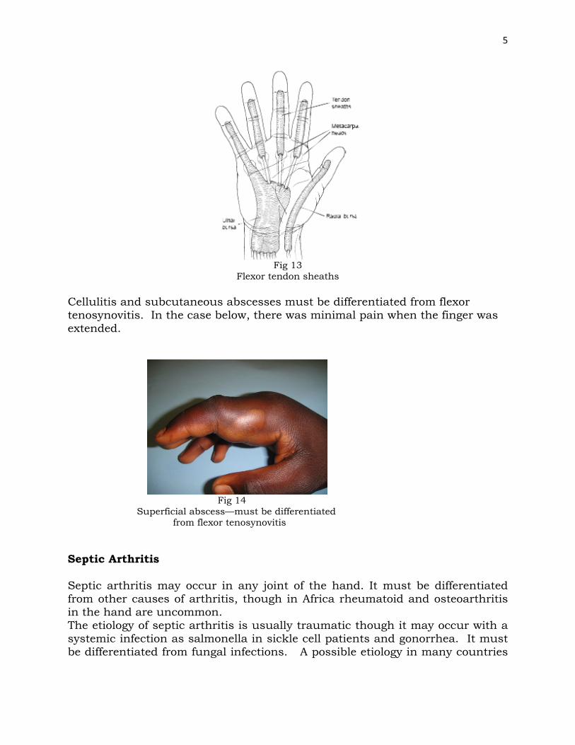
Flexor tendon sheaths
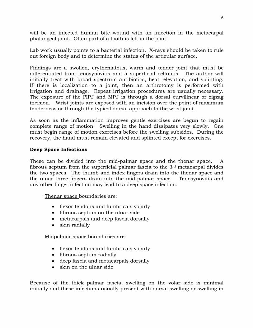
Cellulitis and subcutaneous abscesses must be differentiated from flexor tenosynovitis. In the case below, there was minimal pain when the finger was extended.
Fig 14 Superficial abscess—must be differentiated from flexor tenosynovitis Septic Arthritis Septic arthritis may occur in any joint of the hand. It must be differentiated from other causes of arthritis, though in Africa rheumatoid and osteoarthritis in the hand are uncommon. The etiology of septic arthritis is usually traumatic though it may occur with a systemic infection as salmonella in sickle cell patients and gonorrhea. It must be differentiated from fungal infections. A possible etiology in many countries
6
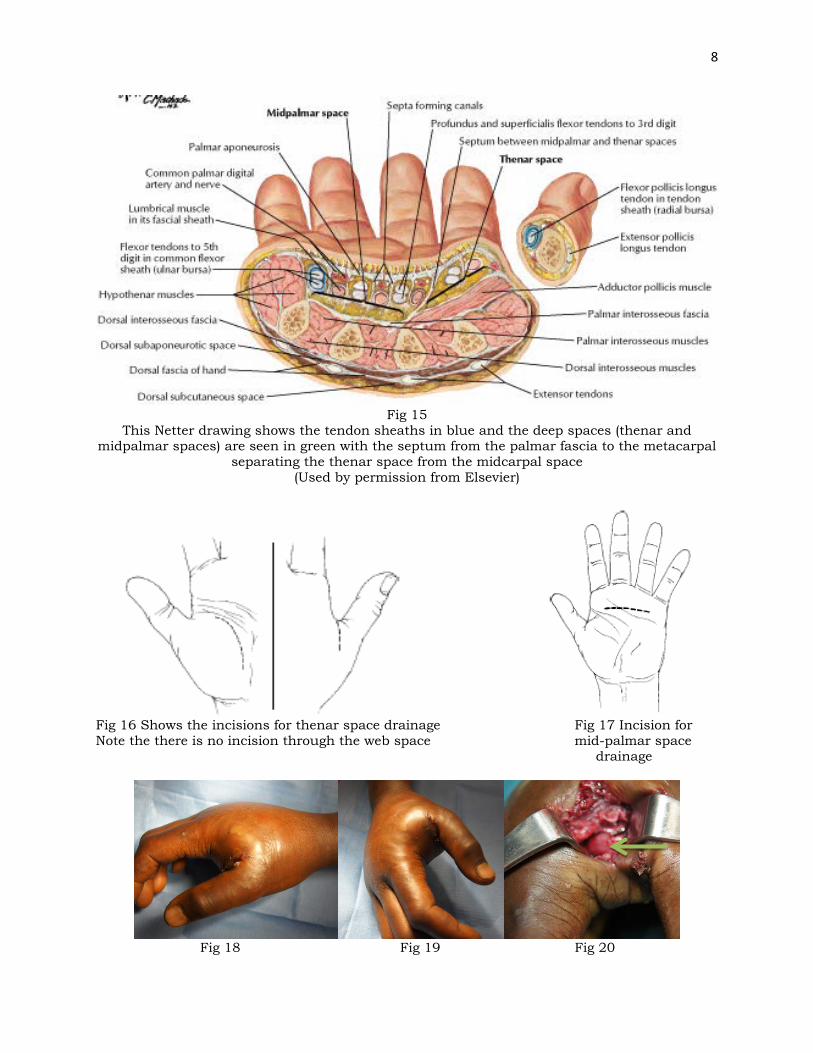
will be an infected human bite wound with an infection in the metacarpal phalangeal joint. Often part of a tooth is left in the joint. Lab work usually points to a bacterial infection. X-rays should be taken to rule out foreign body and to determine the status of the articular surface. Findings are a swollen, erythematous, warm and tender joint that must be differentiated from tenosynovitis and a superficial cellulitis. The author will initially treat with broad spectrum antibiotics, heat, elevation, and splinting. If there is localization to a joint, then an arthrotomy is performed with irrigation and drainage. Repeat irrigation procedures are usually necessary. The exposure of the PIPJ and MPJ is through a dorsal curvilinear or zigzag incision. Wrist joints are exposed with an incision over the point of maximum tenderness or through the typical dorsal approach to the wrist joint. As soon as the inflammation improves gentle exercises are begun to regain complete range of motion. Swelling in the hand dissipates very slowly. One must begin range of motion exercises before the swelling subsides. During the recovery, the hand must remain elevated and splinted except for exercises. Deep Space Infections These can be divided into the mid-palmar space and the thenar space. A fibrous septum from the superficial palmar fascia to the 3rd metacarpal divides the two spaces. The thumb and index fingers drain into the thenar space and the ulnar three fingers drain into the mid-palmar space. Tenosynovitis and any other finger infection may lead to a deep space infection. Thenar space boundaries are:
• flexor tendons and lumbricals volarly • fibrous septum on the ulnar side • metacarpals and deep fascia dorsally • skin radially
Midpalmar space boundaries are:
• flexor tendons and lumbricals volarly • fibrous septum radially • deep fascia and metacarpals dorsally • skin on the ulnar side
Because of the thick palmar fascia, swelling on the volar side is minimal initially and these infections usually present with dorsal swelling or swelling in
7
the first web space. Routine lab work and x-rays should be performed, the latter to rule out a foreign body. Both of these deep space infections are closed space infections with little way for the purulent fluid to escape. Once a diagnosis is suspected, urgent exploration with drainage should be performed. Surgery
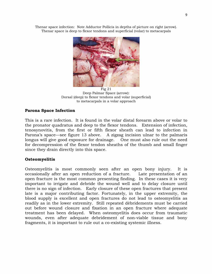
• The incision for the thenar space decompression is on the dorsal side of the first web space. The incision is vertical extending up to but not through the web between the thumb and index finger. This gives excellent exposure to the thenar space which lies just below the tendons to the index finger. After irrigation and drainage, a Penrose drain is left in the space for several days.
• The mid-palmar space is approached through a transverse incision just proximal to the distal palmar crease. Once through the skin and superficial fascia, a vertical dissection is then used to dissect through the soft tissues and between the vessels and tendons until the space is reached just below or dorsal to the tendons. Often once the incision is made through the subcutaneous tissue, a hemostat can be bluntly inserted where there is bulging and between the flexor tendons. One should attempt to identify the digital vessels and nerves though in an edematous hand this may be difficult. After irrigation a drain should be left in place for several days. These deep space infections may require a second look procedure to ensure adequate decompression.
• Postoperatively, the hand must be elevated and early gentle exercises carried out. When the drainage diminishes, the drains may be removed, usually between 3-7 days. Antibiotics should be continued for 7-10 days.
In all hand infections, culture and sensitivity studies may be done if available. If not, antibiotics such as a cephalosporin are usually adequate except in cases of immune-compromised patients and abscesses secondary to farm injuries. In such cases, gram negative and anaerobic infections may need to be covered. These infections will commonly occur in the diabetic or AIDS patient. See below.
8
Fig 15
This Netter drawing shows the tendon sheaths in blue and the deep spaces (thenar and midpalmar spaces) are seen in green with the septum from the palmar fascia to the metacarpal
separating the thenar space from the midcarpal space (Used by permission from Elsevier)
Fig 16 Shows the incisions for thenar space drainage Fig 17 Incision for Note the there is no incision through the web space mid-palmar space drainage
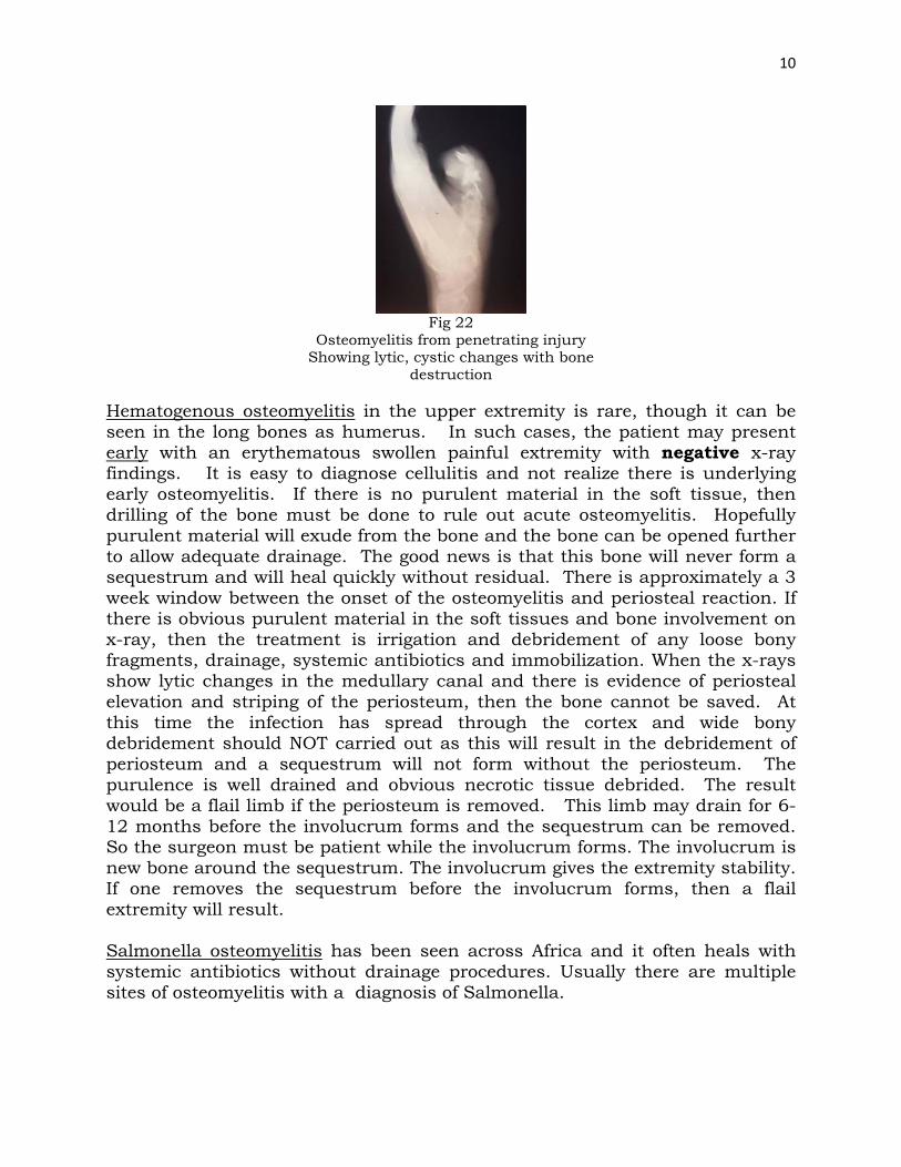
Fig 18 Fig 19 Fig 20
9
Thenar space infection: Note Adductor Pollicis in depths of picture on right (arrow). Thenar space is deep to flexor tendons and superficial (volar) to metacarpals
Fig 21
Deep Palmar Space (arrow): Dorsal (deep) to flexor tendons and volar (superficial)
to metacarpals in a volar approach Parona Space Infection This is a rare infection. It is found in the volar distal forearm above or volar to the pronator quadratus and deep to the flexor tendons. Extension of infection, tenosynovitis, from the first or fifth flexor sheath can lead to infection in Parona’s space—see figure 13 above. A zigzag incision ulnar to the palmaris longus will give good exposure for drainage. One must also rule out the need for decompression of the flexor tendon sheaths of the thumb and small finger since they drain directly into this space. Osteomyelitis Osteomyelitis is most commonly seen after an open bony injury. It is occasionally after an open reduction of a fracture. Late presentation of an open fracture is the most common presenting finding. In these cases it is very important to irrigate and debride the wound well and to delay closure until there is no sign of infection. Early closure of these open fractures that present late is a major contributing factor. Fortunately, in the upper extremity, the blood supply is excellent and open fractures do not lead to osteomyelitis as readily as in the lower extremity. Still repeated dèbridements must be carried out before wound closure and fixation in an open fracture where adequate treatment has been delayed. When osteomyelitis does occur from traumatic wounds, even after adequate debridement of non-viable tissue and bony fragments, it is important to rule out a co-existing systemic illness.
10
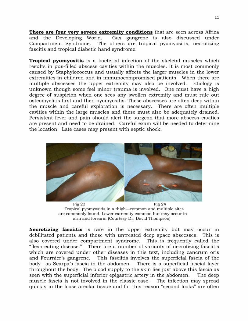
Fig 22
Osteomyelitis from penetrating injury Showing lytic, cystic changes with bone
destruction
Hematogenous osteomyelitis in the upper extremity is rare, though it can be seen in the long bones as humerus. In such cases, the patient may present early with an erythematous swollen painful extremity with negative x-ray findings. It is easy to diagnose cellulitis and not realize there is underlying early osteomyelitis. If there is no purulent material in the soft tissue, then drilling of the bone must be done to rule out acute osteomyelitis. Hopefully purulent material will exude from the bone and the bone can be opened further to allow adequate drainage. The good news is that this bone will never form a sequestrum and will heal quickly without residual. There is approximately a 3 week window between the onset of the osteomyelitis and periosteal reaction. If there is obvious purulent material in the soft tissues and bone involvement on x-ray, then the treatment is irrigation and debridement of any loose bony fragments, drainage, systemic antibiotics and immobilization. When the x-rays show lytic changes in the medullary canal and there is evidence of periosteal elevation and striping of the periosteum, then the bone cannot be saved. At this time the infection has spread through the cortex and wide bony debridement should NOT carried out as this will result in the debridement of periosteum and a sequestrum will not form without the periosteum. The purulence is well drained and obvious necrotic tissue debrided. The result would be a flail limb if the periosteum is removed. This limb may drain for 6-12 months before the involucrum forms and the sequestrum can be removed. So the surgeon must be patient while the involucrum forms. The involucrum is new bone around the sequestrum. The involucrum gives the extremity stability. If one removes the sequestrum before the involucrum forms, then a flail extremity will result. Salmonella osteomyelitis has been seen across Africa and it often heals with systemic antibiotics without drainage procedures. Usually there are multiple sites of osteomyelitis with a diagnosis of Salmonella.
11
There are four very severe extremity conditions that are seen across Africa and the Developing World. Gas gangrene is also discussed under Compartment Syndrome. The others are tropical pyomyositis, necrotizing fascitis and tropical diabetic hand syndrome. Tropical pyomyositis is a bacterial infection of the skeletal muscles which results in pus-filled abscess cavities within the muscles. It is most commonly caused by Staphylococcus and usually affects the larger muscles in the lower extremities in children and in immunocompromised patients. When there are multiple abscesses the upper extremity may also be involved. Etiology is unknown though some feel minor trauma is involved. One must have a high degree of suspicion when one sees any swollen extremity and must rule out osteomyelitis first and then pyomyositis. These abscesses are often deep within the muscle and careful exploration is necessary. There are often multiple cavities within the large muscles and these must also be adequately drained. Persistent fever and pain should alert the surgeon that more abscess cavities are present and need to be drained. Careful exam will be needed to determine the location. Late cases may present with septic shock.
Fig 23 Fig 24
Tropical pyomyositis in a thigh—common and multiple sites are commonly found. Lower extremity common but may occur in arm and forearm (Courtesy Dr. David Thompson) Necrotizing fasciitis is rare in the upper extremity but may occur in debilitated patients and those with untreated deep space abscesses. This is also covered under compartment syndrome. This is frequently called the “flesh-eating disease.” There are a number of variants of necrotizing fasciitis which are covered under other diseases in this text, including cancrum oris and Fournier’s gangrene. This fasciitis involves the superficial fascia of the body—as Scarpa’s fascia in the abdomen. There is a superficial fascial layer throughout the body. The blood supply to the skin lies just above this fascia as seen with the superficial inferior epigastric artery in the abdomen. The deep muscle fascia is not involved in the classic case. The infection may spread quickly in the loose areolar tissue and for this reason “second looks” are often
12
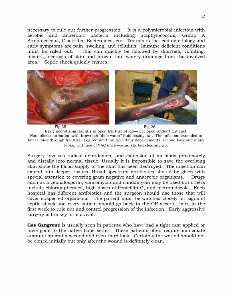
necessary to rule out further progression. It is a polymicrobial infection with aerobic and anaerobic bacteria including Staphylococcus, Group A Streptococcus, Clostridia, Bacteroides, etc. Trauma is the leading etiology and early symptoms are pain, swelling, and cellulitis. Immune deficient conditions must be ruled out. This can quickly be followed by diarrhea, vomiting, blisters, necrosis of skin and brown, foul watery drainage from the involved area. Septic shock quickly ensues.
Fig 25 Fig 26
Early necrotizing fasciitis in open fracture of leg—developed under tight cast. Note blister formation with brownish “dish water” fluid oozing out. The infection extended to
lateral side through fracture. Leg required multiple daily débridements, second look and many looks, with use of VAC once wound started cleaning up.
Surgery involves radical débridement and extension of incisions proximately and distally into normal tissue. Usually it is impossible to save the overlying skin since the blood supply to the skin has been destroyed. The infection can extend into deeper tissues. Broad spectrum antibiotics should be given with special attention to covering gram negative and anaerobic organisms. Drugs such as a cephalosporin, vancomycin and clindamycin may be used but others include chloramphenicol, high doses of Penicillin G, and metronidazole. Each hospital has different antibiotics and the surgeon should use those that will cover suspected organisms. The patient must be watched closely for signs of septic shock and every patient should go back to the OR several times in the first week to rule out and control progression of the infection. Early aggressive surgery is the key for survival. Gas Gangrene is usually seen in patients who have had a tight cast applied or have gone to the native bone setter. These patients often require immediate amputation and a second and even third look. Certainly the wound should not be closed initially but only after the wound is definitely clean.
13
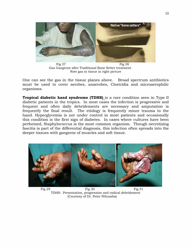
Fig 27 Fig 28 Gas Gangrene after Traditional Bone Setter treatment
Note gas in tissue in right picture One can see the gas in the tissue planes above. Broad spectrum antibiotics must be used to cover aerobes, anaerobes, Clostridia and microaerophilic organisms. Tropical diabetic hand syndrome (TDHS) is a rare condition seen in Type II diabetic patients in the tropics. In most cases the infection is progressive and frequent and often daily debridements are necessary and amputation is frequently the final result. The etiology is frequently minor trauma to the hand. Hyperglycemia is not under control in most patients and occasionally this condition is the first sign of diabetes. In cases where cultures have been performed, Staphylococcus is the most common organism. Though necrotizing fascitis is part of the differential diagnosis, this infection often spreads into the deeper tissues with gangrene of muscles and soft tissue.
Fig 29 Fig 30 Fig 31
TDHS: Presentation, progression and radical debridement (Courtesy of Dr. Peter Nthumba)
14
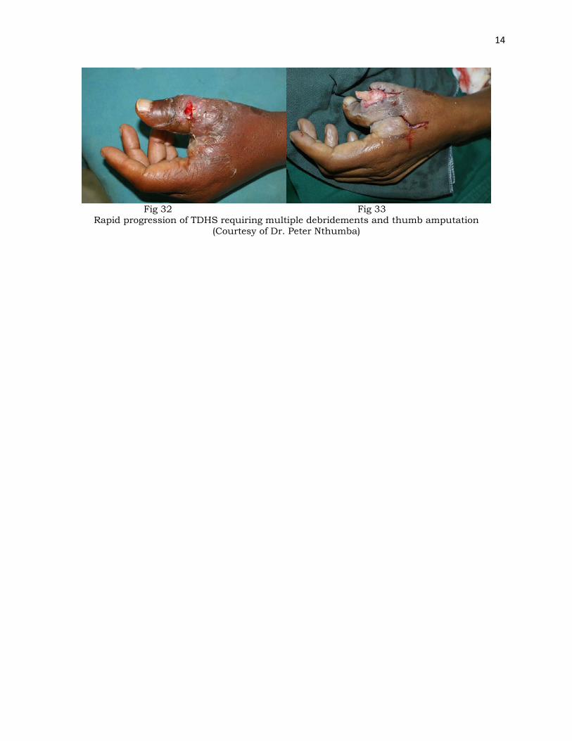
Fig 32 Fig 33
Rapid progression of TDHS requiring multiple debridements and thumb amputation (Courtesy of Dr. Peter Nthumba)