Embed Size (px)
Citation preview

Chapter 25: Burns
Missy Thurlow, MBA, OTR/L, CHT and Patricia Anderson, OTR/L, CHT
Test Prep for the CHT Exam, 3rd Edition 1
Chapter 23: Burns Missy Thurlow, MBA, OTR/L, CHT and Patricia Anderson, OTR/L, CHT
I. Introduction Hands are the most frequent site of thermal injury1 Flame burns are the most frequent cause of burn injury in adults2 Scald burns are the most frequent cause of burns in children2
The skin covering the hand is equivalent to 5% of the total body’s surface area1
A burn of any depth to the hand is classified as a major injury by the American Burn Association1
Most common complications after a burn injury to the hand are post burn edema, compartment syndrome, joint deformities, sensory impairment, restricted use of the hand 1
Burns of the hand and upper extremity can interfere with functional performance in ADL’s and work activities2
II. Wound Classification
Types of Burns Cutaneous2
Caused by application of heat, cold or caustic chemicals Depth of burn varies based on length of contact to heat element and temperature Depth of burn determines healing potential and need for surgical intervention
Cold (frostbite)3
Skin and underlying tissues damaged Blood flow interrupted, hypertonic tissue environment noted
Chemical Burns4 Caustic reactions seen, cellular membranes disrupted
Toxic effects on metabolic process Duration of exposure, chemical agent determines severity Systemic absorption of some chemicals can be deadly
Electrical Burns5 Electrical energy is transformed into thermal energy as current passes
through tissues Injury to cell membranes disrupts membrane potential and function Severity of injury depends on current pathway, resistance to current and strength and duration of current flow
Burn Depth and Classification6 Significant determinant of mortality Primary determinant of the patient's long-term appearance and
functional outcome Epidermal (1st degree)6
Usually sunburn Only involves the epidermis
American Society of Hand TherapistsTM2
Chapter 25: BurnsMissy Thurlow, MBA, OTR/L, CHT and Patricia Anderson, OTR/L, CHT
No blisters, but red and very painful 2-3 days until redness and pain subside.
Partial Thickness Burns (2nd degree)6 Does not extend entirely through the dermis
Epithelial-lined appendages - sweat glands, hair follicles and sebaceous glands remain intact When dead tissue removed, epithelial cells migrate from the surface of each dermal appendage to other epithelial cells from neighboring appendages, forming new, fragile epidermis on top of thin residual dermal bed Deeper burn takes longer to heal and scarring can be severe If burns heal spontaneously within 2-3 weeks, no hypertrophic scarring or functional impairment is noted, but pigmentation changes may be apparent Burns that take longer than 3 weeks to heal may produce hypertrophic scars and functional impairment. A thin, fragile epithelial covering will be present many weeks. Often accumulate a layer of fibrinous exudate and necrotic debris on surface Deep partial thickness burns extend into lower layers of dermis. These heal within 3-9 weeks. Scar formation is common and often is hypertrophic. Joint function may be impaired. Excision and grafting is usually needed.
Full Thickness Burn (3rd degree)6
Involves all layers of dermis and underlying subcutaneous adipose tissue Burn eschar (dead and denatured dermis) is present. When left in situ, eschar separates from the underlying viable tissue, leaving an unhealed bed of granulation tissue Deeper structures may be involved such as muscle, tendon, ligament and bone, and might be classified as deep full thickness or 4th degree Coverage options include skin grafts, biologic dressings and skin substitutes Amputation or tissue transfers may be required (Fig. 1)
III. Surgical Management/Wound Coverage6
Escharotomy Surgical incision through burn eschar. Usually done within 24 hours of
burn injury. When the burn eschar circumferentially surrounds any body structure, the tissues are subject to increasing interstitial pressures which limit venous outflow and decrease arterial inflow. Compartment syndrome may occur which leads to ischemia or necrosis within or distal to that body part within hours. The restricted tissues are released with the incisions usually on the medial and lateral side of the extremity.
Test Prep for the CHT Exam, 3rd Edition 3
Chapter 25: BurnsMissy Thurlow, MBA, OTR/L, CHT and Patricia Anderson, OTR/L, CHT
Once the patient is stable, the eschar can be removed until a viable underlying tissue level is visible.
Debridement of Burn Wounds6 Removal of loose, devitalized, necrotic or contaminated wound tissue
using mechanical or sharp technique. Debridement cleans off the wound surfaces to optimize wound healing. This is usually done on shallow burns that are expected to heal without the need for skin grafting.
Excision of Burn Wounds6 Excision is a surgical procedure requiring incision through the deep
dermis of open wounds, burn eschar, or burn scars and all necrotic tissue. This may be used in preparation for surgical reconstruction Options following surgical excision include topical antimicrobials, temporary biologic covers, immediate grafting, flap closure or other reconstructive procedures. Performed by experienced burn surgeons Single stage excision and grafting requires donor sites for skin harvest, excision of the burn wound and harvesting the donor skin. Donor skin may be meshed to expand the coverage area and is applied to the wound bed with suture, staples, fibrin glue, synthetic adhesives or tapes. Dressings or orthoses may be used to avoid shearing of the grafts and to maintain proper positioning. Multiple stage excision and grafting is used when the burn wound is large and more than one operation is needed as with facial wounds or extensive burns. With a facial burn, a temporary covering follows excision. In several days, skin autografts are placed. This slower process allows for inspection for problems and can result in often 100% graft take. In patients that have extensive burns, staged excision and grafting may be advantageous as the donor sites have time to reepithelialize between harvesting sessions. Temporary wound substitutes may be used when wound is too large to be closed in one procedure or there is not enough donor skin available. Cadaver Allograft is the gold standard temporary skin substitute. Allografts adhere and induce vascularization on an appropriately prepared wound bed. Pain is reduced; loss of fluids, proteins and electrolytes are reduced. Allografts are obtained from a skin bank and may be fresh, refrigerated tissue or frozen. Skin Xenografts are obtained from various animals but mostly pigs.
Burn Wound Coverage6 Small burn wounds are excised and covered by either a full thickness skin
graft or a split thickness skin graft. A full thickness graft will contract less but is harder to heal. Skin substitutes are defined as a biomaterial, engineered tissue or
American Society of Hand TherapistsTM4
Chapter 25: BurnsMissy Thurlow, MBA, OTR/L, CHT and Patricia Anderson, OTR/L, CHT
combination of materials and cells or tissues that can be used for skin autograft or allograft. Skin replacement: A tissue or graft that permanently replaces lost skin with healthy skin. (Fig. 2)
IV. Scar
Scar development and scar contraction are the most significant impediments in recovery from a burn injury.3
Hypertrophic scar Characterized by the three “R”s: red, raised, rigid
during the initial phase of development7
Occurs when excessive collagen forms within the boundary of the burn wound8
Results from imbalance between collagen synthesis and collagen lysis9 Collagen is formed in whorl-type patterns instead of parallel linear patterns of normal skin9
Occurs in burn depth that reaches the reticular layer of the dermis2
Often found in areas of motion/joints as the tension along the length of the scar encourages collagen depositions over collagen lysis9 Often occurs when full thickness burn wounds are closed with re-epithelialization, granulation tissue formation, and contraction9 Burn wounds that are closed by split thickness skin grafting typically do not form hypertrophic scarring over the graft site, however may develop hypertrophic scarring along the seams of the graft or along the edges of the graft and the non-injured skin9 May take up to two years to reach maturation9
Keloid Scar Controversy exists whether there is a difference
between keloids and hypertrophic scar7 Classic definition of a keloid is a scar that forms excessive collagen that does not conform to the boundaries of the wound8
Difficult to distinguish histologic differences between hypertrophic scar and keloids8 Collagen synthesis is greater in keloid scars than in hypertrophic scar8 Hypertrophic scars may recede over time; however this does not typically occur with keloids8
Risk Factors of Hypertrophic Scar Formation

Test Prep for the CHT Exam, 3rd Edition 5
Chapter 25: BurnsMissy Thurlow, MBA, OTR/L, CHT and Patricia Anderson, OTR/L, CHT
Formation of hypertrophic scar after a burn injury cannot be entirely predicted, however several risk factors exist to help predict whether a burn will develop hypertrophic scar7
Length of time for wound closure8 Most important factor in predicting hypertrophy Burns requiring longer than three weeks to close are at risk
Race - darker pigmented skin generally are at risk8
Age - clinically it has been noted that younger people under the age of 30 form more scar than other age groups8 Depth - burns reaching into the reticular dermis scar more than more superficial burns that involve the epidermis or papillary dermis8
Scar Contraction/Scar Contracture Scar contraction is the active process of the shortening of scar
tissue2 Scar contraction will continue to occur until it meets an equal opposing force8 Scar contracture is the end result of scar contraction in which shortened scar tissue has limited the motion of a joint and will require surgical correction8 Full thickness skin grafts will greatly diminish scar contraction8 Contraction occurring with split thickness skin grafts is proportional to the thickness of the graft; the thinner the graft, the more contraction occurs7 (Fig. 3)
Scar Maturation8 Scar development begins with the initiation of wound healing
The first 6 months following injury is the most active building phase of hypertrophic scar development Following the initial 6 months scar maturation can continue for up to 2 years post injury Longer maturation time is dependent on wound depth and length of time to wound closure Scar maturation in children can take 12 to 24 months Scar maturation in adults can take 6 to 24 months The three “P”s of scar maturation are pale, planer, and pliable
Scar Complex Normal skin can stretch up to 60% of its resting length as
compared to immature burn scar that can only stretch up to 15% of its length9,10 Burn injuries that encompass more than one joint require composite ROM of all joints in combination to place a full stretch to the total burn scar9
American Society of Hand TherapistsTM6
Chapter 25: BurnsMissy Thurlow, MBA, OTR/L, CHT and Patricia Anderson, OTR/L, CHT
scar complex is the composite scar of all involved joints combined9
V. Burn Evaluation8
Referral to OT or PT services should be obtained. If the patient is hospitalized, reviewing the record is necessary for optimal understanding of burn location and depth, inhalation, life threatening burns and/or other medical conditions At the time of injury, initial evaluation may be limited by patient’s clinical status or severity of wounds. As the patient progresses, more thorough evaluation can be performed7
Burn Wound Documentation of the extent of the burns, depth, type, and location
should be noted. If possible, wounds assessed without dressings10 Document the date of injury as well as the date of wound closure.
Length of time to wound closure affects scar formation1
In the hand, skin is not uniform in thickness; depth of the wound may differ depending on the location of the burn. A circumferential burn may produce a full thickness wound to the dorsal surface and partial thickness to the palmar surface2 Note how the burn wound was closed: skin grafting, skin substitute, or epithelialization1 Document any open wounds, their location and how they are being managed.
Edema Can be severe during the first days following burn injury1 Edema documented by description or circumferential measurements if wounds closed10
If joints or hands are involved, orthotics necessary to maintain good functional position. Orthoses require modification as edema decreases.8
Pain8 Pain medications documented including dosage and frequency
Perceived pain documented on a pain scale As wounds close, pain typically decreases however itching becomes a common problem and can be more intense than the sensations of pain and is often described as a type of pain. These sensations should be noted as well
Sensibility Testing of the median, ulnar and radial nerves included if
the burn is an electrical injury10 the burn is severe and/or circumferential at risk for
compartment syndrome7 Manual Muscle Test7
Should be assessed in an electrical injury to rule out damage to deep musculature and nerves10

Test Prep for the CHT Exam, 3rd Edition 7
Chapter 25: BurnsMissy Thurlow, MBA, OTR/L, CHT and Patricia Anderson, OTR/L, CHT
Chapter 25 Figures
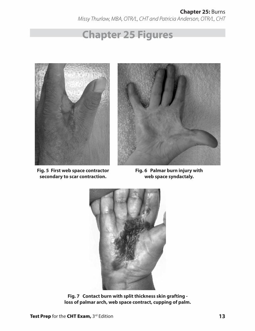
Fig. 1 Full thickness electrical burn with tendon exposure.
Fig. 3 Palmar burn scar contraction with scar bands crossing palm and web spaces.
Fig. 4 Full thickness burn with split thickness skin grafting and tendon
involvement.
Fig. 2 Mixture of wound healing with split thickness
skin grafting and granulation tissue.
American Society of Hand TherapistsTM8
Chapter 25: BurnsMissy Thurlow, MBA, OTR/L, CHT and Patricia Anderson, OTR/L, CHT
Grip and pinch recorded if patient able to perform test and there is no risk of rupture to digital extensors with gripping7
Range of Motion: Active and Passive7 Should not be aggressive or forceful to avoid tearing of fragile tissues
Note any fractures or exposed tendons that will limit PROM No ROM or composite digital flexion should be performed on deep burns to the dorsum of the digits until the digital extensor apparatus is determined intact
ROM measured with goniometer if wounds are closed If wounds are open over a joint, visual estimation or a lateral goniometric measurement are acceptable methods for documentation Goniometric measurements recorded for each individual joint Composite digital flexion and extension should be measured in the hand Limitations of combined motion of all joints involved in the burn should be noted and measured (within the scar complex); most often measured using goniometry, measuring tape, or photography1
Activities Daily Living Scar7
No universal standard instrument is currently used to evaluate scar Vancouver Scar Scale is the most widely used assessment Other methods of documenting scar are standardized photography, written description, and diagrams Descriptive documentation should include: pliability, vascularity, height, appearance, surface texture, scar pain, severity of itching, and location of scar bands
VI. Treatment of the Burned Hand
Acute phase1 Edema formation in burned hands hinders motion and may be a factor in
later contracture formation. Hands must be elevated above heart level to prevent edema. Probably most important initial step in hand burn management without hindering resuscitation, pulmonary, or other critical care management in any size burn
Tendon Exposure7
Most common location for tendon exposure is dorsum of digits at PIP joint. Often results in loss of central slip, which causes volar pull on lateral bands by the lumbricals which positions the digit in PIP flexion and DIP hyperextension, resulting in a boutonniere deformity Important to keep moist dressings over any exposed tendons at all times. Xeroform may be used or any hydrophobic dressing may be adequate. If the tendon becomes dry and brittle, it will rupture with resultant deformity.
Test Prep for the CHT Exam, 3rd Edition 9
Chapter 25: BurnsMissy Thurlow, MBA, OTR/L, CHT and Patricia Anderson, OTR/L, CHT
Overstretching tendon should be avoided during time of exposure. (Fig. 4)
Range of motion7 Without exposed tendon, passive movement can be initiated
immediately post injury. Exercise goals include: elongation of the healed skin and scar tissue, maintaining or increasing range of motion of the burned extremity, promoting function in activities of daily living, restoring dexterity and coordination and resuming endurance and strength for return to preinjury activities. Active range of motion should be encouraged throughout the acute stage when patient is able to participate.
Orthoses7,8 Begins early during the acute stage and continues throughout the
rehabilitation process. It is important orthoses do not impinge vascular flow. Therapist begins orthotic fabrication in the acutely burned patient immediately. Orthotics place the patient in a position that will reduce the chance of contracture. Orthoses also used to protect skin grafts and to decrease movement following surgery. Orthotics initially position the hand to minimize contracture development secondary to edema and wound healing. They may be altered or modified to provide elongation of scar tissue and muscle tendon units. In the early acute stage, orthotics are held in place with the use of Kling, gauze or ace wraps; later, with velcro. Close monitoring of the orthotic fit is important because of edema changes and tissue tightness
Outpatient Therapy (Post Acute) Modalities12
Modalities including fluidotherapy, moist heat, paraffin baths, or ultrasound with combined stretching to increase tissue elongation can be used on an extremity with no open wounds. If open wounds are present, it is important to protect the areas from modalities.
Range of Motion to the Upper Extremity8 Both active, active assistive and passive range of motion is
effective during the outpatient stage. Self ROM is encouraged in the outpatient clinic and at home. Entire upper extremity motion is necessary to prevent shoulder and elbow tightness.
Strength and coordination8,11 Strength and coordination tasks are important as well as
activities of daily living for independent self care. Fine and gross motor task performance is necessary for independent use of the burned extremity and for return to work/home activities.
American Society of Hand TherapistsTM10
Chapter 25: BurnsMissy Thurlow, MBA, OTR/L, CHT and Patricia Anderson, OTR/L, CHT
Edema8 Edema may be managed through light pressure garments, such
as Tubigrip or Isotoner gloves, and active range of motion exercises to help “milk” edema from the burned hand.
Scar Management8 Pressure therapy applied to burn wounds when no or small
open areas remain. Tubigrip or custom compression garments needed to manage scar development in deep 2nd or 3rd degree burns. Orthotics designed to decrease deformities, stretch tightened skin or assist with gaining range of motion are continued from the acute stage. Static progressive or dynamic orthoses may be used to increase ROM in the hand.
VII. Hand Burn Orthoses7,8
Types of Orthoses Static Orthoses - maintain fixed position, indicated for skin graft
protection after surgery or anti-contracture positioning if adequate ROM is not gained by exercise alone. The splints can be modified to account for increased tissue length after exercise or positioning. Static Progressive Orthoses - usually used if sufficient ROM not obtained with static positioning and exercise. Used for correction of contractures and provide inelastic stress to tissues at end range. Allow adjustment to stress as tissue lengthens via stress relaxation. Dynamic Orthoses - provide a continual stress to tissue over time. Usually used after the acute burn stage to manage joint contractures.
Orthosis Application7 Splints are initially applied within 24 hours to position the hand. Design
may be altered or changed to elongate the muscle tendon unit and scar tissue. Orthotics should be worn at all times except therapist assisted ROM initially, and then weaned to night and between exercise. Designed to minimize the common hand deformities associated with severe hand burns.
VIII. Common Hand Burn Deformities1
Most frequent burn deformities: Thumb web space contracture
PIP joint flexion contracture 5th digit boutonniere deformity (Fig. 5)
MCP Joint Hyperextension Deformity “Claw hand deformity”: hyperextension of MCPs with flexion of PIPs
Severity may be depend on extent of dorsal hand edema Dorsal skin becomes taught and palmar arches flatten as edema
Test Prep for the CHT Exam, 3rd Edition 11
Chapter 25: BurnsMissy Thurlow, MBA, OTR/L, CHT and Patricia Anderson, OTR/L, CHT
flows into the extravascular tissues Tension is transferred to digital extensor mechanism with PIP flexion
High incidence occurring in 4th and 5th digits Boutonniere Deformity
Most common deformity in hand burns. Central extensor slip damaged, lateral bands allowed to migrate volar to the axis of motion in the PIP joint. PIP joint flexes which extends the distal IP joint Most commonly seen in 5th digit Occurs in dorsal hand and digital burns Occurs in deep partial or full thickness burns Causes:
Thermal injury - may be direct burn to extensor apparatus Tendon ischemia - extensor tendon compressed between eschar and proximal phalanx with PIP flexion Scar banding - volar to axis of PIP joint Desecration of exposed extensor tendon from bacteria and infection
Involvement of extensor apparatus should be assumed and protected until viability of tendon is confirmed
PIP Flexion Deformity Occurs from long term dorsal post burn edema with MP hyper extension
and PIP flexion Can occur secondary to deep burns and scar contraction on the volar or lateral surfaces of digits or palm
Mallet Finger Loss of DIP extension from injury to terminal slip of extensor tendon Occurs with direct thermal injury
Burn over dorsum of the DIP joint often causes extensor tendon injury or detachment Can occur secondary to tendon ischemia with injured tendon compressed between eschar and base of distal phalanx during DIP flexion
Swan Neck Deformity Hyperextension of PIP joint with flexion of DIP joint
Most prominent in 3rd digit May occur with scarring and contraction of non-grafted dorsal digital burn wounds May occur from delayed skin grafting
May occur as a result of lateral digital scar tissue Burn Syndactyly2
Deformity anticipated in any deep partial or full thickness injuries involving the dorsal hand Can occur in any concave digital web surface where scar contraction pulled distally
American Society of Hand TherapistsTM12
Chapter 25: BurnsMissy Thurlow, MBA, OTR/L, CHT and Patricia Anderson, OTR/L, CHT
Loss of digital web spaces occurs secondary to burn wound contraction As digital web spaces contract, width of the transverse volar arch of the hand is reduced, with loss of MP extension and finger abduction Results in an inability to lay hand flat or cup hand in spherical grasp First dorsal web space contracture results in limitation of thumb radial and palmar abduction, with scar band crossing web space and lateral index finger (Fig. 6)
Cupping of the Palm2 Occurs with deep burn to palmar surface of hand Scar contraction pulls longitudinal and palmar arches together,
increasing concavity of the palm May include thumb flexion and adduction Early prolonged positioning and orthotic use should help counteract this deformity and should include full extension of the wrist, palm and digits with the thumb extended and abducted. Low load, prolonged stretch counteracts this deformity
Scar Band Deformities2 Occurs with deep burns that contract leaving thick, tight scar bands
Scar contraction contributes to this deformity Orthoses, elongation, and positioning the scar on stretch assist with reduction of scar banding. May require surgery to release scar contracture Often seen in delayed healing or full thickness burns (Fig. 7)
Scar Management2 Scar Development
Scar tissue begins during wound closure and is the result of collagen synthesis through myofibroblasts. Myofibroblasts usually present around day 3 of the burn and assist with contracting the wound to a smaller size to expedite healing.
Severity of Scar Development9 Burn depth dependent - 1st degree: no significant scar tissue, 2nd and
3rd degree: may result in significant scar tissue, will require pressure therapy application Healing time - if less than 2 weeks healing time usually see minimal scar tissue. 3 weeks or more produces significant scar tissue and requires pressure garments. Pediatrics vs Geriatrics - Younger patients at risk for more scar development than older patients Skin tones - Darker skin tones usually produce greater scar tissue with potential for keloid scars Prolonged inflammatory wound healing phase will usually produce hypertrophic scarring Superficial partial thickness burns result in blistering, painful
Test Prep for the CHT Exam, 3rd Edition 13
Chapter 25: BurnsMissy Thurlow, MBA, OTR/L, CHT and Patricia Anderson, OTR/L, CHT
Chapter 25 Figures
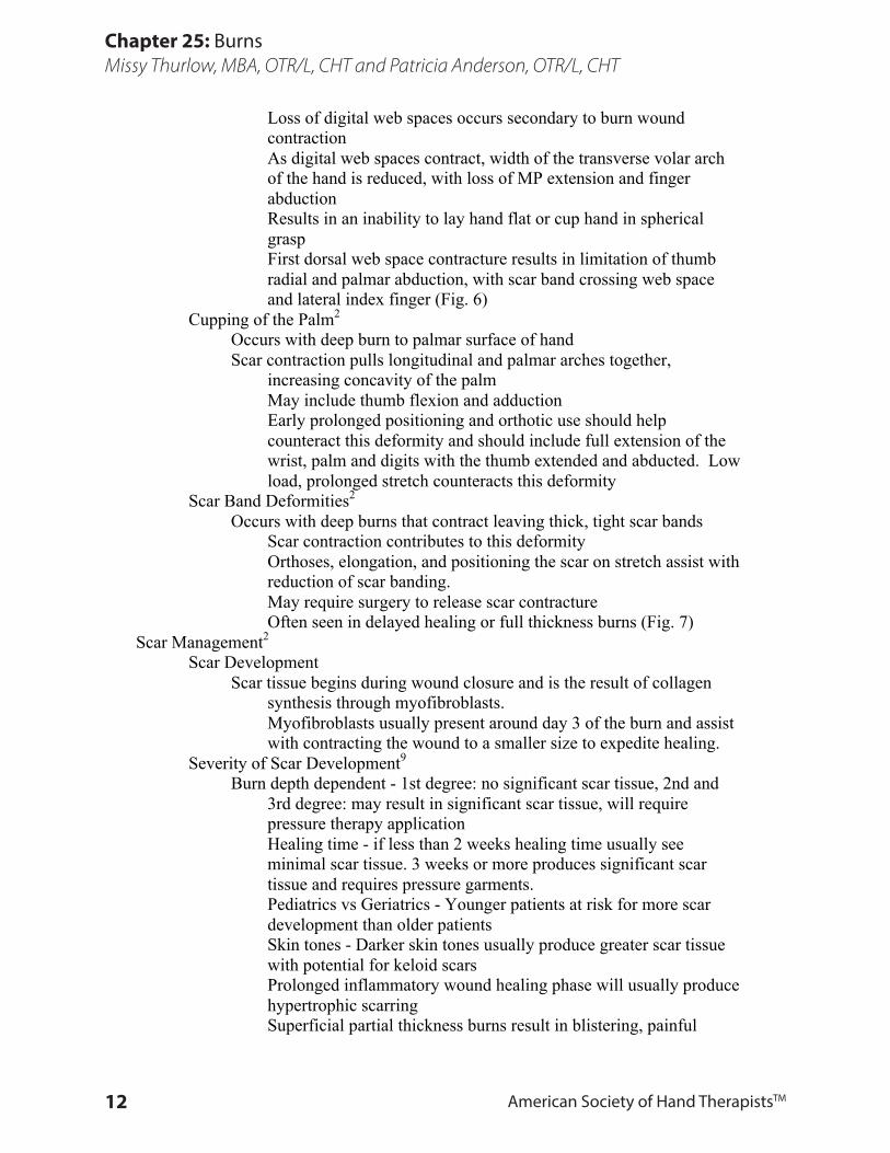
Fig. 5 First web space contractor secondary to scar contraction.
Fig. 6 Palmar burn injury with web space syndactaly.
Fig. 7 Contact burn with split thickness skin grafting - loss of palmar arch, web space contract, cupping of palm.
American Society of Hand TherapistsTM14
Chapter 25: BurnsMissy Thurlow, MBA, OTR/L, CHT and Patricia Anderson, OTR/L, CHT
wounds that should epithelialize within two weeks without complications. Deep partial thickness burns have less blistering, are painful and may take up to three weeks or longer to heal. Significant scar risk is noted. Full thickness burns destroy the epidermis and dermis and result in less painful burn wounds but may have necrotic eschar with high risk of scarring noted.
Scar Evaluation8 Often subjective, done by the clinician. Therapist observes:
Height of the scar: raised or flat Pliability of the scar; categories include normal, supple, yielding, firm, rope or contracture Vascularity assessed based on color: normal tone, pink, red, or purple Other evaluations include pain, stiffness, scar thickness or itching
Decision regarding pressure garments for scarring based on scar evaluation Objective measurements using specific instruments are not commonly used
Pressure Therapy Interim pressure therapy may be needed for red raised wounds or
grafted areas with delayed healing. Interim pressure garments include: Tubigrip or compressive bandages such as ace wraps apply initial pressure to area. Silicone gel or inserts may be placed inside pressure garments for more effective pressure.
Custom pressure garments8 Indicated for raised,red and deep second and third degree burns.
Garments are custom measured. Several companies provide burn garments: Jobst and Bioconcepts are best known manufacturers. Garments worn 23 hours per day to provide optimal pressure. Patients are remeasured for new garments every 3-4 months to provide appropriate fit as burns change. Garments should be worn a minimum of 6 months and upward of 2 years depending on scar maturation. Garments may be fabricated with zippers to minimize shearing to recently healed grafts and to assist with donning and doffing of the garment. Gloves with open fingertips allow use of the hands during ADLs
X. Return to Work/School7,8
Work May benefit from work simulation and work conditioning
Intolerance to extreme temperatures is common after burn injury therefore hot or cold environments should be avoided

Test Prep for the CHT Exam, 3rd Edition 15
Chapter 25: BurnsMissy Thurlow, MBA, OTR/L, CHT and Patricia Anderson, OTR/L, CHT
Pressure garments should be worn to work Sunscreen should be applied to minimize skin discoloration with sun exposure May need work accommodation based on work site evaluation May benefit from initial return to work part time with progression to full time Surgical intervention may be needed in the future, employer must be made aware May benefit from Work Capacity Evaluation Analysis of potential barriers should be identified prior to return to work Provide psychological intervention if needed
School7,8 Pressure garments should be worn to school
Classes and teachers should be notified of student returning May include adaptation of school work, depending on hand function Analysis of potential barriers should be identified prior to return to school Psychological intervention Support of family, therapy team and school teachers necessary for successful return

American Society of Hand TherapistsTM16
Chapter 25: BurnsMissy Thurlow, MBA, OTR/L, CHT and Patricia Anderson, OTR/L, CHT
References1. Howell JW: Management of the burned hand. In: Richards R, Staley M, eds. Burn Care and Rehabilita-
tion: Principles and Practice. Philadelphia, PA: FA Davis;1994: 531-537. 2. Simpson R, Gartner, M. Management of burns in the upper extremity. In: Makin EJ, Callahan A, Skir-
ven TM, Schneider L, Osterman L, eds. Rehabilitation of the Hand.and Upper Extremity. 5th ed. Vol 2. St. Louis, MO: Mosby; 2002:1475-1491. (ch 25).
3. Sammer,D. Acute Care and Rehabilitation of the Hand After Cold Injury. In Skirven,T., Osterman, A., Fedorczyk, J. & Amadio, P, eds. Rehabilitation of the Hand and the Upper Extremity, 6th ed. Vol. 1. Philadelphia, PA: Elsevier Mosby, 2011: 345-351.
4. Rizzo, M. Complex Injuries of the Hand. In Skirven,T.,Osterman, A., Fedorczyk, J. & Amadio, P, eds. Rehabilitation of the Hand and the Upper Extremity, 6th ed. Vol. 2. Philadelphia, PA: Elsevier Mosby, 2011. (Chapter 94).
5. Tham, A. Iiyas, A. Electrical Injuries to the Upper Extremity. In Skirven, T.,Osterman, A., Fedorczyk, J. & Amadio, P, eds. Rehabilitation of the Hand and the Upper Extremity, 6th ed. Vol. 2. Philadelphia, PA: Elsevier Mosby, 2011 (Chapter 100).
6. American Burn Association, White Paper, 2009, American Burn Association. 7. Deshaies, L,: Burns. In Cooper, C. (Ed.) Fundamentals of Hand Therapy – Clinical Reasoning and
Treatment Guidelines for Common Diagnoses of the Upper Extremity, 6th ed. St. Louis, MO. Mosby Elsevier, 2007: 389-403 (Chapter 19).
8. Grigsby, deLind L, Knothe B: Therapist’s Management of the Burned Hand. In: Makin EJ, Callahan A, Skirven TM, Schneider L, Osterman L, eds. Rehabilitation of the Hand.and Upper Extremity. Vol 2. 5th ed. St. Louis, MO: Mosby; 2002:1492-1522.
9. Staley AJ, Richard RL: Scar management. In: Richards R, Staley M, eds In.Burn Care and Rehabilita-tion: Principles and Practice. Philadelphia, PA: FA Davis;1994:380-415.
10. Richard R, Lester M, Miller S, Baily K, Hedman T, Dewey W, Greer M, Renz E, Wold S, Blackbourne L. Identification of Cutaneous Functional Units Related to Burn Scar Contracture Development. J Burn Care and Research. 2009; 30: 625-631.
11. Richard LR, Staley MJ: Burn patient evaluation and treatment planning. In: Richards R, Staley M, eds.Burn Care and Rehabilitation: Principles and Practice. Philadelphia, PA: FA Davis;1994:201-207.
12. http://www.researchutilization.org/matrix/resources/burn/burnguide.html

Test Prep for the CHT Exam, 3rd Edition 17
Chapter 25: BurnsMissy Thurlow, MBA, OTR/L, CHT and Patricia Anderson, OTR/L, CHT
Multiple Choice Questions1. An African – American patient has a severe burn on his shoulder that requires
split-thickness skin grafts. After several months, the patient develops large raised ke-loid areas. What would be a good course of practice for this person?
A. Provide stretching and attempt to remold the scar with TubigripB. Silicone gel for nighttime wear to decrease the scarringC. Referral back to the physician for immediate surgeryD. Provide pressure garments with 30 mmHg of pressure to attempt to decrease the keloid scar
2. A therapist fabricates an orthotic for a severe hand and wrist burn that requires daily dressing changes and has severe edema. The orthotic will initially be held in place with:
A. Soft strapsB. Velcro strapsC. Ace wrapsD. Coban wrap
3. A patient has healing areas that appear hypertrophic. These burns most likely took over:
A. 3 weeks or more to healB. 2 weeks to healC. 1 week to healD. Healed quickly
4. A person with a full thickness circumferential burn to the forearm, elbow and upper arm would most likely benefit from an orthosis that:
A. Positions the elbow in – 30 degrees of extension B. Positions the elbow in full extension C. Positions the elbow in 90 of flexionD. No orthosis indicated
5. A 50% TBSA burn patient s/p skin grafting is doing well and is ready to return to work in the construction field. It is most important that the person:
A. Protects the burn areas with a long sleeve shirt B. No special care is neededC. Completely avoid contact with sunlight for 12 monthsD. Protects healed burn areas with sunscreen and understands sweating ability may have been
affected
6. A patient needs a custom pressure garment for the hand. The patient does not have full digital mobility and will need assistance donning the glove. The best option/modi-fication for this glove is to:
A. Order a non custom edema glove from a catalog until the patient can manage getting the glove off and on independently
B. Provide a garment with less pressure to assist in application C. Provide a zipper on the dorsum of the glove to assist with application D. Use Tubigrip and coban wrapping to increase ease of application and to provide some pressure

18
Chapter 25: BurnsMissy Thurlow, MBA, OTR/L, CHT and Patricia Anderson, OTR/L, CHT
Multiple Choice Questions7. During the initial phase of edema post-burn (48-72 hours):
A. AROM and gentle PROM are encouraged B. AROM should not be done C. Compression is used to control edemaD. Only AROM is done
8. The purpose of an orthosis for an acutely burned hand is to:A. Assist with edema control B. Prevent contracturesC. Promote venous blood and lymph return into the central circulation D. Immobilize the hand to promote healing
9. Remodeling scar tissue is best accomplished by:A. Remodeling of collagen into less organized pliable alignmentB. Increasing patient’s tolerance to forceful stretchingC. Increasing vascular supply to aid in tissue healingD. Low load sustained pressure
10. The following is common for managing burn scar:A. Compressive dressingB. CPMC. ElectrotherapyD. Blocking exercises
11. Scar management begins when:A. Edema is under control B. Skin is closed with only small open wounds remainingC. Open areas require daily dressing changesD. The patient expresses concern over the appearance of the scar
12. The term scar syndactally refers to:A. Web space scar that results in decreased digital abduction B. The production of microscopically similar collagen in the different layers of healing tissueC. Scars on adjacent digits that result in similar functional deficits D. The type of scar resulting from a hand burn requiring safe position orthotics
13. The best predictor for determining if a burn wound will form hypertrophic scarring is:A. Race B. Burn depthC. Age of the patient D. Length of time to wound closure
Multiple Choice Question Answer KeyChapter 25
1-D, 2-C, 3-A, 4-B, 5-D, 6-C, 7-A, 8-B, 9-D, 10-A, 11-B, 12-A, 13-D