Embed Size (px)
Citation preview
Chapter 2 Cell
Zhou Li Prof.
Dept. of Histology and Embryology
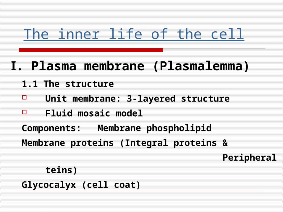
1.1 The structure Unit membrane: 3-layered structure Fluid mosaic model
Components: Membrane phospholipid
Membrane proteins (Integral proteins &
Peripheral proteins)
Glycocalyx (cell coat)
Ⅰ. Plasma membrane (Plasmalemma)
The inner life of the cell
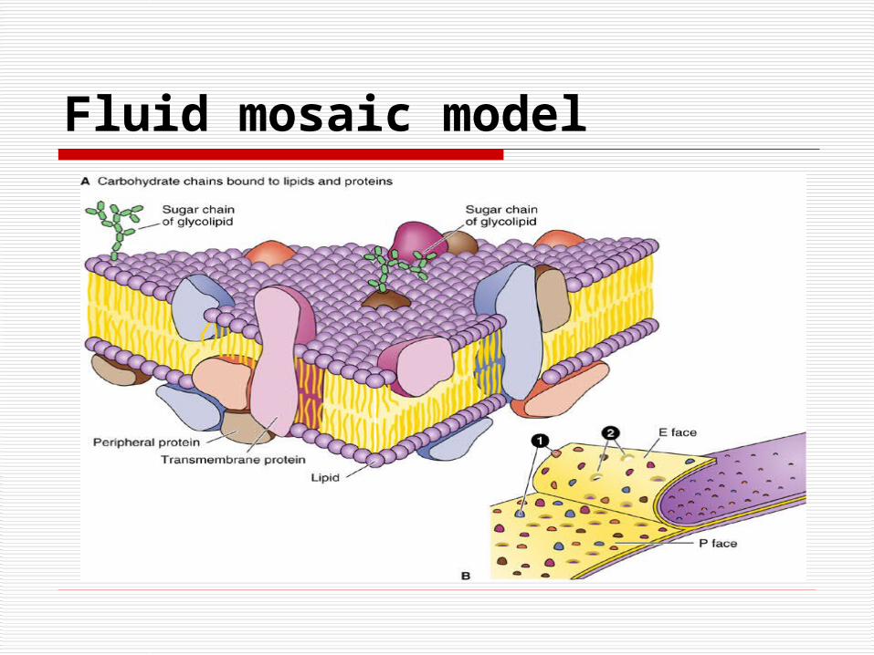
Fluid mosaic model

The functions of cell membrane1. Transmemebrane transport
Passive transport Active transport Transport of macromolecules and partic
les
Endocytosis: Pinocytosis, phagocytosis
Receptor mediated endocytosis
Exocytosis
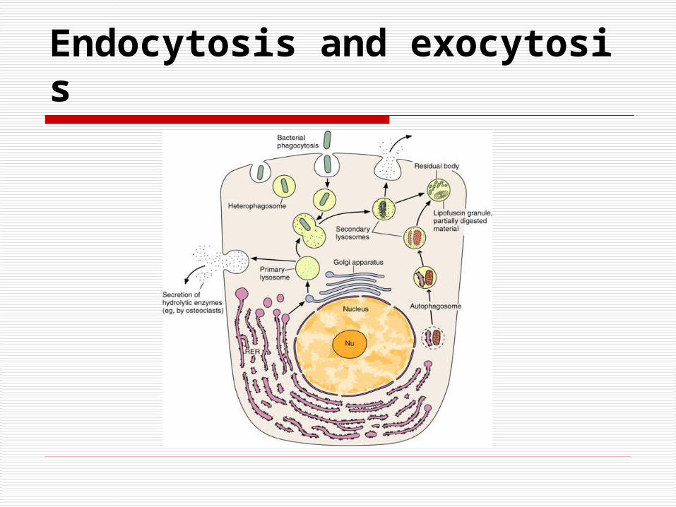
Endocytosis and exocytosis
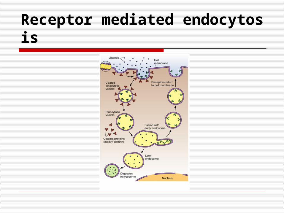
Receptor mediated endocytosis
Ⅱ.Cytoplasm
1. Matrix (Cytosol)
(1) Components
(2) Functions
① Coordinates the intracellular movements of organelles
② Provides a framework for the organization of enzyme and substrates
2.1 Ribosomes
(1) Structure
Small electron-dense particles
Free ribosome & attached ribosome
Polyribosome
(2) Function
Take part in protein synthesis
2. Organelle
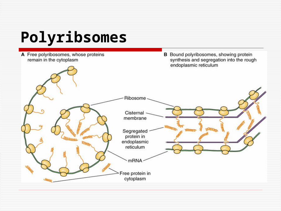
Polyribsomes
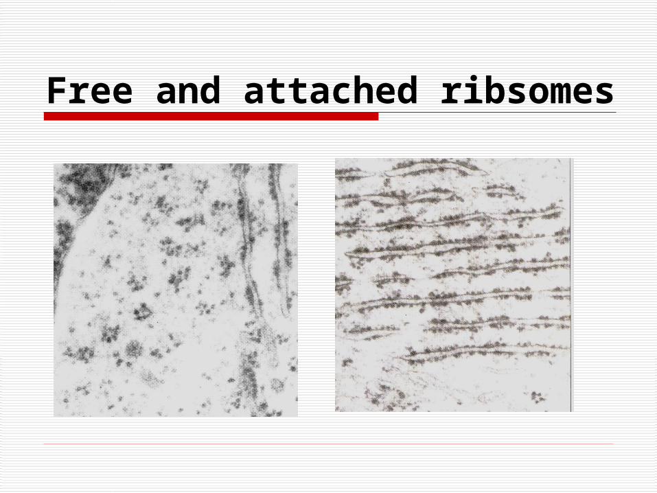
Free and attached ribsomes
2.2 Endoplasmic Reticulum (ER)
(1) Rough Endoplasmic Reticulum (RER)
Structure: Saclike and parallel stacks of fl
attened cisternae, Polyribosomes on the
cytosolic surface
Functions
Synthesis of Secretory proteins
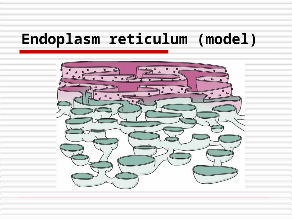
Endoplasm reticulum (model)
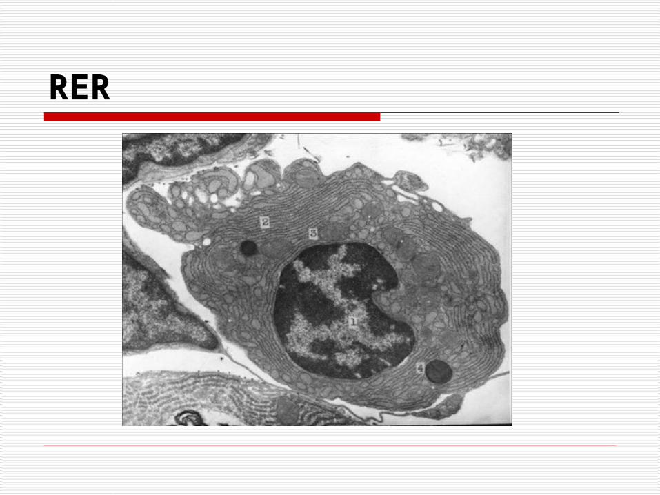
RER
(2) Smooth Endoplasmic Reticulum (SER)
Structure: smooth tubular or vesicle and lacks polyribosomes
Function: varying function of cell from enzymes synthesis of steroid hormone①s neutralizing noxious substances ② ③Synthesizes phospholipids the contra④ction process in muscle cells
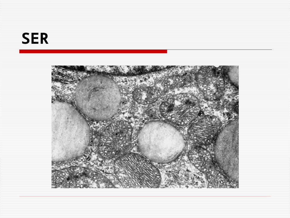
SER
2.3 Golgi Complex (Golgi Apparatus)
Structure: vesicles (Transport vesicles) saccule, vacuoles (Condensing vacuoles)
Forming face ,
maturing face
Functions: initiates packing, glycosylation and concentration of secretory products (including secretory granules and lysosome)
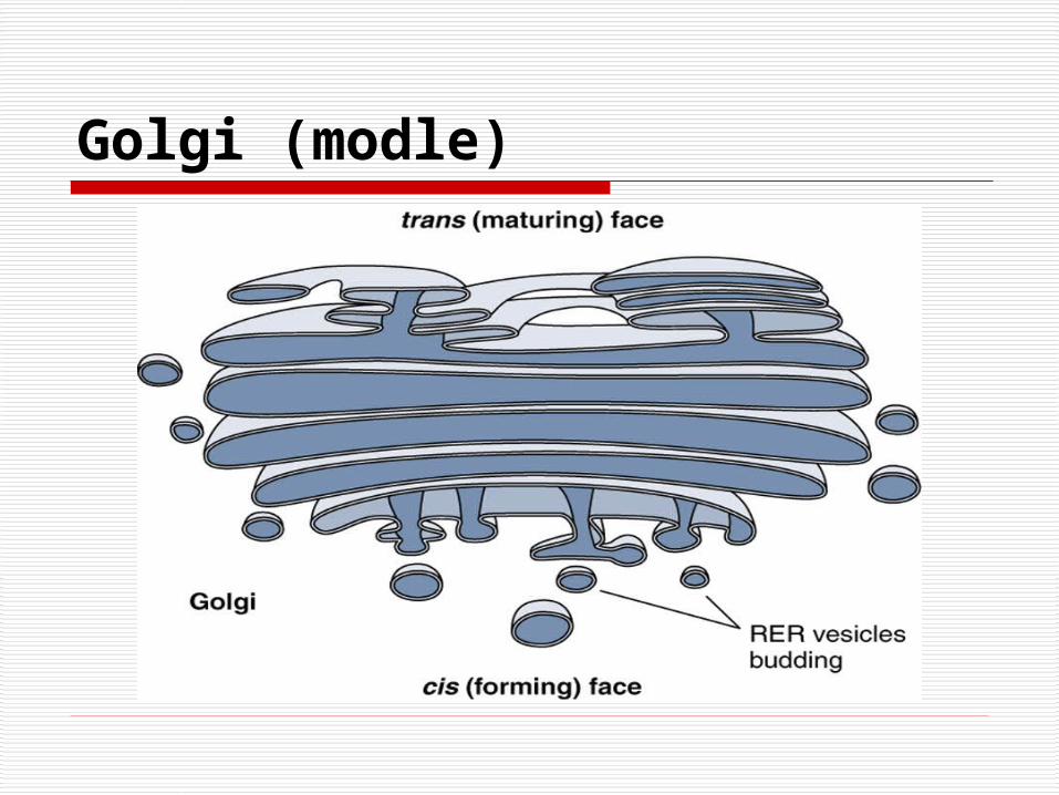
Golgi (modle)
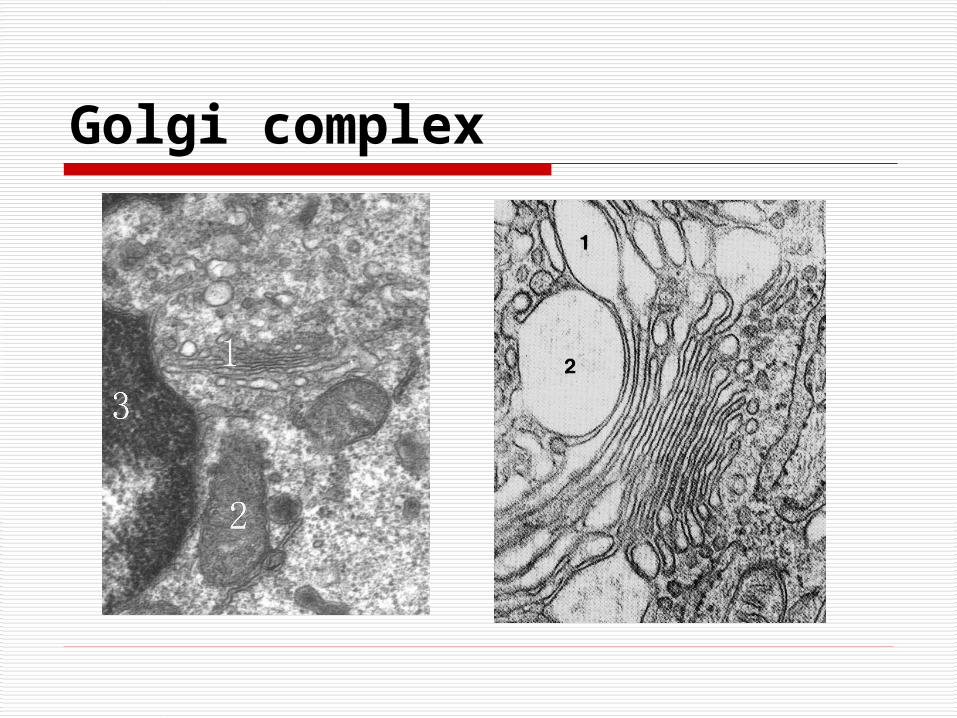
Golgi complex
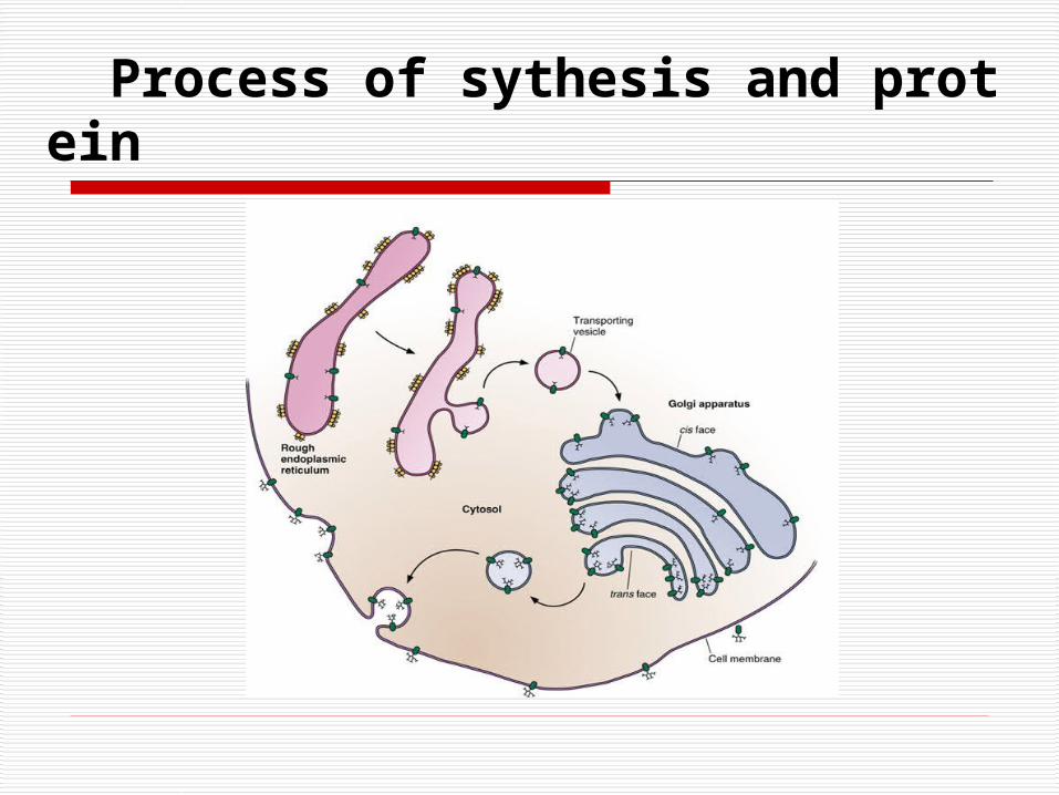
Process of sythesis and protein
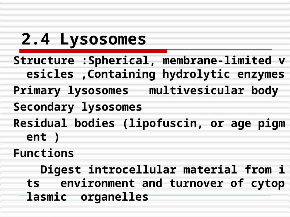
2.4 LysosomesStructure :Spherical, membrane-limited vesicles ,
Containing hydrolytic enzymes
Primary lysosomes multivesicular body
Secondary lysosomes
Residual bodies (lipofuscin, or age pigment )
Functions
Digest introcellular material from its environment and turnover of cytoplasmic organelles
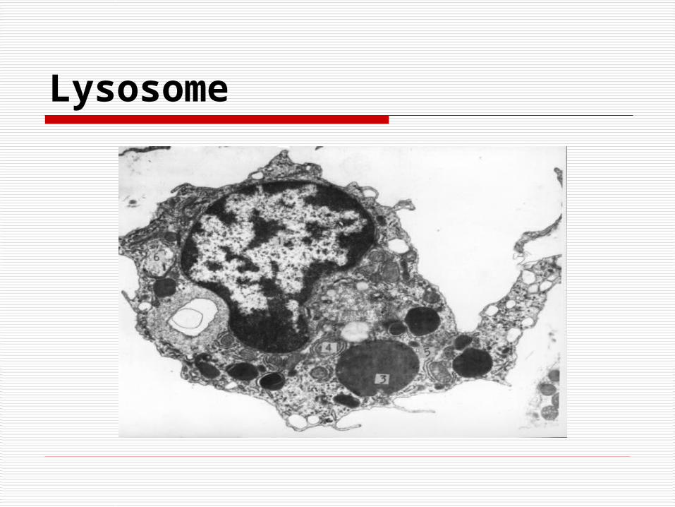
Lysosome
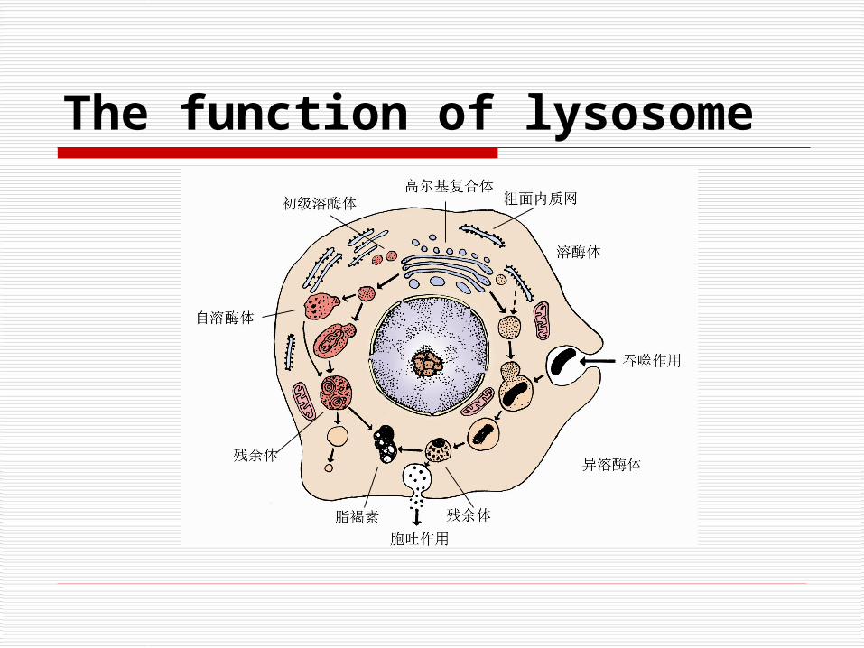
The function of lysosome
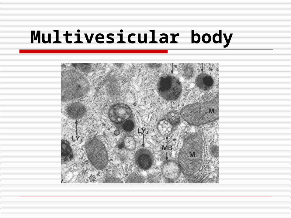
Multivesicular body
2.5 Peroxisomes or Microbodies
Structure:
Spherical membrane-limited organelles, Contain catalase
Functions
① Eliminate hydrogen peroxide
② Degrade toxic molecules in liver and kidney
③ Participate in lipid metabolism
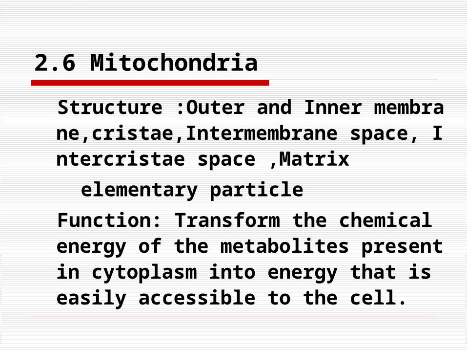
2.6 Mitochondria
Structure :Outer and Inner membrane,cristae,Intermembrane space, Intercristae space ,Matrix
elementary particle
Function: Transform the chemical energy of the metabolites present in cytoplasm into energy that is easily accessible to the cell.
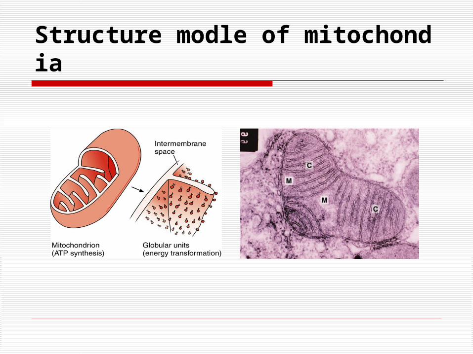
Structure modle of mitochondia
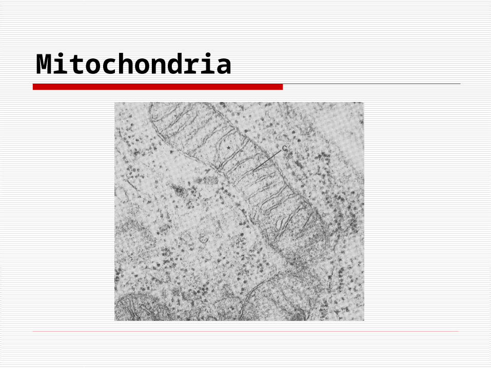
Mitochondria
2.7 Centrosome
Structure: Centrosome is made of a pair of centrioles surrounded by a granular material.
Centriole shows nine sets of microtubules arranged in triplets.
Function: Participate in the organization of the mitotic spindle.
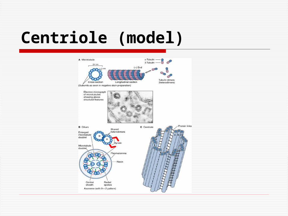
Centriole (model)
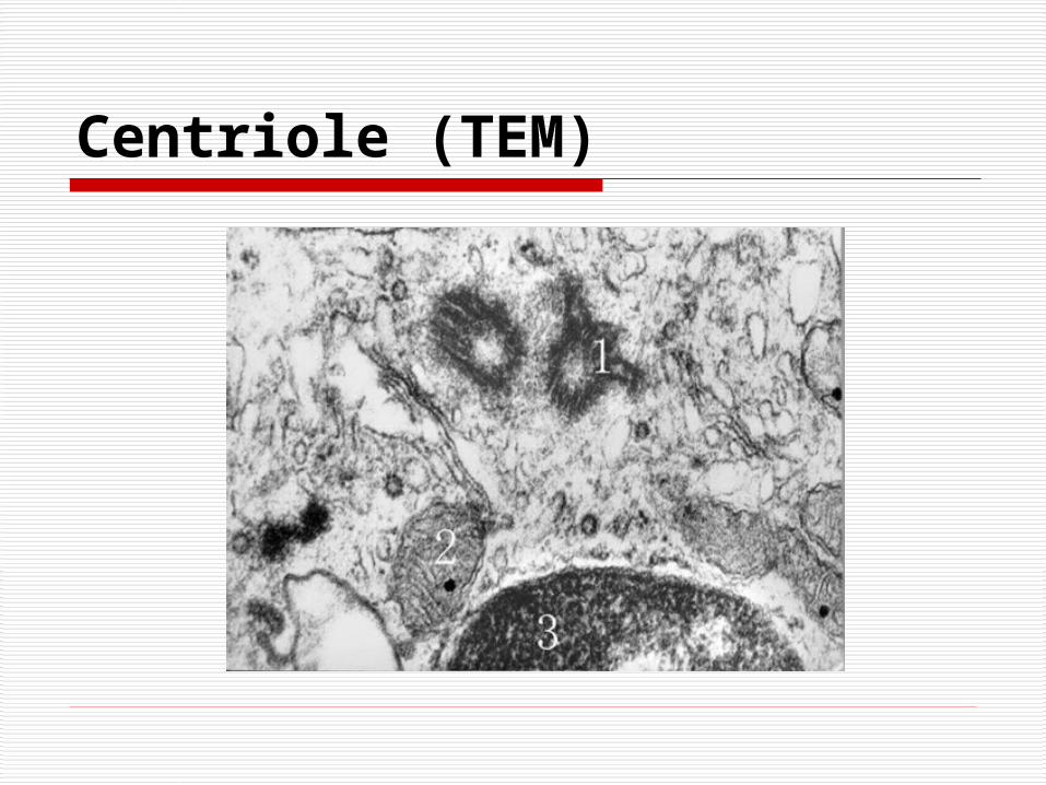
Centriole (TEM)
Cytoskeleton
Microtubules
(1) Structure The subunit is a heterodimer compo
sed of α and β tubulin molecules. Microtubule-organizing centers (cili
a, basal bodies, and centrosomes)

Microtubule and microfiliment

Microtubules ( transversal )

Microfilaments (Actin filaments)
Structure Thin filament (Actin filament, be com
posed of actin) Thick filament (Myosin filament, be co
mposed of myosin)Function Form a meshwork to maintain the sha
pe of the cell

Glycogen granules and microbody (peroxisome)

Inclusion
1. Glycogen granule
2. Lipid droplet
3. Secretory granule or secretory vesicles
4. Pigments (Lipofuscin)

Lipid droplets

Intermediate filaments
Classification:
Keratin filament (Tonofilament)
Desmin filament
Vimentin filament
Neurofilament
Neuroglial filament

Vimentin filament

Intermediate filament

Ⅲ. Cell Nucleus
1. Nuclear envelope
Outer nuclear membrane
Inner nuclear membrane
Perinuclear cisterna
Fibrous lamina
Nuclear pores

Neuclues (model)

2. Chromatin
Components: DNA and Proteins
Classification
(1) Heterochromatin
LM: basophilic clumps
EM: coarse granules
(2) Euchromatin
LM: lightly stained basophilic areas
EM: finely dispersed granular material

Neuclues (TEM)

3. Nucleolus
Components: rRNA and Proteins
4. Nuclear matrix

The Highlight This Chapter
1. Structure and function of the organelles2. What are Euchromatin and Heterochromati
n ?