Embed Size (px)
Citation preview

207
Pelvic exenteration, the en bloc removal of the pelvic organs, is indicated for central recurrent or persistent gynecologic cancer, including cervical, endometrial, vaginal, or vulvar cancer. Even when performed in the setting of specialized centers by highly skilled surgeons, pelvic exenteration is associated with signifi -cant morbidity and mortality. Since the initial series published by Brunschwig 1 in 1948, there has been a dramatic change in the type and frequency of the complications associated with this procedure. A number of factors have infl uenced the outcomes over the past several years, and these include the integration of broad-spectrum antibiotics, thromboembolic prophylaxis, vessel-sealing devices, multiteam surgical expertise, and critical care teams. In addition, modifi cations of urinary diversion and pelvic reconstruction procedures have paved the way to provide improved outcomes and lower complications rates. Neverthe-less, this operation remains a challenge for all patients, and all involved with the care of the patient must recognize that it is a life-changing experience that aff ects physical, psychological, and sexual function and leads to major changes in quality of life. Th e report of a series by Maggioni and colleagues 2 showed that the overall morbidity aft er pelvic exenteration was 66%, with 48% of patients having early complications (<30 days) and 48.5% of patients having late complications. Th e MD Anderson Cancer Center published a report on 160 patients who under-went pelvic exenteration for gynecologic malignancies and noted that the postoperative complication rate was as high as 94%, with 60% of all complications described as a potentially life-threatening event. Th e same group also noted a mortality rate of 1.3%. 3 However, such variation may be secondary to the crite-ria set forth in the respective studies to defi ne complications in the perioperative period. Overall, it is imperative to ensure care-ful patient selection, preoperative and postoperative care, and optimal surgical expertise in a tertiary cancer center to improve not only surgical outcomes but also survival for patients under-going this procedure.
Th is chapter addresses the potential medical and surgi-cal complications that may arise aft er pelvic exenteration. Th e emphasis is on the most common signs and symptoms, detection of such complications, and subsequent management options, highlighting surgical versus nonsurgical options. Th is chapter is intended as a reference guide to aid gynecologic oncologists in assessing the most common complications that arise aft er pelvic exenteration, and accordingly we do empha-size that appropriate consultation with indicated services is always encouraged.
Medical Complications Febrile Morbidity One of the most common postoperative complications in patients who have undergone pelvic exenteration is fever. Postoperative fever is defi ned as a temperature above 38°C (100.4°F) on 2 consecutive postoperative days or above 39°C (102.2°F) on any 1 postoperative day. Th e diff erential diag-nosis is strongly infl uenced by the time of onset of the fever. Th e most common cause of fever within the fi rst 48 hours is a pyretic response to the operation, and this is usually self-limiting. Studies have shown that the rate of febrile morbidity aft er pelvic exenteration can be as high as 71%. 4 In the study by Westin and colleagues 3 from MD Anderson, the rate of early sepsis (<60 days) was 8.8%, and the rate beyond this time point was 1.3%.
Among the most common causes of fever are the following: • Infectious: Surgical site infection, pneumonia, urinary tract
infection, and/or intravascular catheter–related infection • Noninfectious: Hematoma or seroma, deep venous throm-
bosis (DVT) or pulmonary embolism (PE), infl ammatory reaction (pancreatitis), vascular complication (hemorrhage, myocardial infarction, bowel ischemia or infarction), med-ications Aft er pelvic exenteration, sepsis may also be a great cause of
morbidity and mortality. To be diagnosed with sepsis, a patient must have two of the following signs plus a confi rmed infection: body temperature above 38.3°C (101°F) or below 36°C (96.8°F), heart rate higher than 90 beats per minute, and respiratory rate higher than 20 breaths per minute. Severe sepsis is diagnosed when a patient has one of the following: decreased urine output, abrupt changes in mental status, thrombocytopenia, dyspnea, myocardial dysfunction, or abdominal pain.
Th e routine workup of febrile morbidity should be targeted based on the organ system or infectious process of highest sus-picion. Th e need for laboratory testing should be defi ned by the fi ndings of a careful history and physical examination. Th e ini-tial approach to the evaluation should include a complete blood count. Chest x-ray examination, urine cultures, and blood cul-tures are not indicated for all postoperative patients with fever. One should take into account the timing and the causes of fever. In patients with persistent febrile episodes aft er pelvic exentera-tion, one should proceed with abdominal and pelvic computed tomography (CT) scanning to rule out the potential possibility of an intraabdominal abscess.
Complications of Pelvic Exenteration PEDRO T. RAMIREZ | GLORIA SALVO
CHAPTER 16

Section 6 Pelvic Exenteration208
Treatment for febrile morbidity should be tailored accord-ing to the source of the fever. Patients with persistent postop-erative fever should be started on broad-spectrum antibiotics aft er cultures have been obtained. Coverage should be against aerobic gram-negative enteric bacilli and anaerobic organisms. If a source of fever is not apparent and blood cultures show no growth aft er 48 hours, then discontinuation of antimicrobials should be considered. If the cultures are positive, then antibiotic coverage should be focused on the known causative organism(s). All unnecessary treatments including medications, nasogastric tubes, and intravascular and urinary catheters should be discon-tinued, when possible, in the febrile patient.
In the setting of sepsis, all patients should be managed with broad-spectrum antibiotics, hemodynamic support such as crystalloids or albumin, vasopressor therapy, blood product administration, and mechanical ventilation, if needed. Discus-sion of goals of care and prognosis with the patient or family is paramount. Palliative care principles should be considered when appropriate. �
Thromboembolic Events Incidence and Guidelines Among women undergoing major gynecologic surgical proce-dures without thromboprophylaxis, the risk of DVT ranges from 17% to 40%. 5 Th is risk is even higher among women undergoing operation for gynecologic cancer. Martino and colleagues 6 esti-mated the incidence of PE among 507 patients with known or suspected gynecologic cancer undergoing intraabdominal oper-ations and found that the risk of postoperative PE in patients with a diagnosis of cancer was 14 times the risk of postoperative PE in those with benign disease.
Current guidelines for thromboprophylaxis are available from a number of groups, including the American College of Chest Physicians (ACCP), 7 American Society of Clinical Oncology (ASCO), 8 National Comprehensive Cancer Net-work (NCCN), 9 and American College of Obstetricians and Gynecologists (ACOG). 10 All of the aforementioned guide-lines support the recommendation that all patients undergo-ing abdominal or pelvic surgical procedures for malignancy receive pharmacologic prophylaxis. Th e ASCO, NCCN, and ACOG guidelines recommend the consideration of continu-ing prophylaxis for up to 28 days aft er operation. Th e recom-mendation for extended prophylaxis in gynecologic cancer patients is derived from two randomized controlled trials indicating that prolonged thromboprophylaxis reduces the incidence of postoperative venous thromboembolism (VTE). Th e fi rst study was a double-blind multicenter trial in which patients undergoing planned curative open procedures for abdominal or pelvic cancer received enoxaparin (40 mg sub-cutaneously) daily for 6 to 10 days. Patients were then ran-domly assigned to receive either enoxaparin or placebo for another 21 days. Th e results showed a 60% relative reduction and a 7% absolute reduction in the risk of postoperative VTEs in the prolonged thromboprophylaxis group. 11 In a subse-quent study, the investigators evaluated the effi cacy and safety of thromboprophylaxis with low-molecular-weight hepa-rin (LMWH) (dalteparin) administered for 28 days versus 7 days aft er major abdominal surgery for cancer. Th e results showed that the cumulative incidence of VTEs was reduced from 16.3% among patients receiving short-term throm-boprophylaxis to 7.3% among patients receiving prolonged thromboprophylaxis. 12
In patients undergoing pelvic exenteration, the study by Westin and colleagues showed that the rate of thromboembolic events before 60 days was 1.9% and 5% beyond that time. 3 In a study by Jurado and colleagues, 13 the authors reported a rate of DVT among 45 patients who underwent pelvic exenteration of 11% and a rate of PE of 6.7%. Barakat and colleagues 14 reported a mortality rate of 4.5% from PE aft er pelvic exenteration. It is interesting to note that in a study by Iglesias and colleagues 15 from MD Anderson Cancer Center, the authors showed that the rate of thromboembolic events was not aff ected by patient body mass index. �
Signs and Symptoms Th e most common symptoms associated with acute PE include dyspnea (73%), pleuritic chest pain (66%), cough (37%), and hemoptysis (13%). Th e most common signs are tachypnea (70%), rales (51%), tachycardia (30%), fourth heart sound (24%), accentuated pulmonic component of second heart sound (23%), and circulatory collapse (8%). 16 �
Evaluation of Thromboembolic Events Once a medical history has been taken and physical examina-tion performed, it is recommended that patients undergo a complete blood count, liver and kidney function tests, and chest radiography and electrocardiography as part of the initial evalu-ation. In patients with PE, the white blood cell (WBC) count may be normal or elevated, with a WBC count as high as 20,000 K/μL noted in some patients. A chest radiograph may be abnor-mal in most patients with PE, but the fi ndings are not specifi c. Common radiographic abnormalities include atelectasis, pleu-ral eff usion, parenchymal opacities, and elevation of a hemidia-phragm. It is important to note that a normal-appearing chest radiograph in a patient with severe dyspnea and hypoxemia, but without bronchospasm or cardiac shunt, is strongly suggestive of PE. Th e most common electrocardiographic abnormalities in the setting of PE are tachycardia and nonspecifi c ST-T wave abnormalities.
It is important to note that the D-dimer test has limited use-fulness in the setting of cancer and thus is not routinely rec-ommended in the workup of thromboembolic events in such patients. Similarly, although arterial blood gas determination may show hypoxemia, hypocapnia, and respiratory alkalosis in patients with a PE, it is not routinely used because of its very low predictive value.
Th e diagnostic study of choice for DVT is compression ultra-sonography. When a DVT is present, the veins do not collapse when pressure is applied. However, it is important to note that a negative ultrasound Doppler result does not rule out DVT, because a number of DVTs may occur in areas that are inac-cessible to the ultrasound evaluation. For the diagnosis of PE, the ideal choice of study is computed tomographic pulmonary angiography ( Fig. 16.1 ). Th is is for patients with a suspected diagnosis of PE and who are hemodynamically stable. How-ever, in patients who are not stable, bedside echocardiography may be used to obtain a presumptive diagnosis to justify the administration of potentially lifesaving therapies. Ventilation-perfusion (V
./Q.) scanning may be used when CT scanning is not
available or if the patient has a contraindication to CT scan or use of intravenous contrast material. Brain natriuretic peptides (BNPs) are neither sensitive (60%) nor specifi c (62%); how-ever, patients with PE tend to have higher BNP levels. Elevated levels tend to be associated with increased risk of subsequent

Chapter 16 Complications of Pelvic Exenteration 209
complications and mortality in patients with PE. BNP testing is not routinely recommended as part of the standard evaluation of PE. �
Treatment of Thromboembolic Events Th e approach to a patient with a thromboembolic event is to ensure that the patient’s condition has been stabilized aft er assessment of hemodynamic stability. Th e fi rst steps should be to provide adequate oxygen supplementation (targeting O 2 saturation ≥ 90%), obtain peripheral intravenous access, and begin empiric anticoagulation. Th e ACCP guidelines recom-mend starting LMWH or subcutaneous heparin. Once-daily treatment is the preferred choice. Th e length of anticoagula-tion for DVT is 3 months, and the recommended length of therapy for PE is 6 months. Th e ACCP guidelines recommend that thrombolytic therapy should be used in patients with acute PE associated with hypotension (systolic blood pressure BP below 90 mm Hg) who do not have a high risk of bleed-ing. Embolectomy is recommended in patients with massive PE who have a contraindication to fi brinolysis or who remain unstable aft er receiving fi brinolysis. It may also be considered in patients with evidence of right ventricular enlargement or dysfunction on transthoracic echocardiogram. Inferior vena cava fi lters are indicated in the setting of patients with an absolute contraindication to anticoagulant therapy (hemor-rhagic stroke or active bleeding). It is also indicated when recurrent embolism is present even aft er adequate anticoagu-lant therapy. �
Acute Renal Events Acute kidney injury (AKI) is the abrupt loss of kidney function, resulting in the retention of urea and other nitrogenous waste products and in the dysregulation of extracellular volume and electrolytes. Th is term has replaced acute renal failure (ARF) aft er consideration that even small decrements in kidney func-tion are of substantial clinical relevance and are associated with increased morbidity and mortality. In the study by Westin and colleagues, 3 the authors reported that the rate of ARF or AKI aft er pelvic exenteration was 3.8%.
AKI has multiple possible causes, and it is most commonly due to acute tubular necrosis (ATN) from ischemia, nephro-toxin exposure, or sepsis. Other frequent causes include volume depletion, urinary obstruction, rapidly progressive glomeru-lonephritis, and acute interstitial nephritis. AKI is typically detected by means of an increase in serum creatinine and/or a decrease in urine output. Among hospitalized patients, ATN and prerenal disease are the most common causes.
Several consensus defi nitions of AKI have been developed to provide a uniform defi nition of AKI. Th e RIFLE criteria are described here; they consist of three graded levels of kidney dys-function (risk, injury, and failure), based on the magnitude of increase in serum creatinine or urine output, and two outcome measures (loss and end-stage renal disease [ESRD]). Th e RIFLE strata are described in Table 16.1 .
It has been shown that, compared with patients who did not have AKI, patients in the RIFLE stages of “risk,” “injury,” and “failure” had increased relative mortality risks of 2.4 (confi dence interval [CI], 1.94–2.97), 4.15 (CI, 3.14–5.48), and 6.37 (CI, 5.14–7.9), respectively.
Initial Evaluation After Diagnosis All patients with AKI must be carefully evaluated both for reversible causes (hypotension, volume depletion, or obstruc-tion) and for the presence of complications (volume overload, hyperkalemia, metabolic acidosis, hypocalcemia, and hyper-phosphatemia). Th e initial evaluation of the patient with AKI is directed at determining the cause and identifying the complica-tions that may require immediate attention. Th e timing of onset oft en suggests the underlying cause. A careful review of medica-tions is imperative. Oft en, nephrotoxic medications have been started before the onset of AKI, which suggests a cause. In addi-tion, even long-standing medications (particularly angiotensin-converting enzyme [ACE] inhibitors or angiotensin receptor blockers) render patients vulnerable to AKI from prerenal fac-tors or ATN. �
Patient Evaluation Th e initial assessment should include the careful evaluation of vol-ume status and measurement of serum electrolytes, particularly potassium and bicarbonate, and serum phosphate, calcium, and
FIG. 16.1 Spiral computed tomography image of the chest with in-travenous contrast showing an acute pulmonary embolism (arrow) in the thrombus in the segmental branches of the right lobe of the pul-monary artery.
TABLE 16.1 RIFLE Criteria for Acute Renal Compromise 18 Risk 1.5-fold increase in the serum cre-
atinine, or glomerular � ltration rate (GFR) decrease by 25%, or urine output <0.5 mL/kg/h for 6 h
Injury Twofold increase in the serum creati-nine, or GFR decrease by 50%, or urine out-put <0.5 mL/kg/h for 12 h
Failure Threefold increase in the serum cre-atinine, or GFR decrease by 75%, or urine output of <0.3 mL/kg/h for 24 h, or anuria for 12 h
Loss Complete loss of kidney function (e.g., need for renal replacement therapy) for more than 4 weeks
End-stage renal disease
Complete loss of kidney function (e.g., need for renal replacement therapy) for more than 3 months

Section 6 Pelvic Exenteration210
albumin. One should also check serum uric acid and magnesium and perform a complete blood count. Initial testing should include reagent strip urinalysis (dipstick) with automated urine microscopy and the quantifi cation of urine protein or albumin (by random or “spot” protein-to-creatinine ratio or albumin-to-creatinine ratio).
A physical examination may reveal the cause. Signs of vol-ume contraction suggest a prerenal cause of AKI. An ultrasound examination could be an option if renal function does not improve; ultrasonography is the most commonly used imaging technique in patients with AKI. Ultrasonography is safe, easy to perform, and sensitive for obstruction. Magnetic resonance imaging (MRI) with gadolinium should be avoided in patients with AKI because of the nephrotoxicity of the agent. In patients with moderate to advanced kidney disease with estimated glo-merular fi ltration rate (eGFR) below 30 mL/min, the adminis-tration of gadolinium has been associated with the potentially severe syndrome of nephrogenic systemic fi brosis (NSF).
Th e results of the urinalysis and ultrasound examination generally direct the remainder of the diagnostic evaluation. Patients who have evidence of obstruction require further inves-tigation and usually intervention to relieve the obstruction and determine the cause. For patients who have normal renal imag-ing fi ndings, minimal proteinuria, benign urine sediment on urinalysis and microscopy (no red cells or cellular casts), and no clear explanation for AKI, further evaluation is determined by the severity of disease and rate of further decline. • If the creatinine level is persistently elevated or if an initially
mild increase in the creatinine level worsens over the course of days, then a kidney biopsy should be performed. A biopsy is oft en performed when the diagnosis is uncertain. A biopsy usually enables a more defi nitive tissue diagnosis and may allow a therapeutic intervention to prevent ESRD.
• In patients who have signs and symptoms of rapidly progres-sive or unexplained systemic disease, a renal biopsy is war-ranted, even if the eGFR remains stable aft er initial increase.
• In patients who have mild decrements in eGFR (e.g., to 45 to 60 mL/min/1.73 m 2 ) where the eGFR subsequently remains stable, one should just follow the serum creatinine. If the cre-atinine level remains stable, one should continue to follow creatinine level, the results of urine studies (urinalysis, mi-croscopic studies, urine protein and creatinine), and blood pressure until a clear temporal pattern has been established.
Urinalysis Th e urinalysis involves both use of a urine dipstick and micro-scopic examination of the urine sediment. Th e dipstick can be used to test for protein (albumin), pH, glucose, hemoglobin (or myo-globin), leukocyte esterase (refl ecting pyuria), and specifi c gravity. �
Urine Sodium Excretion Th e fractional excretion of sodium (FENa) measures the per-cent of fi ltered sodium that is excreted in the urine. • Th e FENa is commonly used to assist in diff erentiating pre-
renal disease (a reduction in glomerular fi ltration rate [GFR] due solely to decreased renal perfusion) from ATN, the two most common causes of AKI.
• In patients with suspected prerenal disease or ATN, it is rec-ommended that the FENa be measured. A value of the FENa below 1% commonly indicates prerenal disease; in compari-son, a value between 1% and 2% may be seen with either disorders, and a value above 2% usually indicates ATN. �
Urine Volume Trends in and comparisons between the volumes of fl uid going into and coming out of a patient (including urine output) are helpful physiologic parameters in patients with AKI. Oliguria (typically defi ned as <0.3 mL/kg/h or <500 mL/day of urine output) may or may not occur in patients with AKI. Normal urine output can be maintained even with an abnormally low GFR in patients with nonoliguric ATN. Th e prognosis of non-oliguric AKI is generally better than that of oliguric or anuric disease. 19,20 �
Management Volume Issues An assessment of volume status is performed in all patients with AKI because correction of volume depletion or volume overload (especially when associated with worsening cardiac output) may reverse or ameliorate AKI. �
Volume Depletion Unless contraindicated, the patient with a clinical history con-sistent with fl uid loss (such as vomiting and diarrhea), physical examination fi ndings consistent with hypovolemia (hypoten-sion and tachycardia), and/or oliguria should receive intra-venous fl uid therapy. Th is fl uid challenge attempts to identify prerenal failure that can progress to AKI if not treated promptly. Studies have shown that prompt reversal of volume depletion may prevent or limit kidney injury due to ATN. However, such fl uid infusion is contraindicated in those with obvious volume overload or heart failure. Fluids may be either crystalloid or col-loid. One should begin with 1 to 3 L of fl uid, with careful and repeated clinical assessment to evaluate the patient’s response to this therapy. Fluid therapy should be targeted to physiologic end points. �
Volume Overload Hypervolemia may be present at initial evaluation or may occur because of excessive fl uid administration in the setting of impaired ability to excrete sodium and water. Th is is especially true for patients with sepsis, who commonly receive aggressive intravenous fl uid resuscitation. �
Hyperkalemia Hyperkalemia is a common and potentially life-threatening complication of AKI. In general, all patients with AKI and hyperkalemia that is refractory to medical therapy should be dialyzed unless hyperkalemia is mild (i.e., <5.5 mEq/L) and the cause of AKI is known to be easily reversed (such as prerenal AKI due to volume depletion or ACE inhibitors). �
Prognosis Most patients with AKI recover renal function, with recovery manifesting with an increase in urine output and a gradual decrease in the blood urea nitrogen (BUN) and serum creati-nine concentration. However, in many patients, including those with previously normal renal function, renal function does not return to baseline levels. In addition, many studies have demon-strated an increase in the risk of chronic kidney disease (CKD) and ESRD in patients who recover from AKI. Even small, acute rises in serum creatinine as low as 0.3 mg/dL (27 µ mol/L) are associated with both short-term and long-term increases in mortality. �
Chapter 16 Complications of Pelvic Exenteration 211
Surgical Complications Wound-Related Events Superfi cial Wound Separation A frequent complication in patients who undergo pelvic exen-teration is wound complication. Th e rate of such complications has ranged from 5.6% to 29.4% in the largest published series. However, one must consider that these include both abdominal wound complications and perineal wound issues. 2–4 �
Dehiscence and Evisceration Complete fascial dehiscence is associated with a mortality rate of 10%. Early postoperative fascial dehiscence is a surgi-cal emergency and should be addressed promptly. Th e risk fac-tors for fascial disruption are advanced age, chronic pulmonary disease, anemia, postoperative coughing, wound infection, and complexity of surgery. Other factors include malignancy, obe-sity, sepsis, hypoalbuminemia or poor nutrition, and chronic glucocorticoid therapy. Herniation is more common when the incision length exceeds 18 cm. 21 Dehiscence is most likely due to placement of the suture too close to the edge or under tension. To minimize this complication, elective midline abdominal clo-sure should be performed with continuous absorbable sutures.
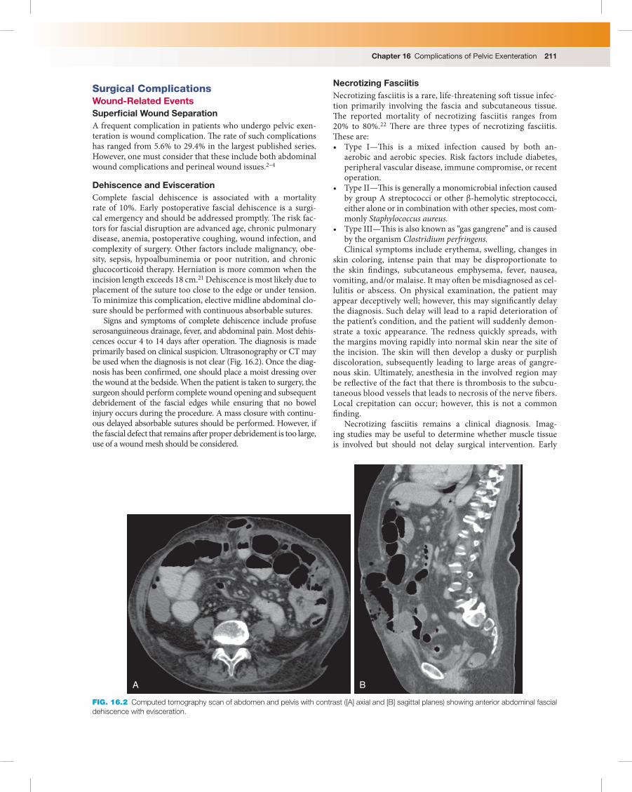
Signs and symptoms of complete dehiscence include profuse serosanguineous drainage, fever, and abdominal pain. Most dehis-cences occur 4 to 14 days aft er operation. Th e diagnosis is made primarily based on clinical suspicion. Ultrasonography or CT may be used when the diagnosis is not clear ( Fig. 16.2 ). Once the diag-nosis has been confi rmed, one should place a moist dressing over the wound at the bedside. When the patient is taken to surgery, the surgeon should perform complete wound opening and subsequent debridement of the fascial edges while ensuring that no bowel injury occurs during the procedure. A mass closure with continu-ous delayed absorbable sutures should be performed. However, if the fascial defect that remains aft er proper debridement is too large, use of a wound mesh should be considered. �
Necrotizing Fasciitis Necrotizing fasciitis is a rare, life-threatening soft tissue infec-tion primarily involving the fascia and subcutaneous tissue. Th e reported mortality of necrotizing fasciitis ranges from 20% to 80%. 22 Th ere are three types of necrotizing fasciitis. Th ese are: • Type I—Th is is a mixed infection caused by both an-
aerobic and aerobic species. Risk factors include diabetes, peripheral vascular disease, immune compromise, or recent operation.
• Type II—Th is is generally a monomicrobial infection caused by group A streptococci or other β -hemolytic streptococci, either alone or in combination with other species, most com-monly Staphylococcus aureus.
• Type III—Th is is also known as “gas gangrene” and is caused by the organism Clostridium perfringens. Clinical symptoms include erythema, swelling, changes in
skin coloring, intense pain that may be disproportionate to the skin fi ndings, subcutaneous emphysema, fever, nausea, vomiting, and/or malaise. It may oft en be misdiagnosed as cel-lulitis or abscess. On physical examination, the patient may appear deceptively well; however, this may signifi cantly delay the diagnosis. Such delay will lead to a rapid deterioration of the patient’s condition, and the patient will suddenly demon-strate a toxic appearance. Th e redness quickly spreads, with the margins moving rapidly into normal skin near the site of the incision. Th e skin will then develop a dusky or purplish discoloration, subsequently leading to large areas of gangre-nous skin. Ultimately, anesthesia in the involved region may be refl ective of the fact that there is thrombosis to the subcu-taneous blood vessels that leads to necrosis of the nerve fi bers. Local crepitation can occur; however, this is not a common fi nding.
Necrotizing fasciitis remains a clinical diagnosis. Imag-ing studies may be useful to determine whether muscle tissue is involved but should not delay surgical intervention. Early
A B
FIG. 16.2 Computed tomography scan of abdomen and pelvis with contrast ([A] axial and [B] sagittal planes) showing anterior abdominal fascial dehiscence with evisceration.

Section 6 Pelvic Exenteration212
radiographic fi ndings include soft tissue thickness and opac-ity. CT scans may show dermal thickening, increased soft tissue attenuation, infl ammatory fat stranding, and possible superfi -cial or deep crescentic fl uid or air in the subfascial planes. 23
Th e treatment of patients with necrotizing fasciitis consists of adequate and aggressive surgical debridement, supportive care, and broad-spectrum antibiotics. Surgical exploration is the only way to defi nitely establish the diagnosis. At operation the fi ndings will reveal that the normal skin and subcutaneous tis-sue become loosened from the rapidly spreading deep necrotic fascia. It is important to note that fascial necrosis is usually more advanced than the appearance suggests. Without prompt treat-ment, secondary involvement of the deeper muscle layers may occur, resulting in myositis or myonecrosis. It is important to recognize that surgical debridement is associated with a lower mortality when performed within 24 hours of diagnosis. 24 �
Urinary Diversion Complications During a total or anterior pelvic exenteration, urinary diversion is routinely performed. Patients can choose either continent or incontinent urinary diversion, and there are advantages and dis-advantages associated with both of these techniques. Incontinent diversion is faster and less technically challenging than continent diversion; also, incontinent diversion may have the advantage of requiring less maintenance eff ort and self-care by the patient. Th e incidences of early and late complications of incontinent urinary diversion have been reported to be 33% and 28%, respectively. 25 Th e most commonly reported complications are anastomotic leakage (3%), fi stula formation (3%–19%), need for reoperation (8%–19%), renal insuffi ciency (6%–17%), urostomy stricture (7%), and ureteral obstruction (7%) ( Fig. 16.3 ).
Continent urinary diversion off ers better cosmetic results than incontinent diversion; however, overall complication rates with continent diversion remain signifi cant and range from 37% to 66%. Th e most common complications associated with conti-nent urinary diversion are pyelonephritis (13%–42%), diffi culty with catheterization (12%–54%), ureteral (anastomotic) stric-ture (2%–22%), urostomy stricture (4%–22%), incontinence (7%–13.3%), urinary stone formation (7%–18%) ( Fig. 16.4 ), ureteral (anastomotic) leaks (2%–14%), fi stula (2%–15%), and permanent renal failure (3%). Th ere is also the potential risk of development of hyperchloremic metabolic acidosis.
In a study by Ramirez and colleagues 26 from MD Anderson Cancer Center, the authors reported on 133 patients who under-went total pelvic exenteration. Ninety-nine patients (74.4%) underwent a total pelvic exenteration, and 34 (25.6%) underwent an anterior pelvic exenteration. In 46 patients (34.6%), conti-nent urinary diversion was performed, and incontinent urinary diversion was performed in 87 patients (65.4%). Th e mean age at exenteration was 47.6 years (range, 30–73 years) in the continent urinary diversion group and 57.2 years (range, 27–86 years) in the incontinent urinary diversion group ( P < .0001). Median fol-low-up time aft er exenteration was 28.5 months (range, 2.3–185.7 months) for patients with continent urinary diversion and 28.1 months (range, 1.4–187.1 months) for patients with incontinent urinary diversion. Th e most common postoperative complica-tion was pyelonephritis or urosepsis, which occurred in 32.6% of the patients with continent urinary diversion and 37.9% of the patients with incontinent urinary diversion ( P = .58). Th e second most common complication was urinary stone formation, which occurred in 34.8% of the patients with continent urinary diver-sion and 2.3% of the patients with incontinent urinary diversion
( P = .001). No stone formation was observed in the fi rst 60 days aft er continent urinary diversion. Of the 16 patients with stone formation and continent urinary diversion, 11 were asymptom-atic and did not require intervention. Th ree patients underwent laparotomy for stone removal—one because of an enterocutane-ous (pouch-to-skin) fi stula, possibly secondary to infection and an obstructive mucous plug, and the other two because of large size (one patient) and number of stones (n = 1). One patient had bilateral nephrostomy tubes placed because of urinary obstruc-tion and poor functional status, and one patient was treated successfully with cystolitholapaxy. Both patients with stone for-mation and incontinent urinary diversion were asymptomatic and did not require intervention.
No signifi cant diff erences were observed between the con-tinent and incontinent urinary diversion groups for rates of ureteral (anastomotic) leakage, ureteral (anastomotic) stric-ture, renal insuffi ciency, fi stula formation, conduit reopera-tion, or pyelonephritis or urosepsis. No statistical signifi cance in urostomy stricture formation was found aft er multivari-ate analysis ( P = .08). In patients with at least one episode of pyelonephritis or urosepsis, there was no signifi cant diff erence between the groups ( P = .20). Th ere was also no signifi cant diff erence between the groups for the number of hospitaliza-tions required because of complications related to the urinary diversion ( P = .45). When the analysis was limited to patients who had received preoperative pelvic radiation, there was an increased incidence of urostomy stricture aft er 60 days in patients with continent urinary diversion on univariate analy-sis. Of the patients with continent urinary diversion, 28.3% reported incontinence, and 15.2% reported diffi culty with catheterization.
In that study, the authors concluded that patients undergoing pelvic exenteration have a high risk of complications and that there is no diff erence in postoperative complication rates related to urinary diversion except that urinary stone formation is more common among patients with a continent urinary diversion. Continent urinary diversion is also associated with the potential for additional complications: incontinence and diffi culty with catheterization. �
Bowel-Related Complications Patients undergoing a pelvic exenteration are at a higher risk of developing postoperative bowel complications as a result of various factors that aff ect healing, such as poor nutritional sta-tus and prior history of radiation therapy. It has been shown that the rate of bowel-related complications aft er pelvic exenteration is approximately 10%. 3,4
Postoperative Ileus Postoperative paralytic ileus refers to obstipation and intoler-ance of oral intake due to nonmechanical factors that disrupt the normal coordinated propulsive motor activity of the gastro-intestinal tract following abdominal or nonabdominal surgical procedures. Aft er abdominal operation, “normal” physiologic postoperative ileus due to postoperative gut dysmotility is widely reported as lasting 0 to 24 hours in the small intestine, 24 to 48 hours in the stomach, and 48 to 72 hours in the colon. 27
Th e multiple defi nitions of “prolonged” postoperative ileus have included: • No return of bowel function postoperatively (ranging from
postoperative days 4 to 6) • Absence of fl atus or stool by postoperative day 6

Chapter 16 Complications of Pelvic Exenteration 213
• Postoperative nausea or vomiting necessitating cessation of oral intake, intravenous support, or nasogastric tube place-ment by postoperative day 5
• Return of bowel function aft er postoperative day 5 • Absence of fl atus and/or bowel movement prolonging hos-
pitalization beyond discharge goal (ranging from postopera-tive days 6 to 8)
• Lack of bowel activity more than 5 days aft er operation
Among the most common nonsurgical risk factors are opioid use, antihypertensive agents, antidiarrheal or antiemetic agents, any drug with an anticholinergic property, muscle relaxants, and atropine products. Th ere are also a number of medical conditions that may predispose the patient to postoperative ileus. Th ese include pancre-atitis, gastroenteritis, spinal cord injury, myocardial infarction, stroke,
pneumonia, diabetes, diabetic ketoacidosis, botulism, or Parkinson disease. When considering surgical factors, one must consider that lower abdominal procedures with large incisions and with intestinal manipulation (e.g., colorectal, gynecologic [exenteration]) are asso-ciated with a higher risk of postoperative ileus, whereas abdominal procedures with smaller incisions and minimal visceral manipula-tion (e.g., cholecystectomy) are associated with a lower risk.
Th e most common symptoms are abdominal distention, bloating, diff use abdominal pain, nausea and/or vomiting, inability to pass fl atus, and inability to tolerate a regular oral diet. On examination, the patient may have abdominal distention and a tympanitic abdomen with reduced bowel sounds and some degree of tenderness.
Th e diagnosis is established based on clinical fi ndings and plain abdominal fi lms. Th ese may show dilated loops of bowel
Right ureter
Conduit
Right ureterLeft ureter
A B
C FIG. 16.3 (A) Digital subtraction angiogram showing fi lling of the conduit without evidence of leakage of the right ureteral anastomosis (normal right ureter drainage). (B) Left posterior oblique view demonstrating contrast leaking from left ureter into the pelvis (arrows). (C) Computed tomog-raphy scan of the abdomen and pelvis with contrast. Urinary leak at ureteric anastomosis with the ileal conduit in the posterior left pelvis.

Section 6 Pelvic Exenteration214
but with evidence of air in the colon and rectum without a tran-sition zone that would suggest bowel obstruction ( Fig. 16.5A ). Th ere should also not be any evidence of free air that is asso-ciated with perforation. Th e diagnosis is established when the signs and symptoms persist for more than 3 to 5 days. When in doubt, CT of the abdomen will help diff erentiate small bowel obstruction from ileus, given that it has a sensitivity and speci-fi city of 90% to 100%.
Th erapy for patients with postoperative ileus should focus on removal of any recognized inciting factors, maintenance and replacement of fl uid therapy, electrolyte replacement, bowel rest and bowel decompression (as needed), and serial abdominal examinations. �
Bowel Obstruction Small bowel obstruction can be functional or mechanical. Th e small bowel is involved in about 80% of cases of mechanical bowel obstruction. 28 Th ere are several causes for small bowel obstruction; however, in the postoperative period aft er pelvic exenteration, the most common cause of small bowel obstruc-tion is adhesion formation. It is imperative to diagnose the bowel obstruction early so that the appropriate management may be initiated. In simple mechanical obstruction, blockage occurs without vascular compromise. Th e normal secretory and absorptive functions of the mucosa are depressed, and the bowel wall becomes edematous and congested. Th ere may also be transudative loss of fl uid from the intestinal lumen into the peritoneal cavity. Electrolyte loss is common in this setting, leading to metabolic alkalosis, and the fl uid loss may result in hypovolemia. If the bowel obstruction is not recognized and properly addressed, the obstruction will lead to vascular com-promise, and the blood fl ow to the bowel will diminish. Venous obstruction occurs fi rst, followed by arterial occlusion, result-ing in rapid ischemia of the bowel wall. Th e ischemic bowel becomes edematous and infarcts, leading to gangrene and per-foration. Acute mechanical small bowel obstruction is a com-mon surgical emergency.
Signs and Symptoms Patients with bowel obstruction may have an abrupt onset of abdominal pain, nausea, vomiting, cramping, and abdominal distention. Patients with partial obstruction may have intermit-tent episodes of diarrhea; however, more commonly, patients with complete obstruction will have obstipation at presentation. One should note that the presence of diarrhea in the setting of bowel obstruction does not automatically indicate resolution of the obstruction. At inspection of the abdomen, the physi-cal examination may reveal evidence of distention, hyperactive bowel sounds secondary to high-pitched peristalsis, and tender-ness. In multiple retrospective reviews, abdominal distention was the most frequent physical fi nding on clinical examina-tion, occurring in 56% to 65% of patients. 29,30 With signifi cant bowel distention, bowel sounds may become muffl ed, and as the bowel distention progresses, the bowel sounds may become hypoactive. Th e degree of tenderness is dependent on the level of obstruction and whether there is evidence of bowel ischemia. Fever may be associated with complications of obstruction such as ischemia or necrosis. �
Diagnosis In general, the diagnosis of small bowel obstruction is a clinical and radiologic diagnosis. Th e initial evaluation should include supine and upright abdominal radiographs. X-ray fi ndings are diagnostic in 50% to 60% of patients; equivocal in about 20% to 30%; and normal, nonspecifi c, or misleading in 10% to 20%. 31 Th e key radiographic signs that allow distinction between a high-grade small bowel obstruction and a low-grade obstruc-tion are the presence of small bowel distention, with maximal dilated loops averaging 36 mm in diameter and exceeding 50% of the caliber of the largest visible colon loop, in addition to a 2.5-times increase in the number of distended loops in the abdo-men compared with the normal number. Other fi ndings that are most signifi cant and predictive of high-grade small bowel obstruction are the presence of more than two air-fl uid levels, air-fl uid levels wider than 2.5 cm, and air-fl uid levels diff ering more than 2 cm in height from one another within the same small bowel loop. 32 It is oft en diffi cult to diff erentiate postoperative ileus from an obstruction based solely on fi ndings on abdomi-nal radiographs. In that setting, a defi nitive diagnosis is attained based on both clinical suspicion and CT scan fi ndings (see Fig. 16.5B and C ). It should be noted that in patients with necrosis or gangrene, the abdominal imaging may demonstrate gas in the bowel wall, also known as pneumatosis intestinalis ( Fig. 16.6 ). Th is is an ominous sign and a surgical emergency because immi-nent bowel perforation is usually seen in this setting.
Standard CT is the ideal imaging modality for evaluation of small bowel obstruction, with sensitivity of 90% to 96%, speci-fi city of 96%, and accuracy of 95%. Newer multidetector CT scanners with multiplanar reformation capability are considered more eff ective in evaluation of small bowel obstruction. Th ere-fore, CT is considered the best modality for determining which patients would benefi t from conservative management and close follow-up and which patients would benefi t from immediate surgical intervention. 33 CT criteria for small bowel obstruction are the presence of dilated small bowel loops (diameter >2.5 cm from outer wall to outer wall) proximally to normal-caliber or collapsed loops distally. It should be noted that multidetector CT usually does not require oral contrast mate-rial because the retained intraluminal fl uid serves as a natural
FIG. 16.4 Computed tomography scan of the abdomen and pelvis with contrast. Multiple urinary continent conduit calculi. Arrow demon-strates site of stones.

Chapter 16 Complications of Pelvic Exenteration 215
negative contrast agent, and it allows assessment of extramural areas that would not be visible at contrast-enhanced studies.
If CT scan is unavailable, sonography can sometimes serve as a useful substitute. Sonography is not commonly used for the evaluation of small bowel obstruction, mainly because most of the time the bowel loops are fi lled with gas, producing nondi-agnostic sonograms, and because adhesions, the most common cause of mechanical small bowel obstruction, are not detected with this technique. 34 However, when the obstructed bowel seg-ments are dilated and fi lled with fl uid, not only can the level of obstruction be recognized but the cause of the obstruction can also be demonstrated with the use of the fl uid-fi lled bowel as a sonic window. �
Treatment Aggressive intravenous fl uid therapy and correction of electro-lyte imbalance are crucial in the initial management of acute
small bowel obstruction. A Foley catheter and occasionally a central venous catheter are needed to monitor fl uid resuscita-tion. Blood tests identify electrolyte imbalance, elevated leu-kocyte count, abnormal liver function test results, elevated amylase level, acidosis, anemia, and bleeding tendency. A naso-gastric tube allows decompression of the stomach and prevents aspiration. Traditionally, it has been recommended that patients with small bowel obstruction (without indications for immedi-ate surgical exploration) should be observed for no longer than 12 to 24 hours, aft er which time, if no improvement is seen, the patient should undergo exploration. However, as long as there remain no fi ndings on serial clinical evaluation to suggest a complicated obstruction, the patient may be observed for a lon-ger period of time. Repeated examination of the patient during this period is extremely important.
Data regarding nonoperative management suggest it to be successful in 65% to 81% of partial small bowel obstruction
A B
C FIG. 16.5 (A) Air-fl uid levels (arrows) consistent with postoperative ileus. (B) Prominent small bowel loops consistent with distal small bowel obstruction (yellow line) . (C) Computed tomography scan of the abdomen and pelvis. Small bowel obstruction with evidence of transition point (arrow).

Section 6 Pelvic Exenteration216
cases. All patients suspected of having complicated bowel obstruction (complete obstruction, closed-loop obstruc-tion, bowel ischemia, necrosis, or perforation) based on clinical and radiologic examination should be taken to the operating room for abdominal exploration ( Fig. 16.7 ). It should be noted that for patients who ultimately require an operation, a delay of more than 1 day has been iden-tified as a risk factor for requiring bowel resection 35
( Fig. 16.8 ). �
Anastomotic Leaks Th e overall incidence of anastomotic leaks is approximately 2% to 7%. 36 Th e lowest leak rates are found with ileocolic anastomo-sis (1%–3%), and the highest rates are found in coloanal anasto-mosis (10%–20%). 37 Th e mortality rate for an anastomotic leak in the literature typically is in the 10% to 15% range. 38 In the study by Maggioni and colleagues, 2 the rate of leaks in patients undergoing pelvic exenteration was 2.8%. In the setting of pelvic exenteration, the anastomotic leak usually occurs as a result of small bowel anastomosis when a segment of ileum is used as the incontinent urostomy. Th e anastomotic leak may also be seen in the setting of ileocolonic anastomosis when a continent conduit is performed aft er the distal ileum and ascending colon have been used for the urinary conduit.
Most anastomotic leaks usually become apparent 5 to 7 days postoperatively. Th e majority of the literature defi nes
A
B FIG. 16.6 Computed tomography scan of the abdomen and pelvis (coronal [A] and axial [B] views). Air in small bowel wall consistent with pneumatosis intestinalis (red arrows) and closed-loop obstruction of small bowel (yellow arrow) .
A
B
FIG. 16.7 Pneumoperitoneum demonstrating free air within abdomi-nal cavity. (A) Abdominal radiograph. (B) Computed tomography scan of the abdomen and pelvis (arrow) .