Embed Size (px)
Citation preview
267
Chapter 10
Preventing Infection at Mucosal Surfaces
Most infectious diseases suffered by humans are caused by pathogens much smaller than a human cell. For these microbes, the human body constitutes a vast resource-rich environment in which to live and reproduce. In facing such threats, the body deploys a variety of defense mechanisms that have accumu-lated over hundreds of millions of years of invertebrate and vertebrate evolu-tion. In considering mechanisms of innate immunity in Chapters 2 and 3 and of adaptive immunity in Chapters 4–11, we principally used the example of a bacterial pathogen that enters the body through a skin wound, causing an innate immune response in the infected tissue that then leads to an adaptive immune response in the draining lymph node. The merits of this example are that it is simple and involves a tissue for which we have all observed the effects of wounds, infection, and inflammation. Until recently, these were the only responses studied by most immunologists, who usually administered their experimental antigens by subcutaneous injection. But in the real world, only a fraction of human infections are caused by pathogens that enter the body’s tissues by passage through the skin. Many more infections, including all of those caused by viruses, make their entry by passage through one of the mucosal surfaces. Although the immune response to infection of mucosal tis-sue has strategies and principles in common with those directed at infections of skin and connective tissue, there are important differences, both in the cells and molecules involved, as well as the ways in which they are used. Appreciation of the extent of these differences has led to the concept that the human immune system actually consists of two semi-autonomous parts: the systemic immune system, which defends against pathogens penetrating the skin, and the mucosal immune system, which defends against pathogens breaching mucosal surfaces. This chapter focuses on mucosal immunity and how it dif-fers from systemic immunity.
10-1 The communication functions of mucosal surfaces render them vulnerable to infection
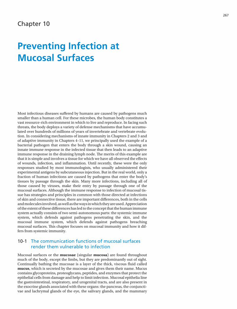
Mucosal surfaces or the mucosae (singular mucosa) are found throughout much of the body, except the limbs, but they are predominantly out of sight. Continually bathing the mucosae is a layer of the thick, viscous fluid called mucus, which is secreted by the mucosae and gives them their name. Mucus contains glycoproteins, proteoglycans, peptides, and enzymes that protect the epithelial cells from damage and help to limit infection. Mucosal epithelia line the gastrointestinal, respiratory, and urogenital tracts, and are also present in the exocrine glands associated with these organs: the pancreas, the conjuncti-vae and lachrymal glands of the eye, the salivary glands, and the mammary
Chapter 10: Preventing Infection at Mucosal Surfaces268
glands of the lactating breast (Figure 10.1). These tissues are all sites of com-munication, where material and information are passed between the body and its environment. Because of their physiological functions of gas exchange (lungs), food absorption (gut), sensory activity (eyes, nose, mouth, and throat), and reproduction (uterus, vagina, and breast), the mucosal surfaces are by necessity dynamic, thin, permeable barriers to the interior of the body. These properties make the mucosal tissues particularly vulnerable to subversion and breach by pathogens. This fragility, combined with the vital functions of mucosae, has driven the evolution of specialized mechanisms for their defense.
The combined area of a person’s mucosal surfaces is vastly greater than that of the skin: the small intestine alone has a surface area 200 times that of the skin. Reflecting this difference, three-quarters of the body’s lymphocytes and plasma cells are to be found in secondary lymphoid tissues serving mucosal surfaces. A similar proportion of all the antibodies made by the body is secreted at mucosal services as the dimeric form of IgA, also known as secretory IgA or SIgA (see Chapter 9). A distinctive feature of the gut mucosa is its constant contact with the large populations of commensal microorganisms that inhabit the lumen of the gut and constitute the gut microbiota. Other major contents of the gut are the proteins, carbohydrates, lipids, and nucleic acids derived from the plants and animals that contribute to our diet. In this situation, the major challenge is to make immune responses that eliminate pathogenic microorganisms and restrict the growth and location of commensal microor-ganisms, but do not interfere with our food and nutrition. As most research on mucosal immunity has been on the gut, this will provide our principal exam-ple of a mucosal tissue, but first we will examine the constituents and proper-ties of the mucus.
IS4 i10.01/10.01
lachrymal gland conjunctiva
esophagus
stomach
intestine
salivary glandsinus
trachea respiratorytract
urogenitaltract
gastrointestinaltract
Mucosal tissues of the human body
lungs
pancreas
mammary gland
kidney
uterusbladdervagina
oral cavity
Figure 10.1 Distribution of mucosal tissues. This diagram of a woman shows the mucosal tissues. The mammary glands are a mucosal tissue only after pregnancy, when the breast is lactating. Red, gastrointestinal tract; blue, respiratory tract; green, urinary tract; yellow, genital tract; orange, secretory glands.
269
10-2 Mucins are gigantic glycoproteins that endow the mucus with the properties to protect epithelial surfaces
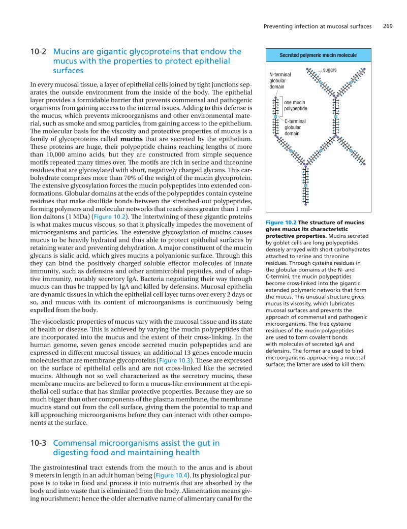
In every mucosal tissue, a layer of epithelial cells joined by tight junctions sep-arates the outside environment from the inside of the body. The epithelial layer provides a formidable barrier that prevents commensal and pathogenic organisms from gaining access to the internal issues. Adding to this defense is the mucus, which prevents microorganisms and other environmental mate-rial, such as smoke and smog particles, from gaining access to the epithelium. The molecular basis for the viscosity and protective properties of mucus is a family of glycoproteins called mucins that are secreted by the epithelium. These proteins are huge, their polypeptide chains reaching lengths of more than 10,000 amino acids, but they are constructed from simple sequence motifs repeated many times over. The motifs are rich in serine and threonine residues that are glycosylated with short, negatively charged glycans. This car-bohydrate comprises more than 70% of the weight of the mucin glycoprotein. The extensive glycosylation forces the mucin polypeptides into extended con-formations. Globular domains at the ends of the polypeptides contain cysteine residues that make disulfide bonds between the stretched-out polypeptides, forming polymers and molecular networks that reach sizes greater than 1 mil-lion daltons (1 MDa) (Figure 10.2). The intertwining of these gigantic proteins is what makes mucus viscous, so that it physically impedes the movement of microorganisms and particles. The extensive glycosylation of mucins causes mucus to be heavily hydrated and thus able to protect epithelial surfaces by retaining water and preventing dehydration. A major constituent of the mucin glycans is sialic acid, which gives mucins a polyanionic surface. Through this they can bind the positively charged soluble effector molecules of innate immunity, such as defensins and other antimicrobial peptides, and of adap-tive immunity, notably secretory IgA. Bacteria negotiating their way through mucus can thus be trapped by IgA and killed by defensins. Mucosal epithelia are dynamic tissues in which the epithelial cell layer turns over every 2 days or so, and mucus with its content of microorganisms is continuously being expelled from the body.
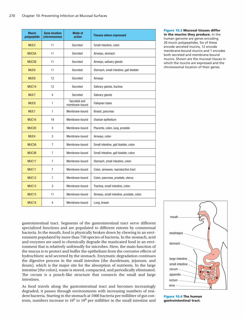
The viscoelastic properties of mucus vary with the mucosal tissue and its state of health or disease. This is achieved by varying the mucin polypeptides that are incorporated into the mucus and the extent of their cross-linking. In the human genome, seven genes encode secreted mucin polypeptides and are expressed in different mucosal tissues; an additional 13 genes encode mucin molecules that are membrane glycoproteins (Figure 10.3). These are expressed on the surface of epithelial cells and are not cross-linked like the secreted mucins. Although not so well characterized as the secretory mucins, these membrane mucins are believed to form a mucus-like environment at the epi-thelial cell surface that has similar protective properties. Because they are so much bigger than other components of the plasma membrane, the membrane mucins stand out from the cell surface, giving them the potential to trap and kill approaching microorganisms before they can interact with other compo-nents at the surface.
10-3 Commensal microorganisms assist the gut in digesting food and maintaining health
The gastrointestinal tract extends from the mouth to the anus and is about 9 meters in length in an adult human being (Figure 10.4). Its physiological pur-pose is to take in food and process it into nutrients that are absorbed by the body and into waste that is eliminated from the body. Alimentation means giv-ing nourishment; hence the older alternative name of alimentary canal for the
Preventing infection at mucosal surfaces
Secreted polymeric mucin molecule
C-terminalglobulardomain
N-terminalglobulardomain
one mucinpolypeptide
sugars
IS4 n10.100/10.02
Figure 10.2 The structure of mucins gives mucus its characteristic protective properties. Mucins secreted by goblet cells are long polypeptides densely arrayed with short carbohydrates attached to serine and threonine residues. Through cysteine residues in the globular domains at the N- and C-termini, the mucin polypeptides become cross-linked into the gigantic extended polymeric networks that form the mucus. This unusual structure gives mucus its viscosity, which lubricates mucosal surfaces and prevents the approach of commensal and pathogenic microorganisms. The free cysteine residues of the mucin polypeptides are used to form covalent bonds with molecules of secreted IgA and defensins. The former are used to bind microorganisms approaching a mucosal surface; the latter are used to kill them.
Chapter 10: Preventing Infection at Mucosal Surfaces270
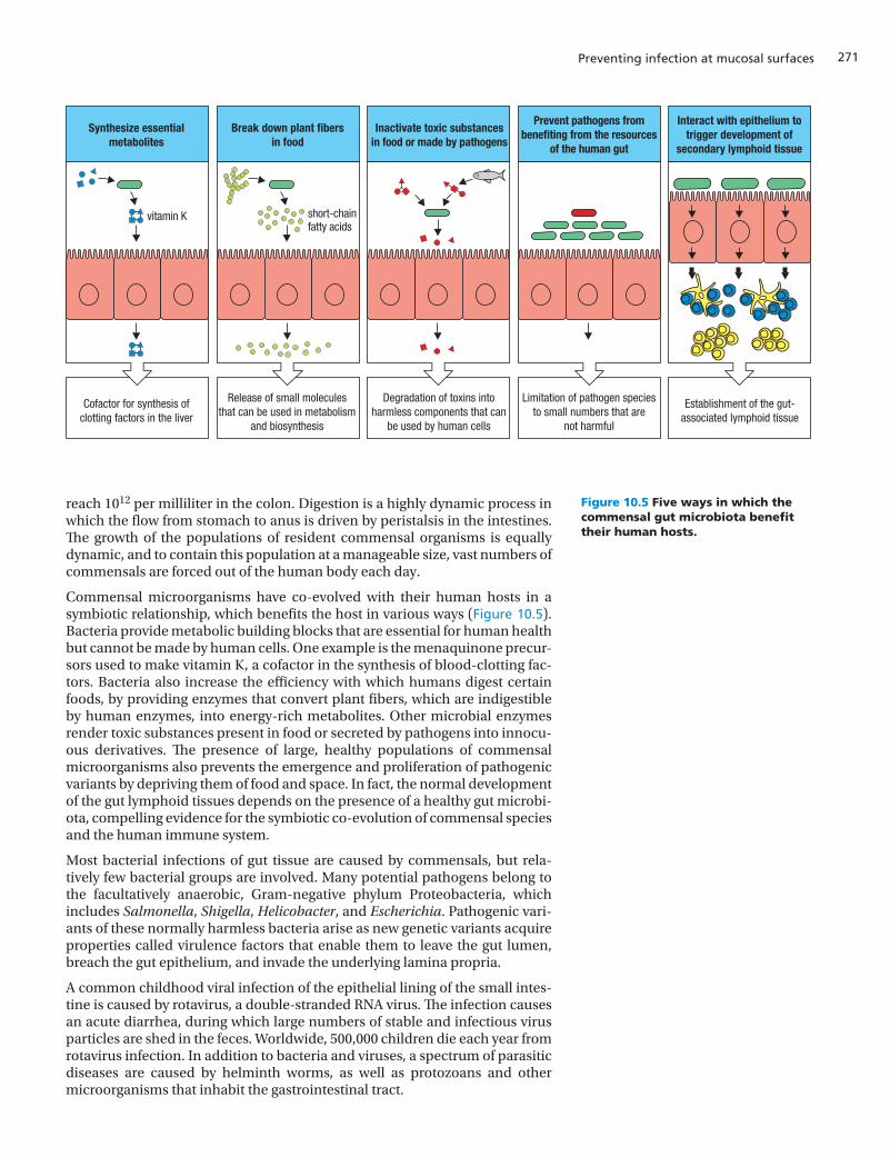
gastrointestinal tract. Segments of the gastrointestinal tract serve different specialized functions and are populated to different extents by commensal bacteria. In the mouth, food is physically broken down by chewing in an envi-ronment populated by more than 750 species of bacteria. In the stomach, acid and enzymes are used to chemically degrade the masticated food in an envi-ronment that is relatively unfriendly for microbes. Here, the main function of the mucus is to protect and buffer the epithelium from the corrosive effects of hydrochloric acid secreted by the stomach. Enzymatic degradation continues the digestive process in the small intestine (the duodenum, jejunum, and ileum), which is the major site for the absorption of nutrients. In the large intestine (the colon), waste is stored, compacted, and periodically eliminated. The cecum is a pouch-like structure that connects the small and large intestines.
As food travels along the gastrointestinal tract and becomes increasingly degraded, it passes through environments with increasing numbers of resi-dent bacteria. Starting in the stomach at 1000 bacteria per milliliter of gut con-tents, numbers increase to 105 to 108 per milliliter in the small intestine and
Mucinpolypeptide
Gene location(chromosome)
Mode ofaction Tissues where expressed
Secreted Small intestine, colonMUC2 11
Secreted Airways, stomachMUC5A 11
Secreted Airways, salivary glandsMUC5B 11
Secreted Stomach, small intestine, gall bladderMUC6 11
Secreted AirwaysMUC8 12
Secreted Salivary glands, tracheaMUC19 12
Secreted Salivary glandsMUC7 4
Secreted andmembrane-bound Fallopian tubesMUC9 1
Membrane-bound Breast, pancreasMUC1 1
Membrane-bound Placenta, colon, lung, prostateMUC20 3
Membrane-bound Airways, colonMUC4 3
Membrane-bound Small intestine, gall bladder, colonMUC3A 7
Membrane-bound Small intestine, gall bladder, colonMUC3B 7
Membrane-bound Stomach, small intestine, colonMUC17 7
Membrane-bound Colon, airwaves, reproductive tractMUC11 7
Membrane-bound Colon, pancreas, prostate, uterusMUC12 7
Membrane-bound Trachea, small intestine, colonMUC13 3
Membrane-bound Airways, small intestine, prostate, colonMUC15 11
Membrane-bound Lung, breastMUC18 4
IS4 n10.101/10.03
Membrane-bound Ovarian epitheliumMUC16 19
mouth
esophagus
stomach
large intestine
small intestine
cecum
appendix
rectum
anus
IS4 i10.02/10.04
Figure 10.3 Mucosal tissues differ in the mucins they produce. In the human genome are genes encoding 20 mucin polypeptides. Six of these encode secreted mucins, 12 encode membrane-bound mucins and 1 encodes both secreted and membrane-bound mucins. Shown are the mucosal tissues in which the mucins are expressed and the chromosomal location of their genes.
Figure 10.4 The human gastrointestinal tract.
271
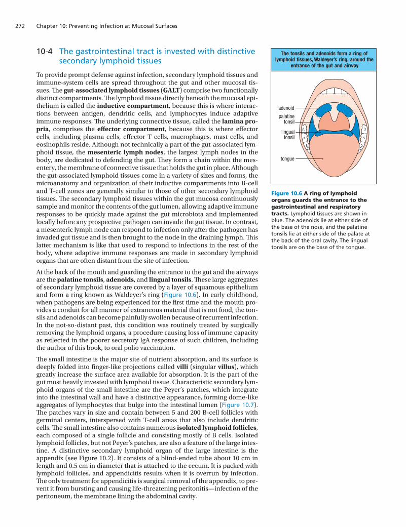
reach 1012 per milliliter in the colon. Digestion is a highly dynamic process in which the flow from stomach to anus is driven by peristalsis in the intestines. The growth of the populations of resident commensal organisms is equally dynamic, and to contain this population at a manageable size, vast numbers of commensals are forced out of the human body each day.
Commensal microorganisms have co-evolved with their human hosts in a symbiotic relationship, which benefits the host in various ways (Figure 10.5). Bacteria provide metabolic building blocks that are essential for human health but cannot be made by human cells. One example is the menaquinone precur-sors used to make vitamin K, a cofactor in the synthesis of blood-clotting fac-tors. Bacteria also increase the efficiency with which humans digest certain foods, by providing enzymes that convert plant fibers, which are indigestible by human enzymes, into energy-rich metabolites. Other microbial enzymes render toxic substances present in food or secreted by pathogens into innocu-ous derivatives. The presence of large, healthy populations of commensal microorganisms also prevents the emergence and proliferation of pathogenic variants by depriving them of food and space. In fact, the normal development of the gut lymphoid tissues depends on the presence of a healthy gut microbi-ota, compelling evidence for the symbiotic co-evolution of commensal species and the human immune system.
Most bacterial infections of gut tissue are caused by commensals, but rela-tively few bacterial groups are involved. Many potential pathogens belong to the facultatively anaerobic, Gram-negative phylum Proteobacteria, which includes Salmonella, Shigella, Helicobacter, and Escherichia. Pathogenic vari-ants of these normally harmless bacteria arise as new genetic variants acquire properties called virulence factors that enable them to leave the gut lumen, breach the gut epithelium, and invade the underlying lamina propria.
A common childhood viral infection of the epithelial lining of the small intes-tine is caused by rotavirus, a double-stranded RNA virus. The infection causes an acute diarrhea, during which large numbers of stable and infectious virus particles are shed in the feces. Worldwide, 500,000 children die each year from rotavirus infection. In addition to bacteria and viruses, a spectrum of parasitic diseases are caused by helminth worms, as well as protozoans and other microorganisms that inhabit the gastrointestinal tract.
Preventing infection at mucosal surfaces
IS4 n10.102/10.05
Synthesize essentialmetabolites
Cofactor for synthesis ofclotting factors in the liver
Release of small moleculesthat can be used in metabolism
and biosynthesis
Degradation of toxins intoharmless components that can
be used by human cells
Limitation of pathogen speciesto small numbers that are
not harmful
Establishment of the gut-associated lymphoid tissue
Break down plant fibersin food
Inactivate toxic substancesin food or made by pathogens
Prevent pathogens frombenefiting from the resources
of the human gut
Interact with epithelium totrigger development of
secondary lymphoid tissue
vitamin K short-chainfatty acids
Figure 10.5 Five ways in which the commensal gut microbiota benefit their human hosts.
Chapter 10: Preventing Infection at Mucosal Surfaces272
10-4 The gastrointestinal tract is invested with distinctive secondary lymphoid tissues
To provide prompt defense against infection, secondary lymphoid tissues and immune-system cells are spread throughout the gut and other mucosal tis-sues. The gut-associated lymphoid tissues (GALT) comprise two functionally distinct compartments. The lymphoid tissue directly beneath the mucosal epi-thelium is called the inductive compartment, because this is where interac-tions between antigen, dendritic cells, and lymphocytes induce adaptive immune responses. The underlying connective tissue, called the lamina pro-pria, comprises the effector compartment, because this is where effector cells, including plasma cells, effector T cells, macrophages, mast cells, and eosinophils reside. Although not technically a part of the gut-associated lym-phoid tissue, the mesenteric lymph nodes, the largest lymph nodes in the body, are dedicated to defending the gut. They form a chain within the mes-entery, the membrane of connective tissue that holds the gut in place. Although the gut-associated lymphoid tissues come in a variety of sizes and forms, the microanatomy and organization of their inductive compartments into B-cell and T-cell zones are generally similar to those of other secondary lymphoid tissues. The secondary lymphoid tissues within the gut mucosa continuously sample and monitor the contents of the gut lumen, allowing adaptive immune responses to be quickly made against the gut microbiota and implemented locally before any prospective pathogen can invade the gut tissue. In contrast, a mesenteric lymph node can respond to infection only after the pathogen has invaded gut tissue and is then brought to the node in the draining lymph. This latter mechanism is like that used to respond to infections in the rest of the body, where adaptive immune responses are made in secondary lymphoid organs that are often distant from the site of infection.
At the back of the mouth and guarding the entrance to the gut and the airways are the palatine tonsils, adenoids, and lingual tonsils. These large aggregates of secondary lymphoid tissue are covered by a layer of squamous epithelium and form a ring known as Waldeyer’s ring (Figure 10.6). In early childhood, when pathogens are being experienced for the first time and the mouth pro-vides a conduit for all manner of extraneous material that is not food, the ton-sils and adenoids can become painfully swollen because of recurrent infection. In the not-so-distant past, this condition was routinely treated by surgically removing the lymphoid organs, a procedure causing loss of immune capacity as reflected in the poorer secretory IgA response of such children, including the author of this book, to oral polio vaccination.
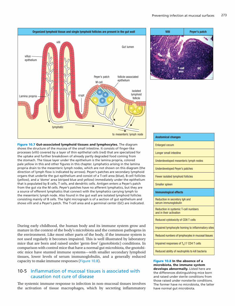
The small intestine is the major site of nutrient absorption, and its surface is deeply folded into finger-like projections called villi (singular villus), which greatly increase the surface area available for absorption. It is the part of the gut most heavily invested with lymphoid tissue. Characteristic secondary lym-phoid organs of the small intestine are the Peyer’s patches, which integrate into the intestinal wall and have a distinctive appearance, forming dome-like aggregates of lymphocytes that bulge into the intestinal lumen (Figure 10.7). The patches vary in size and contain between 5 and 200 B-cell follicles with germinal centers, interspersed with T-cell areas that also include dendritic cells. The small intestine also contains numerous isolated lymphoid follicles, each composed of a single follicle and consisting mostly of B cells. Isolated lymphoid follicles, but not Peyer’s patches, are also a feature of the large intes-tine. A distinctive secondary lymphoid organ of the large intestine is the appendix (see Figure 10.2). It consists of a blind-ended tube about 10 cm in length and 0.5 cm in diameter that is attached to the cecum. It is packed with lymphoid follicles, and appendicitis results when it is overrun by infection. The only treatment for appendicitis is surgical removal of the appendix, to pre-vent it from bursting and causing life-threatening peritonitis—infection of the peritoneum, the membrane lining the abdominal cavity.
The tonsils and adenoids form a ring oflymphoid tissues, Waldeyer’s ring, around the
entrance of the gut and airway
adenoid
palatinetonsil
lingualtonsil
tongue
IS4 i10.03/10.06
Figure 10.6 A ring of lymphoid organs guards the entrance to the gastrointestinal and respiratory tracts. Lymphoid tissues are shown in blue. The adenoids lie at either side of the base of the nose, and the palatine tonsils lie at either side of the palate at the back of the oral cavity. The lingual tonsils are on the base of the tongue.
273
Organized lymphoid tissue and single lymphoid follicles are present in the gut wall
to mesenteric lymph node
isolatedlymphoid
follicle
Gut lumen
villi
Peyer’s patch
M cell
cryptLamina propria
villusepithelium
lymphatic
T-cellarea
GC
dome
IS4 i10.04/10.07
Villi Peyer’s patch
follicle-associatedepithelium
During early childhood, the human body and its immune system grow and mature in the context of the body’s microbiota and the common pathogens in the environment. Like most other parts of the body, if the immune system is not used regularly it becomes impaired. This is well illustrated by laboratory mice that are born and raised under ‘germ-free’ (gnotobiotic) conditions. In comparison with control mice that have a normal gut microbiota, the gnotobi-otic mice have stunted immune systems—with smaller secondary lymphoid tissues, lower levels of serum immunoglobulin, and a generally reduced capacity to make immune responses (Figure 10.8).
10-5 Inflammation of mucosal tissues is associated with causation not cure of disease
The systemic immune response to infection in non-mucosal tissues involves the activation of tissue macrophages, which by secreting inflammatory
Preventing infection at mucosal surfaces
Figure 10.7 Gut-associated lymphoid tissues and lymphocytes. The diagram shows the structure of the mucosa of the small intestine. It consists of finger-like processes (villi) covered by a layer of thin epithelial cells (red) that are specialized for the uptake and further breakdown of already partly degraded food coming from the stomach. The tissue layer under the epithelium is the lamina propria, colored pale yellow in this and other figures in this chapter. Lymphatics arising in the lamina propria drain to the mesenteric lymph nodes, which are not shown on this diagram (the direction of lymph flow is indicated by arrows). Peyer’s patches are secondary lymphoid organs that underlie the gut epithelium and consist of a T-cell area (blue), B-cell follicles (yellow), and a ‘dome’ area (striped blue and yellow) immediately under the epithelium that is populated by B cells, T cells, and dendritic cells. Antigen enters a Peyer’s patch from the gut via the M cells. Peyer’s patches have no afferent lymphatics, but they are a source of efferent lymphatics that connect with the lymphatics carrying lymph to the mesenteric lymph node. Also found in the gut wall are isolated lymphoid follicles consisting mainly of B cells. The light micrograph is of a section of gut epithelium and shows villi and a Peyer’s patch. The T-cell area and a germinal center (GC) are indicated.
Anatomical changes
Enlarged cecum
Longer small intestine
Underdeveloped mesenteric lymph nodes
Underdeveloped Peyer’s patches
Fewer isolated lymphoid follicles
Smaller spleen
Immunological effects
Reduction in secretory IgA and serum immunoglobulin
Reduction in systemic T-cell numbers and in their activation
Reduced cytotoxicity of CD8 T cells
Impaired lymphocyte homing to inflammatory sites
Reduced numbers of lymphocytes in mucosal tissues
Impaired responses of TH17 CD4 T cells
Reduced ability of neutrophils to kill bacteria
IS4 n10.103/10.08
Figure 10.8 In the absence of a microbiota, the immune system develops abnormally. Listed here are the differences distinguishing mice born and raised under sterile conditions from those raised under nonsterile conditions. The former have no microbiota, the latter have normal gut microbiota.
Chapter 10: Preventing Infection at Mucosal Surfaces274
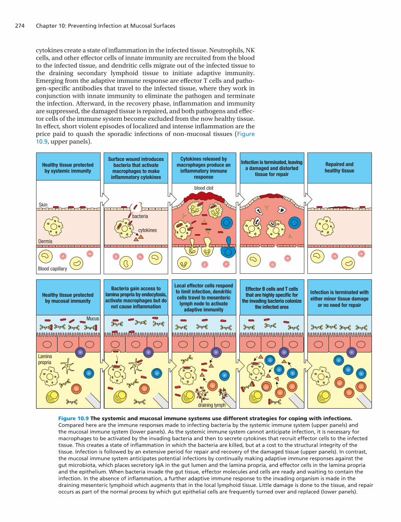
cytokines create a state of inflammation in the infected tissue. Neutrophils, NK cells, and other effector cells of innate immunity are recruited from the blood to the infected tissue, and dendritic cells migrate out of the infected tissue to the draining secondary lymphoid tissue to initiate adaptive immunity. Emerging from the adaptive immune response are effector T cells and patho-gen-specific antibodies that travel to the infected tissue, where they work in conjunction with innate immunity to eliminate the pathogen and terminate the infection. Afterward, in the recovery phase, inflammation and immunity are suppressed, the damaged tissue is repaired, and both pathogens and effec-tor cells of the immune system become excluded from the now healthy tissue. In effect, short violent episodes of localized and intense inflammation are the price paid to quash the sporadic infections of non-mucosal tissues (Figure 10.9, upper panels).
IS4 n10.104/10.09
Healthy tissue protectedby systemic immunity
Surface wound introducesbacteria that activatemacrophages to make
inflammatory cytokines
Cytokines released bymacrophages produce an
inflammatory immuneresponse
Infection is terminated, leavinga damaged and distorted
tissue for repair
Repaired andhealthy tissue
Skin
Dermis
Blood capillary
blood clot
cytokines
bacteria
Healthy tissue protectedby mucosal immunity
Laminapropria
Mucus
draining lymph
Bacteria gain access tolamina propria by endocytosis,activate macrophages but do
not cause inflammation
Local effector cells respondto limit infection, dendriticcells travel to mesenteric
lymph node to activateadaptive immunity
Effector B cells and T cellsthat are highly specific for
the invading bacteria colonizethe infected area
Infection is terminated witheither minor tissue damage
or no need for repair
Figure 10.9 The systemic and mucosal immune systems use different strategies for coping with infections. Compared here are the immune responses made to infecting bacteria by the systemic immune system (upper panels) and the mucosal immune system (lower panels). As the systemic immune system cannot anticipate infection, it is necessary for macrophages to be activated by the invading bacteria and then to secrete cytokines that recruit effector cells to the infected tissue. This creates a state of inflammation in which the bacteria are killed, but at a cost to the structural integrity of the tissue. Infection is followed by an extensive period for repair and recovery of the damaged tissue (upper panels). In contrast, the mucosal immune system anticipates potential infections by continually making adaptive immune responses against the gut microbiota, which places secretory IgA in the gut lumen and the lamina propria, and effector cells in the lamina propria and the epithelium. When bacteria invade the gut tissue, effector molecules and cells are ready and waiting to contain the infection. In the absence of inflammation, a further adaptive immune response to the invading organism is made in the draining mesenteric lymphoid which augments that in the local lymphoid tissue. Little damage is done to the tissue, and repair occurs as part of the normal process by which gut epithelial cells are frequently turned over and replaced (lower panels).
275
In contrast to non-mucosal tissues, which interact only occasionally with the microbial world, the mucosal tissues have close and continuous contact with numerous and diverse commensal microorganisms, all of which are a poten-tial source of pathogens. For the gut, any significant breach of the epithelial layer could lead to a massive influx of bacteria and infection of the type that occurs in peritonitis (see Section 10-4). To avoid this, the mucosal immune system adopts two complementary strategies. First, rather than being reactive like systemic immunity, the mucosal immune response is proactive and is constantly making adaptive immune responses against the microorganisms populating the gut. The result is that healthy gut tissue is populated with effec-tor T cells and B cells that stand guard and are poised to respond to any invader from the gut lumen (Figure 10.9, lower panels). The advantage of a proactive strategy is that infections can be stopped earlier and with greater force than is possible in non-mucosal tissues.
The second strategy of the mucosal immune system is to be sparing in the acti-vation of inflammation, because the molecular and cellular weapons of the inflammatory response inevitably cause damage to the tissues where they work, which for mucosal tissues, and particularly the gut, is more likely to exacerbate the infection than clear it up. Inflammation in the gut is the cause of a variety of chronic human diseases.
Of several strategies used to prevent inflammation in mucosal tissues, one is the use of regulatory T cells (CD4 Treg) to turn off inflammatory T cells. IL-10 is a cytokine secreted by Treg that suppresses inflammation by turning off the synthesis of inflammatory cytokines. Rare immunodeficient patients who lack a functional receptor for IL-10 suffer from a chronic inflammatory disease of the gut mucosa that resembles the more prevalent Crohn’s disease and is mediated by inflammatory TH1 and TH17 subsets of CD4 T cells. Another inflammatory condition, celiac disease, is caused by an immune response in the gut lymphoid tissue that damages the intestinal epithelium and reduces the capacity of those affected to absorb nutrients from their food. This condi-tion can arrest the growth and development of children, and in adults causes unpleasant symptoms including diarrhea and stomach pains and general ill health. Celiac disease is caused by an adaptive immune response to the pro-teins of gluten, a major component of grains such as wheat, barley, and rye, which are dietary staples for some human populations. Proving this cause-and-effect relationship, the symptoms of celiac disease disappear when patients adopt a strict gluten-free diet, but quickly come back if they consume gluten again. In healthy gut tissue a compromise is made between the compet-ing demands of nutrition and defense. In celiac patients the truce is broken when a staple food is mistakenly perceived as a dangerous pathogen, which ‘infects’ the gut with every square meal.
The qualitatively different responses of the mucosal and systemic immune sys-tems to microorganisms correlates with their developmental origin. During fetal development, the mesenteric lymph nodes and Peyer’s patches differen-tiate independently of the spleen and the lymph nodes that supply systemic immunity. The distinctive development of the secondary lymphoid tissues of mucosal and systemic immunity occurs under the guidance of different sets of chemokines and receptors for cytokines in the tumor necrosis factor (TNF) family. The differences between the gut-associated lymphoid tissues and the systemic lymphoid organs are thus imprinted early on in life.
10-6 Intestinal epithelial cells contribute to innate immune responses in the gut
Intestinal epithelial cells are very active in the uptake of nutrients and other materials from the gut lumen. They also have Toll-like receptors on their apical and basolateral surfaces, for example TLR5, which recognizes flagellin, the
Preventing infection at mucosal surfaces
Celiac disease
Chapter 10: Preventing Infection at Mucosal Surfaces276
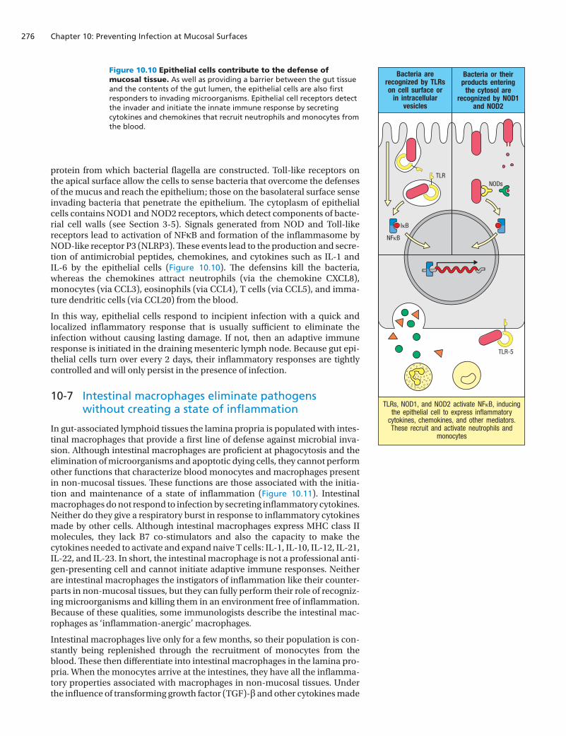
protein from which bacterial flagella are constructed. Toll-like receptors on the apical surface allow the cells to sense bacteria that overcome the defenses of the mucus and reach the epithelium; those on the basolateral surface sense invading bacteria that penetrate the epithelium. The cytoplasm of epithelial cells contains NOD1 and NOD2 receptors, which detect components of bacte-rial cell walls (see Section 3-5). Signals generated from NOD and Toll-like receptors lead to activation of NFκB and formation of the inflammasome by NOD-like receptor P3 (NLRP3). These events lead to the production and secre-tion of antimicrobial peptides, chemokines, and cytokines such as IL-1 and IL-6 by the epithelial cells (Figure 10.10). The defensins kill the bacteria, whereas the chemokines attract neutrophils (via the chemokine CXCL8), monocytes (via CCL3), eosinophils (via CCL4), T cells (via CCL5), and imma-ture dendritic cells (via CCL20) from the blood.
In this way, epithelial cells respond to incipient infection with a quick and localized inflammatory response that is usually sufficient to eliminate the infection without causing lasting damage. If not, then an adaptive immune response is initiated in the draining mesenteric lymph node. Because gut epi-thelial cells turn over every 2 days, their inflammatory responses are tightly controlled and will only persist in the presence of infection.
10-7 Intestinal macrophages eliminate pathogens without creating a state of inflammation
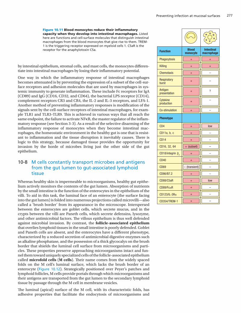
In gut-associated lymphoid tissues the lamina propria is populated with intes-tinal macrophages that provide a first line of defense against microbial inva-sion. Although intestinal macrophages are proficient at phagocytosis and the elimination of microorganisms and apoptotic dying cells, they cannot perform other functions that characterize blood monocytes and macrophages present in non-mucosal tissues. These functions are those associated with the initia-tion and maintenance of a state of inflammation (Figure 10.11). Intestinal macrophages do not respond to infection by secreting inflammatory cytokines. Neither do they give a respiratory burst in response to inflammatory cytokines made by other cells. Although intestinal macrophages express MHC class II molecules, they lack B7 co-stimulators and also the capacity to make the cytokines needed to activate and expand naive T cells: IL-1, IL-10, IL-12, IL-21, IL-22, and IL-23. In short, the intestinal macrophage is not a professional anti-gen-presenting cell and cannot initiate adaptive immune responses. Neither are intestinal macrophages the instigators of inflammation like their counter-parts in non-mucosal tissues, but they can fully perform their role of recogniz-ing microorganisms and killing them in an environment free of inflammation. Because of these qualities, some immunologists describe the intestinal mac-rophages as ‘inflammation-anergic’ macrophages.
Intestinal macrophages live only for a few months, so their population is con-stantly being replenished through the recruitment of monocytes from the blood. These then differentiate into intestinal macrophages in the lamina pro-pria. When the monocytes arrive at the intestines, they have all the inflamma-tory properties associated with macrophages in non-mucosal tissues. Under the influence of transforming growth factor (TGF)-β and other cytokines made
IS4 n10.105/10.10
NFκB
TLRNODs
IκB
TLR-5
TLRs, NOD1, and NOD2 activate NFκB, inducing the epithelial cell to express inflammatory
cytokines, chemokines, and other mediators. These recruit and activate neutrophils and
monocytes
Bacteria are recognized by TLRs on cell surface or
in intracellular vesicles
Bacteria or their products entering
the cytosol are recognized by NOD1
and NOD2
Figure 10.10 Epithelial cells contribute to the defense of mucosal tissue. As well as providing a barrier between the gut tissue and the contents of the gut lumen, the epithelial cells are also first responders to invading microorganisms. Epithelial cell receptors detect the invader and initiate the innate immune response by secreting cytokines and chemokines that recruit neutrophils and monocytes from the blood.
277
by intestinal epithelium, stromal cells, and mast cells, the monocytes differen-tiate into intestinal macrophages by losing their inflammatory potential.
One way in which the inflammatory response of intestinal macrophages becomes attenuated is by preventing the expression of a subset of the cell-sur-face receptors and adhesion molecules that are used by macrophages in sys-temic immunity to generate inflammation. These include Fc receptors for IgA (CD89) and IgG (CD16, CD32, and CD64), the bacterial LPS receptor (CD14), complement receptors CR3 and CR4, the IL-2 and IL-3 receptors, and LFA-1. Another method of preventing inflammatory responses is modification of the signals sent by the cell-surface receptors of intestinal macrophages, for exam-ple TLR1 and TLR3–TLR9. This is achieved in various ways that all reach the same endpoint, the failure to activate NFκB, the master regulator of the inflam-matory response (see Section 3-3). As a result of the selective disarming of the inflammatory response of monocytes when they become intestinal mac-rophages, the homeostatic environment in the healthy gut is one that is resist-ant to inflammation and the tissue disruption it inevitably causes. There is logic to this strategy, because damaged tissue provides the opportunity for invasion by the horde of microbes living just the other side of the gut epithelium.
10-8 M cells constantly transport microbes and antigens from the gut lumen to gut-associated lymphoid tissue
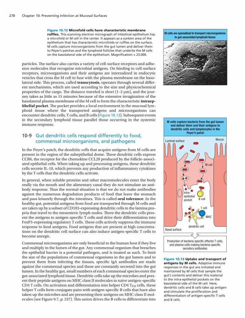
Whereas healthy skin is impermeable to microorganisms, healthy gut epithe-lium actively monitors the contents of the gut lumen. Absorption of nutrients by the small intestine is the function of the enterocytes in the epithelium of the villi. To aid in this task, the luminal face of an enterocyte (the surface facing into the gut lumen) is folded into numerous projections called microvilli—also called a ‘brush border’ from its appearance in the microscope. Interspersed between the enterocytes are goblet cells, which secrete mucus, and in the crypts between the villi are Paneth cells, which secrete defensins, lysozyme, and other antimicrobial factors. The villous epithelium is thus well defended against microbial invasion. By contrast, the follicle-associated epithelium that overlies lymphoid tissues in the small intestine is poorly defended. Goblet and Paneth cells are absent, and the enterocytes have a different phenotype, characterized by a reduced secretion of antimicrobial digestive enzymes such as alkaline phosphatase, and the possession of a thick glycocalyx on the brush border that shields the luminal cell surface from microorganisms and parti-cles. These properties preserve approaching microorganisms intact and fun-nel them toward uniquely specialized cells of the follicle-associated epithelium called microfold cells (M cells). Their name comes from the widely spaced folds on the M cell’s luminal surface, which lacks the brush border of an enterocyte (Figure 10.12). Strategically positioned over Peyer’s patches and lymphoid follicles, M cells provide portals through which microorganisms and their antigens are transported from the gut lumen to the secondary lymphoid tissue by passage through the M cell in membrane vesicles.
The luminal (apical) surface of the M cell, with its characteristic folds, has adhesive properties that facilitate the endocytosis of microorganisms and
Preventing infection at mucosal surfaces
IS4 n10.106/10.11
CD4
CD11a, b, c
CD14
CD16, 32, 64
CD18/integrin β2
CD40
CD69
CD86/B7.2
CD88/C5aR
CD89/FcαR
CD123/IL-3Rα
CD354/TREM-1
low
–
–
–
–
–
–
–
low
–
–
–
+
+
+
+ (subset)
+
+
– (transient)
+
+
+
+
+
Phagocytosis
Killing
Chemotaxis
Respiratoryburst
Antigenpresentation
Cytokineproduction
Co-stimulation
+
+
–
–
?
–
–
+
+
+
+
+
+
+
Phenotype
Function Bloodmonocyte
Intestinalmacrophage
Figure 10.11 Blood monocytes reduce their inflammatory capacity when they develop into intestinal macrophages. Listed here are functions and cell-surface molecules that distinguish intestinal macrophages from the blood monocytes that give rise to them. TREM-1 is the triggering receptor expressed on myeloid cells 1. C5aR is the receptor for the anaphylotoxin C5a.
Chapter 10: Preventing Infection at Mucosal Surfaces278
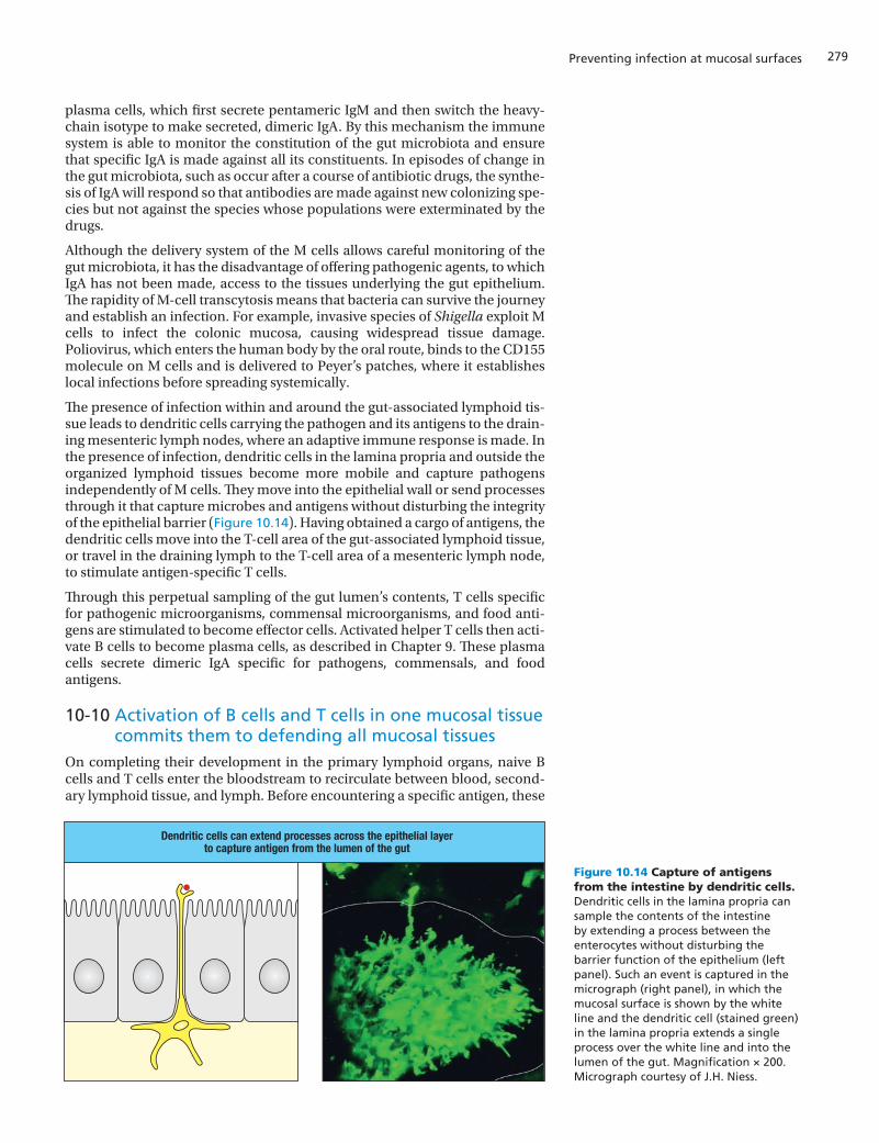
particles. The surface also carries a variety of cell-surface receptors and adhe-sion molecules that recognize microbial antigens. On binding to cell-surface receptors, microorganisms and their antigens are internalized in endocytic vesicles that cross the M cell to fuse with the plasma membrane on the baso-lateral side. This process, called transcytosis, operates through several differ-ent mechanisms, which are used according to the size and physicochemical properties of the cargo. The distance traveled is short (1–2 μm), and the jour-ney takes as little as 15 minutes because of the extensive invagination of the basolateral plasma membrane of the M cell to form the characteristic intraep-ithelial pocket. The pocket provides a local environment in the mucosal lym-phoid tissue where the transported antigens and microorganisms can encounter dendritic cells, T cells, and B cells (Figure 10.13). Subsequent events in the secondary lymphoid tissue parallel those occurring in the systemic immune response.
10-9 Gut dendritic cells respond differently to food, commensal microorganisms, and pathogens
In the Peyer’s patch, the dendritic cells that acquire antigens from M cells are present in the region of the subepithelial dome. These dendritic cells express CCR6, the receptor for the chemokine CCL20 produced by the follicle-associ-ated epithelial cells. When taking up and processing antigens, these dendritic cells secrete IL-10, which prevents any production of inflammatory cytokines by the T cells that the dendritic cells activate.
In general, when soluble proteins and other macromolecules enter the body orally via the mouth and the alimentary canal they do not stimulate an anti-body response. Thus the normal situation is that we do not make antibodies against the numerous degradation products of food that leave the stomach and pass leisurely through the intestines. This is called oral tolerance. In the healthy gut, potential antigens from food are transported through M cells and are taken up by a subset of CD103-expressing dendritic cells in the lamina pro-pria that travel to the mesenteric lymph nodes. There the dendritic cells pres-ent the antigens to antigen-specific T cells and drive their differentiation into FoxP3-expressing regulatory T cells. These cells actively suppress the immune response to food antigens. Food antigens that are present at high concentra-tions on the dendritic cell surface can also induce antigen-specific T cells to become anergic.
Commensal microorganisms are only beneficial to the human host if they live and multiply in the lumen of the gut. Any commensal organism that breaches the epithelial barrier is a potential pathogen, and is treated as such. To limit the size of the populations of commensal organisms in the gut lumen and to prevent them from infecting the tissues, specific IgA antibodies are made against the commensal species and these are constantly secreted into the gut lumen. In the healthy gut, small numbers of each commensal species enter the gut-associated lymphoid tissue. Dendritic cells take up the microbes and pres-ent their peptide antigens on MHC class II molecules to naive antigen-specific CD4 T cells. On activation and differentiation into helper CD4 TFH cells, these helper T cells form conjugate pairs with antigen-specific B cells that have also taken up the microbes and are presenting their antigens on MHC class II mol-ecules (see Figure 9.7, p. 237). This union drives the B cells to differentiate into
M cells are specialized to transport microorganismsto gut-associated lymphoid tissue
M cell
IS4 i10.05/10.12
IS4 n10.107/10.13
Basal surfacedendritic cell
Luminal surface
T cell
B cell
MHC
Mucus
M cell
intra-epithelialpocket
M cells capture bacteria from the gut lumenand deliver them and their antigens todendritic cells and lymphocytes in the
Peyer’s patch
Production of bacteria-specific effector T cells,and plasma cells making bacteria-specific
secretory antibodies
Figure 10.12 Microfold cells have characteristic membrane ruffles. This scanning electron micrograph of intestinal epithelium has a microfold or M cell in the center. It appears as a sunken area of the epithelium that has characteristic microfolds or ruffles on the surface. M cells capture microorganisms from the gut lumen and deliver them to Peyer’s patches and the lymphoid follicles that underlie the M cells on the basolateral side of the epithelium. Magnification × 23,000.
Figure 10.13 Uptake and transport of antigens by M cells. Adaptive immune responses in the gut are initiated and maintained by M cells that sample the gut’s contents and deliver this material to the intra-epithelial pockets on the basolateral side of the M cell. Here, dendritic cells and B cells take up antigen and stimulate the proliferation and differentiation of antigen-specific T cells and B cells.
279
plasma cells, which first secrete pentameric IgM and then switch the heavy-chain isotype to make secreted, dimeric IgA. By this mechanism the immune system is able to monitor the constitution of the gut microbiota and ensure that specific IgA is made against all its constituents. In episodes of change in the gut microbiota, such as occur after a course of antibiotic drugs, the synthe-sis of IgA will respond so that antibodies are made against new colonizing spe-cies but not against the species whose populations were exterminated by the drugs.
Although the delivery system of the M cells allows careful monitoring of the gut microbiota, it has the disadvantage of offering pathogenic agents, to which IgA has not been made, access to the tissues underlying the gut epithelium. The rapidity of M-cell transcytosis means that bacteria can survive the journey and establish an infection. For example, invasive species of Shigella exploit M cells to infect the colonic mucosa, causing widespread tissue damage. Poliovirus, which enters the human body by the oral route, binds to the CD155 molecule on M cells and is delivered to Peyer’s patches, where it establishes local infections before spreading systemically.
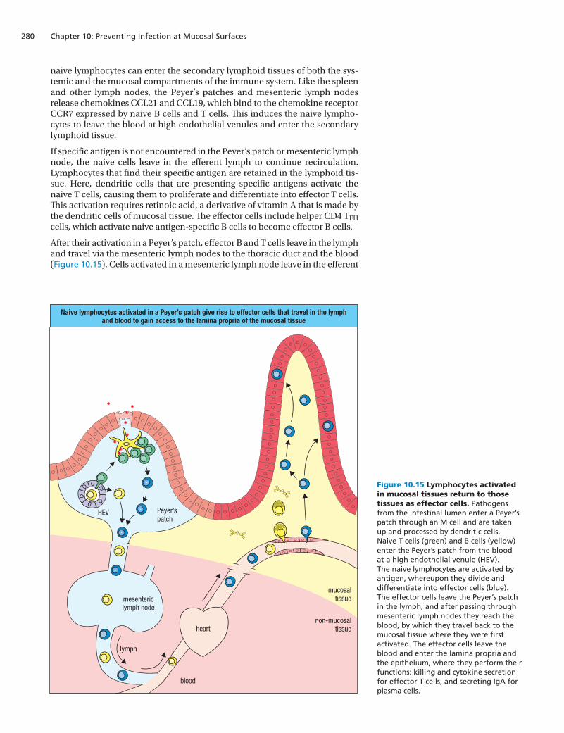
The presence of infection within and around the gut-associated lymphoid tis-sue leads to dendritic cells carrying the pathogen and its antigens to the drain-ing mesenteric lymph nodes, where an adaptive immune response is made. In the presence of infection, dendritic cells in the lamina propria and outside the organized lymphoid tissues become more mobile and capture pathogens independently of M cells. They move into the epithelial wall or send processes through it that capture microbes and antigens without disturbing the integrity of the epithelial barrier (Figure 10.14). Having obtained a cargo of antigens, the dendritic cells move into the T-cell area of the gut-associated lymphoid tissue, or travel in the draining lymph to the T-cell area of a mesenteric lymph node, to stimulate antigen-specific T cells.
Through this perpetual sampling of the gut lumen’s contents, T cells specific for pathogenic microorganisms, commensal microorganisms, and food anti-gens are stimulated to become effector cells. Activated helper T cells then acti-vate B cells to become plasma cells, as described in Chapter 9. These plasma cells secrete dimeric IgA specific for pathogens, commensals, and food antigens.
10-10 Activation of B cells and T cells in one mucosal tissue commits them to defending all mucosal tissues
On completing their development in the primary lymphoid organs, naive B cells and T cells enter the bloodstream to recirculate between blood, second-ary lymphoid tissue, and lymph. Before encountering a specific antigen, these
Preventing infection at mucosal surfaces
IS4 i10.07/10.14
Dendritic cells can extend processes across the epithelial layerto capture antigen from the lumen of the gut
Figure 10.14 Capture of antigens from the intestine by dendritic cells. Dendritic cells in the lamina propria can sample the contents of the intestine by extending a process between the enterocytes without disturbing the barrier function of the epithelium (left panel). Such an event is captured in the micrograph (right panel), in which the mucosal surface is shown by the white line and the dendritic cell (stained green) in the lamina propria extends a single process over the white line and into the lumen of the gut. Magnification × 200. Micrograph courtesy of J.H. Niess.
Chapter 10: Preventing Infection at Mucosal Surfaces280
naive lymphocytes can enter the secondary lymphoid tissues of both the sys-temic and the mucosal compartments of the immune system. Like the spleen and other lymph nodes, the Peyer’s patches and mesenteric lymph nodes release chemokines CCL21 and CCL19, which bind to the chemokine receptor CCR7 expressed by naive B cells and T cells. This induces the naive lympho-cytes to leave the blood at high endothelial venules and enter the secondary lymphoid tissue.
If specific antigen is not encountered in the Peyer’s patch or mesenteric lymph node, the naive cells leave in the efferent lymph to continue recirculation. Lymphocytes that find their specific antigen are retained in the lymphoid tis-sue. Here, dendritic cells that are presenting specific antigens activate the naive T cells, causing them to proliferate and differentiate into effector T cells. This activation requires retinoic acid, a derivative of vitamin A that is made by the dendritic cells of mucosal tissue. The effector cells include helper CD4 TFH cells, which activate naive antigen-specific B cells to become effector B cells.
After their activation in a Peyer’s patch, effector B and T cells leave in the lymph and travel via the mesenteric lymph nodes to the thoracic duct and the blood (Figure 10.15). Cells activated in a mesenteric lymph node leave in the efferent
HEV Peyer’spatch
mesentericlymph node
lymph
blood
mucosaltissue
non-mucosaltissueheart
Naive lymphocytes activated in a Peyer’s patch give rise to effector cells that travel in the lymphand blood to gain access to the lamina propria of the mucosal tissue
IS4 i10.10/10.15
Figure 10.15 Lymphocytes activated in mucosal tissues return to those tissues as effector cells. Pathogens from the intestinal lumen enter a Peyer’s patch through an M cell and are taken up and processed by dendritic cells. Naive T cells (green) and B cells (yellow) enter the Peyer’s patch from the blood at a high endothelial venule (HEV). The naive lymphocytes are activated by antigen, whereupon they divide and differentiate into effector cells (blue). The effector cells leave the Peyer’s patch in the lymph, and after passing through mesenteric lymph nodes they reach the blood, by which they travel back to the mucosal tissue where they were first activated. The effector cells leave the blood and enter the lamina propria and the epithelium, where they perform their functions: killing and cytokine secretion for effector T cells, and secreting IgA for plasma cells.
281
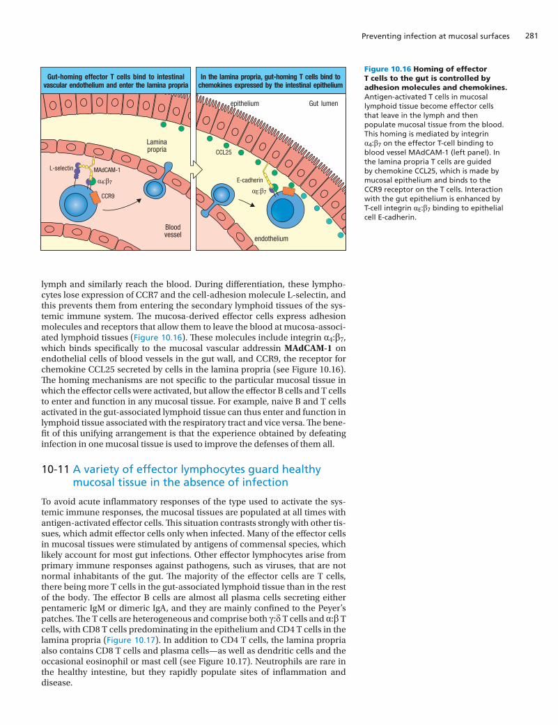
lymph and similarly reach the blood. During differentiation, these lympho-cytes lose expression of CCR7 and the cell-adhesion molecule L-selectin, and this prevents them from entering the secondary lymphoid tissues of the sys-temic immune system. The mucosa-derived effector cells express adhesion molecules and receptors that allow them to leave the blood at mucosa-associ-ated lymphoid tissues (Figure 10.16). These molecules include integrin α4:β7, which binds specifically to the mucosal vascular addressin MAdCAM-1 on endothelial cells of blood vessels in the gut wall, and CCR9, the receptor for chemokine CCL25 secreted by cells in the lamina propria (see Figure 10.16). The homing mechanisms are not specific to the particular mucosal tissue in which the effector cells were activated, but allow the effector B cells and T cells to enter and function in any mucosal tissue. For example, naive B and T cells activated in the gut-associated lymphoid tissue can thus enter and function in lymphoid tissue associated with the respiratory tract and vice versa. The bene-fit of this unifying arrangement is that the experience obtained by defeating infection in one mucosal tissue is used to improve the defenses of them all.
10-11 A variety of effector lymphocytes guard healthy mucosal tissue in the absence of infection
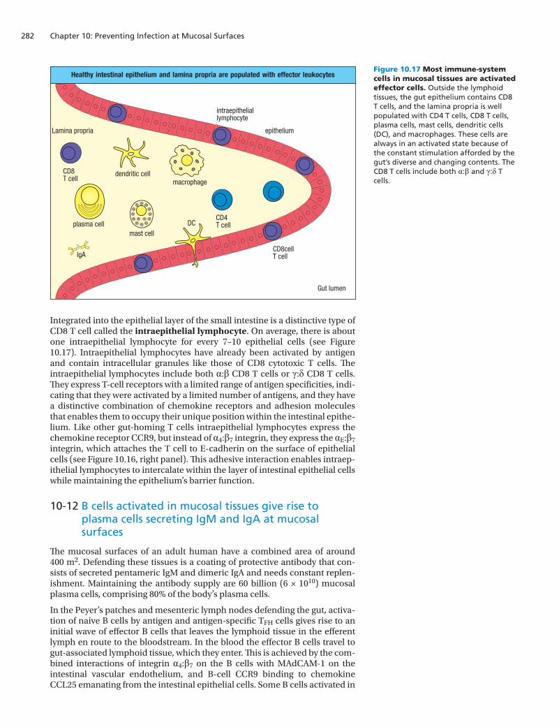
To avoid acute inflammatory responses of the type used to activate the sys-temic immune responses, the mucosal tissues are populated at all times with antigen-activated effector cells. This situation contrasts strongly with other tis-sues, which admit effector cells only when infected. Many of the effector cells in mucosal tissues were stimulated by antigens of commensal species, which likely account for most gut infections. Other effector lymphocytes arise from primary immune responses against pathogens, such as viruses, that are not normal inhabitants of the gut. The majority of the effector cells are T cells, there being more T cells in the gut-associated lymphoid tissue than in the rest of the body. The effector B cells are almost all plasma cells secreting either pentameric IgM or dimeric IgA, and they are mainly confined to the Peyer’s patches. The T cells are heterogeneous and comprise both γ:δ T cells and α:β T cells, with CD8 T cells predominating in the epithelium and CD4 T cells in the lamina propria (Figure 10.17). In addition to CD4 T cells, the lamina propria also contains CD8 T cells and plasma cells—as well as dendritic cells and the occasional eosinophil or mast cell (see Figure 10.17). Neutrophils are rare in the healthy intestine, but they rapidly populate sites of inflammation and disease.
Preventing infection at mucosal surfaces
endothelium
Bloodvessel
Laminapropria
epithelium Gut lumen
CCL25
E-cadherin
CCR9
MAdCAM-1
α4:β7
αE:β7
L-selectin
Gut-homing effector T cells bind to intestinalvascular endothelium and enter the lamina propria
In the lamina propria, gut-homing T cells bind tochemokines expressed by the intestinal epithelium
IS4 i10.11/10.16
Figure 10.16 Homing of effector T cells to the gut is controlled by adhesion molecules and chemokines. Antigen-activated T cells in mucosal lymphoid tissue become effector cells that leave in the lymph and then populate mucosal tissue from the blood. This homing is mediated by integrin α4:β7 on the effector T-cell binding to blood vessel MAdCAM-1 (left panel). In the lamina propria T cells are guided by chemokine CCL25, which is made by mucosal epithelium and binds to the CCR9 receptor on the T cells. Interaction with the gut epithelium is enhanced by T-cell integrin αE:β7 binding to epithelial cell E-cadherin.
Chapter 10: Preventing Infection at Mucosal Surfaces282
Integrated into the epithelial layer of the small intestine is a distinctive type of CD8 T cell called the intraepithelial lymphocyte. On average, there is about one intraepithelial lymphocyte for every 7–10 epithelial cells (see Figure 10.17). Intraepithelial lymphocytes have already been activated by antigen and contain intracellular granules like those of CD8 cytotoxic T cells. The intraepithelial lymphocytes include both α:β CD8 T cells or γ:δ CD8 T cells. They express T-cell receptors with a limited range of antigen specificities, indi-cating that they were activated by a limited number of antigens, and they have a distinctive combination of chemokine receptors and adhesion molecules that enables them to occupy their unique position within the intestinal epithe-lium. Like other gut-homing T cells intraepithelial lymphocytes express the chemokine receptor CCR9, but instead of α4:β7 integrin, they express the αE:β7 integrin, which attaches the T cell to E-cadherin on the surface of epithelial cells (see Figure 10.16, right panel). This adhesive interaction enables intraep-ithelial lymphocytes to intercalate within the layer of intestinal epithelial cells while maintaining the epithelium’s barrier function.
10-12 B cells activated in mucosal tissues give rise to plasma cells secreting IgM and IgA at mucosal surfaces
The mucosal surfaces of an adult human have a combined area of around 400 m2. Defending these tissues is a coating of protective antibody that con-sists of secreted pentameric IgM and dimeric IgA and needs constant replen-ishment. Maintaining the antibody supply are 60 billion (6 × 10¹⁰) mucosal plasma cells, comprising 80% of the body’s plasma cells.
In the Peyer’s patches and mesenteric lymph nodes defending the gut, activa-tion of naive B cells by antigen and antigen-specific TFH cells gives rise to an initial wave of effector B cells that leaves the lymphoid tissue in the efferent lymph en route to the bloodstream. In the blood the effector B cells travel to gut-associated lymphoid tissue, which they enter. This is achieved by the com-bined interactions of integrin α4:β7 on the B cells with MAdCAM-1 on the intestinal vascular endothelium, and B-cell CCR9 binding to chemokine CCL25 emanating from the intestinal epithelial cells. Some B cells activated in
macrophagedendritic cell
mast cellplasma cell
CD8cellT cell
CD8T cell
CD4T cellDC
IgA
Healthy intestinal epithelium and lamina propria are populated with effector leukocytes
Lamina propria epithelium
IS4 i10.08/10.17
Gut lumen
intraepitheliallymphocyte
Figure 10.17 Most immune-system cells in mucosal tissues are activated effector cells. Outside the lymphoid tissues, the gut epithelium contains CD8 T cells, and the lamina propria is well populated with CD4 T cells, CD8 T cells, plasma cells, mast cells, dendritic cells (DC), and macrophages. These cells are always in an activated state because of the constant stimulation afforded by the gut’s diverse and changing contents. The CD8 T cells include both α:β and γ:δ T cells.
283
gut-associated lymphoid tissue return to their tissue of origin, but most take up residence in other areas of the gut and in different mucosal tissues. This strategy enables all the mucosal tissues to benefit from the antibody produced in one of them. Effector B cells settle in the lamina propria, where they com-plete their differentiation into plasma cells that make pentameric IgM and secrete it into the subepithelial space. Here the J chain of the IgM molecule binds to the poly-Ig receptor expressed by immature epithelial cells, also called stem cells, located at the base of intestinal crypts (see Figure 2.18, p. 42). By transcytosis, the poly-Ig receptor carries the antibody from the basal side to the luminal side of the cell, where the IgM is released and bound by the mucus. This transport mechanism for secretory IgM is the same as that used for secre-tory IgA (see Figure 9.18, p. 248).
Only some of the antigen-activated B cells differentiate into plasma cells secreting IgM. The others remain in the B-cell area of the gut-associated lym-phoid tissue, where they undergo affinity maturation and isotype switching. The switch is usually to the IgA isotype, the dominant class of immunoglobulin in mucosal secretions. Switching to IgA is orchestrated by TGF-β and uses the same genetic mechanisms as those described in Chapter 4 for isotype switch-ing and somatic hypermutation in the spleen and lymph nodes (see Sections 4-14 and 4-15). Several other soluble factors enhance switching to the IgA iso-type. These include inducible nitric oxide synthase (iNOS), which is produced by dendritic cells and induces increased expression of the B cells’ TGF-β receptor, the vitamin A derivative retinoic acid, IL-4, IL-10, B-cell-activating factor (BAFF), and a proliferation-inducing ligand (APRIL). Both APRIL and BAFF are made by dendritic cells in gut lymphoid tissue and, in combination with IL-4, strongly bias isotype switching toward IgA.
Under the influence of these factors, effector B cells are programmed to make dimeric IgA that has higher affinities for antigen than the IgM antibodies made by the first wave of plasma cells. The isotype-switched cells constitute a second wave of effector B cells that, like the first, travel to the lamina propria of mucosal tissues throughout the body and differentiate into plasma cells. Plasma cells of the second wave make dimeric IgA that is secreted and transported across the mucosal epithelium by the poly-Ig receptor. Comparison of the sequences of IgA secreted by plasma cells of systemic and mucosal immunity shows a more extensive somatic hypermutation in the variable regions of mucosal IgA than in systemic IgA.
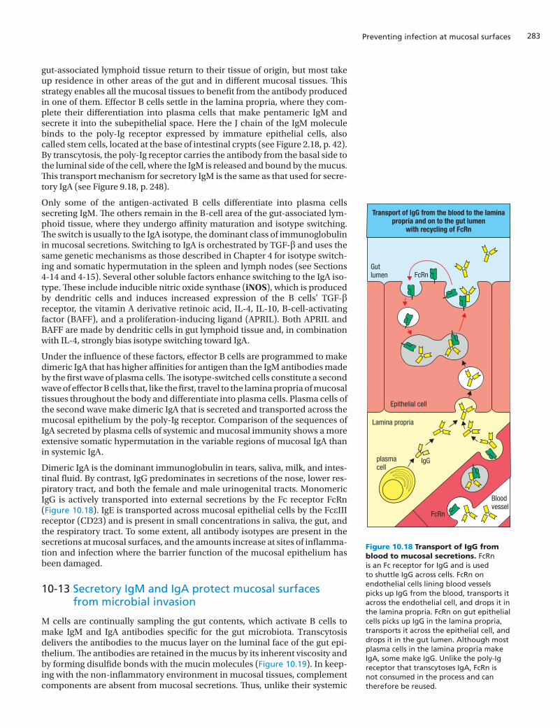
Dimeric IgA is the dominant immunoglobulin in tears, saliva, milk, and intes-tinal fluid. By contrast, IgG predominates in secretions of the nose, lower res-piratory tract, and both the female and male urinogenital tracts. Monomeric IgG is actively transported into external secretions by the Fc receptor FcRn (Figure 10.18). IgE is transported across mucosal epithelial cells by the FcεIII receptor (CD23) and is present in small concentrations in saliva, the gut, and the respiratory tract. To some extent, all antibody isotypes are present in the secretions at mucosal surfaces, and the amounts increase at sites of inflamma-tion and infection where the barrier function of the mucosal epithelium has been damaged.
10-13 Secretory IgM and IgA protect mucosal surfaces from microbial invasion
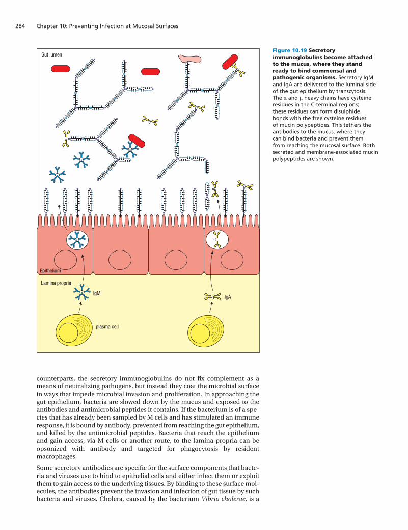
M cells are continually sampling the gut contents, which activate B cells to make IgM and IgA antibodies specific for the gut microbiota. Transcytosis delivers the antibodies to the mucus layer on the luminal face of the gut epi-thelium. The antibodies are retained in the mucus by its inherent viscosity and by forming disulfide bonds with the mucin molecules (Figure 10.19). In keep-ing with the non-inflammatory environment in mucosal tissues, complement components are absent from mucosal secretions. Thus, unlike their systemic
Preventing infection at mucosal surfaces
IS4 n10.108/10.18
FcRn
IgG
Bloodvessel
Epithelial cell
Lamina propria
plasmacell
Gutlumen
Transport of IgG from the blood to the laminapropria and on to the gut lumen
with recycling of FcRn
FcRn
Figure 10.18 Transport of IgG from blood to mucosal secretions. FcRn is an Fc receptor for IgG and is used to shuttle IgG across cells. FcRn on endothelial cells lining blood vessels picks up IgG from the blood, transports it across the endothelial cell, and drops it in the lamina propria. FcRn on gut epithelial cells picks up IgG in the lamina propria, transports it across the epithelial cell, and drops it in the gut lumen. Although most plasma cells in the lamina propria make IgA, some make IgG. Unlike the poly-Ig receptor that transcytoses IgA, FcRn is not consumed in the process and can therefore be reused.
Chapter 10: Preventing Infection at Mucosal Surfaces284
IS4 n10.109/10.19
Lamina propria
Epithelium
Gut lumen
plasma cell
IgAIgM
counterparts, the secretory immunoglobulins do not fix complement as a means of neutralizing pathogens, but instead they coat the microbial surface in ways that impede microbial invasion and proliferation. In approaching the gut epithelium, bacteria are slowed down by the mucus and exposed to the antibodies and antimicrobial peptides it contains. If the bacterium is of a spe-cies that has already been sampled by M cells and has stimulated an immune response, it is bound by antibody, prevented from reaching the gut epithelium, and killed by the antimicrobial peptides. Bacteria that reach the epithelium and gain access, via M cells or another route, to the lamina propria can be opsonized with antibody and targeted for phagocytosis by resident macrophages.
Some secretory antibodies are specific for the surface components that bacte-ria and viruses use to bind to epithelial cells and either infect them or exploit them to gain access to the underlying tissues. By binding to these surface mol-ecules, the antibodies prevent the invasion and infection of gut tissue by such bacteria and viruses. Cholera, caused by the bacterium Vibrio cholerae, is a
Figure 10.19 Secretory immunoglobulins become attached to the mucus, where they stand ready to bind commensal and pathogenic organisms. Secretory IgM and IgA are delivered to the luminal side of the gut epithelium by transcytosis. The α and μ heavy chains have cysteine residues in the C-terminal regions; these residues can form disulphide bonds with the free cysteine residues of mucin polypeptides. This tethers the antibodies to the mucus, where they can bind bacteria and prevent them from reaching the mucosal surface. Both secreted and membrane-associated mucin polypeptides are shown.
285
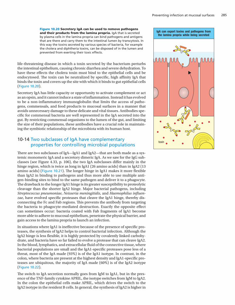
life-threatening disease in which a toxin secreted by the bacterium perturbs the intestinal epithelium, causing chronic diarrhea and severe dehydration. To have these effects the cholera toxin must bind to the epithelial cells and be endocytosed. The toxin can be neutralized by specific, high affinity IgA that binds the toxin and covers up the site with which it binds to gut epithelial cells (Figure 10.20).
Secretory IgA has little capacity or opportunity to activate complement or act as an opsin, and it cannot induce a state of inflammation. Instead it has evolved to be a non-inflammatory immunoglobulin that limits the access of patho-gens, commensals, and food products to mucosal surfaces in a manner that avoids unnecessary damage to these delicate and vital tissues. Antibodies spe-cific for commensal bacteria are well represented in the IgA secreted into the gut. By restricting commensal organisms to the lumen of the gut, and limiting the size of their populations, these antibodies have a crucial role in maintain-ing the symbiotic relationship of the microbiota with its human host.
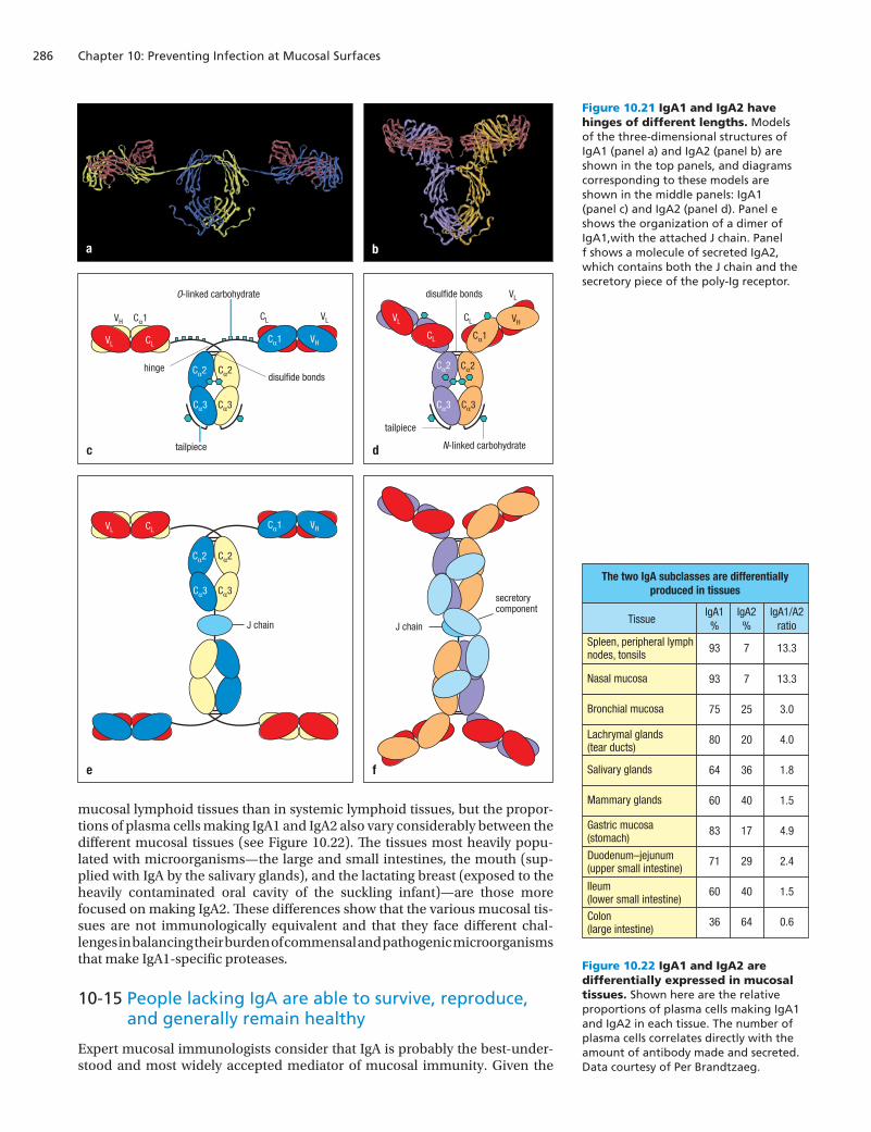
10-14 Two subclasses of IgA have complementary properties for controlling microbial populations
There are two subclasses of IgA—IgA1 and IgA2—that are both made as a sys-temic monomeric IgA and a secretory dimeric IgA. As we saw for the IgG sub-classes (see Figure 4.33, p. 106), the two IgA subclasses differ mainly in the hinge region, which is twice as long in IgA1 (26 amino acids) than in IgA2 (13 amino acids) (Figure 10.21). The longer hinge in IgA1 makes it more flexible than IgA2 in binding to pathogens and thus more able to use multiple anti-gen-binding sites to bind to the same pathogen and deliver it to a phagocyte. The drawback to the longer IgA1 hinge is its greater susceptibility to proteolytic cleavage than the shorter IgA2 hinge. Major bacterial pathogens, including Streptococcus pneumoniae, Neisseria meningitidis, and Haemophilus influen-zae, have evolved specific proteases that cleave the IgA1 hinge, thereby dis-connecting the Fc and Fab regions. This prevents the antibody from targeting the bacteria to phagocyte-mediated destruction. Exactly the opposite effect can sometimes occur: bacteria coated with Fab fragments of IgA1 become more able to adhere to mucosal epithelium, penetrate the physical barrier, and gain access to the lamina propria to launch an infection.
In situations where IgA1 is ineffective because of the presence of specific pro-teases, the synthesis of IgA2 helps to control bacterial infection. Although the IgA2 hinge is less flexible, it is highly protected by covalently linked carbohy-drate, and bacteria have so far failed to evolve a protease that can cleave IgA2. In the blood, lymphatics, and extracellular fluid of the connective tissue, where bacterial populations are small and the IgA1-specific proteases pose less of a threat, most of the IgA made (93%) is of the IgA1 isotype. In contrast, in the colon, where bacteria are present at the highest density and IgA1-specific pro-teases are ubiquitous, the majority of IgA made (60%) is of the IgA2 isotype (Figure 10.22).
The switch to IgA secretion normally goes from IgM to IgA1, but in the pres-ence of the TNF-family cytokine APRIL, the isotype switches from IgM to IgA2. In the colon the epithelial cells make APRIL, which drives the switch to the IgA2 isotype in the resident B cells. In general, the synthesis of IgA2 is higher in
Preventing infection at mucosal surfaces
IS4 i10.12/10.20
IgA can export toxins and pathogens fromthe lamina propria while being secreted
toxin
Figure 10.20 Secretory IgA can be used to remove pathogens and their products from the lamina propria. IgA that is secreted by plasma cells in the lamina propria can bind pathogens and antigens that are there and carry them to the intestinal lumen by transcytosis. In this way the toxins secreted by various species of bacteria, for example the cholera and diphtheria toxins, can be disposed of in the lumen and prevented from exerting their toxic effects.
Chapter 10: Preventing Infection at Mucosal Surfaces286
mucosal lymphoid tissues than in systemic lymphoid tissues, but the propor-tions of plasma cells making IgA1 and IgA2 also vary considerably between the different mucosal tissues (see Figure 10.22). The tissues most heavily popu-lated with microorganisms—the large and small intestines, the mouth (sup-plied with IgA by the salivary glands), and the lactating breast (exposed to the heavily contaminated oral cavity of the suckling infant)—are those more focused on making IgA2. These differences show that the various mucosal tis-sues are not immunologically equivalent and that they face different chal-lenges in balancing their burden of commensal and pathogenic microorganisms that make IgA1-specific proteases.
10-15 People lacking IgA are able to survive, reproduce, and generally remain healthy
Expert mucosal immunologists consider that IgA is probably the best-under-stood and most widely accepted mediator of mucosal immunity. Given the
IS4 n10.110/10.21
VL CL
Cα2
Cα3
Cα2
Cα1
Cα1 VH
Cα3
CL VL
VL
CLVH
Cα2
Cα1
VH
Cα3
Cα2
Cα3
O-linked carbohydrate
N-linked carbohydrate
disulfide bonds
disulfide bonds
hinge
tailpiece
tailpiece
VL
CL
VL CL
Cα2
Cα3
Cα2
Cα1 VH
Cα3
c d
e f
J chain J chain
secretorycomponent
a b
The two IgA subclasses are differentiallyproduced in tissues
IgA1/A2ratio
IgA2%
IgA1%
Tissue
13.3793Spleen, peripheral lymphnodes, tonsils
13.3793Nasal mucosa
3.02575Bronchial mucosa
4.02080Lachrymal glands(tear ducts)
1.83664Salivary glands
1.54060Mammary glands
4.91783Gastric mucosa(stomach)
2.42971Duodenum–jejunum(upper small intestine)
1.54060Ileum(lower small intestine)
0.66436Colon(large intestine)
IS4 i10.13/10.22
Figure 10.21 IgA1 and IgA2 have hinges of different lengths. Models of the three-dimensional structures of IgA1 (panel a) and IgA2 (panel b) are shown in the top panels, and diagrams corresponding to these models are shown in the middle panels: IgA1 (panel c) and IgA2 (panel d). Panel e shows the organization of a dimer of IgA1,with the attached J chain. Panel f shows a molecule of secreted IgA2, which contains both the J chain and the secretory piece of the poly-Ig receptor.
Figure 10.22 IgA1 and IgA2 are differentially expressed in mucosal tissues. Shown here are the relative proportions of plasma cells making IgA1 and IgA2 in each tissue. The number of plasma cells correlates directly with the amount of antibody made and secreted. Data courtesy of Per Brandtzaeg.
287
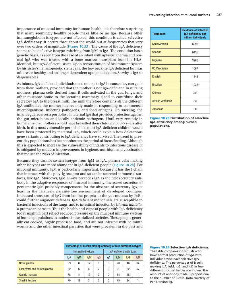
importance of mucosal immunity for human health, it is therefore surprising that many seemingly healthy people make little or no IgA. Because other immunoglobulin isotypes are not affected, this condition is called selective IgA deficiency. It occurs throughout the world but at frequencies that vary over two orders of magnitude (Figure 10.23). The cause of the IgA deficiency seems to be defective isotype switching from IgM to IgA. The condition has a genetic basis, as seen from the case of an infant with aplastic anemia and nor-mal IgA who was treated with a bone marrow transplant from his HLA-identical, but IgA-deficient, sister. Upon reconstitution of his immune system by his sister’s hematopoietic stem cells, the boy became IgA deficient but was otherwise healthy and no longer dependent upon medication. So why is IgA so dispensable?
As infants, IgA-deficient individuals need not make IgA because they can get it from their mothers, provided that the mother is not IgA-deficient. In nursing mothers, plasma cells derived from B cells activated in the gut, lungs, and other mucosae home to the lactating mammary gland to contribute their secretory IgA to the breast milk. The milk therefore contains all the different IgA antibodies the mother has recently made in responding to commensal microorganisms, infecting pathogens, and food antigens. On suckling, the infant’s gut receives a portfolio of maternal IgA that provides protection against the gut microbiota and locally endemic pathogens. Until very recently in human history, mothers would have breastfed their children for 3–7 years after birth. In this most vulnerable period of life, most IgA-deficient children would have been protected by maternal IgA, which could explain how deleterious gene variants contributing to IgA deficiency have survived. The trend in pres-ent-day populations has been to shorten the period of breastfeeding. Although this is expected to increase the vulnerability of infants to infectious disease, it is mitigated by modern improvements in hygiene, nutrition, and vaccination that reduce the risks of infection.
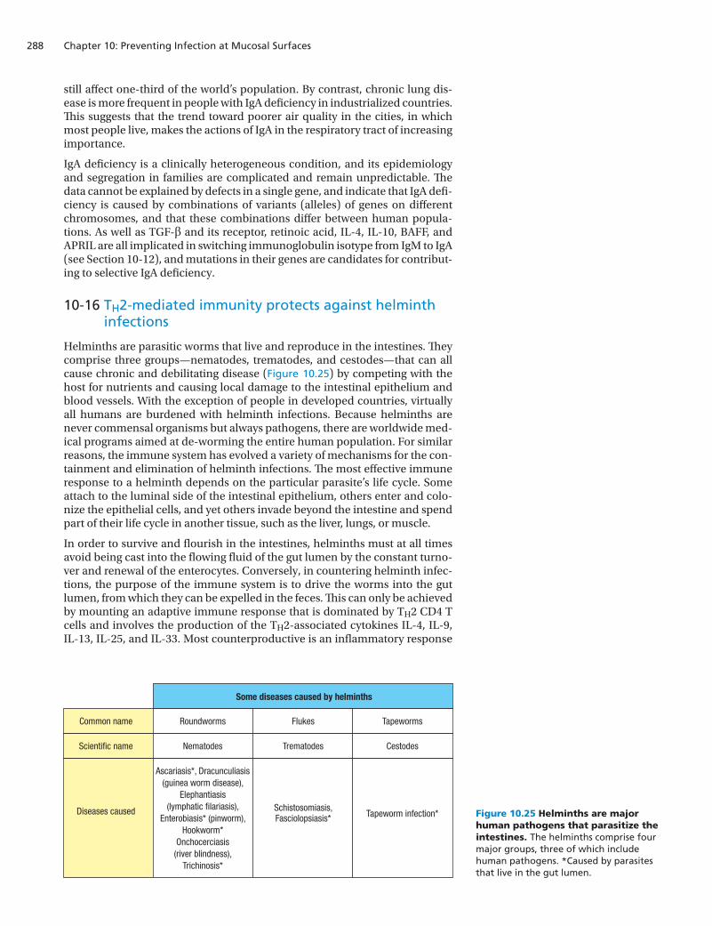
Because they cannot switch isotype from IgM to IgA, plasma cells making other isotypes are more abundant in IgA-deficient people (Figure 10.24). For mucosal immunity, IgM is particularly important, because it has the J chain that interacts with the poly-Ig receptor and so can be secreted at mucosal sur-faces, like IgA. Moreover, IgM always precedes IgA as the first secretory anti-body in the adaptive responses of mucosal immunity. Increased secretion of pentameric IgM probably compensates for the absence of secretory IgA, at least in the relatively parasite-free environment of developed countries. Increased transport of IgG from lamina propria to the gut mucosa by FcRn could further augment defenses. IgA-deficient individuals are susceptible to bacterial infections of the lungs, and to intestinal infection by Giardia lamblia, a protozoan parasite. Thus the health and vigor of people with IgA deficiency today might in part reflect reduced pressure on the mucosal immune systems of human populations in modern industrialized societies. These people gener-ally eat cooked, highly processed food, and are not infested with helminth worms and the other intestinal parasites that were prevalent in the past and
Preventing infection at mucosal surfaces
Figure 10.23 Distribution of selective IgA deficiency among human populations.
Figure 10.24 Selective IgA deficiency. The table compares individuals who have normal production of IgA with individuals who have selective IgA deficiency. The percentages of B cells making IgA, IgM, IgG, and IgD in four different mucosal tissues are shown. The amount of antibody made is proportional to the number of B cells. Data courtesy of Per Brandtzaeg.
PopulationIncidence of selective
IgA deficiency permillion individuals
6993Saudi Arabian
6135Spanish
3968Nigerian
1667US Caucasian
1143English
1036Brazilian
253Chinese
83African-American
60Japanese
IS4 n10.111/10.23
IgDIgGIgMIgAIgDIgGIgMIgA
IgA-deficient individualsNormal individuals
Percentage of B cells making antibody of four different isotypes
3446200817669
572221075682
1356400131176
124750031879Small intestine
Nasal glands
Lachrymal and parotid glands
Gastric mucosa
IS4 i10.14/10.24
Chapter 10: Preventing Infection at Mucosal Surfaces288
still affect one-third of the world’s population. By contrast, chronic lung dis-ease is more frequent in people with IgA deficiency in industrialized countries. This suggests that the trend toward poorer air quality in the cities, in which most people live, makes the actions of IgA in the respiratory tract of increasing importance.
IgA deficiency is a clinically heterogeneous condition, and its epidemiology and segregation in families are complicated and remain unpredictable. The data cannot be explained by defects in a single gene, and indicate that IgA defi-ciency is caused by combinations of variants (alleles) of genes on different chromosomes, and that these combinations differ between human popula-tions. As well as TGF-β and its receptor, retinoic acid, IL-4, IL-10, BAFF, and APRIL are all implicated in switching immunoglobulin isotype from IgM to IgA (see Section 10-12), and mutations in their genes are candidates for contribut-ing to selective IgA deficiency.
10-16 TH2-mediated immunity protects against helminth infections
Helminths are parasitic worms that live and reproduce in the intestines. They comprise three groups—nematodes, trematodes, and cestodes—that can all cause chronic and debilitating disease (Figure 10.25) by competing with the host for nutrients and causing local damage to the intestinal epithelium and blood vessels. With the exception of people in developed countries, virtually all humans are burdened with helminth infections. Because helminths are never commensal organisms but always pathogens, there are worldwide med-ical programs aimed at de-worming the entire human population. For similar reasons, the immune system has evolved a variety of mechanisms for the con-tainment and elimination of helminth infections. The most effective immune response to a helminth depends on the particular parasite’s life cycle. Some attach to the luminal side of the intestinal epithelium, others enter and colo-nize the epithelial cells, and yet others invade beyond the intestine and spend part of their life cycle in another tissue, such as the liver, lungs, or muscle.
In order to survive and flourish in the intestines, helminths must at all times avoid being cast into the flowing fluid of the gut lumen by the constant turno-ver and renewal of the enterocytes. Conversely, in countering helminth infec-tions, the purpose of the immune system is to drive the worms into the gut lumen, from which they can be expelled in the feces. This can only be achieved by mounting an adaptive immune response that is dominated by TH2 CD4 T cells and involves the production of the TH2-associated cytokines IL-4, IL-9, IL-13, IL-25, and IL-33. Most counterproductive is an inflammatory response
Figure 10.25 Helminths are major human pathogens that parasitize the intestines. The helminths comprise four major groups, three of which include human pathogens. *Caused by parasites that live in the gut lumen.
Some diseases caused by helminths
Common name Roundworms Flukes Tapeworms
Scientific name Nematodes Trematodes Cestodes
Diseases caused Schistosomiasis,Fasciolopsiasis* Tapeworm infection*
IS4 n10.112/10.25
Ascariasis*, Dracunculiasis(guinea worm disease),
Elephantiasis(lymphatic filariasis),
Enterobiasis* (pinworm),Hookworm*
Onchocerciasis(river blindness),
Trichinosis*
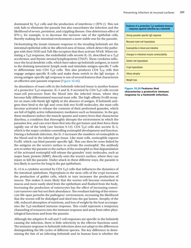
289
dominated by TH1 cells and the production of interferon-γ (IFN-γ). This not only fails to eliminate the parasite but also exacerbates the infection and the likelihood of severe, persistent, and crippling disease. One deleterious effect of IFN-γ, for example, is to decrease the turnover rate of the epithelial cells, thereby making the intestinal environment a more stable one for the parasite.
Orchestrating the innate immune response to the invading helminth are the intestinal epithelial cells in the affected area of tissue, which detect the patho-gen with their NOD and Toll-like receptors that then activate NFκB. When ini-tiating a TH2 response, the endothelial cells secrete IL-33, described as a TH2 accelerator, and thymic stromal lymphopoietin (TSLP). These cytokines influ-ence the local dendritic cells, which have taken up helminth antigens, to travel to the draining mesenteric lymph node and stimulate antigen-specific T cells to differentiate into CD4 TH2 cells. This also produces CD4 TFH cells that engage antigen-specific B cells and make them switch to the IgE isotype. A strong antigen-specific IgE response is one of several features that characterize an effective anti-parasite response (Figure 10.26).
An abundance of mast cells in the helminth-infected tissue is another feature of a protective TH2 response. IL-3 and IL-9 secreted by CD4 TH2 cells recruit mast-cell precursors from the blood into the infected tissue, where they become fully differentiated mucosal mast cells. The high-affinity FcεRI recep-tor on mast cells binds IgE tightly in the absence of antigen. If helminth anti-gens then bind to the IgE and cross-link two FcεRI molecules, the mast cells become activated to release the contents of their preformed granules, which are full of highly active inflammatory mediators such as histamine. In the gut, these mediators induce the muscle spasms and watery feces that characterize diarrhea, a condition that thoroughly disrupts the environment in which the parasites live, and can evict them first into the gut lumen and then force them rapidly out of the body (see Section 9-13). CD4 TH2 cells also secrete IL-5, which is the major cytokine controlling eosinophil development and function. During a helminth infection, the IL-5 increases the numbers of eosinophils in the blood and in the infected gut tissue. Like mast cells, eosinophils express FcεRI, which can bind parasite-specific IgE. This can then be cross-linked by the antigens on the worm’s surface to activate the eosinophil. The antibody acts to tether the parasite to the surface of the eosinophil so that degranulation of the activated eosinophil will release the granules’ toxic molecules, such as major basic protein (MBP), directly onto the worm’s surface, where they can injure or kill the parasite. Under attack in these different ways, the parasite is less likely to survive for long in the gut epithelium.
IL-13 is a cytokine secreted by CD4 TH2 cells that influences the dynamics of the intestinal epithelium. Hyperplasia in the stem cells of the crypt increases the production of goblet cells, which in turn increases the production of mucus. This makes it more likely that the worms will become enmeshed in mucus and more easily shed from the epithelium and flushed from the body. Increasing the production of enterocytes has the effect of increasing entero-cyte turnover rate but not their abundance. The resultant halving of the entero-cyte life-span perturbs the pathogens’ environment, increasing the likelihood that the worms will be dislodged and shed into the gut lumen. Atrophy of the villi, reduced absorption of nutrients, and loss of weight by the host accompa-nies the TH2-mediated immune response. This could represent a temporary channeling of resources into the immune response and away from other phys-iological functions and from the parasite.
Although the adaptive B-cell and T-cell responses are specific to the helminth causing the infection, there is little selectivity in the effector functions used. The immune response to helminth infections does not adapt to the differences distinguishing the life cycles of different species. The key difference in deter-mining the fate of an infecting helminth and its human host is whether the
Preventing infection at mucosal surfaces
Figure 10.26 Features that characterize a protective immune response to a helminth infection.
Features of a protective TH2-mediated immuneresponse against infection by a helminth
Strong parasite-specific IgE response
Mucosal mast-cell hyperplasia
Eosinophilia in blood and intestine
Changes in intestinal muscle contractability
Goblet-cell hyperplasia
Crypt hyperplasia
Villous atrophy
Weight loss
IS4 n10.113/10.26
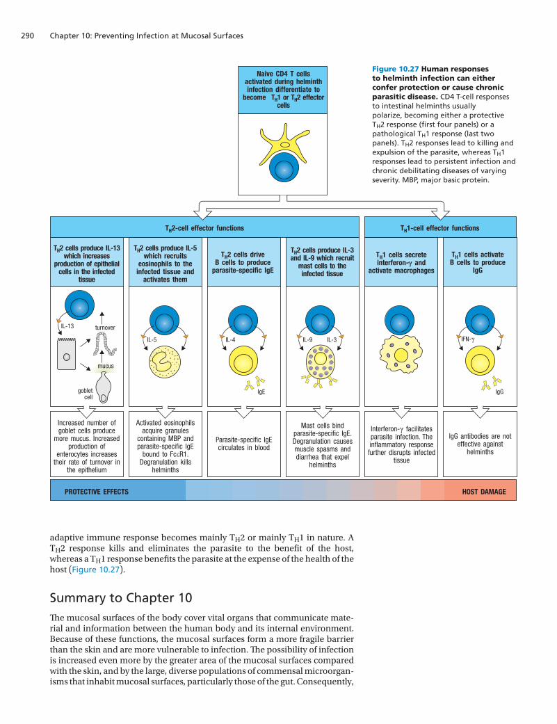
Chapter 10: Preventing Infection at Mucosal Surfaces290
IS4 i10.16/10.27
Naive CD4 T cells activated during helminth infection differentiate to
become TH1 or TH2 effector cells
PROTECTIVE EFFECTS HOST DAMAGE
TH2-cell effector functions
TH2 cells produce IL-13which increases
production of epithelialcells in the infected
tissue
TH1 cells secreteinterferon-γ and
activate macrophages
TH1 cells activateB cells to produce
IgG
TH2 cells produce IL-5which recruits
eosinophils to theinfected tissue and
activates them
TH2 cells driveB cells to produce
parasite-specific IgE
TH2 cells produce IL-3and IL-9 which recruit
mast cells to theinfected tissue
TH1-cell effector functions
Increased number of goblet cells produce
more mucus. Increased production of
enterocytes increases their rate of turnover in
the epithelium
Activated eosinophils acquire granules
containing MBP and parasite-specific IgE
bound to FcεR1.Degranulation kills
helminths
Parasite-specific IgE circulates in blood
Mast cells bind parasite-specific IgE. Degranulation causes muscle spasms and diarrhea that expel
helminths
Interferon-γ facilitates parasite infection. The inflammatory response
further disrupts infected tissue
IgG antibodies are not effective against
helminths
turnover
mucus
gobletcell
IL-13
IL-5 IL-4
IgE
IL-9 IL-3 IFN-γ
IgG
adaptive immune response becomes mainly TH2 or mainly TH1 in nature. A TH2 response kills and eliminates the parasite to the benefit of the host, whereas a TH1 response benefits the parasite at the expense of the health of the host (Figure 10.27).
Summary to Chapter 10The mucosal surfaces of the body cover vital organs that communicate mate-rial and information between the human body and its internal environment. Because of these functions, the mucosal surfaces form a more fragile barrier than the skin and are more vulnerable to infection. The possibility of infection is increased even more by the greater area of the mucosal surfaces compared with the skin, and by the large, diverse populations of commensal microorgan-isms that inhabit mucosal surfaces, particularly those of the gut. Consequently,
Figure 10.27 Human responses to helminth infection can either confer protection or cause chronic parasitic disease. CD4 T-cell responses to intestinal helminths usually polarize, becoming either a protective TH2 response (first four panels) or a pathological TH1 response (last two panels). TH2 responses lead to killing and expulsion of the parasite, whereas TH1 responses lead to persistent infection and chronic debilitating diseases of varying severity. MBP, major basic protein.
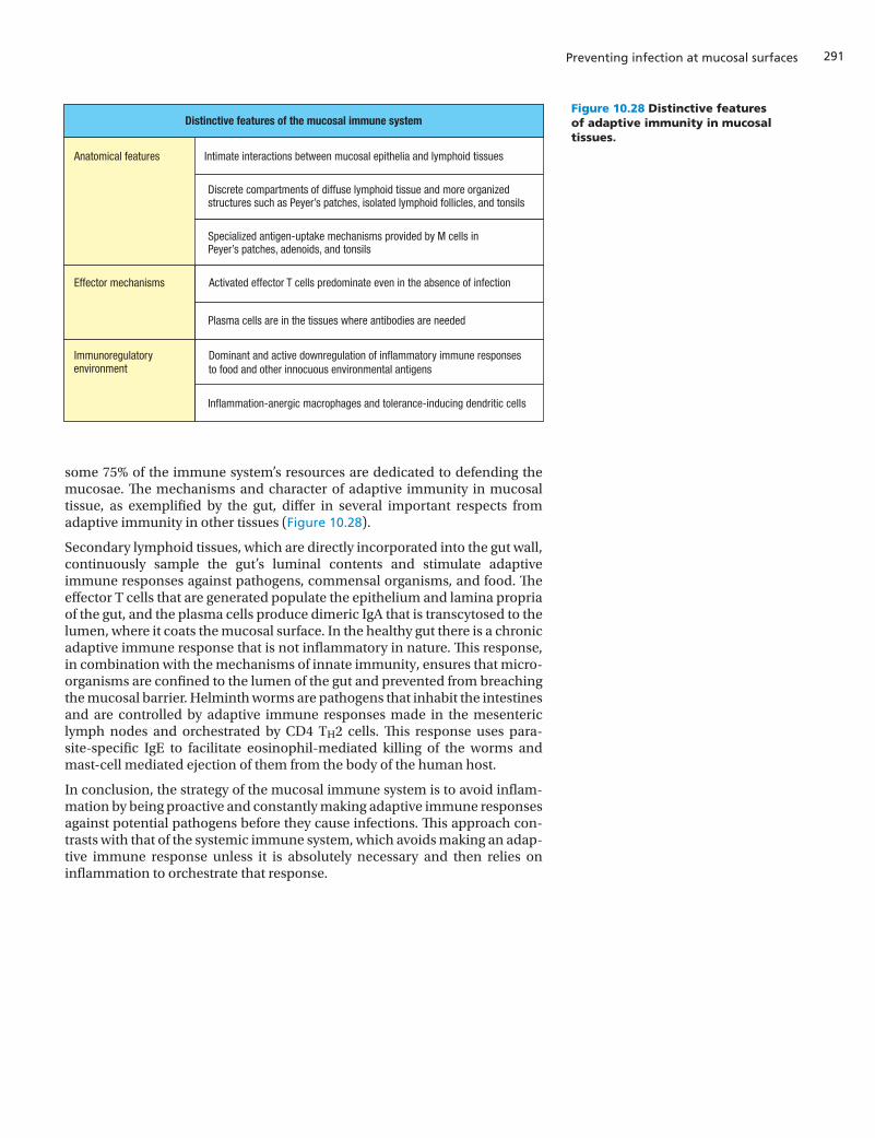
291
some 75% of the immune system’s resources are dedicated to defending the mucosae. The mechanisms and character of adaptive immunity in mucosal tissue, as exemplified by the gut, differ in several important respects from adaptive immunity in other tissues (Figure 10.28).
Secondary lymphoid tissues, which are directly incorporated into the gut wall, continuously sample the gut’s luminal contents and stimulate adaptive immune responses against pathogens, commensal organisms, and food. The effector T cells that are generated populate the epithelium and lamina propria of the gut, and the plasma cells produce dimeric IgA that is transcytosed to the lumen, where it coats the mucosal surface. In the healthy gut there is a chronic adaptive immune response that is not inflammatory in nature. This response, in combination with the mechanisms of innate immunity, ensures that micro-organisms are confined to the lumen of the gut and prevented from breaching the mucosal barrier. Helminth worms are pathogens that inhabit the intestines and are controlled by adaptive immune responses made in the mesenteric lymph nodes and orchestrated by CD4 TH2 cells. This response uses para-site-specific IgE to facilitate eosinophil-mediated killing of the worms and mast-cell mediated ejection of them from the body of the human host.
In conclusion, the strategy of the mucosal immune system is to avoid inflam-mation by being proactive and constantly making adaptive immune responses against potential pathogens before they cause infections. This approach con-trasts with that of the systemic immune system, which avoids making an adap-tive immune response unless it is absolutely necessary and then relies on inflammation to orchestrate that response.
Preventing infection at mucosal surfaces
Figure 10.28 Distinctive features of adaptive immunity in mucosal tissues.
Distinctive features of the mucosal immune system
Anatomical features Intimate interactions between mucosal epithelia and lymphoid tissues
Discrete compartments of diffuse lymphoid tissue and more organized structures such as Peyer’s patches, isolated lymphoid follicles, and tonsils
Specialized antigen-uptake mechanisms provided by M cells in Peyer’s patches, adenoids, and tonsils
Effector mechanisms Activated effector T cells predominate even in the absence of infection
Plasma cells are in the tissues where antibodies are needed
Immunoregulatory Dominant and active downregulation of inflammatory immune responses
Inflammation-anergic macrophages and tolerance-inducing dendritic cells
to food and other innocuous environmental antigens
IS4 i10.17/10.28
environment
Chapter 10: Preventing Infection at Mucosal Surfaces292
Questions
10–1 Mucosae exist in all of the following anatomical locations except _____.
a. lactating breastsb. urogenital tractc. the limbsd. gastrointestinal tracte. salivary glandsf. lachrymal glandsg. the respiratory tracth. the pancreas.
10–2 All of the following are characteristics of some or all mucosal surfaces except _____. (Select all that apply.)
a. the secretion of viscous fluid called mucusb. reproductive activitiesc. absorption of nutrientsd. participation in gas exchangee. participation in sensory activitiesf. collectively constitute approximately 25% of the body’s
immune activitiesg. use of tight junctions to join epithelial layersh. tissue regenerates about every 20–30 days.
10–3 Match the term in column A with its description in col-umn B.
Column A Column B
a. mucosae 1. constitute the gut microbiota
b. cecum 2. epithelial surfaces distributed throughout the body
c. systemic immune system
3. protective epithelial glycoproteins
d. mucins 4. located between the small and large intestines
e. commensal microorganisms
5. defends against pathogens that breach the skin
10–4 The main function of the mucus in the stomach is to _____.
a. trap and kill ingested microorganismsb. enzymatically degrade complex nutrientsc. protect epithelial cells from the acidified environmentd. protect the microbiota from corrosive gastric juicese. delay the digestive process to maximize absorption.
10–5 _____ arises from an adaptive immune response to the proteins of gluten.
a. Cholerab. Celiac diseasec. Selective IgA deficiencyd. Crohn’s disease.
10–6 The “M” used to name M cells of the gastrointestinal tract derives from _____.
a. mesentericb. microfoldc. monocytesd. mucosae. mast cells.
10–7 Match the term in column A with its description in col-umn B.
Column A Column B
a. lamina propria 1. situated at the entrance of the gut and airway
b. Peyer’s patch 2. a chain of lymph nodes in connective tissue of the gastrointestinal tract
c. Waldeyer’s ring 3. dome-like bulging aggregates of lymphocytes that extend into the lumen of the gut
d. M cells 4. transport antigen to pockets on the basolateral side of the gut epithelium
e. mesenteric lymph nodes
5. CD8 T cells with limited range of antigen specificities
f. intraepithelial lymphocytes
6. connective tissue beneath the gut epithelium
10–8 Identify two ways in which the immune responses in gut mucosal tissues contrast with those initiated in systemic non-mucosal tissues.
10–9 Which of the following statements regarding intraepithe-lial lymphocytes is false? (Select all that apply.)
a. They comprise approximately 10% of the cells in the mucosal epithelium.
b. They are composed of both CD4 and CD8 T cells.c. They are separated from the lamina propria by a
basement membrane.d. They are activated effector T cells with a narrow range of
antigen specificities.e. They do not include NK cells.f. They express the α4:β7 integrin that binds to E-cadherin
on epithelial surfaces.
10–10 An important distinction between macrophages that populate the lamina propria of the gut and the macrophages that populate the skin is that the former _____.
a. cannot phagocytose and kill bacterial pathogensb. do not present antigens to T cellsc. do not possess signaling receptors needed for
production of inflammatory cytokinesd. express much higher levels of TLRse. are very rare in the gut mucosa.
10–11 Whereas _____ is the predominant immunoglobulin in intestinal fluid, _____ is the dominant immunoglobulin in the urogenital tract.
a. dimeric IgA; IgEb. IgE; dimeric IgAc. dimeric IgA; pentameric IgMd. dimeric IgA; IgGe. pentameric IgM; monomeric IgA.
10–12 Which of the following pairs is mismatched?a. NOD1: a cytoplasmic receptor of intestinal epitheliumb. NLRP3: assists in the formation of an inflammasomec. intestinal macrophages: professional antigen-presenting
cells
293
d. TLR-5: detects flagellin on apical and basolateral epithelial surfaces
e. neutrophils: attracted by CXCL8.
10–13 A T lymphocyte activated in the GALT will subsequently home to all of the following except _____. (Select all that apply.)
a. mucosal lymphoid tissues of lactating mammary glandsb. the spleenc. mucosal lymphoid tissues of the respiratory tractd. systemic lymph nodese. mucosal lymphoid tissues of the gastrointestinal tractf. lymphoid tissues of the skin.
10–14 Identify which of the following immune responses is piv-otal to the killing and elimination of helminths.
a. killing by cytotoxic T cells in the lamina propriab. TH1-induced inflammationc. TH2-associated cytokinesd. phagocytosis by intestinal macrophagese. systemic immune responsesf. B-cell secretion of IgG.
10–15 Richard Brennan began penicillamine therapy after he was diagnosed with Wilson’s disease (manifested by copper accumulation in the tissues) at age 10 years. Ten months after beginning this treatment he began to experience multiple sinus infections, and one episode of pneumonia. Recently he came to the emergency room with acute diarrhea, vomiting, fever, and foul-smelling intestinal gas. Stool samples revealed the presence of trophozoites of Giardia. Blood tests showed normal levels of B and T cells and normal IgM and IgG concentrations, but mark-edly decreased IgA at 6 mg/dl (normal range 40–400 mg/dl). Richard was treated for his giardiasis with metronidazole. His selective IgA deficiency was associated with penicillamine, shown previously to be a complication in some patients with Wilson’s disease. His IgA levels returned to normal when peni-cillamine was discontinued. This is an example of a drug-in-duced transient form of IgA deficiency. Which of the following antibodies that uses the same transport receptor as dimeric IgA would have been present in the lumen of the gastrointestinal tract and mucosal secretions of Richard while he was taking penicillamine?
a. IgDb. IgMc. IgGd. IgEe. none of the above.

Young boy with measles.