Embed Size (px)
Citation preview

Antibody Structure and Function*
* Special thanks to Dr. L. Scott Rodkey, Ph.D.
Jeffrey K. Actor, Ph.D. Jeffrey K. Actor, Ph.D. Pathology and Laboratory MedicinePathology and Laboratory Medicine
The University of TexasThe University of Texas--Houston Medical SchoolHouston Medical School

Anatomy and Physiology of Antibodies

Antibodies are gamma-globulins

Ig Domain Structures
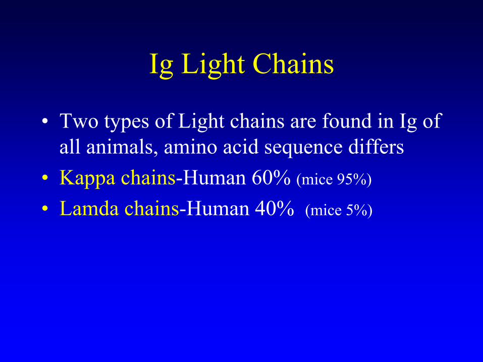
Ig Light Chains
•
Two types of Light chains are found in Ig of all animals, amino acid sequence differs
•
Kappa chains-Human 60% (mice 95%)
•
Lamda
chains-Human 40% (mice 5%)
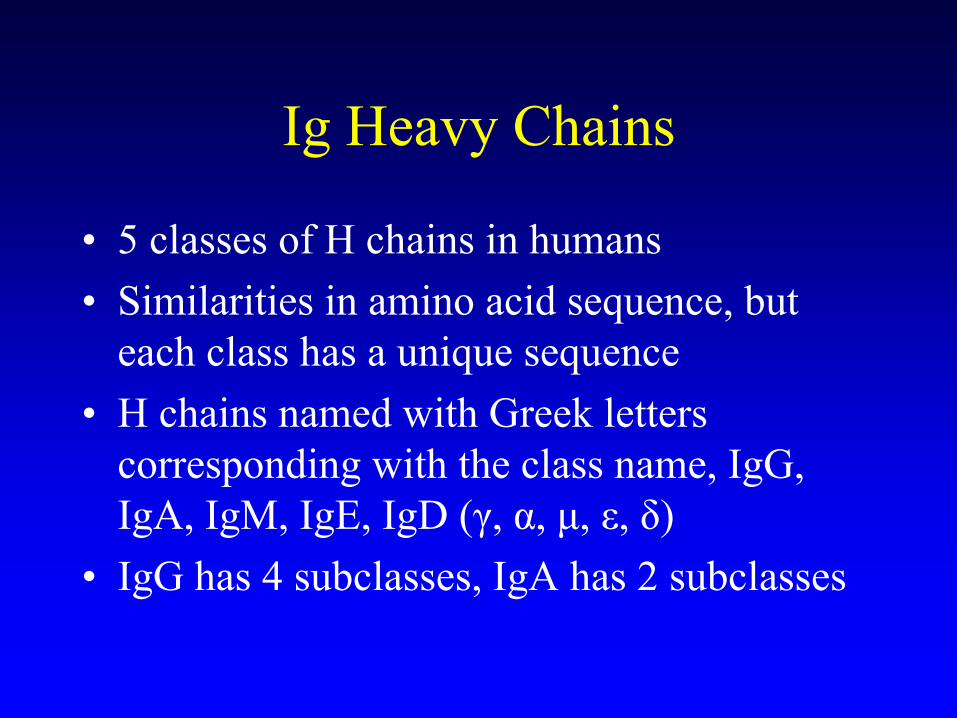
Ig Heavy Chains
•
5 classes of H chains in humans•
Similarities in amino acid sequence, but each class has a unique sequence
•
H chains named with Greek letters corresponding with the class name, IgG, IgA, IgM, IgE, IgD (γ, α, μ, ε, δ)
•
IgG has 4 subclasses, IgA has 2 subclasses

Domains
•
Early studies showed regularity of structure of all the Ig classes
•
Each 100-110 aa
has a 60 aa
S-S loop•
V domains code the paratope, binds Ag
•
C domains code regions important for mediating secondary biological functions, ie binding Complement, crossing the placenta.

D O M A IN F U N C T IO N S O F H U M A N IgG --------------- ---------- ---------- ---------- ---------- ---------- ----------- ---------- ------ --------------- -- D o m ain(s) Fu n ctio n V H + V L A ntig en B in din g C H1 + C L Spa cer be tw een a ntig en -bin ding an d e ffector fu nc tion s C H2 B in ding C 1 q C o ntrol o f ca ta bolic ra te C H3 Interac tion w ith F c receptor o n m a cro p hag e/m on o cyte C H1 + C H3 B in d to Protein A

Ig Variable and Hypervariable Regions
•
Amino terminal aa
sequence was shown to vary from one Light chain to another
•
Kabat
and Wu developed the Variability Plot to measure degree of variation
•
Found 3 Hypervariable regions in both L and H chain V regions.
•
These are epitope contact aa
regions, CDR (complementarity determining regions)

Definition of Variability
•
The ratio of the number of different amino acids at a given position to the frequency of the most common amino acid at that position is defined as VARIABILITY.

Variability Plot

Ig Hinge Regions
•
Hinge regions on IgG, IgA and IgD are coded by distinct exons
•
Short span of amino acids between 1st
and
2nd
C domains
•
Rich in Cys and Pro•
Provides for flexibility of the molecule
•
Is readily accessible to solvent and enzymes

Ig Classes or Isotypes
•
There are 5 major classes or isotypes of Ig in Humans:–
IgG, IgA, IgM, IgE, IgD
–
Ig, Ig, Ig, Ig, Ig
•
There are 4 subclasses of IgG isotypes in Humans–
IgG1, IgG2, IgG3, IgG4
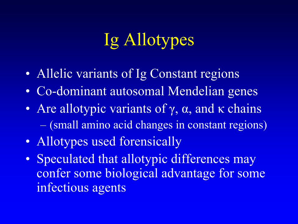
Ig Allotypes
•
Allelic variants of Ig Constant regions•
Co-dominant autosomal Mendelian genes
•
Are allotypic variants of γ, α, and κ
chains–
(small amino acid changes in constant regions)
•
Allotypes used forensically•
Speculated that allotypic differences may confer some biological advantage for some infectious agents
Ig Superfamily
•
There are structural similarities to Ig of the molecules of numerous membrane bound glycoprotein molecules such as the MHC molecules and the T cell receptor molecules. T cell receptors and triggering will be covered in another lecture.
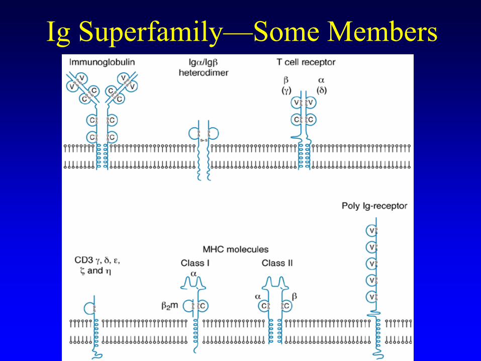
Ig Superfamily—Some Members
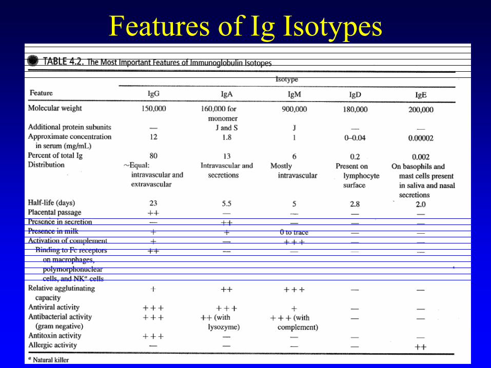
Features of Ig Isotypes

Ig Structural FeaturesG
155 kDa

IgG Structural Features II

IgG Biological Properties
•
Agglutination and Precipitation•
Antibody dependent cell mediated cytotoxicity
•
Complement Activation and Opsonization•
Placental passage (only isotype to do so)
•
Toxin/Viral Neutralization•
Bacterial immobilization
•
Long serum half-life (~23 days)

Effect of Opsonizing Ab and C’•
IgG antibodies are efficient activators of the Complement system.
•
Bacterial antigen-antibody interactions trigger a series of enzymes collectively known as Complement.
•
Some of the by-products of these reactions can act as opsonins and other components are chemotactic (attract phagocytic cells).
•
Net effect is greater uptake of pathogenic bacteria and clearance by phagocytic cells.

Antibody Dependent Cell- Mediated Cytotoxicity (ADCC)

Important Differences Between Human IgG SubclassesIgG1 IgG2 IgG3 IgG4
Occurrence (% total IgG) 70 20 7 3
Half-life 23 23 7 23
Complement binding + + +++ —
Placental passage ++ ± ++ ++
Binding of monocytes +++ + +++ ±

IgG is Recycled by Fc Receptor Protector (FcRp)
Receptor recycling assists in increased half-life of IgG in circulation/serum

•
Rh antigens, also called Rhesus antigens, are transmembrane proteins expressed at the surface of erythrocytes (involved in carbon dioxide and/or ammonia transport across the plasma membrane).
•
Red blood cells that express surface antigen D (RhD
antigen) are called “Rh positive”.
•
About 15% of the population have no RhD
antigens (“Rh negative”).
Clinical Tidbit: IgG and Placental Passage
•
A Rh negative mother who carries a Rh positive fetus runs the risk of producing immune antibodies to the Rh antigens on the fetal RBC. The exposure during primary pregnancy is minimized. However, the mother may generate Rh antibodies after birth if the mother comes into contact with fetal blood cells during placenta rupture.
•
Upon subsequent pregnancies, the next Rh positive fetus will be at risk since the mother will retain a low level of circulating antibodies against the Rh antigen. Destruction of fetal erythrocytes will ensue by passive immune transfer of maternal antibodies to fetus, resulting in erythroblastosis fetalis (hemolytic disease of the newborn).
Clinical Tidbit: IgG and Placental Passage
•
It is of great clinical importance to identify Rh mismatched mother and fetus; typically an indirect agglutination test is performed to identify isohemmaglutination.
•
If positive, the mother is clinically treated immediately after giving birth with anti-Rh antibodies (Rh immune globulin (RhIG)
or
Rhogam) which reacts with the fetal RBC. Ensuing antibody-antigen complexes are removed prior to maternal recognition of foreign Rh antigen.
Clinical Tidbit: IgG and Placental Passage

Clinical Correlation
•
Hemolytic disease of the fetus/newborn. Maternal IgG antibodies specific for RhD are actively transported across the placenta, opsonize fetal RhD+ RBC for phagocytosis
by liver cells, fetal hematocrit drops to dangerous or fatal levels.

IgG Neutralization
•
TOXIN NEUTRALIZATION: –
Bacterial toxins bind to specific cellular receptors to gain entry to the cell and then exert toxic effects intracellularly.
–
The strategy to protect the host from toxins is to make a variety of antibodies specific for different epitopes on the toxin to immobilize it in the form of an antigen-antibody aggregate, thus preventing the toxin from reaching the cell receptor.
–
The Ab-Ag aggregates can be easily phagocytosed (via Fc receptors) and the toxins degraded and rendered non-toxic by acid proteases in the phagosomes.
–
Note: this is the basis for using antigenically similar TOXOIDS to make vaccines that will elicit a strong immune response with no toxic effects.

IgG Neutralization•
BACTERIAL IMMOBILIZATION–
Motile bacteria have movement arrested by IgG antibodies by cross-linking flagella or clumping them via flagella. The antibody functions like handcuffs in stopping the waving of flagella. The result is that the bacteria are less invasive and
less
efficient in spreading through tissue.
•
VIRAL NEUTRALIZATION–
Most viruses utilize some form of cellular receptor for initial binding to gain entry into the cell. IgG antibodies specific for
viral structures that bind to cell receptors inhibit or eliminate initial binding to the cell, thereby protecting the cell from viral entry. The binding of IgG also facilitates phagocytosis
of the
organism, targeting for removal and destruction.
Review IgGIgG
Anatomy: Gamma Heavy Chains, /
Light Chains
IgG Physiology: Agglutination, Placental Passage, Opsonization, ADCC, Complement Binding, Toxin/Viral Neutralization, Bacterial Immobilization, Recycled to allow greater serum half-life
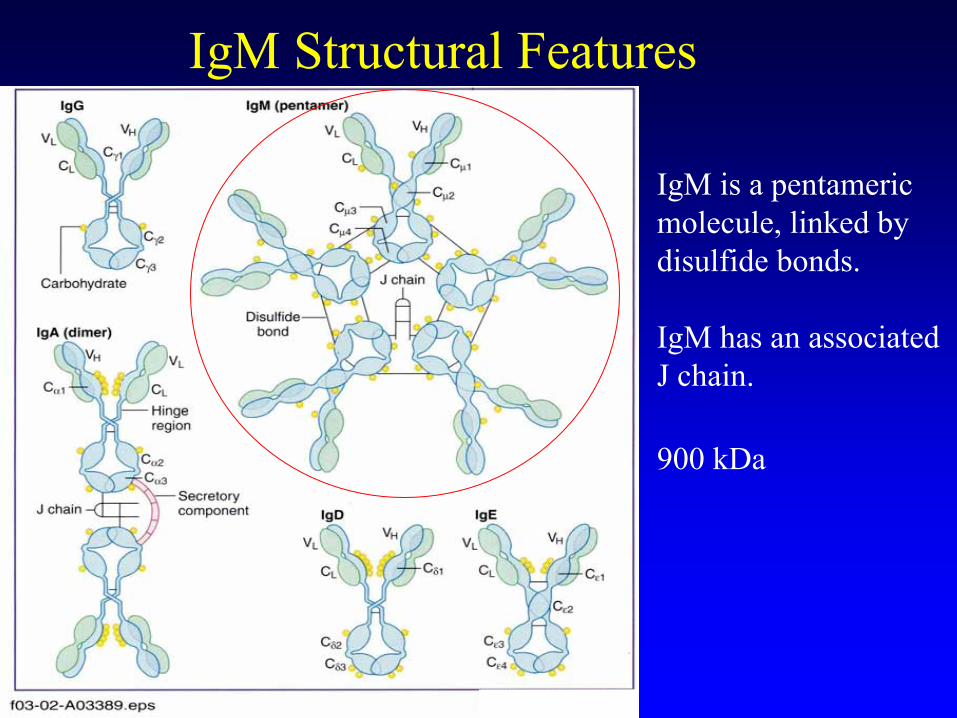
IgM Structural Features
IgM is a pentameric molecule, linked by
disulfide bonds.
IgM has an associated J chain.
900 kDa

IgM Biological Properties
•
Efficient bacterial/viral agglutinator, potentially 10 paratopes/molecule
•
Has a half-life of 5-10 days in serum•
Most efficient Ig for mediating Complement fixation
•
Very effective at toxin neutralization•
Isohemagglutinin-naturally present Ab reactive with A/B blood groups, barrier to random transfusion & transplantation

IgM Antigen Binding
IgG
AgAg
IgM
AgAg
Ag
Ag
Efficient for agglutination, complement fixation, neutralization.

Blood Groups, IgM and Isohemagglutinins
•
ABO Blood Groups. The ABO blood groups were first identified in 1901. They represent important antigens to be accounted for to assure safe blood transfusions.
•
The ABO antigens represent carbohydrate moieties present on erythrocytes.
•
Individuals naturally develop IgM antibodies (called isoantibodies) specific for ABO antigens that they do not express (cross-reactive to bacterial polysaccharides).
•
If the individual receives a transfusion of blood that contains non- compatible ABO antigens, isoantibodies will causes agglutination
of the donor cells.
•
This process is referred to as isohemagglutination; the antigens are sometimes called isohemagglutinins.

Blood Group
ErythrocyteAntigens
Serum Antibodies Genotypes
A A Anti-B AA or AOB B Anti-A BB or BO
AB A and B Neither AB O Neither Anti-A and anti-B OO
Blood Groups and Isohemagglutinins
Sugar moieties found on RBCs
O = universal donor
Review IgMIgM
Anatomy: Pentameric, high molecular weight
IgM Physiology: Agglutination, no Placental Passage, Opsonization, Complement fixation, Toxin/Viral Neutralization, Isohemagglutination

IgA Structural Features
160 kDax2

IgA Subclasses
Two subclasses: IgA1 and IgA2.• Differ in disulfide links between heavy and light chains.
•IgA has J chain.•IgA has hinge region.•IgA has associated secretory component.

Biological Properties of IgA Antibodies
•
IgA is abundant on mucosal surfaces as a “First Line Defense”.
•
IgA is bactericidal for Gram negative organisms in the presence of Lysozyme.
•
IgA is an efficient viral agglutinator, preventing viral attachment to epithelial cell viral receptors.

IgA Secretion Mechanism

Review IgAIgA
Anatomy: Dimer
IgA Physiology: Mucosal surfaces, bactericidal, viral agglutinator, does not fix complement
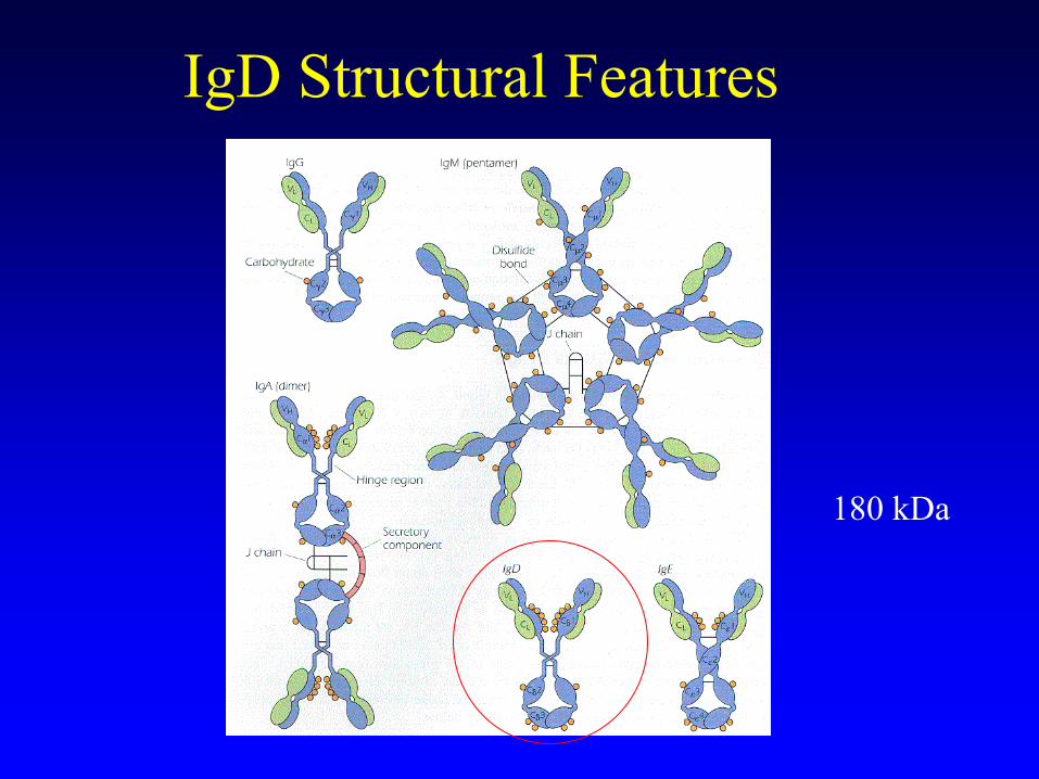
IgD Structural Features
180 kDa

Biological Properties of IgD Antibodies
•
IgD is not found in significant amounts in serum; primarily found on surface of B cells.
•
Principal function involves initial Ag triggering of B cells while bound to the membrane on the surface of B cells.

IgE Structural Features
200 kDa
Biological Properties of IgE Antibodies
•
IgE mediates Type I hypersensitivity reactions.
•
IgE antibodies bind to Fcε
receptors on Mast cells.
•
Ag binding with the IgE antibody induces degranulation, secretion of histamine, heparin, and other pharmacologic agents.
•
High IgE levels occur during “large” parasitic infections (eg. helminths).

IgE Cross-linking Leads to Mast Cell Triggering and Degranulation
IgE Anti-DNP
Antibody to IgE Receptor

Coda
•
The function of antibodies, like the rest of the body, is only understood with a firm foundation in (molecular) anatomy.
•
The 5 Ig isotypes each mediate specific biological effects, due to different C region amino acid sequences in their respective H chain.–
Review Chart: Features of Ig Isotypes!


Which immunoglobulin isotype has the highest molecular weight?A.
IgM
B.
IgD C.
IgG
D.
IgE E.
IgA
Option A (IgM) is correct. IgM is found on the surface of B lymphocytes, and secreted as a pentameric
molecule of approximately
970 kdaltons. IgM is extremely effective at fixing complement, and is an effective agglutinator of particulate antigens because of the high number (10) of antigen specific binding sites.
A 34 year old caucasian
woman gives birth to a first child who is identified as being Rh-positive. Although the mother is Rh-negative, she was not treated after birth with Rhogram
(anti-Rh antibodies). During the next pregnancy, the
fetus is found to have erythroblastosis fetalis. Which isotype of maternal immunoglobulin is responsible for the hemolytic disease of the newborn?A.
IgM
B.
IgDC.
IgE
D.
IgGE.
IgA
Option D (IgG) is correct. A Rh negative mother who carries a Rh positive fetus runs the risk of producing immune antibodies to the Rh antigens on the fetal RBC. The exposure during primary pregnancy is minimized; the mother can generate anti-Rh antibodies after birth if she contacts fetal blood cells during placenta rupture. Upon subsequent pregnancies, the next Rh positive fetus will be at risk since the mother will retain a low level of circulating antibodies against the Rh antigen. Maternal IgG antibodies specific for RhD
are actively transported across the placenta, opsonize fetal RhD+ RBC for phagocytosis
by liver cells, fetal hematocrit drops to dangerous or fatal levels. Only IgG has significant placental passage to cause this
effect.

A person having Type O blood is best described as: A. Having erythrocyte antigens A and B, and having anti-A and anti-
B antibodies. B. Having erythrocyte antigens A and B, but not having anti-A and anti-B antibodies. C. Having no erythrocyte antigens, but not having anti-A and anti-B antibodies. D. Having no erythrocyte antigens, and having anti-A and anti-B antibodies.
Option D (Having no erythrocyte antigens, and having anti-A and anti-B antibodies) is correct. The ABO blood groups represent important carbohydrate moieties present on erythrocytes to be accounted for to assure safe blood transfusions. Individuals naturally develop antibodies (called isoantibodies) specific for ABO antigens that they do not express (due to cross-
reactivity with bacterial polysaccharides). If the individual receives a transfusion of blood that contains non-compatible ABO antigens, isoantibodies will causes agglutination of the donor cells, a process referred
to as
isohemagglutination; the antigens are sometimes called isohemagglutinins.

Which organ serves as both a primary and secondary lymphoid organ?
A.
Peyer’s Patches B.
Tonsils
C.
Appendix D.
Thymus
E.
Bone Marrow
Option E (Bone Marrow) is correct. Islands of haemopoietic
progenitor cells in the adult bone marrow give rise directly to polymorphonuclear cells, mononcytes, dendritic cells, B lymphocytes and precursor T lymphocytes. Although the bone marrow is technically a primary lymphoid organ, recirculation due to vascularization enable entry of circulating leukocytes from peripheral tissue, thus also allowing bone marrow to serve a secondary organ function.