Embed Size (px)
Citation preview
1
10-1
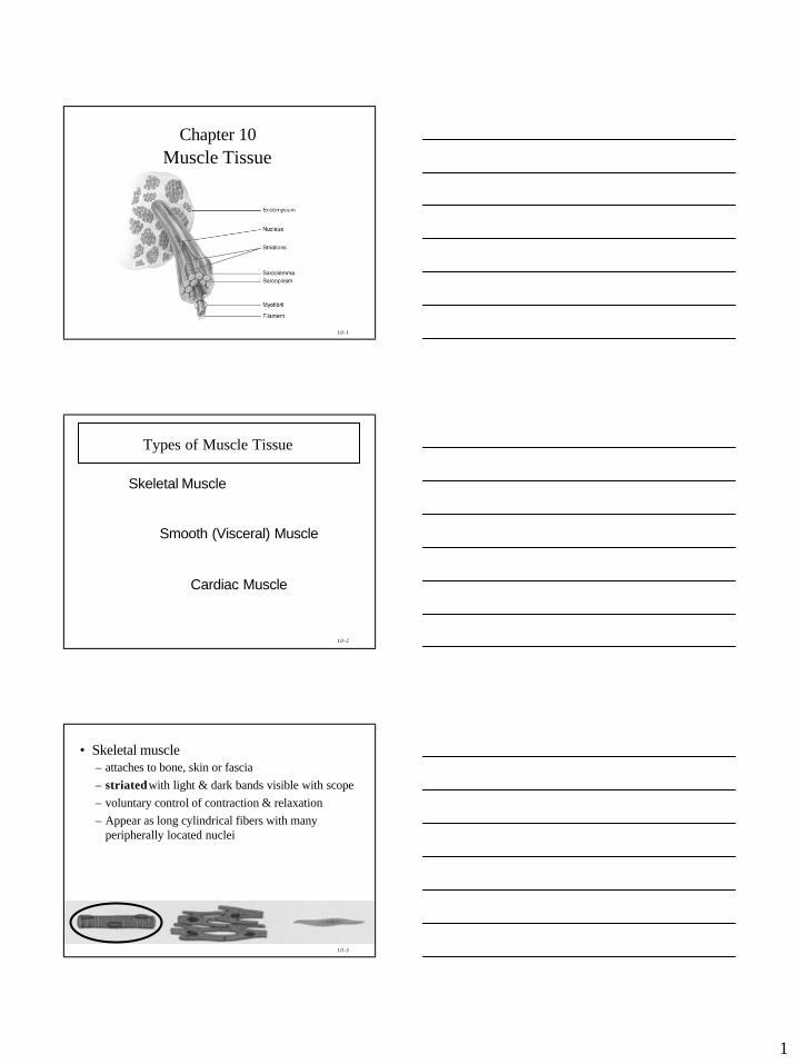
Chapter 10Muscle Tissue
10-2
Types of Muscle Tissue
Skeletal Muscle
Smooth (Visceral) Muscle
Cardiac Muscle
10-3
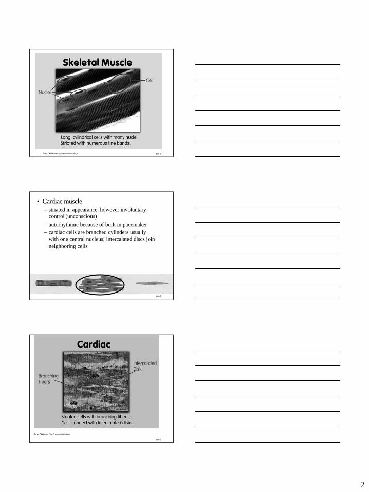
• Skeletal muscle– attaches to bone, skin or fascia– striatedwith light & dark bands visible with scope – voluntary control of contraction & relaxation– Appear as long cylindrical fibers with many
peripherally located nuclei
2
10-4From Oklahoma City Community College
10-5
• Cardiac muscle– striated in appearance, however involuntary
control (unconscious)– autorhythmic because of built in pacemaker– cardiac cells are branched cylinders usually
with one central nucleus; intercalated discs join neighboring cells
10-6
From Oklahoma City Community College
3
10-7
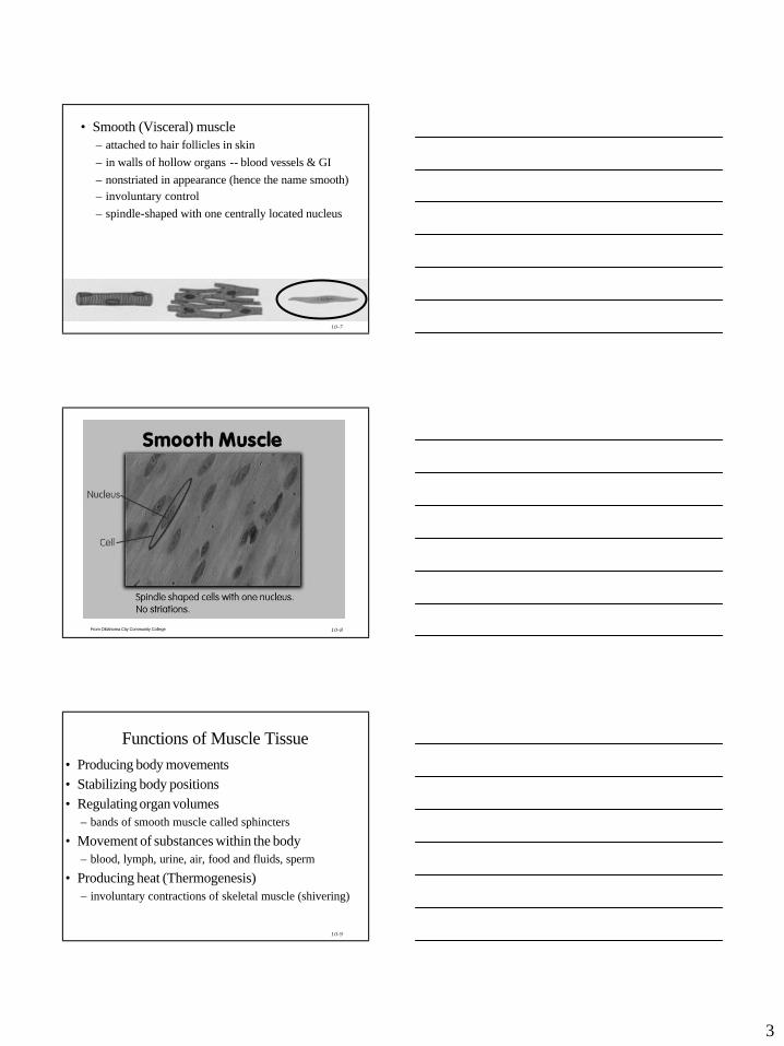
• Smooth (Visceral) muscle – attached to hair follicles in skin– in walls of hollow organs -- blood vessels & GI– nonstriated in appearance (hence the name smooth)– involuntary control– spindle-shaped with one centrally located nucleus
10-8From Oklahoma City Community College
10-9
Functions of Muscle Tissue• Producing body movements• Stabilizing body positions• Regulating organ volumes
– bands of smooth muscle called sphincters
• Movement of substances within the body– blood, lymph, urine, air, food and fluids, sperm
• Producing heat (Thermogenesis)– involuntary contractions of skeletal muscle (shivering)
4
10-10
Properties of Muscle Tissue• Excitability
– Ability to respond to certain stimuli by producing electrical signals called action potentials (impulses)
• Conductivity– ability to propagate the electrical signals over membrane
• Contractility– ability to shorten and thicken (contract) generate force
• Extensibility– ability to be stretched without damaging the tissue
• Elasticity– ability to return to original shape after being stretched
10-11
Skeletal Muscle: Fascia• Two types of Fascia: superficial and deep• Superficial fascia is loose connective tissue & fat
underlying the skin• it stores water and fat. Much of the fat of an
overweight person is in the superficial fascia.• it reduces the rate of heat loss• it provides mechanical protection against traumatic
blows• it provides a framework for nerves and blood
vessels to enter and exit muscle.
10-12
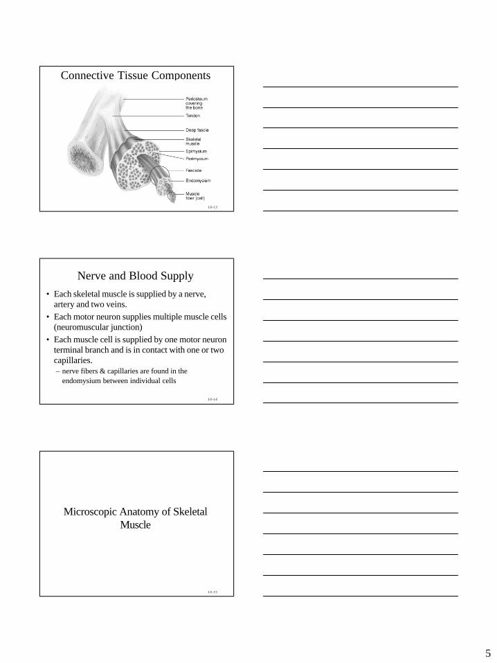
Skeletal Muscle: Fascia• Deep fascia = dense irregular connective tissue
around muscle– Epimysium = surrounds the whole muscle – Perimysium = surrounds bundles (fascicles) of
10-100 muscle cells– Endomysium = separates individual muscle
cells• All these connective tissue layers extend beyond
the muscle belly to form the tendon• Deep fascia also separates muscles into groups,
support nerves, blood vessels, and lymph vessels and fills in spaces between muscles.
5
10-13
Connective Tissue Components
10-14
Nerve and Blood Supply • Each skeletal muscle is supplied by a nerve,
artery and two veins.• Each motor neuron supplies multiple muscle cells
(neuromuscular junction)• Each muscle cell is supplied by one motor neuron
terminal branch and is in contact with one or two capillaries.– nerve fibers & capillaries are found in the
endomysium between individual cells
10-15
Microscopic Anatomy of Skeletal Muscle
6
10-16
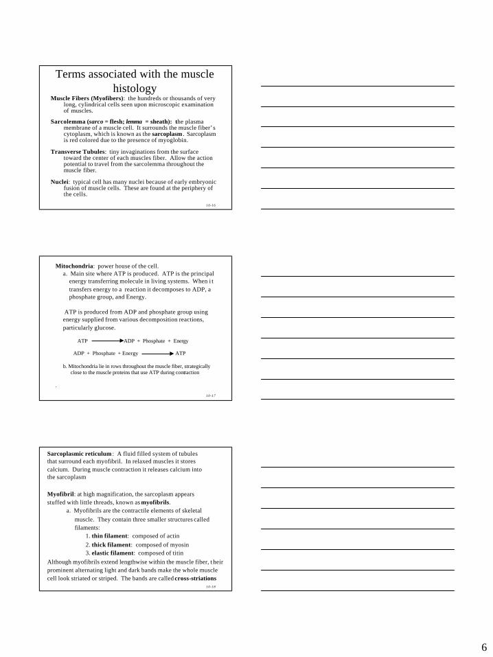
Terms associated with the muscle histology
Muscle Fibers (Myofibers): the hundreds or thousands of very long, cylindrical cells seen upon microscopic examination of muscles.
Sarcolemma (sarco = flesh; lemma = sheath): the plasma membrane of a muscle cell. It surrounds the muscle fiber’s cytoplasm, which is known as the sarcoplasm . Sarcoplasm is red colored due to the presence of myoglobin.
Transverse Tubules: tiny invaginations from the surface toward the center of each muscles fiber. Allow the action potential to travel from the sarcolemma throughout the muscle fiber.
Nuclei: typical cell has many nuclei because of early embryonic fusion of muscle cells. These are found at the periphery of the cells.
10-17
Mitochondria: power house of the cell. a. Main site where ATP is produced. ATP is the principal
energy transferring molecule in living systems. When i t transfers energy to a reaction it decomposes to ADP, aphosphate group, and Energy.
ATP is produced from ADP and phosphate group using energy supplied from various decomposition reactions, particularly glucose.
ATP ADP + Phosphate + Energy
ADP + Phosphate + Energy ATP
b. Mitochondria lie in rows throughout the muscle fiber, strategically close to the muscle proteins that use ATP during contraction
.
10-18
Sarcoplasmic reticulum : A fluid filled system of tubules that surround each myofibril. In relaxed muscles it stores calcium. During muscle contraction it releases calcium into the sarcoplasm
Myofibril: at high magnification, the sarcoplasm appears stuffed with little threads, known as myofibrils.
a. Myofibrils are the contractile elements of skeletal muscle. They contain three smaller structures called filaments:
1. thin filament: composed of actin 2. thick filament: composed of myosin3. elastic filament: composed of titin
Although myofibrils extend lengthwise within the muscle fiber, t heir prominent alternating light and dark bands make the whole musclecell look striated or striped. The bands are called cross-striations
7
10-19
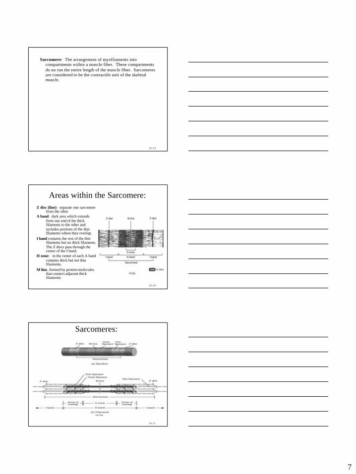
Sarcomere: The arrangement of myofilaments into compartments within a muscle fiber. These compartments do no run the entire length of the muscle fiber. Sarcomeres are considered to be the contractile unit of the skeletal muscle.
10-20
Areas within the Sarcomere:Z disc (line): separate one sarcomere
from the other A band: dark area which extends
from one end of the thick filaments to the other and includes portions of the thin filaments where they overlap.
I band contains the rest of the thin filaments but no thick filaments. The Z discs pass through the center of the I band.
H zone: in the center of each A band contains thick but not thin filaments.
M line , formed by protein molecules that connect adjacent thick filaments
10-21
Sarcomeres:
8
10-22
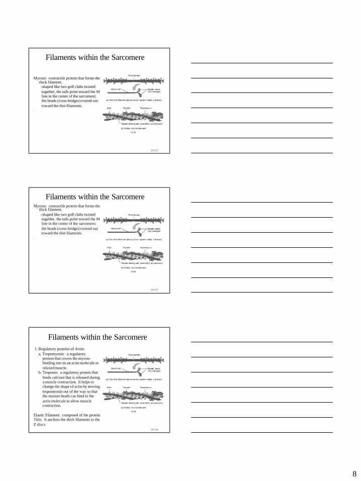
Filaments within the Sarcomere
Myosin: contractile protein that forms the thick filament.-shaped like two golf clubs twisted together, the tails point toward the M line in the center of the sarcomere; the heads (cross bridges) extend out toward the thin filaments.
.
10-23
Filaments within the SarcomereMyosin: contractile protein that forms the
thick filament.-shaped like two golf clubs twisted together, the tails point toward the M line in the center of the sarcomere; the heads (cross bridges) extend out toward the thin filaments.
.
10-24
Filaments within the Sarcomere1. Regulatory proteins of Actin:
a. Tropomyosin: a regulatory protein that covers the myosin binding site on an actin molecule in relaxed muscle.
b. Troponin: a regulatory protein that binds calcium that is released during a muscle contraction. It helps to change the shape of actin by moving tropomyosin out of the way so that the myosin heads can bind to the actin molecule to allow muscle contraction.
Elastic Filament: composed of the protein Titin. It anchors the thick filaments to the Z discs
9
10-25
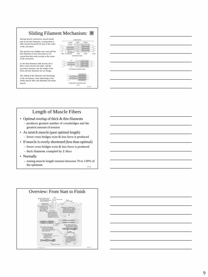
Sliding Filament Mechanism:During muscle contraction, myosin heads pull on the thin filaments, causing them to slide inward toward the H zone at the center of the sarcomere
The myosin cross bridges may even pull the thin filaments of each sarcomere so far inward that their ends overlap in the center of the sarcomere.
As the thin filaments slide inward, the Z discs come toward each other, and the sarcomere shortens, but the length of the thick and thin filaments do not change.
The sliding of the filaments and shortening of the sarcomeres cause shortening of the whole muscle fiber and ultimately the entire muscle
10-26
• Optimal overlap of thick & thin filaments– produces greatest number of crossbridges and the
greatest amount of tension
• As stretch muscle (past optimal length)– fewer cross bridges exist & less force is produced
• If muscle is overly shortened (less than optimal)– fewer cross bridges exist & less force is produced– thick filaments crumpled by Z discs
• Normally– resting muscle length remains between 70 to 130% of
the optimum
Length of Muscle Fibers
10-27
Overview: From Start to Finish
10
10-28
Role of CalciumThe sliding filament mechanism explains the mechanism of contraction, but the substance which starts and stops contraction is calcium: An increase in calcium in the sarcoplasm causes thesliding filament to start and a decrease in calcium in the sarcoplasm causes the sliding filaments to turn off.
Calcium affects the muscle contraction in 6 steps:1. a muscle action potential travels along the sarcolemma2. calcium release channels open up in the SR3. calcium floods the sarcoplasm around the thick and thin
filaments4. calcium combines with troponin5. this causes the troponin/tropomyosin complex to change
shape and uncover the myosin binding site on the actin molecules.
6. the muscle contracts
10-29
The Power Stroke and the Role of ATP in Muscle Contraction
5 Steps of the Power Stroke:
1. ATP from the mitochondria attaches to the myosin cross bridges (heads)2. ATP is slit into ADP and Phosphate via an enzyme ATPase. The Energy
reorients the myosin heads placing the myosin cross bridges in an activated state.
3. When calcium levels increase the troponin/tropomyosin complex moves allowing the activated myosin heads to bind to the actin molecules.
4. The myosin heads then swivel toward the center of the sarcomere. This swiveling is called the power stroke. As the heads swivel they release ADP, allowing another molecule of ATP to attach to the myosin head.
5. This process repeats over and over as long as there is enough ATP and as long as there is enough calcium.
10-30
Relaxation of Muscle Tissue6 Steps:
1. The action potential stops2. Acetylcholineesterase breaks down any ACh left in the
synaptic cleft3. Calcium release channels close in the SR4. Calcium active transport pumps turn on in the SR and
remove calcium from the sarcoplasm
5. Calsequestrin (calcium binding protein) binds to calcium in the SR
6. As calcium levels decrease, troponin/tropomyosin complex slides back into place over myosin binding sites.
11
10-31
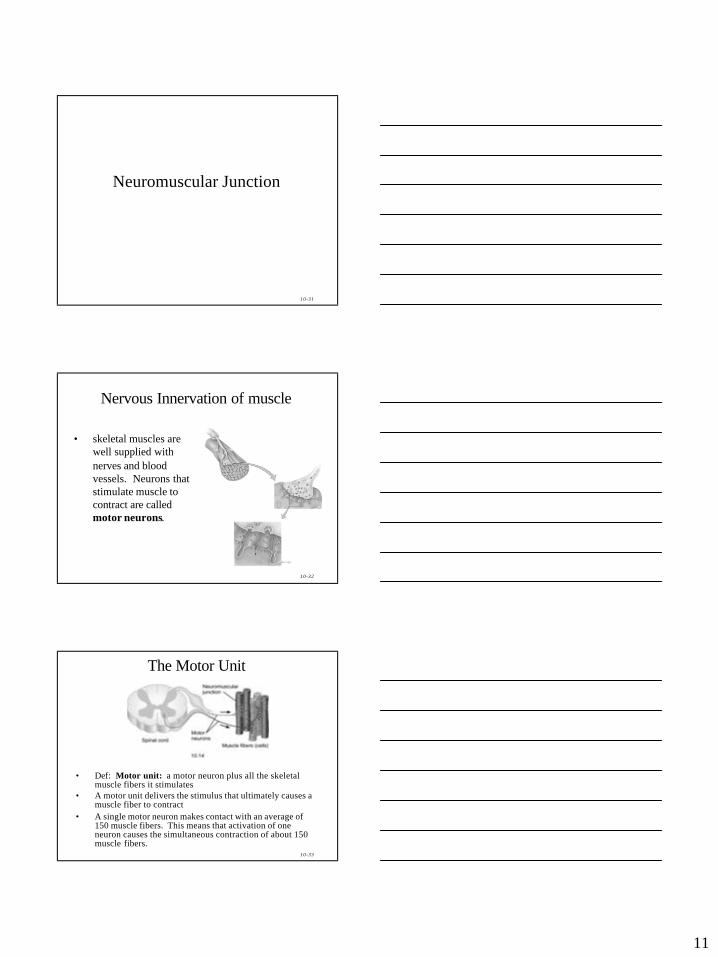
Neuromuscular Junction
10-32
Nervous Innervation of muscle
• skeletal muscles are well supplied with nerves and blood vessels. Neurons that stimulate muscle to contract are called motor neurons.
10-33
The Motor Unit
• Def: Motor unit: a motor neuron plus all the skeletal muscle fibers it stimulates
• A motor unit delivers the stimulus that ultimately causes a muscle fiber to contract
• A single motor neuron makes contact with an average of 150 muscle fibers. This means that activation of one neuron causes the simultaneous contraction of about 150 muscle fibers.
12
10-34
• all the muscle fibers of a motor unit contract and relax together.
• muscles that control precise movements have many small motor units. For example, muscles of the larynx that control voice production have as few as two or three muscle fibers per motor unit and muscles controlling eye movements may have 10-20 muscles fibers per motor unit.
• muscles of the body that are responsible for powerful gross movements, such as the biceps brachii in the arm and gastrocnemius in the leg, have some motor units with as many as 2000 muscle fibers each.
• stimulation of a motor neuron produces a contraction in all muscle fibers of a particular motor unit. Accordingly, the total strength of a contraction depends, in part, on which motor units are activated.
10-35
• since skeletal muscle fibers often are very long cells, the neuromuscular junction usually is located near the midpoint of the fiber.
• muscle action potentials then spread from the center of the fiber toward both ends. This arrangement permits nearly simultaneous contraction of all parts of the fiber
10-36
Neuromuscular Junction (NMJ) or Synapse
• NMJ = myoneural junction– end of axon nears the surface of a muscle fiber at its motor
end plate region (remain separated by synaptic cleft or gap)
13
10-37
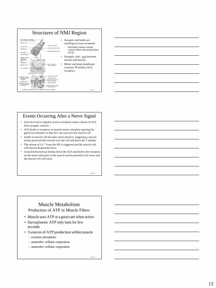
Structures of NMJ Region• Synaptic end bulbs are
swellings of axon terminals– End bulbs contain synaptic
vesicles filled with acetylcholine (ACh)
• Synaptic cleft: gap between neuron and muscle
• Motor end plate membrane contains 30 million ACh receptors.
10-38
Events Occurring After a Nerve Signal• Arrival of nerve impulse at nerve terminal causes release of ACh
from synaptic vesicles• ACh binds to receptors on muscle motor end plate opening the
gated ion channels so that Na+ can rush into the muscle cell
• Inside of muscle cell becomes more positive, triggering a muscleaction potential that travels over the cell and down the T tubules
• The release of Ca +2 from the SR is triggered and the muscle cell will shorten & generate force
• Acetylcholinesterase breaks down the ACh attached to the recepto rs on the motor end plate so the muscle action potential will cease and the muscle cell will relax.
10-39
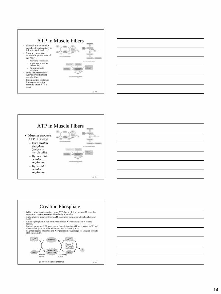
Muscle MetabolismProduction of ATP in Muscle Fibers
• Muscle uses ATP at a great rate when active• Sarcoplasmic ATP only lasts for few
seconds• 3 sources of ATP production within muscle
– creatine phosphate– anaerobic cellular respiration– anaerobic cellular respiration
14
10-40
ATP in Muscle Fibers• Skeletal muscle quickly
switches from inactivity to full activity & back.
• Muscle contraction requires huge amounts of ATP for:– Powering contraction– Pumping Ca+ into SR
(relaxation)– Other metabolic
reactions• Only a few seconds of
ATP is present inside muscle fibers.
• If contraction continues for more than a few seconds, more ATP is made.
10-41
ATP in Muscle Fibers
• Muscles produce ATP in 3 ways:– From creatine
phosphate(unique to muscle cells).
– By anaerobic cellular respiration
– By aerobic cellular respiration.
10-42
Creatine Phosphate• While resting, muscle produces more ATP than needed so excess ATP is used to
synthesize creatine phosphate (found only in muscle).• A phosphate is transferred from ATP to creatine forming creatine phosphate and
ADP.• Creatine phosphate is 3-6x more plentiful than ATP in sarcoplasm of relaxed
muscle.• During contraction ADP starts to rise (muscle is using ATP and creating ADP) and
creatine then gives back the phosphate to ADP creating ATP.• Together creatine phosphate and ATP provide enough energy for about 15 seconds
(100-meter dash).
15
10-43
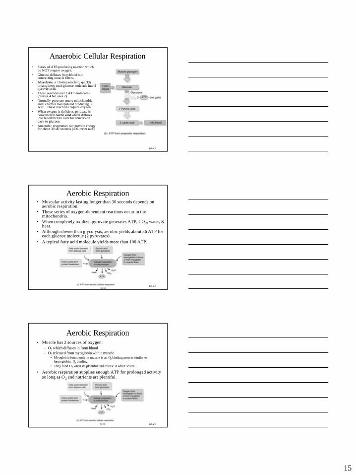
Anaerobic Cellular Respiration• Series of ATP-producing reaction which
do NOT require oxygen.• Glucose diffuses from blood into
contracting muscle fibers.• Glycolysis, a 10-step reaction, quickly
breaks down each glucose molecule into 2 pyruvic acid.
• These reactions net 2 ATP molecules (creates 4 but uses 2).
• Normally pyruvate enters mitochondria and is further manipulated producing 36 ATP. These reactions require oxygen.
• When oxygen is deficient, pyruvate is converted to lactic acid which diffuses into blood then to liver for conversion back to glucose.
• Anaerobic respiration can provide energy for about 30 -40 seconds (400 -meter race)
10-44
Aerobic Respiration• Muscular activity lasting longer than 30 seconds depends on
aerobic respiration.• These series of oxygen-dependent reactions occur in the
mitochondria.• When completely oxidize, pyruvate generates ATP, CO 2, water, &
heat.• Although slower than glycolysis, aerobic yields about 36 ATP for
each glucose molecule (2 pyruvates).• A typical fatty acid molecule yields more than 100 ATP.
10-45
Aerobic Respiration• Muscle has 2 sources of oxygen:
– O2 which diffuses in from blood– O2 released from myoglobin within muscle.
• Myoglobin found only in muscle is an O2 binding protein similar to hemoglobin. O2 binding
• They bind O2 when its plentiful and release it when scarce.
• Aerobic respiration supplies enough ATP for prolonged activity so long as O 2 and nutrients are plentiful.
16
10-46
Muscle Fatigue• Inability of a muscle to contract forcefully after a
prolonged activity.• Although precise mechanisms are not clear,
several factor are thought to contribute to muscle fatigue:– Inadequate release of Ca+ from SR– Decline of Ca+ concentration in SR.– Depletion of creatine phosphate.– O2 depletion– Lactic acid/ADP buildup– Surprisingly, ATP levels in fatigued muscles are not
much lower than resting muscles.
10-47
Oxygen Debt• After prolonged activity, heavy breathing
continues for a while and O2 consumption remains above resting level.
• Recovery period may be just a few minutes or several hours.
• Oxygen debt refers to O2 over and above the resting O2 need.
• Extra O2 was used to “pay back” or restore metabolic conditions to resting levels in 3 ways:– Convert lactic acid back to glycogen– Resynthesize creatine phosphate and ATP– Replace O 2 removed from myoglobin.
10-48
Oxygen Debt cont.• It is now know that:
– Only a small amount of glycogen resynthesis occurs from lactic acid
– Most glycogen is made much later from eating carbs.– Lactic acid which remains is converted back to pyruvic
acid and used for ATP via aerobic respiration in heart, liver, kidneys, skeletal muscle.
– Post exercise O 2 use is boosted by:• Elevated temperature after strenuous exercise increases
increase rate of chemical reactions.– Faster reactions use more ATP and require more O2
• For these reasons, recovery oxygen uptake is a better term.
17
10-49
Control of Muscle Tension• Motor units:
– Increasing strength requires increasing motor unit recruitment .
– Typically different motor units in a whole muscle are not stimulated to contract in unison. While some are contracting others are relaxing.
• Weakest motor units are used first than additional motor units a re added as needed until full contraction occurs.
• Twitch contraction:– Brief contraction of all muscle fibers in a motor unit in
response to a single impulse.• Lasts from 20-200ms
10-50
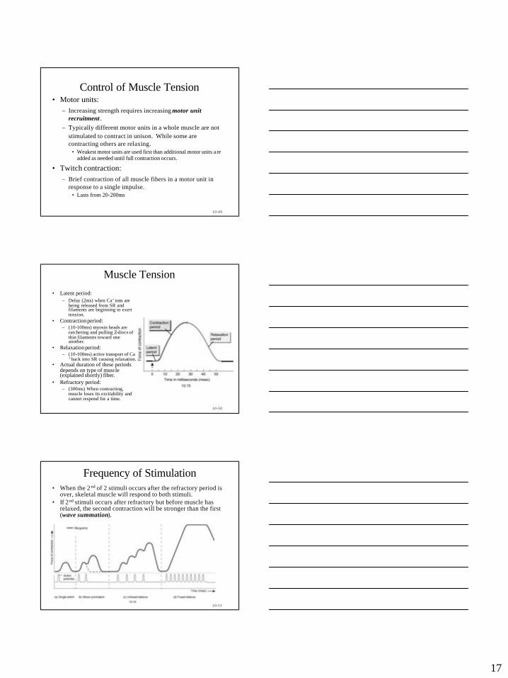
Muscle Tension• Latent period:
– Delay (2ms) when Ca+ ions are being released from SR and filaments are beginning to exert tension.
• Contraction period:– (10-100ms) myosin heads are
ratcheting and pulling Z-discs of thin filaments toward one another.
• Relaxation period:– (10-100ms) active transport of Ca
+ back into SR causing relaxation.• Actual duration of these periods
depends on type of muscle (explained shortly) fiber.
• Refractory period:– (300ms) When contracting,
muscle loses its excitability and cannot respond for a time.
10-51
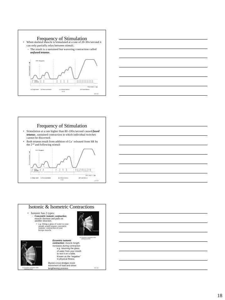
Frequency of Stimulation• When the 2 nd of 2 stimuli occurs after the refractory period is
over, skeletal muscle will respond to both stimuli.• If 2nd stimuli occurs after refractory but before muscle has
relaxed, the second contraction will be stronger than the first (wave summation).
18
10-52
Frequency of Stimulation• When skeletal muscle is stimulated at a rate of 20-30x/second it
can only partially relax between stimuli.– The result is a sustained but wavering contraction called
unfused tetanus .
10-53
Frequency of Stimulation• Stimulation at a rate higher than 80 -100x/second caused fused
tetanus , sustained contraction in which individual twitches cannot be discerned.
• Both tetanus result from addition of Ca + released from SR by the 2 nd and following stimuli
10-54
Isotonic & Isometric Contractions• Isotonic has 2 types:
– Concentric isotonic contraction: muscle shortens and pulls on another structure.
• e.g. lifting a glass of water to your mouth would require concentric isotonic contraction of your biceps muscle.
Eccentric isotonic contraction: muscle length increases during contraction
e.g. returning the glass of water from your mouth to rest it on a table. Known as the “negative ”in physical fitness.
Myosin-cross-bridges resist movement of load and slows lengthening process
19
10-55
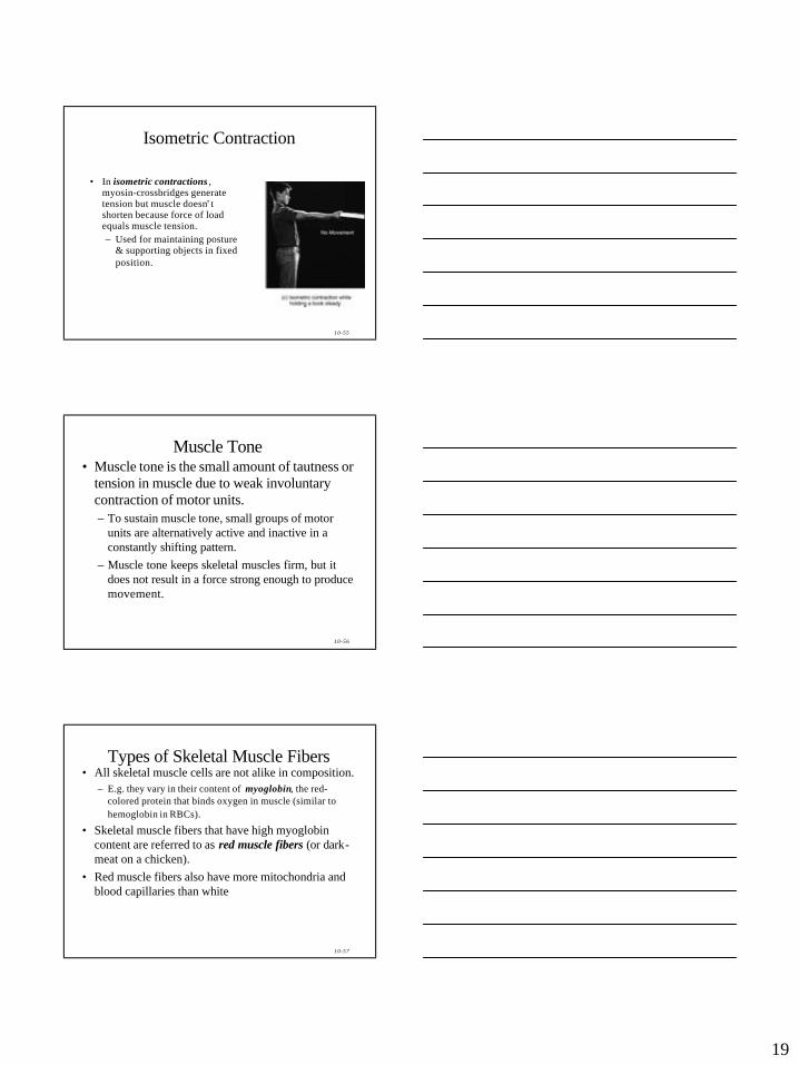
Isometric Contraction
• In isometric contractions , myosin-crossbridges generate tension but muscle doesn’ t shorten because force of load equals muscle tension.– Used for maintaining posture
& supporting objects in fixed position.
10-56
Muscle Tone• Muscle tone is the small amount of tautness or
tension in muscle due to weak involuntary contraction of motor units.– To sustain muscle tone, small groups of motor
units are alternatively active and inactive in a constantly shifting pattern.
– Muscle tone keeps skeletal muscles firm, but it does not result in a force strong enough to produce movement.
10-57
Types of Skeletal Muscle Fibers• All skeletal muscle cells are not alike in composition.
– E.g. they vary in their content of myoglobin, the red-colored protein that binds oxygen in muscle (similar to hemoglobin in RBCs).
• Skeletal muscle fibers that have high myoglobin content are referred to as red muscle fibers (or dark-meat on a chicken).
• Red muscle fibers also have more mitochondria and blood capillaries than white
20
10-58
Types of Skeletal Muscle Fibers
• Muscle fibers with low myoglobin content are called white muscle fibers (or white meat).
• Skeletal muscle fibers also contract and relax at different speeds.– Categorized as either slow or fast depending on how
rapidly ATPase in myosin heads splits ATP to ADP driving the myosin head to repeated pull actin.
• Skeletal muscle vary in which metabolic reactions they use to generate this ATP and how quickly they fatigue.
10-59
Types of Skeletal Muscle Fibers
• Based on these characteristics skeletal fibers are classified into 3 main types:
1. Slow oxidative fibers2. Fast oxidative-glycolytic fibers
3. Fast glycolytic fibers.
10-60
Slow Oxidative (SO) fibers• Smallest in diameter i.e. least powerful• Appear dark red
– Large myoglobin content & many capillaries• Many large mitochondria i.e. produce much ATP• Created ATP mainly via aerobic cellular respiration i.e. why
they are called oxidative use much oxygen to create ATP.• Slow since contraction cycle or “ rowing of myosin heads” is
slower pace than “ fast” fibers.• Twitch contraction is 100-200ms.• Very fatigue resistant• Capable of prolonged, sustained contractions for many hours• Used in posture muscles, & aerobic, endurance-type activities.
21
10-61
Fast Oxidative-Glycolytic (FOG) Fibers• Intermediate diameter (between SO & FG)• Large amounts of myoglobin & many blood vessels i.e.
also dark red.• Create much ATP via aerobic cellular respiration.• Moderately fatigue resistant .• Also have high glycogen content i.e. generate ATP via
glycolysis (anaerobic cellular respiration.• Speed of contraction faster than SO since they breakdown
ATP 3 -5x faster than SO. – Myosin heads row faster.
• Reach peak tension more quickly than SO but for briefer duration
• FOG used in activities such as walking & sprinting.
10-62
Fast Glycolytic (FG) Fibers• Largest diameter fibers
– Contain most myofibrils.• Generate most powerful contractions.• Low myoglobin content & few capillaries.• Contain large amounts of glycogen and generate ATP mainly
via glycolysis (anaerobic cellular respiration).• Fast-twitch fibers used for short-duration of intense strength
e.g. throwing a ball or weight lifting.• Weight lifting produces increases in size, strength, & glycogen
content of fast glycolytic fibers.• FG fibers of weight lifters may be 50% larger than sedentary
people.• Fatigue quickly.
10-63
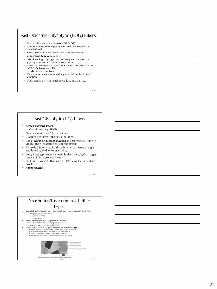
Distribution/Recruitment of Fiber Types
• Most whole skeletal muscles are a mixture of all three types of fibers (SO, FOG, FG).– The proportions vary depending on:
• Action of the muscle• Person’s training regimen• Genetic factors.
• Postural muscles have higher proportion of SO fibers.• Muscles of arms/shoulder have high proportion of FG• Legs have large numbers of both SO & FOG.• Skeletal muscle fibers of any given motor unit are all the same type.
– Different motor units are recruited in specific order, depending on need.– If weak contraction is suffice only SO motor units are activated .– If more force is needed, FOG motor units are recruited.– If maximal force is required FG motor units are called into action .
22
10-64
Exercise & Skeletal Muscle
• Ratio of fast-twitch & slow-twitch in each muscle is genetically determined and helps account for individual differences in physical performance.
• Various type of exercises can induce changes in fiber types.• Endurance type (aerobic ) exercises, cause gradual
transformation of some FG to FOG.– These exercises also result in CV and respiratory changes so ske letal
muscles receive better supplies of oxygen and nutrients.
• Strength type exercises produce a size & strength of FG fibers.– Size increases is due to increased synthesis of thick & thin filaments
resulting in hypertrophy .
10-65
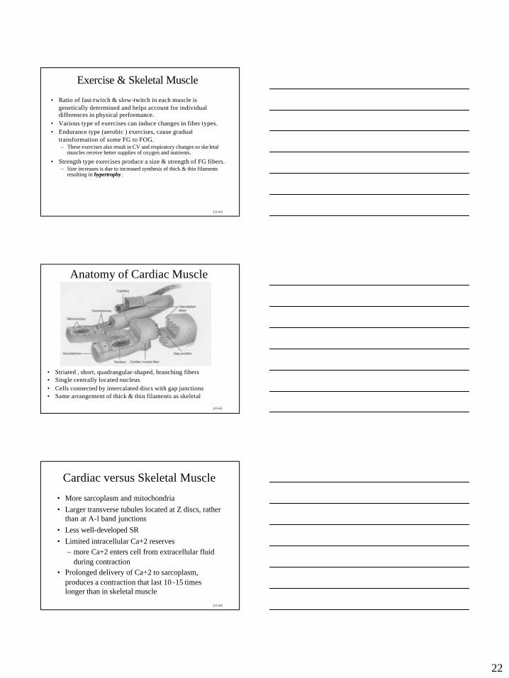
Anatomy of Cardiac Muscle
• Striated , short, quadrangular-shaped, branching fibers • Single centrally located nucleus• Cells connected by intercalated discs with gap junctions• Same arrangement of thick & thin filaments as skeletal
10-66
Cardiac versus Skeletal Muscle
• More sarcoplasm and mitochondria• Larger transverse tubules located at Z discs, rather
than at A-l band junctions• Less well-developed SR• Limited intracellular Ca+2 reserves
– more Ca+2 enters cell from extracellular fluid during contraction
• Prolonged delivery of Ca+2 to sarcoplasm, produces a contraction that last 10 -15 times longer than in skeletal muscle
23
10-67
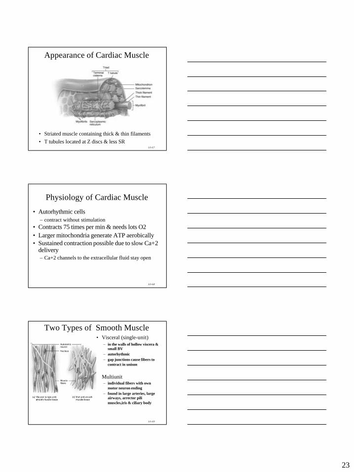
Appearance of Cardiac Muscle
• Striated muscle containing thick & thin filaments• T tubules located at Z discs & less SR
10-68
Physiology of Cardiac Muscle
• Autorhythmic cells– contract without stimulation
• Contracts 75 times per min & needs lots O2 • Larger mitochondria generate ATP aerobically • Sustained contraction possible due to slow Ca+2
delivery– Ca+2 channels to the extracellular fluid stay open
10-69
Two Types of Smooth Muscle• Visceral (single-unit)
– in the walls of hollow viscera & small BV
– autorhythmic– gap junctions cause fibers to
contract in unison
• Multiunit – individual fibers with own
motor neuron ending– found in large arteries, large
airways, arrector pili muscles,iris & ciliary body
24
10-70
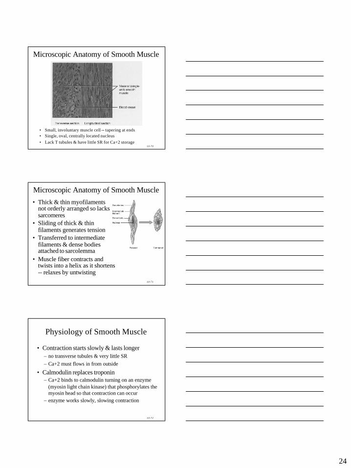
Microscopic Anatomy of Smooth Muscle
• Small, involuntary muscle cell -- tapering at ends• Single, oval, centrally located nucleus• Lack T tubules & have little SR for Ca+2 storage
10-71
Microscopic Anatomy of Smooth Muscle• Thick & thin myofilaments
not orderly arranged so lacks sarcomeres
• Sliding of thick & thin filaments generates tension
• Transferred to intermediate filaments & dense bodies attached to sarcolemma
• Muscle fiber contracts and twists into a helix as it shortens -- relaxes by untwisting
10-72
Physiology of Smooth Muscle
• Contraction starts slowly & lasts longer– no transverse tubules & very little SR– Ca+2 must flows in from outside
• Calmodulin replaces troponin– Ca+2 binds to calmodulin turning on an enzyme
(myosin light chain kinase) that phosphorylates the myosin head so that contraction can occur
– enzyme works slowly, slowing contraction
25
10-73
Smooth Muscle Tone
• Ca+2 moves slowly out of the cell– delaying relaxation and providing for state of
continued partial contraction – sustained long-term
• Useful for maintaining blood pressure or a steady pressure on the contents of GI tract
10-74
Regulation of Contraction• Regulation of contraction due to
– nerve signals from autonomic nervous system– changes in local conditions (pH, O2, CO2, temperature
& ionic concentrations)– hormones (epinephrine -- relaxes muscle in airways &
some blood vessels)
• Stress-relaxation response– when stretched, initially contracts & then tension
decreases to what is needed– stretch hollow organs as they fill & yet pressure
remains fairly constant– when empties, muscle rebounds & walls firm up
10-75
Regeneration of Muscle• Skeletal muscle fibers cannot divide after 1st year
– growth is enlargement of existing cells
– repair• satellite cells & bone marrow produce some new cells
• if not enough numbers---fibrosis occurs most often• Cardiac muscle fibers cannot divide or regenerate
– all healing is done by fibrosis (scar formation)• Smooth muscle fibers (regeneration is possible)
– cells can grow in size (hypertrophy)– some cells (uterus) can divide (hyperplasia)
– new fibers can form from stem cells in BV walls
26
10-76
Muscle Tissue Regeneration
• Immature skeletal muscle fibers lose their ability to divide i.e. skeletal muscle growth after birth is due to hypertrophy rather than hyperplasia .
• When significant skeletal muscle damage or degeneration occurs, skeletal muscle undergoes fibrosis, replacement of muscle fibers by fibrous scar tissue.– i.e. skeletal muscle can only regenerate to a limited
extent.