Embed Size (px)
Citation preview
1
Changes of parvalbumin expression in the spinal cord
after peripheral inflammation
Gisela Zacharova, David Sojka and Jiri Palecek
Department of Functional Morphology, Institute of Physiology,
Academy of Sciences of the Czech Republic, Prague, Czech Republic.
17 pages, 5 Figures
Short title: PARVALBUMIN EXPRESSION IN THE SPINAL CORD
Corresponding author: Jiri Palecek M.D., Ph.D. Department of Functional Morphology Institute of Physiology ASCR Videnska 1083 142 20 Praha 4 Czech Republic e-mail: [email protected] office: (-420) 2 4106 2664 fax: (-420) 2 4106 2488

2
SUMMARY
Parvalbumin (PV) is a calcium binding protein that is expressed by numerous neuronal
subpopulations in the central nervous system. Staining for PV was often used in
neuroanatomical studies in the past. Recently, several studies have suggested that PV acts in
neurons as a mobile endogenous calcium buffer that affects temporo-spatial characteristics of
calcium transients and is involved in modulation of synaptic transmission. In our experiments,
expression of PV in the lumbar dorsal horn spinal cord was evaluated using densitometric
analysis of immunohistological sections and Western-blot techniques in control and arthritic
rats. There was a significant reduction of PV immunoreactivity in the superficial dorsal horn
region ipsilateral to the arthritis after induction of the peripheral inflammation. The ipsilateral
area and intensity of PV staining in this area were reduced to 38% and 37% respectively, out of
the total PV staining on both sides. It is suggested that this reduction may reflect decreased
expression of PV in GABAergic inhibitory neurons. Reduction of PV concentration in the
presynaptic GABAergic terminals could lead to potentiation of inhibitory transmission in the
spinal cord. Our results suggest that changes in expression of calcium binding proteins in spinal
cord dorsal horn neurons may modulate nociceptive transmission.
Key words: arthritis, calcium buffer, hyperalgesia, GABA, parvalbumin

3
INTRODUCTION
Numerous proteins and receptors were shown to play an important role in modulation of
nociceptive signaling at spinal cord level. Parvalbumin (PV) is a calcium binding protein (CBP)
that has often been used as a marker of specific neuronal populations in neuroanatomical and
immunohistological studies in the past. Recently several studies have considered the biophysical
consequences of the presence of PV in the cells as a calcium buffer (Neher and Augustine 1992;
Zhou and Neher 1993). Synaptic activity as well as number of other cellular processes are
regulated by local and/or global changes of intracellular calcium level. During propagation of
action potentials, intracellular calcium level is increased mainly due to calcium influx through
voltage dependent calcium channels. Other important sources of extracellular calcium in the
dendrites during synaptic activity are calcium permeable NMDA and AMPA channels. The
intracellular calcium concentration is further regulated by several processes, such as calcium
uptake into intracellular stores, extrusion mechanisms and endogenous calcium buffering
(Berridge et al. 2003).
CBPs such as PV, calbindin and others represent mobile endogenous buffers that can
substantially affect the temporo-spatial characteristics of any intracellular calcium concentration
change, depending on their respective biophysical calcium binding characteristics. It was shown
in other neuronal systems that a low level of CBP leads to high amplitude short lasting calcium
concentration changes, while the presence of a high level of calcium buffers in the cell leads to
low amplitude, longer duration calcium transients under the same circumstances (Chard et al.
1993; Palecek et al. 1999). While both fast (calbindin) and slow (PV) calcium buffers can
reduce the calcium transient peak, PV selectively increases the fast component of the calcium
transient decay (Chard et al. 1993; Caillard et al. 2000; Lee et al. 2000; Schmidt et al. 2003;
Muller et al. 2007).
It is acknowledged that in some chronic pain states associated with the presence of
pathologically increased sensitivity such as allodynia and hyperalgesia, central sensitization due

4
to synaptic modulation at the spinal cord level may play an important role (Woolf and
Thompson 1991; Willis 2001). It was previously suggested that this process is calcium
dependent (Willis 2001; see also Malenka and Nicoll 1999). The presence or absence of CBPs
acting as calcium buffers in the pre- or postsynaptic part of the synapse could thus effectively
modulate synaptic transmission at the spinal cord level. Studies of PV distribution in the lumbar
spinal cord have shown that it is mostly located in a strongly immunopositive band of small
neuronal cell bodies and neuropil in the inner part of lamina II (Antal et al. 1990; Ren and Ruda
1994) and at the border of laminae II/III (Yamamoto et al.1989). There is also strong PV
immunostaining present in the neck of the spinal cord grey matter in the ventro-medial region
(lamina VII). Numerous PV positive neurons are present throughout the deeper laminae and also
in the ventral horn (Ren and Ruda 1994).
In this study the changes of PV expression in the lumbar spinal cord dorsal horn after
experimentally induced peripheral inflammation were observed in rats. The level of PV
expression was assessed by densitometry in the superficial dorsal horn and in the ventro-medial
part of the spinal cord on immunohistochemically stained sections from lumbar segment L5.
Further confirmation of the changes in PV expression after peripheral inflammation was
provided by western blot analysis of the spinal cord tissue.
METHODS
All procedures used in these experiments were reviewed and approved by the Institutional
Animal Care and Use Committee and were consistent with the guidelines of the International
Association for the Study of Pain for the care and use of laboratory animals. Adult male Wistar
rats (250 – 350 g) were kept in plastic cages with soft bedding, free access to food and water and
were maintained on a 12 hr light, 12 hr dark cycle. In total, 16 animals were used in this study.
Experimental arthritis was induced by unilateral intra-articular knee injection of a 3%
mixture of kaolin and carrageenan in saline solution under ether anesthesia. The animals were

5
left to recover in their home cages. Animals were deeply anesthetized with sodium pentobarbital
(80 mg/kg intraperitoneally) 28h later and perfused transcardially with heparinized saline (0.9%
NaCl) followed by 500 ml of ice cold 4% paraformaldehyde in 0.1 M phosphate buffer. Spinal
cord tissue was removed after the perfusion, the control side marked with a pin, and post-fixed
overnight in the same fixative and cryoprotected 24h in 30% sucrose solution in phosphate
buffer (PB). Transverse cryostat sections (30 μm) from the L5 spinal cord segment were
obtained from 5 experimental and 3 intact control rats. Every 3rd slice was collected and
immunostained for PV by the streptavidin-biotin-peroxidase complex method. First, the sections
were washed with a solution of 0.1 M phosphate-buffered-saline (PBS) 3 times for 10 min and
then incubated in blocking solution of 3% normal donkey serum (NDS) for 30 min at room
temperature, followed by overnight incubation at 4°C in primary antibody solution (mouse
monoclonal anti-parvalbumin; Sigma). After incubation, tissue sections were washed in 1%
NDS 3 times for 10 min and incubated for 2 hrs at room temperature in biotinylated secondary
antibody solution (1:400 for 2 hrs biotin-SP-conjugated donkey anti-mouse IgG; Jackson
Immuno Research Laboratories, Inc., USA). Then the sections were washed in PB twice for 10
min and incubated in peroxidase-conjugated streptavidin (dilution 1:600, peroxidase-conjugated
streptavidin; Jackson Immuno Research) for 2 hrs at room temperature. The reaction product
was visualized with 0.01% hydrogen peroxide and 0.05% diaminobenzidine as the chromogen.
After rinsing in 0.1M PB for 10 min, the sections were mounted onto gelatinized slides and
coverslipped. The whole spinal cord section was photographed with an Olympus BX-80 and
5Mpix Digital camera in one image. Off line densitometric analysis of the spinal cord images
was performed using the AIDA software package (see Fig.1). Two symmetrical areas on each
side in the superficial dorsal horn and in the ventromedial part were selected for analysis (Fig.
1). A small area in the part of the tissue without positive signal was selected to determine the
threshold value for the immunopositive signal intensity. The software calculated the area of
positivity within the pre-selected region, the sum of intensity in these areas, and the quotient of

6
these values in absolute values and in percent. The percent values on a given side of the
analyzed section were calculated as the percentage of immunopositive area or intensity,
compared to the sum of positive areas or intensities on both sides. Percent values were used for
statistical analysis to account for the differences between animals. The differences in the
intensity of immunostaining in a side to side comparison were evaluated using paired t-tests.
For further confirmation of PV expression levels, SDS-PAGE and western blotting were
used in another group of animals. Rats were anesthetized with sodium pentobarbital (80 mg/kg
ip) and laminectomy was performed. Dissected lumbar spinal cord was rapidly immersed in ice-
cold physiological solution and hemisected. Left and right side spinal cord parts (L3-5) were
weighed and homogenized with a hand-held pellet pestle in 30 mM Tris-HCl buffer (w/v 1:10)
containing 0,1 μl of an inhibitor of proteases (Sigma P8340) per 1 mg of tissue. Prepared
samples were left on the ice for 10 min and centrifuged at 5000 rpm for 10 min at 4°C.
Supernatant was removed and centrifuged again at 13000 rpm for 20 min. An aliquot was taken
for spectrophotometric total protein quantification using an Eppendorf BioPhotometer
(Eppendorf, Germany). The protein concentration for each sample was determined by the
Bradford assay using a Bio-Rad Protein Assay kit according to the manufacturer’s
recommendations. For immunodetection of parvalbumin, protein samples and molecular mass
standards (Bio-Rad) were resolved on 12% Tris/Tricine gels. Tris/Tricine SDS-PAGE was
performed according to the method of Schägger and Jagow (1991). Proteins were then
transferred to a 0.2 µm nitrocellulose membrane with blotting apparatus (Bio-Rad), at 15V
overnight. Western blots were developed as described by Towbin et al. (1979). Mouse
monoclonal anti-parvalbumin (Sigma) was used as primary antibody at a dilution of 1:2000. The
secondary antibody conjugated to horseradish peroxidase and/or biotinylated antibody followed
by streptavidin peroxidase (Jackson Immuno Research Laboratories, Inc., USA) were used at a
dilution of 1:6000, 1:32000 and 1:20000 respectively. The proteins were visualized with an ECL
kit (Bio-Rad) and data were captured using Fuji LAS-1000 Imaging System CCD Camera.

7
Quantitative analysis of immunoblots was performed using the image analysis program ImageJ
(version 1.35h).
RESULTS
In this study the intensity of immunostaining for parvalbumin in ipsilateral and contralateral
sides of the spinal cord in rats with unilateral experimental knee joint arthritis was analyzed.
Two regions with high intensity immunostaining for PV within the dorsal horn were examined
with densitometric analysis. One was in the superficial dorsal horn (substantia gelatinosa) and
the second was in the ventro-medial part of the dorsal horn. Five experimental and 3 control,
intact animals were used in the study. From each rat, 10 sections from lumbar segment L5 were
analyzed.
Lower intensity and/or smaller area of positive parvalbumin staining in the substantia
gelatinosa on the arthritic side was clearly evident in about 2/3 of the sections examined (Fig.2).
The staining was present mostly in the neuropil with occasional immunopositive cell bodies, as
can be seen on the higher magnification picture of the dorsal horn area (Fig.3). The differences
in the density and in the area of PV staining between the control side and the side ipsilateral to
the induced arthritis were assessed by densitometric analysis. In the superficial dorsal horn, both
area and the intensity of the PV immunostaining were confirmed to be significantly decreased
on the side of the experimental arthritis when compared to the contralateral side (Fig. 4). The
difference in the staining was even more significant when it was evaluated as an intensity/area
quotient. In the ventro-medial part of the dorsal horn no apparent difference was obvious. The
densitometric analysis also did not reveal any significant difference in the area and intensity of
the PV staining in the ventro-medial part of the spinal cord. However, the quotient of the
intensity/area was significantly reduced (P < 0.05) on the side of the experimental arthritis (Fig.
4). In control rats, no significant difference was revealed by the densitometric measurement in
any of the measured parameters when left and right sides of the spinal cord were compared in
8
the same way as in the experimental animals. The percent values were 49.91% to 50.09% for the
area and 49.82% to 50.18% for the intensity of the total in a left to right side comparison for the
substantia gelatinosa region and 49.87% to 50.13% (area) and 49.81% to 50.19 (intensity) for
the ventromedial region.
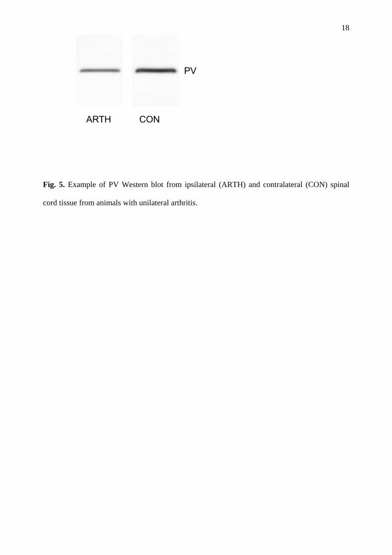
Western blot analysis of PV content in the spinal cord samples was used to confirm the
densitometric immunohistochemical analysis (Fig. 5). Tissue from ipsilateral and contralateral
sides of the spinal cord lumbar enlargement (L3-L5) in 5 animals was analysed. The PV content
in the spinal cord tissue ipsilateral to the arthritis was significantly reduced to 68 ± 12% (SEM,
p<0.05, paired t-test) when compared to the control side. There was no significant difference in
the PV content of the left and right sides in the control animals (n=3).
DISCUSSION
Our study demonstrated that peripheral inflammation can induce decreased expression of
PV in the spinal cord dorsal horn. The possible functional consequences of this change would be
preferentially dependent on its precise morphological location and on the biophysical properties
of PV.
It was shown previously that unilateral peripheral inflammation can induce bilateral
changes in expression of some molecules at the spinal cord level after peripheral inflammation.
However, in most of the studies the effect was much more pronounced on the side ipsilateral to
the unilateral inflammation (Traub et al. 1994; Castro–Lopez et al. 1994; Zhang et al. 2002).
These changes may be also dependent on the exact model of inflammation used (complete
Freund’s adjuvant vs. carageenan) and the time when the changes were evaluated after the
induction of inflammation. In our experiments we did not observe any obvious changes on the
contralateral side when compared to the control animals. In densitometric studies it is usually
difficult to compare density of staining in different tissue slices, unless a very significant change

9
in staining is present. Therefore we did not attempt to do densitometric comparisons between the
control and experimental animals.
The presence of different calcium binding proteins such as parvalbumin or calbindin in the
spinal dorsal horn was described in detailed morphological studies at light (Yamamoto et al.
1989; Antal et al. 1990; Zhang et al. 1990; Ren and Ruda 1994) as well as electron-microscopic
levels (Antal et al. 1990). In general, these calcium binding proteins, which can act as calcium
buffers and have impact on intracellular calcium kinetics, were found to be present in all layers
of the spinal cord. However, the distribution within the spinal cord was different for each of the
CBP. Parvalbumin appears to be predominantly expressed in a strongly PV immunopositive
band in the inner part of lamina II (Antal et al. 1990; Ren and Ruda 1994), or along the lamina
II/III border (Yamamoto et al. 1989), which is in good agreement with the present study.
Morphological analysis of the dorsal horn lumbar spinal cord in rats suggested, that PV is
located mainly in the neuropil near spinal cord PV immunoreactive neurons (Yamamoto et al.
1989). This was further supported by the finding that the intensity of the PV immunoreactive
band in the superficial dorsal horn appeared to be unaffected by dorsal rhizotomy or by neonatal
capsaicin treatment (Yamamoto et al. 1989). It was only slightly reduced after ganglionectomy
(Ren and Ruda 1994). Parvalbumin immunoreactivity was found in the postsynaptic as well as
in the presynaptic terminals in the inner part of lamina II (Antal et al. 1990), which indicates a
possible important role for parvalbumin in modulation of sensory inputs both at presynaptic and
postsynaptic levels.
The morphological and functional characterization of neurons in the substantia gelatinosa
is quite complex. However, it is clear from numerous studies that neurons in this region are
heavily involved in pain transmission and modulation (Willis and Coggeshall 2004). It was
shown previously, that about 43% of the neurons in this region are GABA immunoreactive and
are thus presumably GABAergic inhibitory interneurons (Todd and Sullivan 1990). It was
suggested that in other areas of the central nervous system PV containing neurons represent a

10
subpopulation of GABAergic neurons (see Baimbridge et al. 1992 for a review). At the spinal
cord level, it was shown that 5-21% of the GABA immunopositive neurons in the substantia
gelatinosa region coexpress PV (Antal et al. 1991). Similar results were shown also for a
subpopulation of substantia gelatinosa GABAergic neurons expressing green fluorescent protein
in transgenic mice (Hantman et al. 2004). However, the population of PV immunopositive
neurons in this spinal cord dorsal horn region (laminae II + III) that show staining also for
GABA is much higher (75%, Laing et al. 1994; 60-70%, Antal et al. 1991). These data suggest
that a majority of the PV immunopositive neurons in this region of the spinal cord dorsal horn
may be GABAergic inhibitory interneurons. Based on this evidence it seems plausible to
suggest that some of the changes in PV expression in the substantia gelatinosa region seen in our
study were due to reduced expression of PV in GABAergic interneurons.
Calcium ions regulate a number of intracellular processes and play a major role in
transmembrane signalling in neuronal cells (Baimbridge et al. 1992; Berridge et al. 2003). The
role of internal calcium buffers and the temporo-spatial characteristics of intracellular calcium
signalling was originally demonstrated in bovine chromaffin cells (Neher and Augustine 1992).
PV acts as a mobile calcium buffer with relatively slow binding characteristics and its presence
does not reduce the peak of the calcium transient as much as the fast buffers do, but rather
accelerates the initial decay of the calcium transient (Chard et al. 1993; Caillard et al. 2000; Lee
et al. 2000; Schmidt et al. 2003; Muller et al. 2007). PV presence leads to a fast reduction of
calcium concentration and decreased residual calcium concentration after the calcium transient
(Atluri and Regehr 1996). The importance of PV for synaptic modulation was shown at the
interneuron/Purkinje cell synapse, where elimination of PV from the presynaptic ending
changed a depressing synapse into a facilitating one (Caillard et al. 2000; Collin et al. 2005).
Similar results were obtained at the Calyx of Held synapse, where presynaptic PV accelerated
the decay of the calcium transient and short term facilitation (Muller et al. 2007). The increased

11
rate of decay of the calcium transient produced by PV was also shown in rat dorsal root ganglion
neurons (Chard et al. 1993).
A transient decrease of parvalbumin immunoreactivity was described in cultured cortical
neurons, where a time and dose-dependent reversible decrease in parvalbumin and GAD67
immunoreactivity was present after application of an NMDA receptor antagonist specifically in
PV interneurons (Kinney et al. 2006). Marked reduction of PV expression in interneurons was
described also in the hippocampus of spontaneously hypertensive rats with stroke, where it was
correlated with increased immunoreactivity for PKCgamma in pyramidal cells (De Jong et al.
1993). PKCgamma was shown to be considerably up-regulated under peripheral inflammatory
conditions also in lamina II of the lumbar dorsal horn in rats, where it has a close relationship to
but no co-expression with PV immunoreactivity (Martin et al. 1999).
In our study we have shown that peripheral inflammation leads to decreased expression of
PV in the neuropil of the spinal cord dorsal horn, presumably originating from the processes of
GABAergic interneurons. Decreased content of PV in the presynaptic endings of these
interneurons could lead to increased residual calcium level during high frequency bursts of
activity and potentiate facilitation and increased transmitter release. This would lead to
increased potency of inhibitory circuits at the spinal cord level under peripheral inflammatory
conditions. While the exact role of PV in the dorsal horn spinal cord neurons needs to be
examined further, it seems plausible to suggest that changes in expression of calcium binding
proteins may play an important role in the modulation of nociceptive transmission.
ACKNOWLEDGEMENTS
The authors thank Mrs. K. Gabova and M. Pytlova for their excellent technical help.
Supported by GACR 305/06/1115, GACR 304/08/0256, MSMT CR LC554, MYORES LSH-
CT-2004-511978, AV0Z 50110509

12
REFERENCES
ANTAL M, FREUND TF, POLGAR E: Calcium-binding proteins, parvalbumin- and calbindin-D
28k-immunoreactive neurons in the rat spinal cord and dorsal root ganglia: a light and electron
microscopic study. J Comp Neurol 295:467-484, 1990.
ANTAL M, POLGAR E, CHALMERS J, MINSON JB, LLEWELLYN-SMITH I, HEIZMANN CW,
SOMOGYI P: Different populations of parvalbumin- and calbindin-D28k-immunoreactive
neurons contain GABA and accumulate 3H-D-aspartate in the dorsal horn of the rat spinal cord.
J Comp Neurol 314:114-124, 1991.
ATLURI PP, REGEHR WG: Determinants of the time course of facilitation at the granule cell to
Purkinje cell synapse. J Neurosci 16:5661-5671, 1996.
BAIMBRIDGE KG, CELIO MR, ROGERS JH: Calcium-binding proteins in the nervous system.
Trends Neurosci 15:303-308, 1992.
BERRIDGE MJ, BOOTMAN MD, RODERICK HL: Calcium signalling: dynamics, homeostasis
and remodelling. Nat Rev Mol Cell Biol 4:517-529, 2003.
CAILLARD O, MORENO H, SCHWALLER B, LLANO I, CELIO MR, MARTY A: Role of the
calcium-binding protein parvalbumin in short-term synaptic plasticity. Proc Natl Acad Sci U S A
97:13372-13377, 2000.
CASTRO-LOPES JM, TAVARES I, TOLLE TR, COIMBRA A: Carrageenan-induced
inflammation of the hind foot provokes a rise of GABA-immunoreactive cells in the rat spinal
cord that is prevented by peripheral neurectomy or neonatal capsaicin treatment. Pain 56:193-
201, 1994.
CHARD PS, BLEAKMAN D, CHRISTAKOS S, FULLMER CS, MILLER RJ: Calcium buffering
properties of calbindin D28k and parvalbumin in rat sensory neurones. J Physiol 472:341-357,
1993.
COLLIN T, CHAT M, LUCAS MG, MORENO H, RACAY P, SCHWALLER B, MARTY A, LLANO
I: Developmental changes in parvalbumin regulate presynaptic Ca2+ signaling. J Neurosci
25:96-107, 2005.
DE JONG GI, VAN DER ZEE EA, BOHUS B, LUITEN PG: Reversed alterations of

13
hippocampal parvalbumin and protein kinase C-gamma immunoreactivity after stroke in
spontaneously hypertensive stroke-prone rats. Stroke 24:2082-2085; discussion 2086, 1993.
HANTMAN AW, VAN DEN POL AN, PERL ER: Morphological and physiological features of a
set of spinal substantia gelatinosa neurons defined by green fluorescent protein expression. J
Neurosci 24:836-842, 2004.
KINNEY JW, DAVIS CN, TABAREAN I, CONTI B, BARTFAI T, BEHRENS MM: A specific role
for NR2A-containing NMDA receptors in the maintenance of parvalbumin and GAD67
immunoreactivity in cultured interneurons. J Neurosci 26:1604-1615, 2006.
LAING I, TODD AJ, HEIZMANN CW, SCHMIDT HH: Subpopulations of GABAergic neurons in
laminae I-III of rat spinal dorsal horn defined by coexistence with classical transmitters,
peptides, nitric oxide synthase or parvalbumin. Neuroscience 61:123-132, 1994.
LEE SH, SCHWALLER B, NEHER E: Kinetics of Ca2+ binding to parvalbumin in bovine
chromaffin cells: implications for [Ca2+] transients of neuronal dendrites. J Physiol 525 Pt
2:419-432, 2000.
MALENKA RC, NICOLL RA: Long-term potentiation--a decade of progress? Science 285:1870-
1874, 1999.
MARTIN WJ, LIU H, WANG H, MALMBERG AB, BASBAUM AI: Inflammation-induced up-
regulation of protein kinase Cgamma immunoreactivity in rat spinal cord correlates with
enhanced nociceptive processing. Neuroscience 88:1267-1274, 1999.
MULLER M, FELMY F, SCHWALLER B, SCHNEGGENBURGER R: Parvalbumin is a mobile
presynaptic Ca2+ buffer in the calyx of held that accelerates the decay of Ca2+ and short-term
facilitation. J Neurosci 27:2261-2271, 2007.
NEHER E, AUGUSTINE GJ: Calcium gradients and buffers in bovine chromaffin cells. J
Physiol 450:273-301, 1992.
PALECEK J, LIPS MB, KELLER BU: Calcium dynamics and buffering in motoneurones of the
mouse spinal cord. J Physiol 520 Pt 2:485-502, 1999.
REN K, RUDA MA: A comparative study of the calcium-binding proteins calbindin-D28K,
calretinin, calmodulin and parvalbumin in the rat spinal cord. Brain Res Brain Res Rev 19:163-

14
179, 1994.
SCHAGGER H, VON JAGOW G: Blue native electrophoresis for isolation of membrane protein
complexes in enzymatically active form. Anal Biochem 199:223-231, 1991.
SCHMIDT H, STIEFEL KM, RACAY P, SCHWALLER B, EILERS J: Mutational analysis of
dendritic Ca2+ kinetics in rodent Purkinje cells: role of parvalbumin and calbindin D28k. J
Physiol 551:13-32, 2003.
TODD AJ, SULLIVAN AC: Light microscope study of the coexistence of GABA-like and glycine-
like immunoreactivities in the spinal cord of the rat. J Comp Neurol 296:496-505, 1990.
TOWBIN H, STAEHELIN T, GORDON J: Electrophoretic transfer of proteins from
polyacrylamide gels to nitrocellulose sheets: procedure and some applications. Proc Natl Acad
Sci U S A 76:4350-4354, 1979.
TRAUB RJ, SOLODKIN A, GEBHART GF: NADPH-diaphorase histochemistry provides
evidence for a bilateral, somatotopically inappropriate response to unilateral hindpaw
inflammation in the rat. Brain Res 647:113-123, 1994.
WILLIS WD: Role of neurotransmitters in sensitization of pain responses. Ann N Y Acad Sci
933:142-156, 2001.
WILLIS WD, COGGESHALL RE: Sensory mechanisms of the spinal cord. Kluwer Academic /
Plenum Publisher, New York, 2004.
WOOLF CJ, THOMPSON SW: The induction and maintenance of central sensitization is
dependent on N-methyl-D-aspartic acid receptor activation; implications for the treatment of
post-injury pain hypersensitivity states. Pain 44:293-299, 1991.
YAMAMOTO T, CARR PA, BAIMBRIDGE KG, NAGY JI: Parvalbumin- and calbindin D28k-
immunoreactive neurons in the superficial layers of the spinal cord dorsal horn of rat. Brain Res
Bull 23:493-508, 1989.
ZHANG JH, MORITA Y, HIRONAKA T, EMSON PC, TOHYAMA M: Ontological study of
calbindin-D28k-like and parvalbumin-like immunoreactivities in rat spinal cord and dorsal root
ganglia. J Comp Neurol 302:715-728, 1990.
ZHANG YQ, GAO X, JI GC, HUANG YL, WU GC, ZHAO ZQ: Expression of 5-HT1A receptor
15
mRNA in rat lumbar spinal dorsal horn neurons after peripheral inflammation. Pain 98:287-295,
2002.
ZHOU Z, NEHER E: Mobile and immobile calcium buffers in bovine adrenal chromaffin cells. J
Physiol 469:245-273, 1993.

16
Fig. 1. Off line densitometric analysis of L5 spinal cord segments performed using an AIDA
software package. Two symmetrical areas in the dorsal horn and ventromedial region were used
for the densitometric analysis.
Fig. 2. Examples of immuno-staining for PV in the lumbar spinal cord. (A) There was no side to
side change of the PV staining in the control animals. (B) There was a marked reduction of the
staining in the dorsal horn ipsilateral to the inflammation (arrow) in a spinal cord slice from the
experimental animal. (C) No staining was present when primary antibody was omitted as a
control.

17
Fig. 3. Example of dorsal horn immuno-staining for PV on the contralateral (A) and ipsilateral
(B) side to the experimental arthritis. Most of the staining was present in the neuropil, while
some stained cell bodies were also present.
Fig. 4. (A) Densitometric analysis showed that the area of immuno-positivity in the
symmetrical parts of superficial dorsal horn was smaller on the arthritic side when compared to
the side contralateral to side of the arthritis. The sum of the positive areas on both sides
represents 100%. The sum of signal intensity in this area was also lower as well as the quotient
of the intensity/area. (B) There was no significant difference in the area and intensity of staining
in the ventro-medial part of the dorsal horn. However, the quotient of the intensity/area was
significantly reduced on the arthritic side. (* p<0.05, *** p<0.001)
18
Fig. 5. Example of PV Western blot from ipsilateral (ARTH) and contralateral (CON) spinal
cord tissue from animals with unilateral arthritis.