Embed Size (px)
Citation preview

Changes in the Lung Microbiome following LungTransplantation Include the Emergence of Two DistinctPseudomonas Species with Distinct Clinical AssociationsRobert P. Dickson1, John R. Erb-Downward1, Christine M. Freeman1,2, Natalie Walker1, Brittan S. Scales1,3,
James M. Beck4, Fernando J. Martinez1, Jeffrey L. Curtis1,5, Vibha N. Lama1, Gary B. Huffnagle1,3*
1 Division of Pulmonary and Critical Care Medicine, Department of Internal Medicine, University of Michigan Medical School, Ann Arbor, Michigan, United States of
America, 2 Research Service, Department of Veterans Affairs Health Care System, Ann Arbor, Michigan, United States of America, 3 Department of Microbiology and
Immunology, University of Michigan Medical School, Ann Arbor, Michigan, United States of America, 4 Department of Medicine, University of Colorado Denver, Aurora,
Colorado and Medicine Service, Veterans Affairs Eastern Colorado Health Care System, Denver, Colorado, United States of America, 5 Pulmonary & Critical Care Medicine
Section, Medical Service, VA Ann Arbor Healthcare System, Ann Arbor, Michigan, United States of America
Abstract
Background: Multiple independent culture-based studies have identified the presence of Pseudomonas aeruginosa inrespiratory samples as a positive risk factor for bronchiolitis obliterans syndrome (BOS). Yet, culture-independentmicrobiological techniques have identified a negative association between Pseudomonas species and BOS. Our objectivewas to investigate whether there may be a unifying explanation for these apparently dichotomous results.
Methods: We performed bronchoscopies with bronchoalveolar lavage (BAL) on lung transplant recipients (46 procedures in33 patients) and 26 non-transplant control subjects. We analyzed bacterial communities in the BAL fluid using qPCR andpyrosequencing of 16S rRNA gene amplicons and compared the culture-independent data with the clinical metadata andculture results from these subjects.
Findings: Route of bronchoscopy (via nose or via mouth) was not associated with changes in BAL microbiota (p = 0.90).Among the subjects with positive Pseudomonas bacterial culture, P. aeruginosa was also identified by culture-independentmethods. In contrast, a distinct Pseudomonas species, P. fluorescens, was often identified in asymptomatic transplantsubjects by pyrosequencing but not detected via standard bacterial culture. The subject populations harboring these twodistinct pseudomonads differed significantly with respect to associated symptoms, BAL neutrophilia, bacterial DNA burdenand microbial diversity. Despite notable differences in culturability, a global database search of UM Hospital ClinicalMicrobiology Laboratory records indicated that P. fluorescens is commonly isolated from respiratory specimens.
Interpretation: We have reported for the first time that two prominent and distinct Pseudomonas species (P. fluorescens andP. aeruginosa) exist within the post-transplant lung microbiome, each with unique genomic and microbiologic features andwidely divergent clinical associations, including presence during acute infection.
Citation: Dickson RP, Erb-Downward JR, Freeman CM, Walker N, Scales BS, et al. (2014) Changes in the Lung Microbiome following Lung Transplantation Includethe Emergence of Two Distinct Pseudomonas Species with Distinct Clinical Associations. PLoS ONE 9(5): e97214. doi:10.1371/journal.pone.0097214
Editor: Ian C. Davis, The Ohio State University, United States of America
Received January 2, 2014; Accepted April 16, 2014; Published May 15, 2014
Copyright: � 2014 Dickson et al. This is an open-access article distributed under the terms of the Creative Commons Attribution License, which permitsunrestricted use, distribution, and reproduction in any medium, provided the original author and source are credited.
Funding: Funding provided by National Institutes of Health grants T32HL00774921 (RPD), U01HL098961 (JMB, JLC, GBH), R01HL094622 (VNL), R01HL114447(GBH, FJM), T32AI007528 (BSS) and the Biomedical Laboratory and Clinical Science Research & Development Services, Department of Veterans Affairs (CMF, JMB,JLC). The funders had no role in study design, data collection and analysis, decision to publish, or preparation of the manuscript.
Competing Interests: The authors have declared that no competing interests exist.
* E-mail: [email protected]
Introduction
In recent years, novel culture-independent techniques of
microbial identification have permitted analysis of entire bacterial
communities within the airways of patients with various diseases
[1–4]. Lung transplantation is the only therapeutic option for
many end-stage lung diseases [5]. Microbial infection and
colonization have been associated with increased morbidity and
mortality among lung transplant recipients, due to pneumonia or
bronchiolitis obliterans syndrome (BOS) [5–8]. Lung transplanta-
tion and the immunosuppressive therapies it requires result in
numerous changes to host defenses that may alter the microbiota
of the respiratory tract [9,10]. In a recent study of lung transplant
recipients using culture-independent techniques, Willner et. al
observed a negative association between the abundance of
Pseudomonas species (spp). and the diagnosis of BOS [11], a
surprising result given the numerous independent studies demon-
strating that the detection of P. aeruginosa in respiratory cultures is a
positive risk factor for the subsequent development of BOS
[6,12,13]. Additionally, studies of the post-transplant lung
microbiome have conflicted regarding the impact of transplanta-
tion on microbial diversity, with one report finding increased
diversity compared to controls [14] and another reporting
decreased diversity [15]. The source of these conflicting findings,
PLOS ONE | www.plosone.org 1 May 2014 | Volume 9 | Issue 5 | e97214

as well as the clinical significance and associated clinical factors of
post-transplant microbial diversity, remain undetermined.
In this study, we aimed to address these conflicting findings via
culture-independent identification of microbial communities in
BAL samples obtained from lung transplant recipients, stratified
by clinical parameters, and non-transplant control subjects. We
hypothesized that the post-transplant lung microbiome would be
distinct from that of non-transplant controls, and, consistent with
the dichotomous reports, would contain more than one prominent
species of Pseudomonas that would correlate with transplant health.
We also hypothesized that the diversity of post-transplant lung
microbiota would not be uniform among transplant recipients and
would correlate with clinically significant parameters.
Methods
Ethics StatementAll clinical investigations were conducted according to the
principles expressed in the Declaration of Helsinki. The study
protocol was approved by the institutional review boards of the
University of Michigan Healthcare System and the Ann Arbor
Veterans Affairs Healthcare System. All patients provided written
informed consent. The institutional review boards have examined
the protocols and certified that ‘‘The risks are reasonable in
relation to benefits to subjects and the knowledge to be gained.
The risks of the study have been minimized to the extent possible.’’
Subject enrollmentLung transplant recipients. BAL samples were obtained
from lung transplant recipients undergoing bronchoscopy at the
University of Michigan. All lung transplant recipients at the
University of Michigan were eligible for enrollment in the study.
Specimens were collected consecutively between 11/1/2011 and
8/1/2012.
Non-transplant control subjects. Specimens were obtained
from volunteers enrolled in the Lung HIV Microbiome Project
who underwent research bronchoscopy at the VA Ann Arbor
Healthcare System [16,17]. All subjects were HIV-negative.
Clinical dataClinical data regarding lung transplant recipients was abstracted
from the electronic medical record of the University of Michigan
and from the Organ Transplant Information System (OTIS). BOS
was defined by physiologic testing according to the International
Society of Heart and Lung Transplantation guidelines [18].
Sample acquisition and processingPatients received conscious sedation and nebulized lidocaine.
The bronchoscope was advanced via the mouth or nose and
through the vocal cords. After a brief airway exam, the
bronchoscope was wedged in the right middle lobe or lingula of
the allograft (for surveillance bronchoscopies) or, in the case of
symptomatic patients with available imaging, in the segment with
the most evidence of radiographic abnormality. In non-transplant
control subjects, the bronchoscope was wedged in the right middle
lobe and lingula. BAL was performed with instillation of between
120 and 300 ml of sterile isotonic saline. Samples were stored on
ice, centrifuged at 13,000 RPM for 30 minutes (Hermle Z 231 M
microcentrifuge) in dolphin-nosed Eppendorf tubes and stored at
2806C until the time of DNA extraction. All samples obtained
from transplant subjects were processed by the University of
Michigan Microbiology Laboratory for routine microbial analysis
(bacterial, fungal and AFB culture). For bacterial culture, BAL
fluid was plated on chocolate, sheep blood and MacConkey agar
plates and incubated for $72 hours. Bacteria were identified and
reported if they grew more than 104 colony forming units (CFU)
per mL, or if under 104 CFU/mL but were a single gram negative
bacillus species and the only reportable pathogen.
DNA isolationGenomic DNA was extracted from BAL pellets resuspended in
360 ml ATL buffer (Qiagen DNeasy Blood & Tissue kit) and
homogenized in UltraClean fecal DNA bead tubes (MO-BIO,
Carlsbad, CA) using a modified protocol previously demonstrated
to isolate bacterial DNA [19].
Quantitative Polymerase Chain Reaction (qPCR)Quantification of bacterial 16S rDNA was performed by real-
time PCR utilizing TaqMan hydrolysis probes on a Roche 480
LightCycler. Degenerate bacterial 16S rDNA specific primers
were targeted to the V1-V2 regions of the 16S rDNA gene using
the following sequences: 59-AGAGTTTGATCCTGGCTCAG-39
(forward); 59-CTGCTGCCTYCCGTA-39 (reverse); (59-FAM-
TA+ACA+CATG+CA+AGTC+GA- BHQ1-39 (probe). [20–22].
The probe was developed for the primer BSR65/17 landing site
using the following sequence: 59-TCGACTTGCATGTRTTA-39.
16S clones derived from a Haemophilus species were used for
generation of a standard curve. After an initial denaturation of five
minutes at 95uC, 40 cycles of amplification were performed:
30 seconds at 94uC, 30 seconds at 50uC and 30 seconds at 72uC.
A final elongation step was performed at 72uC.
454 PyrosequencingThe V3–V5 hypervariable regions of the bacterial 16S rRNA
gene were sequenced in the V5–V3 direction using barcoded
primer sets corresponding to 357F and 926R [23]. These
barcoded primers were originally developed by the Broad
Institute. Primary PCR cycling conditions were 95uC for two
minutes, followed by 20 cycles of touchdown PCR (95uC20 seconds, followed by an annealing for 30 seconds beginning
at 60uC and decreasing one degree every two cycles until 50uC,
and an elongation of 72uC 45 seconds), then 20 cycles of standard
PCR (95uC for 20 seconds, 50uC for 30 seconds, and 72uC for
45 seconds), and finished with 72uC for 5 minutes. Quality control
and sequencing was carried out at the University of Michigan,
using the Roche 454 GS Junior according to established protocols
[24]. Pre-procedure bronchoscope rinse controls, reagent water
controls and mock community standards were analyzed with each
sequencing run as quality controls.
Data analysisSequence data were processed and analyzed using the software
mothur v.1.27.0 according to the Standard Operating Procedure
for 454 sequence data (http://www.mothur.org) using a minimum
sequence length of 250 basepairs [25]. A shared community file
and a phylotyped (genus-level grouping) file were generated using
operational taxonomic units (OTUs) binned at 97% identity
generated using the dist.seqs, cluster, make.shared and classify.otu
commands in mothur. No subsampling was performed and all
subsequent phylogenetic analysis was performed in R. Amounts of
bacterial DNA detected in reagent water controls were small
relative to BAL and mock community specimens (discussed in
results below). OTUs detected in reagent water controls were
removed from all BAL specimens prior to analysis. OTU numbers
were arbitrarily assigned in the binning process and are referred to
throughout the manuscript in association with their most specified
level of taxonomy.
Two Pseudomonads in the Lung Transplant Microbiome
PLOS ONE | www.plosone.org 2 May 2014 | Volume 9 | Issue 5 | e97214

These files, along with the files containing the taxonomic
information for the OTUs, were imported and further analyzed in
R using the R-package vegan 2.0–4 for diversity analyses and
ordinations, and a custom R script for sorting classification results
into tables. Classification of OTUs was carried out using the
mothur implementation of the Ribosomal Database Project (RDP)
Classifier and the RDP taxonomy training set 9 (fasta reference =
trainset9_032012.pds.fasta, taxonomy reference = train-
set9_032012.pds.tax), available on the mothur website. A mean
of 14766703 high-quality reads were obtained per BAL specimen.
1109 unique OTUs were identified across all the specimens. For
relative abundance and ordination analysis, samples were
normalized to the percent of total reads and we restricted analysis
to OTUs that were present at greater than 1% of the sample
population; all OTUs were included in diversity analysis.
Sequences are available online at the NIH Sequence Read
Archive (http://www.ncbi.nlm.nih.gov/sra, accession numbers
2419687–2419764).
Microbe-Specific PCRSelect BAL specimens were analyzed via PCR using microbe-
specific primers. Pseudomonas aeruginosa-targeted primers used were
PA-SS-F (59-GGG GGA TCT TCG GAC CTC A-39, location:
189-206) and PA-SS-R (59-TCC TTA GAG TGC CCA CCC G-
39, location: 1124-1144) [26]. These primers were targeted at
species-specific signature sequences in the 16S rDNA variable
regions 2 and 8 and validated in our laboratory against numerous
Pseudomonas species. For these primers, the initial denaturization
step was at 95 C for 10 minutes, followed by 25 cycles of 94 C for
20 seconds, 58 C for 20 seconds and 72 C for 40 seconds with a
final extension step of one minute at at 72 C. Pseudomonas fluorescens-
targeted primers used were 16SPSEfluF (59-TGC ATT CAA AAC
TGA CTG-39, location: 493-510) and 16SPSER (59-AAT CAC
ACC GTG GTA ACC G-39, location: 1323–1338) [27]. For these
primers, the initial denaturization step was at 95 C for 10 minutes,
followed by 5 cycles of 94 C for 45 seconds, 55 C for 1 minute and
72 C for 2 minutes. This was followed by 35 cycles of 92 C for
45 seconds, 60 C for 45 seconds, 72 C for 2 minutes and a final
extension step of 72 C for 2 minutes. Final cooling was performed
at 4 C.
Phylogenetic Tree GenerationBacterial genomes were uploaded into DNASTAR SeqBuilder
(Lasergene). Sequences corresponding to the V3–V5 region of the
16S rRNA gene were aligned to each genome. A fasta-format
document containing V3–V5 sequences was uploaded into
MAFFT v.7, an online multiple sequence alignment program
(http://mafft.cbrc.jp/alignment/server/) [28]. Details regarding
the tree-building algorithm can be found at http://mafft.cbrc.jp/
alignment/software/algorithms/algorithms.html. After sequence
alignment, bootstrapping was performed and 1000 re-sample
iterations were executed before generation of the final phyloge-
netic tree.
Statistical analysisStatistical analyses were performed using Prism 5 (GraphPad
Software) for ANOVA, t-test and regression analysis, and vegan
and R for all diversity, rank abundance and ordination analyses.
ANOVA-like permutation testing of constrained ordinations, both
redundancy analysis (RDA) and by canonical correspondence
analysis (CCA), was performed using the anova.cca function in the
R package vegan. Significant differences in community member-
ship identified via constrained ordination were confirmed using
PERMANOVA (permutational multivariate analysis of variance)
via the adonis function in vegan.
Study population46 bronchoscopies were performed on 33 lung transplant
recipients (Table 1). Six patients underwent two bronchoscopies,
two underwent three and one patient underwent four; the
remaining 24 patients underwent one bronchoscopy. Of the 46
specimens, two had minimal 16S bacterial DNA signal and were
excluded from subsequent diversity, ordination, and rank-abun-
dance analysis, but were included in relative 16S rDNA qPCR
comparisons.
26 bronchoscopies were performed on 26 non-transplant
control subjects, all lacking known history of lung disease
(Table 2). All 26 specimens were included in all analyses.
For the purposes of analysis, transplant recipients were
considered asymptomatic if the bronchoscopy was performed as
a scheduled surveillance bronchoscopy (performed routinely at the
University of Michigan following transplantation at six weeks,
three months, six months and 12 months) and the patient had no
acute complaints of cough, dyspnea, fever or increased sputum
production and was not undergoing bronchoscopy for a newly
appreciated radiographic infiltrate or decrease in lung function.
Patient DemographicsMost transplant recipients were male and had undergone
bilateral lung transplantation (1). The most common pre-
transplant diagnosis was pulmonary fibrosis, followed by cystic
fibrosis (CF) and chronic obstructive pulmonary disease (COPD).
Most bronchoscopies (67%) were performed within one year of
transplantation.
When compared to patients who were asymptomatic at the time
of bronchoscopy, symptomatic patients were more likely to be
female and to have a history of BOS (p,0.05, Table 1).
Symptomatic and asymptomatic patients did not differ signifi-
cantly with regard to time since transplant or pre-transplant
diagnosis (p.0.05, Table 1). A higher fraction of symptomatic
subjects had recent antibiotic exposure and positive BAL bacterial
culture (both Pseudomonas and otherwise), though these did not
meet statistical significance (p.0.05, Table 1, Table 3). The
most commonly prescribed classes of antibiotics were fluoroquin-
olones, tetracyclines and macrolides. Only three subjects received
nebulized tobramycin in the time prior to BAL. There were no
significant differences in immunosuppression between the symp-
tomatic and asymptomatic subjects.
Results
BAL from Lung Transplant Recipients Contains DistinctBacterial Microbiota from that of Non-transplant ControlSubjects
Bacteria levels in the samples from the various groups were
determined by quantifying 16S rRNA gene copy numbers in 5 ml
of unfractionated BAL. The amount of bacterial DNA detected in
the pre-procedure bronchoscope rinse was at or near the limit of
detection (Figure 1). In healthy, non-smoking individuals (non-
transplant controls), the amount of bacteria in the BAL was
significantly higher than in the pre-procedure control samples.
Most importantly, the bacteria levels in the BAL specimens from
lung transplant recipients were significantly higher than those in
the healthy controls (Figure 1). On average, there was 15-fold
more bacterial in the BAL specimens from transplant recipients
than in those from non-transplant controls, with some samples
1000 fold higher (Figure 1). There was no significant difference
Two Pseudomonads in the Lung Transplant Microbiome
PLOS ONE | www.plosone.org 3 May 2014 | Volume 9 | Issue 5 | e97214

in bacterial 16S rRNA gene levels between symptomatic and
asymptomatic transplant recipients. Thus, the BAL from lung
transplant recipients contained significantly more bacteria than
that from non-transplant control subjects.
Because bronchoscopies of transplant subjects were performed
both via the nose and mouth, which contain distinct microbiota,
and given the possibility of upper respiratory tract contamination
of BAL specimens, we asked whether route of bronchoscope
insertion was associated with differences in BAL microbiota. The
majority (61%) of bronchoscopies performed on transplant
subjects were performed via nasal insertion of the bronchoscope;
the remainder were performed via an oral route. We employed the
data visualization technique of Principal Components Analysis
(PCA) to compare the bacterial communities in these specimen
groups. We detected no spatial separation of BAL specimens
obtained via the nose and those obtained via the mouth
(Figure 2). The two specimen groups were not statistically
distinct when tested either via ANOVA-like permutation testing of
constrained ordination (both redundancy analysis [RDA] and
canonical correspondence analysis [CCA]) or via the adonis
(PERMANOVA) function in vegan (p = 0.90). Thus route of
bronchoscope insertion had no appreciable effect on BAL
microbiota, arguing against significant contamination via upper
respiratory tract microbiota.
Our next objective was to compare the composition of the BAL
microbiota between transplant subjects and non-transplant con-
trols, as well as between symptomatic and asymptomatic
recipients. Using an unconstrained PCA of all samples
(Figure 3A), we observed spatial separation of the three pre-
specified subject groups (non-transplant controls, asymptomatic
transplant recipients and symptomatic transplant recipients),
though location of each group’s members within the ordination
Table 1. Patient and Bronchoscopy Characteristics.
Patient characteristics
Total (33) Asymptomatic (17) Symptomatic (16) p
Patient demographics: Male 29 (79%) 16 (94%) 10 (62%) 0.04
Age 20–66 (49.9615.8) 22–66 (52.6617.2) 20–62 (47.0614.4) 0.32
Age at transplant 19–65 (48.7615.5) 22–65 (51.6616.6) 20–61 (45.7614.2) 0.29
Days post-transplant 26–2626 (431.76575) 26–2626 (407.46661.8) 58–1827 (457.66487.5) 0.81
Type of transplant: Bilateral lung 26 (79%) 11 (65%) 15 (94%) 0.08
Single lung 13 (21%) 6 (35%) 1 (6%) 0.08
Pre-transplant diagnosis: Pulmonary fibrosis 13 (39%) 6 (35%) 7 (44%) 0.73
Cystic fibrosis 8 (24%) 4 (24%) 4 (25%) 1
COPD 5 (15%) 4 (24%) 2 (12%) 0.65
Other 7 (21%) 3 (18%) 3 (19%) 1
BOS: BOS (ever) 7 (21%) 1 (6%) 6 (38%) 0.04
BOS (at time of bronchoscopy) 4 (12%) 0 (0%) 4 (25%) 0.04
Bronchoscopy characteristics
Total (44) Asymptomatic (23) Symptomatic (21) p
Antibiotics: Antibiotics within one month 28 (64%) 12 (52%) 16 (76%) 0.13
Antibiotics within one week 19 (43%) 8 (35%) 11 (52%) 0.36
Antibiotics at bronchoscopy 16 (36%) 6 (26%) 10 (48%) 0.21
Pathology: Acute cellular rejection (A-grade) 5 (11%) 2 (9%) 3 (14%) 0.66
Acute airway inflammation (B-grade)
2 (4%) 2 (9%) 0 (0%) 0.49
Organizing pneumonia 6 (14%) 3 (13%) 3 (14%) 1
Depending on variable type, data are presented as either N (% of group) or as range (mean 6 standard deviation).doi:10.1371/journal.pone.0097214.t001
Table 2. Non-transplant Control Subject Characteristics (n = 26).
Male 8 (31%)
Age 18–75 (40.2616.6)
Current Smoker 5 (19%)
Former Smoker 4 (15%)
FEV1% predicted 59–150 (103.2616.9)
doi:10.1371/journal.pone.0097214.t002
Two Pseudomonads in the Lung Transplant Microbiome
PLOS ONE | www.plosone.org 4 May 2014 | Volume 9 | Issue 5 | e97214

was heterogenous. This separation of specimens by subject group
was apparent when each group’s centroid was plotted (Figure 3B),
implying differences in the collective microbiota of each group
when compared to the others. To test for statistical significance of
these findings, we performed ANOVA-like permutation testing of
constrained ordination, both by RDA and CCA. RDA demon-
strated that significantly different microbial communities were
associated with each of the subject groups (control, symptomatic
recipients or asymptomatic recipients) (p,0.005, Figure 3C).
This significance persisted when controlled for pretransplant
diagnosis, time since transplant and FEV1 at the time of
bronschoscopy (p,0.005 for all), and was confirmed using adonis
(PERMANOVA) function in vegan (p,0.005) and corresponding
multivariable analyses. When constrained by clinical parameters,
only the presence of BOS at the time of bronchoscopy (p = 0.01) or
of BOS at any point (p = 0.02) were associated with significant
differences in BAL microbiota composition by ordination
(Table 4). The significance of the distinct microbiota detected
in subjects with BOS persisted when tested using adonis (p = 0.03)
and when controlled for FEV1 at the time of bronchoscopy
(p = 0.02) but not when controlled for time since transplant
(p = 0.06). No significant differences were found when ordination
was constrained by any other clinical parameter (p.0.05 for all)
(Table 4). These analyses demonstrated that significant differ-
ences in the BAL bacterial communities could be identified
between non-transplant, asymptomatic and symptomatic subjects,
with BOS associated with significant differences in BAL microbi-
ota among lung transplant recipients.
Decreased Bacterial Diversity is Associated with Evidenceof Acute Bacterial Infection
We also investigated microbial diversity in the BAL from
transplant recipients. The overall microbial diversity, as measured
by the Shannon diversity index, was decreased among lung
transplant recipients as compared to non-transplant controls
(p = 0.001, Figure 4A). The distribution of diversity among
transplant subjects was not uniform on inspection of this figure,
with most subjects comparable to nontransplant control subjects
but with 12 (27.2%) exhibiting markedly decreased bacterial
diversity. When specimens were analyzed according to bronchos-
copy indication, diversity among non-transplant control subjects
was significantly higher than that of symptomatic transplant
recipients (p # 0.001) but not that of asymptomatic transplant
recipients (p.0.05) (Figure 4B). Microbial diversity was not
associated with exposure to antibiotics at the time of BAL or
within seven days or 30 days of BAL (p.0.05 for all) (Figure 4C).
Microbial diversity was significantly and negatively associated with
the presence of BAL neutrophilia (p = 0.017) (Figure 4D),bacterial DNA burden (p#0.0001) (Figure 4E) and positive
bacterial culture (p#0.001) (Figure 4F). Microbial diversity was
not associated with any other tested clinical parameters, including
pre-transplant diagnosis (Table 4). Thus, microbial diversity is not
uniform in the lungs of transplant recipients and is negatively
associated with other culture-independent indices of acute
bacterial respiratory infection.
BAL from Lung Transplant Recipients Contain Increasedlevels of Pseudomonadaceae, Enterobacteriaceae andStaphylococcaceae and Decreased Levels ofPrevotellaceae, Veillonellaceae, and Streptococcaceae
Despite the statistically significant differences in the PCA
clusters between subject groups (Figure 3), some overlap existed
between 1) the non-transplant controls and asymptomatic
transplant recipients as well as between 2) asymptomatic and
symptomatic transplant recipients. To explore whether we could
determine the microbial constituents that were responsible for
clustering of specimens in the PCA, we utilized a biplot analysis of
the PCA (Figure 3D). This revealed that one of the major factors
accounting for the difference in the non-transplant control subjects
and many asymptomatic transplant subjects compared to other
subjects was the abundance of OTU 1054 (Figure 3D, x). This
OTU was classified as a Prevotella sp.. The major factor accounting
for the difference in the heterogenous symptomatic transplant
recipient group was the abundance of a single OTU (1065) in five
Table 3. Culture Results of Lung Transplant BAL Specimens.
Total (44) Asymptomatic (23) Symptomatic (21) p
Positive bacterial growth (any) 35 (80%) 19 (83%) 16 (76%) 0.72
Positive bacterial growth (.10K CFU/mL) 18 (41%) 7 (30%) 11 (52%) 0.22
Positive Pseudomonas aeruginosa growth 9 (20%) 2 (9%) 7 (33%) 0.06
doi:10.1371/journal.pone.0097214.t003
Figure 1. 16S rRNA gene qPCR of DNA prepared fromunfractionated BAL samples. The number of copies of bacterial16S rRNA genes per 5 mL of BAL was measured using qPCR asdescribed in the methods. Specimen groups were compared usingANOVA and Tukey’s multiple comparisons test.doi:10.1371/journal.pone.0097214.g001
Two Pseudomonads in the Lung Transplant Microbiome
PLOS ONE | www.plosone.org 5 May 2014 | Volume 9 | Issue 5 | e97214

of the subjects. This OTU was classified as a Pseudomonas sp.
(Figure 3D, y). Finally, the PCA region that included most
asymptomatic and some symptomatic transplant recipients
(Figure 3D, z) was largely defined by the presence of two other
OTUs (1025 and 1080). OTU 1080 was classified as an Escherichia
sp., while OTU 1025 was classified as a Pseudomonas sp., distinct
from the other Pseudomonas OTU (1065). Thus, the bacteria
identified in the collection of lung transplant recipient BALs in our
study included at least two distinct Pseudomonas spp. that appeared
to be differentially represented in asymptomatic vs. symptomatic
subjects.
We next analyzed the differences in the BAL microbiota among
our subject groups at a taxonomic level. At the levels of phylum
and family taxonomic classification, symptomatic and asymptom-
atic transplant recipients were markedly distinct from non-
transplant controls but indistinguishable from each other
(Figure 5A). Among non-transplant control subjects, the most
commonly observed phyla (in descending order) were Bacteroidetes,
Firmicutes, and Proteobacteria. These three phyla were also those most
commonly observed among lung transplant recipients, though
their relative frequency of detection was reversed, with Proteobac-
teria most commonly observed phylum. The most common
bacterial families detected among non-transplant control subjects
were Prevotellaceae, Veillonellaceae, and Streptococcaceae, while the most
frequently detected families in BAL from transplant recipients
were Pseudomonadaceae, Enterobacteriaceae and Staphylococcaceae
(Figure 5A). There was minimal difference at a family level of
taxonomy between symptomatic and asymptomatic transplant
recipients. The presence of Pseudomonadaceae in the lung transplant
recipients, which was rare in control subjects, was reflected in the
biplot PCA (Figure 3D). Overall, the majority of nontransplant
control subjects had greater than 10% of their BAL microbiota
consisting of Prevotella spp. and Veillonella spp., while this was
infrequent among transplant subjects (Table 5). In contrast, the
majority of transplant subjects had greater than 10% Pseudomonas
spp. and Escherichia spp., which was infrequent among nontrans-
plant control subjects (Table 5).
Two Distinct Pseudomonas Species Can Be Identified inthe BAL from Lung Transplant Recipients
The most abundant OTU among symptomatic transplant
recipients was a Pseudomonas sp. (OTU 1065) that, when present,
was found in very high relative abundance (Table 6, Figure 5B,y; Figure S1). This OTU was the same that defined the cluster of
symptomatic transplant subjects in PCA analysis (Figure 3B, y).
In contrast, the most commonly detected OTU among asymp-
tomatic transplant recipients (OTU 1025) was separately classified
also as a Pseudomonas sp. (Table 6, Figure 5B, z) and was the
same OTU that was the major factor that accounted for the
clustering of asymptomatic transplant subjects in the PCA analysis
(Figure 3B, z). A third OTU (OTU 1024) was also classified as a
Pseudomonas sp. but was relatively infrequent and was detected in
low abundance compared to the other Pseudomonas OTUs
(Table 6, Figure S1). A fourth Pseudomonas-classified OTU
(OTU 955) was detected in only one specimen, for which it
comprised only 0.19% of total reads; it was excluded from
subsequent analysis. Figure S2 shows the representative sequenc-
es for the three most abundant Pseudomonas OTUs as well as their
phylogenetic relationships. As OTU 1024 was not a significant
factor in the clustering of the samples by PCA, we focused our
Figure 2. Comparison of transplant BAL specimens obtained via nasal and oral routes of bronchoscopy. Unsupervised principalcomponent analysis (PCA) labeled by route of bronchoscope insertion. Group dissimilarity tested using ANOVA permutation test of ordinationconstrained by route of insertion.doi:10.1371/journal.pone.0097214.g002
Two Pseudomonads in the Lung Transplant Microbiome
PLOS ONE | www.plosone.org 6 May 2014 | Volume 9 | Issue 5 | e97214

subsequent analysis on the two most common and significant
Pseudomonas OTUs, 1025 and 1065.
In order to more thoroughly characterize the Pseudomonas
OTUs, we utilized the National Center for Biotechnology
Information Basic Local Alignment Search Tool (BLAST)
(http://blast.ncbi.nlm.nih.gov/Blast.cgi). We screened all fully
sequenced microbial genomes within the NCBI database using
the Pseudomonas OTUs’ 97% homologous representative nucleotide
sequences. Of the 10 speciated bacterial strains in the NCBI
database sharing 100% coverage and homology with the
representative sequence of the P. aeruginosa OTU (OTU 1065),
nine were identified as P. aeruginosa (Table 7, Table S1). In
contrast, the speciated bacterial strains sharing 100% coverage
and homology with the other prominent Pseudomonas OTU (OTU
1025) were exclusively members of the P. fluorescens group
(Table 7, Table S1, http://www.uniprot.org/taxonomy/
200451).
Given the potential limitations of BLAST analysis in determin-
ing species-level taxonomic designations using 16S sequences, we
designed a phylogenetic analysis utilizing existing sequences of
clincally-obtained and reference genomes of 42 various Pseudomo-
nas species as well as five non-Pseudomonas species. Using ten
diferent housekeeping genes, we built MLST phylogeny trees
(Figure S3) and compared them to trees generated using only 16S
Figure 3. Ordination analyses of bacterial communities detected in BAL samples. Unsupervised principal component analysis (PCA):labeled by specimen group (A) and with specimen group centroids labeled (B) C: Ordination constrained by specimen group (RDA). Groupdissimilarity tested using ANOVA permutation test. D: Biplot analysis of PCA plot with prominent OTUs labeled.doi:10.1371/journal.pone.0097214.g003
Two Pseudomonads in the Lung Transplant Microbiome
PLOS ONE | www.plosone.org 7 May 2014 | Volume 9 | Issue 5 | e97214

representative sequences (Figure S2). These trees illustrate that
with these Pseudomonas species, MLST- and 16S-generated
phylogenetic trees are similar. P. aeruginosa consistently separates
from members of the P. fluorescens group in both, implying that
enough genomic divergence is present in the 16S region to
distinguish the two species. From our specimens, the P. aeruginosa
OTU (OTU 1065) clustered exclusively with previously sequenced
P. aeruginosa species. The other prominent Pseudomonas OTU (OTU
1025) clustered tightly and exclusively with members of the P.
fluorescens group.
We also analyzed a number of the BAL samples with P.
aeruginosa and P. fluorescens-specific PCR primers [26,27]. Three
BAL specimens from subjects with high abundance of OTU 1065
(and low OTU 1025) and five BAL specimens with high
abundance of OTU 1025 (and low 1065) were analyzed. The
specimens with high abundance of OTU 1065 were PCR-positive
for P. aeruginosa while those with a high abundance of OTU 1025
were PCR-positive for P. fluorescens. All patients who had OTU
1065 as the singular dominant OTU in their BAL microbiota also
grew P. aeruginosa from their BAL (using conventional clinical
microbiology techniques). In contrast, P. aeruginosa was not grown
from any of the 11 BALs for which OTU 1025 was the most
abundant OTU. Thus, when considered in aggregate, the
pyrosequencing, BLAST, phylogenetic-tree analysis, microbe-
specific PCR and culture data all support the conclusion that
OTU 1065 represents P. aeruginosa and OTU 1025 repressents P.
fluorescens.
The Presence of P. aeruginosa and P. fluorescens in theBAL of Lung Transplant Recipients are Associated withDistinct Clinical Features
In order to assess potential differences in clinical significance
associated with the presence of these two pseudomonads, we
directly compared all subjects who had greater than 10% relative
abundance of either prominent Pseudomonas OTU (Figure 6). No
subjects had greater than 10% of both OTU (Figure 6A). When
compared to subjects with greater than 10% P. fluorescens, subjects
with P. aeruginosa had higher levels of BAL neutrophilia
(Figure 6B, p = 0.003), bacterial DNA (Figure 6C,p = 0.001), and lower bacterial diversity (Figure 6D,p,0.001). All of the subjects with greater than 10% P. aeruginosa
also had positive bacterial culture identified as Pseudomonas, while
this was true of only one of the 15 subjects with greater than 10%
P. fluorescens (Figure 6E, p = 0.001). Pre-transplant Pseudomonas
infection was not more common among specimens with greater
than 10% P. aeruginosa (2/6, 33%) than among other specimens
(13/38, 34% (p.0.05). The most striking observation was that all
of the subjects with greater than 10% P. aeruginosa were
symptomatic at the time of bronchoscopy (Table 6), while most
of the subjects with greater then 10% P. fluorescens were
asymptomatic (Figure 6F).
Pseudomonas fluorescens has Strong Positive andNegative Correlations with Prominent Lung Microbes
Given the known ability of P. fluorescens to produce numerous
antimicrobial molecules, we investigated the presence of correla-
tions between its relative abundance and that of other prominent
lung microbes. Significant positive and linear associations were
Table 4. Significance of Comparisons of Lung Transplant Recipients by Diversity Analysis and Constrained Ordination.
Clinical CharacteristicConstrained Ordination Significance(p)
Shannon Diversity IndexSignificance (p)
Transplant/ PatientFactors
Pre-transplant diagnosis .0.05 .0.05
Type of transplant (single/bilateral) .0.05 .0.05
Sex .0.05 .0.05
Age .0.05 .0.05
Time over one year .0.05 .0.05
Antibiotics within 30 days .0.05 .0.05
Antibiotics within 7 days .0.05 .0.05
Antibiotics at time of BAL .0.05 .0.05
Pneumocystis prophylaxis agent .0.05 .0.05
BAL Results Bacterial DNA (16S qPCR) .0.05 p#0.001
BAL neutrophilia .0.05 p = 0.017
Bacterial growth (.104 CFU/mL) .0.05 p#0.001
Bacterial growth (any) .0.05 .0.05
Fungal growth .0.05 .0.05
Acute cellular rejection .0.05 .0.05
Airway Factors FEV1 under 70% predicted .0.05 .0.05
BOS (ever) 0.0225 .0.05
BOS (at bronchoscopy) 0.01 .0.05
Statistical significance of comparisons of Shannon Diversity Indices was determined using Student’s t-test and ANOVA for categorical variables and linear regressionanalysis for continuous variables. Constrained ordination was performed using Canonical Correspondance Analysis, and significance was determined using ANOVA-likepermutation testing.doi:10.1371/journal.pone.0097214.t004
Two Pseudomonads in the Lung Transplant Microbiome
PLOS ONE | www.plosone.org 8 May 2014 | Volume 9 | Issue 5 | e97214

found between P. fluorescens and a common Escherichia OTU (OTU
1080) (p,0.0001, R2 = 0.37), and significant negative nonlinear
associations were found between P. fluorescens and OTUs of
Prevotella and Veillonella (p = 0.003 and 0.01, respectively)
(Figure 7). Among transplant subjects, P. fluorescens also had a
significant, negative and nonlinear association with the presence of
P. aeruginosa (p = 0.05, unpaired t test).
Pseudomonas fluorescens is Commonly Isolated from theAirways of Lung Transplant Recipients and Patients withOther Chronic Lung Diseases
Given the prominence of P. fluorescens in the respiratory
microbiota of our lung transplant recipients, we searched the
database of bacterial culture isolates from the University of
Michigan Clinical Microbiology Laboratory to determine how
frequently it is cultured from respiratory specimens. Between
January 1, 2002 and December 31, 2012, P. fluorescens was cultured
from 242 distinct respiratory specimens, roughly 2 specimens per
month. Of these, 59.9% were cultured using routine laboratory
protocols for respiratory specimens; the remainder were cultured
using a modified protocol adapted for specimens collected from
patients with cystic fibrosis (CF). The majority (53.7%) were
cultured from sputum specimens; 21.1% were cultured from
throat swabs, and 13.2% were cultured from bronchoscopically-
obtained specimens (BAL or brushings). The most common
underlying condition was CF (38.8% of all isolates), followed by
other chronic airway disease (COPD, asthma and non-CF
bronchiectasis: 16.1%) and lung transplantation (7.4%, 18
specimens). 10.7% (26) of the specimens were obtained from
patients with suspected acute pneumonia, and in all but four of
these instances the patient was either chronically immunosup-
pressed or had recent healthcare exposures meeting criteria for
healthcare-associated pneumonia. In most isolates (85.1%), P.
fluorescens was co-isolated with species classified by the laboratory as
"oral flora." Other species commonly co-isolated were P. aeruginosa
(25.6%), Staphylococcus aureus (15.7%) and Stenotrophomonas maltophilia
(11.6%). Thus despite the notable differences observed in the
culturability of pseudomonads in our study, a global database
search of UM Hospital Clinical Microbiology Laboratory records
indicated that P. fluorescens is commonly isolated from respiratory
specimens.
Discussion
Using culture-independent techniques and BAL specimens from
59 total subjects, we identified that two distinct Pseudomonas species
dominate the BAL microbiota of some transplant recipients.
Figure 4. Shannon Diversity Indices of bacterial communities in BAL samples from lung transplant recipients. A-C: Comparison ofbacterial community diversity by transplant status (A), specimen group (symptomatic/transplant status) (B) and antibiotic exposure (C). D-E: Bacterialcommunity diversity correlated with BAL neutrophilia (D) and 16S DNA (E). F: Comparison of bacterial community diversity among transplant subjectsby culture results. Comparison of group means performed with unpaired t-test and ANOVA with Tukey’s multiple comparisons test. Continuousvariables assessed for correlation with linear regression.doi:10.1371/journal.pone.0097214.g004
Two Pseudomonads in the Lung Transplant Microbiome
PLOS ONE | www.plosone.org 9 May 2014 | Volume 9 | Issue 5 | e97214

Figure 5. Taxonomic classification of the bacterial OTUs detected in BAL samples. Families and OTUs are ranked in descending order ofmean relative abundance among all subjects. Box plots are colored according to phylum (see Phyla legend). Outliers are plotted as circles. A: Relativeabundance of the 10 most abundant bacterial families. B: Relative abundance of 20 most abundant bacterial OTUs.doi:10.1371/journal.pone.0097214.g005
Two Pseudomonads in the Lung Transplant Microbiome
PLOS ONE | www.plosone.org 10 May 2014 | Volume 9 | Issue 5 | e97214

Whereas P. aeruginosa, when present, is detected in high abundance
and is associated with clinical evidence of acute infection, P.
fluorescens is commonly detected in moderate abundance and is
rarely associated with parameters of acute infection. We observed
that the decreased diversity of post-transplant lung microbiota is
correlated with features of acute infection and is not uniform
among transplant recipients.
Our results reveal that among post-transplant microbiota, at
least two distinct Pseudomonas species are prominent and have
widely divergent clinical associations, which provides a potential
unifying explanation for the apparently dichotomous reports of the
association between Pseudomonas and the development of BOS.
Multiple independent studies have identified the presence of P.
aeruginosa in respiratory cultures as a positive risk factor for the
subsequent development of BOS [6,12,13]. Yet in the largest
published study to date of lung transplant subjects utilizing culture-
independent techniques of microbial identification, Willner et. al.
observed a negative association between Pseudomonas spp. and the
diagnosis of BOS [11]. While the subjects in our study with
abundant P. aeruginosa exhibited evidence consistent with acute
infection (symptoms of acute infection, BAL neutrophilia,
increased bacterial burden, decreased bacterial diversity), the
many subjects with abundant P. fluorescens exhibited little evidence
of acute infection (Figure 6B-6E). The stark difference in culture
positivity between these pseudomonads (Figure 6E) may explain
the differences between prior culture-based studies and the
culture-independent study by Willner et. al.. The distinctions
between these two Pseudomonas species, while of clear clinical and
biological significance, are unappreciated when analysis is limited
to the family level of taxonomic designation (Figure 5A). Our
results highlight the discriminatory power of incorporating
additional techniques of microbial identification (e.g. culture,
BLAST, phylogenetic tree generation, microbe-specific PCR) to
complement the level of taxonomy provided by pyrosequencing
and other culture-independent techniques.
The lack of detection of P. fluorescens via culture among our
specimens was stark and surprising, especially given the relative
frequency with which it is isolated in our clinical microbiology
laboratory. Multiple explanations are possible. P.fluorescens is well-
described to exist in a viable but not-culturable state in the
environment22,23. All clinical respiratory specimens at our
institution are incubated at 376C, while the optimal growth
temperature of P. fluorescens is below 326C (a temperature range
that is found in the airways [29]). P. fluorescens produces numerous
antimicrobial metabolites and inhibits in vitro growth of other
organisms20,21, and in a one report could only be cultured after the
fluid containing it was dialyzed [30]. Thus its culture-negativity
may reflect production of active culture inhibitors.
Our study is the first to describe the widespread abundance of P.
fluorescens in the BAL of lung transplant recipients. We are aware of
only one published report of the detection of P. fluorescens in BAL
fluid (a single patient with ventilator-associated pneumonia,
detected using 16S analysis and not via BAL culture) [31]. The
Pseudomonas genus is large and contains an extremely diverse group
of bacteria at the genomic level. Much work has been done on the
phylogeny of this genus, and recent studies have demonstrated that
P. fluorescens and P. aeruginosa characterize very distinct genomic
groups [32]. P. aeruginosa strains, when analyzed using either multi-
locus sequence typing (MLST) or 16S rRNA gene sequence
comparisons, cluster tightly within a single related group, while
analysis of P. fluorescens strains has identified three distinct genetic
clades [33]. For this reason, P. fluorescens bacteria have been
suggested to belong in a species-complex rather than a single
species [34]. Members of this P. fluorescens species complex include
all sequenced P. fluorescens genomes, along with numerous other
speciated pseudomonads [33]. P. poae, encountered during our
BLAST analysis (Table S1), is a fluorescent pseudomonad that is
genetically included within the P. fluorescens group [32] and
phylogenetic analysis within our laboratory using MLST of nine
selected housekeeping genes also clusters it within the P. fluorescens
species-complex (manuscript in preparation). Taken together,
analysis using culture, 16S analysis, MLST and PCR all indicate
that the two prominent Pseudomonas OTUs in lung transplant BAL
represent two distinct pseudomonads: P. aeruginosa and P. fluorescens.
Indeed, our review of P. fluorescens isolates from our own clinical
microbiology laboratory found that the organism is cultured from
respiratory specimens of patients with lung transplantation and
other chronic lung diseases with relative frequency.
Other recent culture-independent studies have located P.
fluorescens elsewhere in the aerodigestive tract. A recent culture-
independent analysis of the human salivary microbiome found
increased prevalence of P. fluorescens among solid organ transplant
Table 5. Percentage of Subjects with .10% Relative Abundance of Prominent Genera.
Subject Group Prevotella Veillonella Streptococcus Pseudomonas Escherichia
Nontransplant Control Subjects (26) 65.4% 53.8% 23.1% 3.8% 0.0%
Asymptomatic Transplant Subjects (23) 8.7% 17.4% 30.4% 52.2% 52.2%
Symptomatic Transplant Subjects (21) 4.8% 23.8% 14.3% 57.1% 28.6%
doi:10.1371/journal.pone.0097214.t005
Table 6. Percentage of Subjects with .10% Relative Abundance of Prominent Pseudomonas OTUs.
Subject Group OTU 1065: P. aeruginosa OTU 1025: P. fluorescens OTU 1024
Nontransplant Control Subjects (26) 0.0% 0.0% 3.8%
Asymptomatic Transplant Subjects (23) 0.0% 47.8% 4.3%
Symptomatic Transplant Subjects (21) 28.6% 23.8% 0.0%
doi:10.1371/journal.pone.0097214.t006
Two Pseudomonads in the Lung Transplant Microbiome
PLOS ONE | www.plosone.org 11 May 2014 | Volume 9 | Issue 5 | e97214
recipients, [35] and a separate study found P. fluorescens in the vast
majority (93%) of gastric antrum biopsies taken from patients with
acid-related gastrointestinal disorders [36]. Members of the P.
fluorescens species-complex are obligate aerobic Gram-negative
bacilli that are ubiquitous in plant, soil and water environments
[37]; they are prominent plant commensals and are very rarely
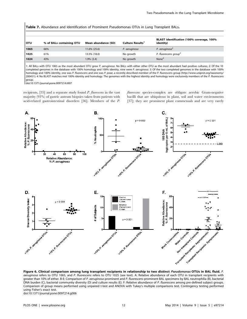
Table 7. Abundance and Identification of Prominent Pseudomonas OTUs in Lung Transplant BALs.
OTU % of BALs containing OTU Mean abundance (SD) Culture Results1BLAST identification (100% coverage, 100%identity)
1065 66% 11.6% (25.6) P. aeruginosa P. aeruginosa2
1025 61% 13.5% (18.0) No growth P. fluorescens group3
1024 43% 1.9% (3.4) No growth None4
1: All BALs with OTU 1065 as the most abundant OTU grew P. aeruginosa. No BALs with either other OTU as the most abundant had positive cultures; 2: Of the 10completed genomes in the database with 100% homology and 100% identity, nine were P. aeruginosa. 3: Of the two completed genomes in the database with 100%homology and 100% identity, one was P. fluorescens and one was P. poae, a recently described member of the P. fluorescens group (http://www.uniprot.org/taxonomy/200451); 4: No BLAST matches met 100% identity and homology. The genomes with the highest identity and homology were exclusively members of the P. fluorescensgroup.doi:10.1371/journal.pone.0097214.t007
Figure 6. Clinical comparison among lung transplant recipients in relationship to two distinct Pseudomonas OTUs in BAL fluid. P.aeruginosa refers to OTU 1065, and P. fluorescens refers to OTU 1025 (see text). A: Relative abundance of each OTU in transplant recipients withgreater than 10% of either. B-E: Comparison of P. aeruginosa-prominent and P. fluorescens-prominent BAL specimens by BAL neutrophilia (B), bacterialDNA burden (C), bacterial community diversity (D) and culture results (E). F: Relative abundance of P. fluorescens among pre-defined subject groups.Comparison of group means performed using unpaired t-test and ANOVA with Tukey’s multiple comparisons test. Contingency testing performedusing Fisher’s exact test.doi:10.1371/journal.pone.0097214.g006
Two Pseudomonads in the Lung Transplant Microbiome
PLOS ONE | www.plosone.org 12 May 2014 | Volume 9 | Issue 5 | e97214
described as pathogenic in humans [38]. They have been
commonly detected in the home environments of patients with
respiratory disease [39]. Increased abundance of P. fluorescens has
been observed in the oropharyngeal microbiota of patients
infected with the H1N1 influenza virus [40]. Antibodies against
P. fluorescens are common among patients with Crohn’s disease
[41,42], though whether it is a marker or mediator of human
disease remains undetermined.
The significant associations (both positive and negative) between
abundance of P. fluorescens and that of other prominent bacteria has
several possible interpretations. P. fluorescens actively inhibits in
vitro growth of other bacteria [43]. It produces numerous
antibiotics (including mupirocin [44]) and is used in agriculture
as a biological control organism [37]. Hence, although the
observed associations may be secondary to the presence of
conditions that are jointly favorable to P. fluorescens and Escherichia
sp. and unfavorable to Prevotella and Veillonella spp., an important
research direction will be to determine whether P. fluorescens
exhibits in vivo pro- and anti-biotic effects in the respiratory tract.
We observed decreased bacterial diversity in the BAL of
transplant recipients when compared to controls and identified
negative associations between BAL diversity and multiple clinical
features associated with acute infection. Recent studies are
divergent regarding changes in BAL microbiota diversity among
lung transplant recipients when compared to non-transplant
controls. Borewicz et al. observed increased BAL microbiota
diversity among transplant recipients [15], while two other
published reports reported decreased diversity [11,14]. In COPD
and asthma, the relationship between disease severity and lung
microbial diversity has been similarly conflicting [1]. Most BAL
microbiota studies to date have either excluded patients with
active infection or have made no distinction in analysis between
symptomatic and asymptomatic subjects (as in the above-cited
lung transplant studies). Our findings are consistent with
observations from the CF literature indicating that decreased
diversity is not a direct consequence of duration of illness or
impaired lung function but is instead driven by other factors [4].
To our knowledge, ours is the first report observing significant
associations between BAL microbiota diversity and other BAL
indices of acute infection. In our population, this decreased
diversity did not appear to be secondary to recent exposure to
antibiotics, as patients who had received antibiotics other than
their Pneumocystis prophylaxis agent in the preceding 30 days, seven
days and at the time of BAL did not have decreased BAL bacterial
diversity indices. These results are surprising and divergent from
the cystic fibrosis literature [4]. The fact that all transplant subjects
are at baseline on antibiotics for Pneumocystis prophylaxis (unlike
subjects with other lung pathologies such as CF) may be relevant
to this difference. While decreased bacterial diversity in the gut has
been associated with obesity and geography [45,46], its signifi-
cance in the lung apart from as a marker of acute infection is
uncertain.
A potential limitation of this study is the absence of concurrent
oral and nasal sampling for identification of "lung-specific"
bacteria. There has been discussion in the literature about the
extent to which microbiota detected in BAL reflect lung-resident
organisms as opposed to oral-derived microbes picked up during
sedation-related aspiration or broncho-scopic carryover. One
study of six healthy subjects observed a similar constitution in
the bacterial communities of the lower and upper respiratory tracts
[20]; however, subsequent published studies from the NIH-funded
LHMP consortium have identified bacterial species by BAL with
significantly disproportionate representation in lower respiratory
tract samples [16,17]. These newer, larger studies suggest that
some bacteria might replicate in or colonize the lower respiratory
tract, with specific selective pressures favoring some species over
others. Such preferential accumulation of certain OTUs supports
the existence of lung-specific microbiota. An important contribu-
tion of the current study is the observation that route of
bronchoscopy insertion (nasal or oral) has no appreciable effect
on BAL microbiota (Figure 2) despite the markedly divergent
microbial communities detected in the human mouth and nose
[47]. This finding suggests that passage of the bronchoscope
through these spaces has little impact on detected BAL microbiota,
and bronchoscopic carryover of upper respiratory tract microbiota
in BAL fluid is minimal. Further, the strong and significant
associations we found between BAL bacterial community diversity
and composition on one hand, and clinically and biologically
significant parameters on the other, is relevant to ongoing
discussions about the diagnostic implications of sampling lung
microbiota via BAL [48]. We believe that the associations we
report between BAL microbiota communities and significant
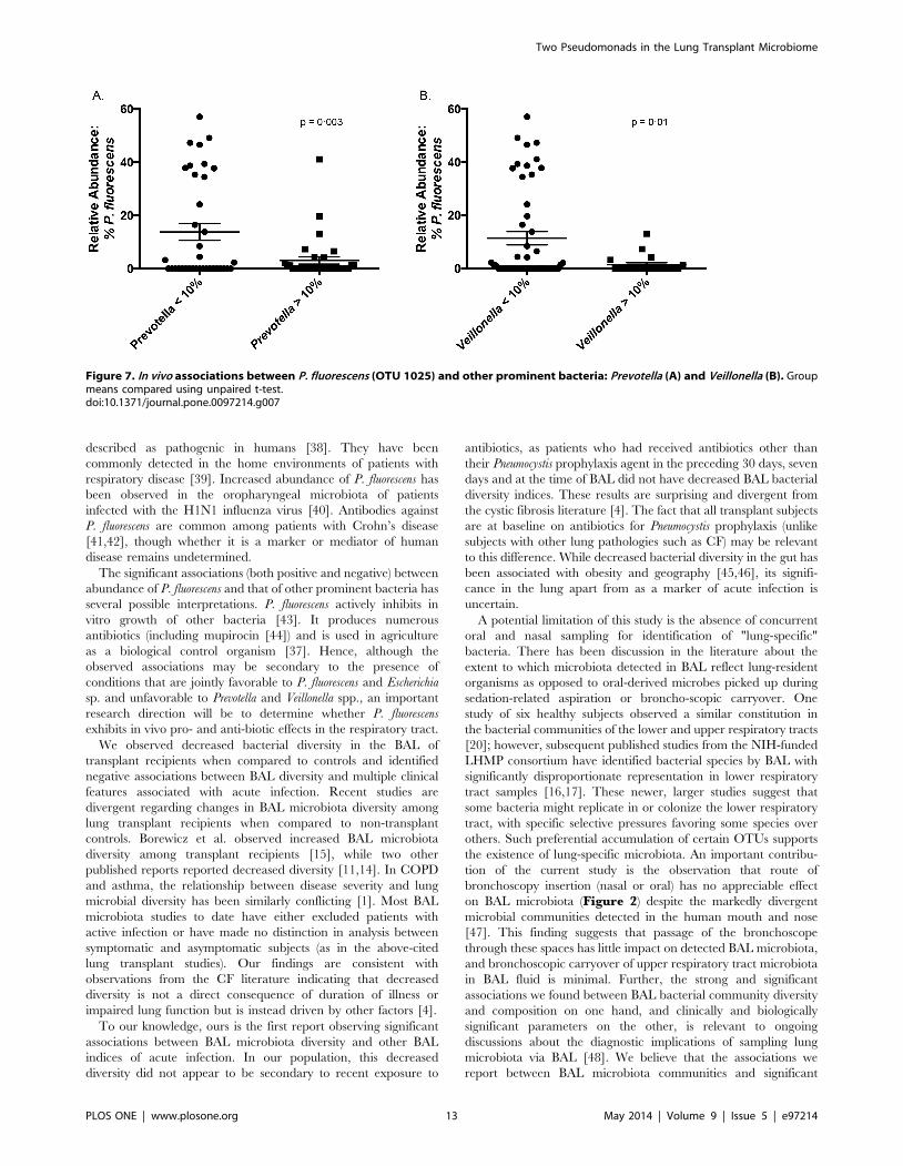
Figure 7. In vivo associations between P. fluorescens (OTU 1025) and other prominent bacteria: Prevotella (A) and Veillonella (B). Groupmeans compared using unpaired t-test.doi:10.1371/journal.pone.0097214.g007
Two Pseudomonads in the Lung Transplant Microbiome
PLOS ONE | www.plosone.org 13 May 2014 | Volume 9 | Issue 5 | e97214

clinical parameters indicate that the microorganisms detected in
BAL specimens have clinical and biological significance, regardless
of their derivation.
Another limitation of this study, common to all studies utilizing
BAL, is the potentially variable effect of dilution on BAL assays.
Despite efforts to standardize the procedure of saline instillation
and collection, the BAL return in each bronchoscopy is variable.
While this may result in variably concentrated samples, our results
indicate that the variation in bacterial DNA burden among
subjects is many-fold higher than that of variation in saline return
(Figure 1), and any confounding effect of dilution is not enough to
overwhelm the clear correlations apparent between bacterial DNA
burden and important clinical parameters (Figures 4E, 6C).
Variation in dilution between BAL samples should have no impact
on relative abundance of bacteria, from which most of this study’s
findings are derived. The use of 40 cycles in our PCR protocol is a
potential source of bias, but our touchdown protocol (described in
Methods) is optimized for low biomass samples and produces a low
fraction of spurious priming [49].
Our results suggest that future analyses of the lung transplant
microbiome should distinguish between P. aeruginosa and other
Pseudomonas species, as well as between symptomatic and asymp-
tomatic subjects and those with evidence of acute respiratory
infection.
Supporting Information
Figure S1 Relative abundance of prominent Pseudomo-nas OTUs among lung transplant recipient BAL speci-mens.(TIF)
Figure S2 Genomic Comparison of Prominent Pseudo-monas OTUs. A: Representative sequences of 16S V3-V5 for
three prominent Pseudomonas OTUs. B: Phylogenetic tree derived
from above sequences. Figures generated using Lasergene
Megalign (Madison, MI).
(TIF)
Figure S3 Phylogenetic Tree of Pseudomonas-classifiedOTUs and Clinically-Obtained and Reference Genomes.MLST phylogeny tree created using DNASTAR SeqBuilder
(Lasergene). Numbers in black are branch length. Numbers in blue
are bootstrap confidence values.
(TIF)
Table S1 NCBI BLAST Results for Prominent Pseudo-monas OTUs. Results restricted to sequences with 100%
coverage and identity and ranked in descending order of total
score.
(DOC)
Acknowledgments
The authors thank Zachary Britt, Sean Crudgington, Nicole Falkowski,
Dayana Rojas and Chinmay Pandit for assistance in tissue processing. The
authors wish to thank Rick Bushman and his lab at the University of
Pennsylvania for providing the 16S qPCR protocol and Duane Newton for
obtaining data on P. fluorescens isolates from the University of Michigan
Clinical Microbiology Laboratory.
Author Contributions
Conceived and designed the experiments: RPD JRE BSS JMB JLC VNL
GBH. Performed the experiments: RPD CMF NW JMB JLC VNL GBH.
Analyzed the data: RPD JRE BSS GBH. Contributed reagents/materials/
analysis tools: RPD JRE FJM JLC VNL GBH. Wrote the paper: RPD
GBH.
References
1. Dickson RP, Erb-Downward JR, Huffnagle GB (2013) The role of the bacterial
microbiome in lung disease. Expert Rev Respir Med 7: 245–257.
2. Erb-Downward JR, Thompson DL, Han MK, Freeman CM, McCloskey L, et
al. (2011) Analysis of the lung microbiome in the "healthy" smoker and in
COPD. PLoS One 6: e16384.
3. Hilty M, Burke C, Pedro H, Cardenas P, Bush A, et al. (2010) Disordered
microbial communities in asthmatic airways. PLoS One 5: e8578.
4. Zhao J, Schloss PD, Kalikin LM, Carmody LA, Foster BK, et al. (2012) Decade-
long bacterial community dynamics in cystic fibrosis airways. Proc Natl Acad
Sci U S A109: 5809–5814.
5. Christie JD, Edwards LB, Kucheryavaya AY, Benden C, Dipchand AI, et al.
(2012) The Registry of the International Society for Heart and Lung
Transplantation: 29th adult lung and heart-lung transplant report-2012.
J Heart Lung Transplant 31: 1073–1086.
6. Botha P, Archer L, Anderson RL, Lordan J, Dark JH, et al. (2008) Pseudomonas
aeruginosa colonization of the allograft after lung transplantation and the risk of
bronchiolitis obliterans syndrome. Transplantation 85: 771–774.
7. Husain S, Singh N (2002) Bronchiolitis obliterans and lung transplantation:
evidence for an infectious etiology. Semin Respir Infect 17: 310–314.
8. Khalifah AP, Hachem RR, Chakinala MM, Schechtman KB, Patterson GA, et
al. (2004) Respiratory viral infections are a distinct risk for bronchiolitis
obliterans syndrome and death. Am J Respir Crit Care Med 170: 181–187.
9. Duncan MD, Wilkes DS (2005) Transplant-related immunosuppression: a
review of immunosuppression and pulmonary infections. Proc Am Thorac Soc
2: 449–455.
10. Kotloff RM, Thabut G (2011) Lung transplantation. Am J Respir Crit Care Med
184: 159–171.
11. Willner DL, Hugenholtz P, Yerkovich ST, Tan ME, Daly JN, et al. (2013) Re-
Establishment of Recipient-Associated Microbiota in the Lung Allograft is
Linked to Reduced Risk of Bronchiolitis Obliterans Syndrome. Am J Respir Crit
Care Med.
12. Vos R, Vanaudenaerde BM, Geudens N, Dupont LJ, Van Raemdonck DE, et
al. (2008) Pseudomonal airway colonisation: risk factor for bronchiolitis
obliterans syndrome after lung transplantation? Eur Respir J 31: 1037–1045.
13. Gottlieb J, Mattner F, Weissbrodt H, Dierich M, Fuehner T, et al. (2009) Impact
of graft colonization with gram-negative bacteria after lung transplantation on
the development of bronchiolitis obliterans syndrome in recipients with cystic
fibrosis. Respir Med 103: 743–749.
14. Charlson ES, Diamond JM, Bittinger K, Fitzgerald AS, Yadav A, et al. (2012)
Lung-enriched organisms and aberrant bacterial and fungal respiratory
microbiota after lung transplant. Am J Respir Crit Care Med 186: 536–545.
15. Borewicz K, Pragman AA, Kim HB, Hertz M, Wendt C, et al. (2012)
Longitudinal Analysis of the Lung Microbiome in Lung Transplantation. FEMS
Microbiol Lett.
16. Lozupone C, Cota-Gomez A, Palmer BE, Linderman DJ, Charlson ES, et al.
(2013) Widespread Colonization of the Lung by Tropheryma whipplei in HIV
Infection. Am J Respir Crit Care Med.
17. Morris A, Beck JM, Schloss PD, Campbell TB, Crothers K, et al. (2013)
Comparison of the respiratory microbiome in healthy nonsmokers and smokers.
Am J Respir Crit Care Med 187: 1067–1075.
18. Estenne M, Maurer JR, Boehler A, Egan JJ, Frost A, et al. (2002) Bronchiolitis
obliterans syndrome 2001: an update of the diagnostic criteria. J Heart Lung
Transplant21: 297–310.
19. Mason KL, Erb Downward JR, Mason KD, Falkowski NR, Eaton KA, et al.
(2012) Candida albicans and bacterial microbiota interactions in the cecum
during recolonization following broad-spectrum antibiotic therapy. Infect
Immun 80: 3371–3380.
20. Charlson ES, Bittinger K, Haas AR, Fitzgerald AS, Frank I, et al. (2011)
Topographical continuity of bacterial populations in the healthy human
respiratory tract. Am J Respir Crit Care Med 184: 957–963.
21. Hill DA, Hoffmann C, Abt MC, Du Y, Kobuley D, et al. (2009) Metagenomic
analyses reveal antibiotic-induced temporal and spatial changes in intestinal
microbiota with associated alterations in immune cell homeostasis. Mucosal
Immunol 3: 148–158.
22. Wilmotte A, Van der Auwera G, De Wachter R (1993) Structure of the 16 S
ribosomal RNA of the thermophilic cyanobacterium Chlorogloeopsis HTF
(’Mastigocladus laminosus HTF’) strain PCC7518, and phylogenetic analysis.
FEBS Lett 317: 96–100.
23. Jumpstart Consortium Human Microbiome Project Data Generation Working
Group (2010) Human Microbiome Consortium 16S 454 Sequencing Protocol.
4.2.2 ed.
Two Pseudomonads in the Lung Transplant Microbiome
PLOS ONE | www.plosone.org 14 May 2014 | Volume 9 | Issue 5 | e97214
24. Daigle D, Simen BB, Pochart P (2011) High-throughput sequencing of PCR
products tagged with universal primers using 454 life sciences systems. Curr
Protoc Mol Biol Chapter 7: Unit7 5.
25. Schloss PD, Westcott SL, Ryabin T, Hall JR, Hartmann M, et al. (2009)
Introducing mothur: open-source, platform-independent, community-supported
software for describing and comparing microbial communities. Appl Environ
Microbiol 75: 7537–7541.
26. Spilker T, Coenye T, Vandamme P, LiPuma JJ (2004) PCR-based assay for
differentiation of Pseudomonas aeruginosa from other Pseudomonas species
recovered from cystic fibrosis patients. J Clin Microbiol 42: 2074–2079.
27. Scarpellini M, Franzetti L, Galli A (2004) Development of PCR assay to identify
Pseudomonas fluorescens and its biotype. FEMS Microbiol Lett 236: 257–260.
28. Katoh K, Misawa K, Kuma Ki, Miyata T (2002) MAFFT: a novel method for
rapid multiple sequence alignment based on fast Fourier transform. Nucleic
Acids Res 30: 3059–3066.
29. Ingenito EP, Solway J, McFadden ER Jr, Pichurko B, Bowman HF, et al. (1987)
Indirect assessment of mucosal surface temperatures in the airways: theory and
tests. J Appl Physiol 63: 2075–2083.
30. Bernstein DI, Lummus ZL, Santilli G, Siskosky J, Bernstein IL (1995) Machine
operator’s lung. A hypersensitivity pneumonitis disorder associated with
exposure to metalworking fluid aerosols. Chest 108: 636–641.
31. Bahrani-Mougeot FK, Paster BJ, Coleman S, Barbuto S, Brennan MT, et al.
(2007) Molecular analysis of oral and respiratory bacterial species associated with
ventilator-associated pneumonia. J Clin Microbiol 45: 1588–1593.
32. Mulet M, Lalucat J, GarcıaValdes E (2010) DNA sequence-based analysis of the
Pseudomonas species. Environ Microbiol12: 1513–1530.
33. Loper JE, Hassan KA, Mavrodi DV, Davis II EW, Lim CK, et al. (2012)
Comparative genomics of plant-associated Pseudomonas spp.: insights into
diversity and inheritance of traits involved in multitrophic interactions. PLoS
genetics 8: e1002784.
34. Silby MW, Winstanley C, Godfrey SA, Levy SB, Jackson RW (2011)
Pseudomonas genomes: diverse and adaptable. FEMS Microbiol Rev 35:
652–680.
35. Diaz PI, Hong BY, Frias-Lopez J, Dupuy AK, Angeloni M, et al. (2013)
Transplantation-Associated Long-Term Immunosuppression Promotes Oral
Colonization by Potentially Opportunistic Pathogens without Impacting Other
Members of the Salivary Bacteriome. Clin Vaccine Immunol 20: 920–930.
36. Patel SK, Pratap CB, Verma AK, Jain AK, Dixit VK, et al. (2013) Pseudomonas
fluorescens-like bacteria from the stomach: A microbiological and molecularstudy. World J Gastroenterol 19: 1056–1067.
37. Paulsen IT, Press CM, Ravel J, Kobayashi DY, Myers GS, et al. (2005)
Complete genome sequence of the plant commensal Pseudomonas fluorescensPf-5. Nat Biotechnol 23: 873–878.
38. Gershman MD, Kennedy DJ, Noble-Wang J, Kim C, Gullion J, et al. (2008)Multistate outbreak of Pseudomonas fluorescens bloodstream infection after
exposure to contaminated heparinized saline flush prepared by a compounding
pharmacy. Clin Infect Dis 47: 1372–1379.39. Mortensen JE, Fisher MC, LiPuma JJ (1995) Recovery of Pseudomonas cepacia
and other Pseudomonas species from the environment. Infect Control HospEpidemiol: 30–32.
40. Leung RK, Zhou JW, Guan W, Li SK, Yang ZF, et al. (2012) Modulation ofpotential respiratory pathogens by pH1N1 viral infection. Clin Microbiol Infect.
41. Bossuyt X (2006) Serologic markers in inflammatory bowel disease. Clin Chem
52: 171–181.42. Wei B, Huang T, Dalwadi H, Sutton CL, Bruckner D, et al. (2002)
Pseudomonas fluorescens encodes the Crohn’s disease-associated I2 sequenceand T-cell superantigen. Infect Immun 70: 6567–6575.
43. Baader A, Garre C (1887) Uber Antagonisten unter den Bacterien.
Correspondenz-Blatt fur Schweizer Arzte 13: 385–392.44. Sutherland R, Boon R, Griffin K, Masters P, Slocombe B, et al. (1985)
Antibacterial activity of mupirocin (pseudomonic acid), a new antibiotic fortopical use. Antimicrob Agents Chemother 27: 495–498.
45. Turnbaugh PJ, Hamady M, Yatsunenko T, Cantarel BL, Duncan A, et al.(2008) A core gut microbiome in obese and lean twins. Nature 457: 480–484.
46. Yatsunenko T, Rey FE, Manary MJ, Trehan I, Dominguez-Bello MG, et al.
(2012) Human gut microbiome viewed across age and geography. Nature 486:222–227.
47. Human Microbiome Project Consortium (2012) Structure, function anddiversity of the healthy human microbiome. Nature 486: 207–214.
48. Twigg HL, Morris A, Ghedin E, Curtis JL, Huffnagle GB, et al. (2013) Use of
bronchoalveolar lavage to assess the respiratory microbiome: signal in the noise.The Lancet Respiratory Medicine 1: 354–356.
49. Don R, Cox P, Wainwright B, Baker K, Mattick J (1991) ’Touchdown’PCR tocircumvent spurious priming during gene amplification. Nucleic Acids Res 19:
4008.
Two Pseudomonads in the Lung Transplant Microbiome
PLOS ONE | www.plosone.org 15 May 2014 | Volume 9 | Issue 5 | e97214