Embed Size (px)
Citation preview
Changes in Kinetochore Structure and Molecular Composition in Response to
Mis-attachment
Muyao Shen
Thesis submitted to the faculty of the Virginia Polytechnic Institute and State
University in partial fulfillment of the requirements for the degree of
Master of Science
In
Biological Sciences
Daniela Cimini, Chair
Richard A. Walker
William R. Huckle
May 27, 2011
Blacksburg, Virginia
Keywords: kinetochore-microtubule (KT-MT) mis-attachment, the spindle
assembly checkpoint (SAC), tension, KT stretching
Copyright 2011, Muyao Shen
Changes in Kinetochore Structure and Molecular Composition in Response to
Mis-attachment
Muyao Shen
Abstract
Each mitotic chromosome is constituted by two sister chromatids whose correct segregation to
the daughter cells is ensured by amphitelic attachment, in which the two sister kinetochores (KTs)
are attached to microtubules (MTs) from opposite mitotic spindle poles. KT mis-attachments can
occur in early mitosis and cause chromosome mis-segregation and aneuploidy if not corrected.
These mis-attachments include monotelic (one attached and one unattached sister KT), syntelic
(both sister KTs attached to the same spindle pole), and merotelic (a single KT attached to MTs
from opposite spindle poles) attachments. A biochemical pathway named the Spindle Assembly
Checkpoint (SAC) is responsible for delaying anaphase onset to allow correction of KT mis-
attachments. SAC activation is believed to occur due to KT localization of certain SAC proteins
and/or lack of tension, but only monotelic attachment has been proven to activate the SAC. To
determine if and how other KT mis-attachments may activate the SAC, we studied how
molecular composition and structure of the KT changes in response to different types of
attachments. Our data suggest that monotelic attachment is the only type of attachment that can
induce a SAC response thanks to the accumulation of the SAC protein Mad2 at the KT. Our data
also indicate that structural changes of the KT, measured as intra- or inter-KT stretching, do not
directly induce a SAC response. Instead, our findings suggest decreased KT stretching,
especially in inter-KT stretching of syntelic chromosomes, may play a key role in bringing
MCAK and other KT substrates closer to Aurora B kinase for rapid and efficient correction of
KT mis-attachments.

iii
Acknowledgements
First, I would like to especially thank my major advisor, Dr. Daniela Cimini, for giving me the
opportunity to work on such an amazing project. Dr. Daniela Cimini has given me guidance,
patience, and encouragement in every aspect of graduate development in the past three years,
from research design to growth as a critical-thinking person. She has been such a great mentor
and advisor who made my time here a great educational experience as well as a lot of fun.
Second, I would like to thank the members of my committee, Dr. Richard Walker and Dr.
William Huckle for all their support and help with every step of my research. They have made
useful suggestions that helped to improve my work a lot during committee meetings. They have
been tremendous source of support and comfort that made me confident about my research.
Third, I would like to thank past and present members of the Cimini lab. Special thanks to
William Silkworth, Joshua Nicholson, Bin He, Anthony Asmar and Isaac Nardi. They have not
only been great lab mates but also great friends. I’m grateful to them for not only helping me
with technical problems such as microscope issues but also sharing joy and happiness with me.
Last, but not the least, special thanks to my parents and all my family who have supported and
encouraged me throughout my academic career. Thanks to Tian Hong for helping me develop
several figures in my thesis and for being a comforting boyfriend during these stressful times.
iv
Table of Contents
CHAPTER 1: LITERATURE REVIEW ................................................................................... 1
MITOSIS ....................................................................................................................................... 1
THE MITOTIC SPINDLE ................................................................................................................. 4
THE KINETOCHORE ...................................................................................................................... 6
Molecular composition and function of the vertebrate inner KT ........................................... 8
Molecular composition and function of the vertebrate outer KT ......................................... 11
Molecular composition and function of the fibrous corona .................................................. 12
Temporal sequence of KT assembly and disassembly in vertebrates ................................... 14
THE SPINDLE ASSEMBLY CHECKPOINT ...................................................................................... 15
KT MIS-ATTACHMENTS .............................................................................................................. 22
CHAPTER 2: INTRODUCTION .............................................................................................. 26
THE BEHAVIOR OF THE SAC IN RESPONSE TO KT-MT MIS-ATTACHMENTS ................................ 26
ATTACHMENT VS. TENSION IN SAC SIGNALING ........................................................................ 27
RATIONALE AND HYPOTHESIS ................................................................................................... 28
CHAPTER 3: MATERIALS AND METHODS ...................................................................... 30
CELL CULTURE AND DRUG TREATMENT ..................................................................................... 30
ANTIBODIES ............................................................................................................................... 30
IMMUNOSTAINING ...................................................................................................................... 31
CONFOCAL MICROSCOPY AND IMAGE ACQUISITION ................................................................... 32
DATA ANALYSIS ......................................................................................................................... 32
CHAPTER 4: RESULTS ........................................................................................................... 36
ATTACHMENT-DEPENDENT CHANGES IN INTER- AND INTRA-KT STRETCHING. .......................... 36
QUANTIFICATION OF MAD2 AT ATTACHED, UNATTACHED, AND MIS-ATTACHED KTS. ............... 38
QUANTIFICATION OF 3F3/2 PHOSPHOEPITOPE AND BUBR1 AT ATTACHED, UNATTACHED, AND
MIS-ATTACHED KTS. .................................................................................................................. 39
QUANTIFICATION OF AURB, INCENP, AND MCAK AT ATTACHED, UNATTACHED, AND MIS-
ATTACHED KTS. ......................................................................................................................... 41
CHAPTER 5: DISCUSSION ..................................................................................................... 45
INTRA-KT STRETCHING PER SE DOES NOT APPEAR TO BE THE KEY FACTOR IN THE RESPONSE TO
SYNTELIC ATTACHMENT. ............................................................................................................ 45
WHAT CAUSES THE REDUCED INTRA-KT DISTANCE IN UNATTACHED, MEROTELIC, AND SYNTELIC
KTS? .......................................................................................................................................... 46
KINETOCHORE ATTACHMENT AND ACCUMULATION OF MAD2, 3F3/2, AND BUBR1. .................. 48
AURORA B, INCENP, AND MCAK AT UNATTACHED AND MIS-ATTACHED KTS. ....................... 49
HOW DO CELLS RESPOND TO DIFFERENT TYPES OF ATTACHMENTS? ........................................... 50
CHAPTER 6: SUMMARY AND FUTURE DIRECTION ..................................................... 53
REFERENCES ............................................................................................................................ 55
v
List of figures
Figures Page number Figure 1.1. Six stages of cell division. 2 Figure 1.2. Diagrammatic representation of the mitotic spindle and the three
classes of MTs. 5
Figure 1.3. Structure of a vertebrate KT. [used with permission]
Cheeseman, I.M., and Desai, A. (2008). Molecular architecture of the
kinetochore-microtubule interface. Nat Rev Mol Cell Bio 9, 33-46.
http://www.nature.com/nrm/journal/v9/n1/full/nrm2310.html
(accessed May 05,2011) Used with permission from Nature
Publishing Group; letter attached.
7
Figure 1.4. Diagrammatic representation of KT associated proteins. [used
with permission]
Musacchio, A., and Salmon, E.D. (2007). The spindle-assembly
checkpoint in space and time. Nat Rev Mol Cell Bio 8, 379-393.
http://www.nature.com/nrm/journal/v8/n5/full/nrm2163.html
(accessed May 05,2011) Used with permission from Nature
Publishing Group; letter attached.
11
Figure 1.5. Schematic representation of temporally ordered KT assembly
and disassembly. [used with permission]
Cheeseman, I.M., and Desai, A. (2008). Molecular architecture of the
kinetochore-microtubule interface. Nat Rev Mol Cell Bio 9, 33-46.
http://www.nature.com/nrm/journal/v9/n1/full/nrm2310.html
(accessed May 05,2011) Used with permission from Nature
Publishing Group; letter attached.
14
Figure 1.6. Schematic representations of attachment- Vs. tension-sensitive
SAC. 19
Figure 1.7. The protein network and signaling pathway of the SAC. [used
with permission]
Musacchio, A., and Salmon, E.D. (2007). The spindle-assembly
checkpoint in space and time. Nat Rev Mol Cell Bio 8, 379-393.
http://www.nature.com/nrm/journal/v8/n5/full/nrm2163.html
(accessed May 05,2011) Used with permission from Nature
Publishing Group; letter attached.
22
Figure 1.8. Types of KT attachments. [used with permission]
Cimini, D. (2008). Merotelic kinetochore orientation, aneuploidy, and
cancer. Biochim Biophys Acta 1786, 32-40.
http://www.sciencedirect.com/science/article/pii/S0304419X08000267
(accessed May 05,2011) Used with permission from Elsevier Limited;
letter attached.
23
Figure 3.1. Diagram of measurement of fluorescence intensities of the signal
(Fs). 33
Figure 3.2. Schematic of intra-KT stretching measurement. 34 Figure 4.1. Examples of different types of KT mis-attachments in PtK1
cells. 36
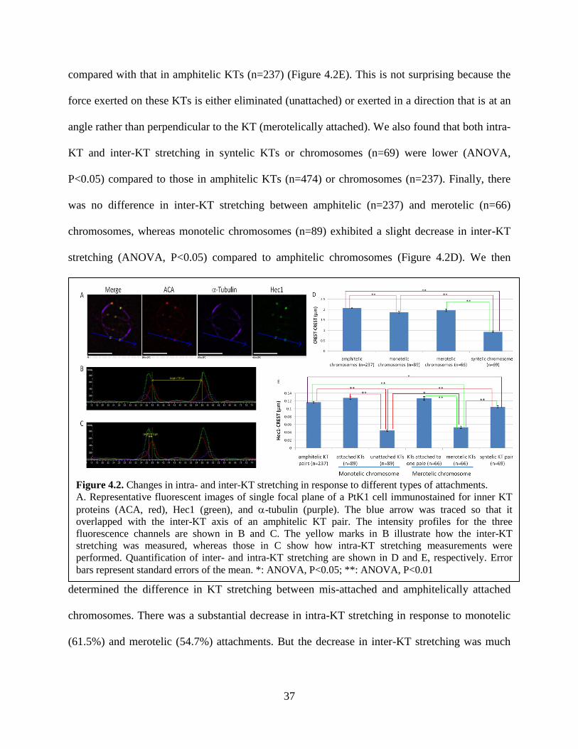
Figure 4.2. Changes in intra- and inter-KT stretching in response to different
types of attachments. 37
vi
Figure 4.3. Quantification of Mad2, 3F3/2 phosphoepitope, and BubR1 at
KTs with different attachments. 39
Figure 4.4. Quantification of AurB, INCENP, and MCAK at KTs with
different attachments. 43
Figure 5.1. Model of elements that cause intra-KT stretching in response to
different attachments. 47
Figure 5.2. Model illustrating how inter-KT stretching contributes to AurB-
dependent error correction. 51
vii
List of tables
Tables Page number
Table 4.1. Summary of the changes in KT
structure and molecular composition in
response to mis-attachments. Amphitelic KTs
are set as control.
44
Table 4.2. Summary of the changes in KT
structure and molecular composition in
response to mis-attachments. Amphitelic
attachment is set as control.
44
1
Chapter 1: Literature Review
Mitosis
Equal partitioning of the genome is achieved through a process called mitosis, during which cells
equally distribute the replicated chromosomes between the two daughter cells. Mitosis occurs
during the M phase of the cell cycle. M phase follows a number of characteristic cell cycle
phases, including G1, S, and G2, which together constitute interphase, and during which the cell
prepares to divide by growing in size (G1) and replicating its DNA (S) (Blow and Tanaka, 2005;
Howard, 1951; Smith and Martin, 1973; Wittmann et al., 2001). In eukaryotic cells, mitosis can
be divided into five distinct stages: prophase, prometaphase, metaphase, anaphase, and telophase.
Mitosis is followed by cytokinesis, which leads to cytoplasm division, and thus completes cell
division (Rieder and Khodjakov, 2003; Wittmann et al., 2001).
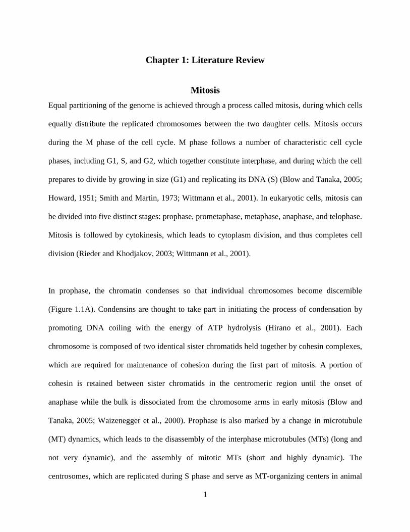
In prophase, the chromatin condenses so that individual chromosomes become discernible
(Figure 1.1A). Condensins are thought to take part in initiating the process of condensation by
promoting DNA coiling with the energy of ATP hydrolysis (Hirano et al., 2001). Each
chromosome is composed of two identical sister chromatids held together by cohesin complexes,
which are required for maintenance of cohesion during the first part of mitosis. A portion of
cohesin is retained between sister chromatids in the centromeric region until the onset of
anaphase while the bulk is dissociated from the chromosome arms in early mitosis (Blow and
Tanaka, 2005; Waizenegger et al., 2000). Prophase is also marked by a change in microtubule
(MT) dynamics, which leads to the disassembly of the interphase microtubules (MTs) (long and
not very dynamic), and the assembly of mitotic MTs (short and highly dynamic). The
centrosomes, which are replicated during S phase and serve as MT-organizing centers in animal

2
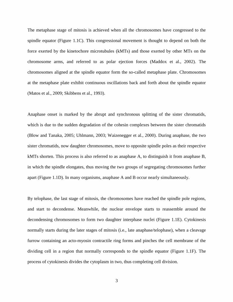
Figure 1.1. Six stages of cell division.
A: prophase; B: prometaphase; C: metaphase; D:
anaphase; E: telophase; F: cytokinesis. DNA is shown in
blue and red, MTs in green.
cells, move apart during prophase, and an aster starts to form around each duplicated centrosome
(Karsenti and Vernos, 2001; Zhai et al., 1996). The separation of centrosomes initiates the
formation of the mitotic spindle outside the nucleus.
The breakdown of the nuclear envelope (not occurring in lower eukaryotes) marks the beginning
of prometaphase (Figure 1.1B). MTs have access to the nuclear region immediately after nuclear
envelope disassembly, and can search
the nuclear space by undergoing
polymerization/depolymerization cycles,
until they encounter a kinetochore (KT),
a specialized protein structure that
mediates chromosome-spindle
attachment. Once attached to a KT,
MTs turn into a stable state, and no longer undergo catastrophe (i.e., switch from growing to
shortening). A MT can initially establish a lateral interaction with a KT, but it will be rapidly
converted to an end-on attachment, the type of attachment responsible for regulating
chromosome movement (Hayden et al., 1990; Tanaka et al., 2005; Tanaka and Desai, 2008).
Because of the stochastic nature of KT-MT encounters, most chromosomes initially become
mono-oriented, with one KT bound to MTs and its sister KT unattached, and move to the pole to
which they are attached (Rieder and Alexander, 1990; Rieder and Salmon, 1998). Once the
unattached sister KT binds MTs from the opposite spindle pole, the chromosome is said to be bi-
oriented, and moves to the spindle equator (Rieder and Salmon, 1998).
3
The metaphase stage of mitosis is achieved when all the chromosomes have congressed to the
spindle equator (Figure 1.1C). This congressional movement is thought to depend on both the
force exerted by the kinetochore microtubules (kMTs) and those exerted by other MTs on the
chromosome arms, and referred to as polar ejection forces (Maddox et al., 2002). The
chromosomes aligned at the spindle equator form the so-called metaphase plate. Chromosomes
at the metaphase plate exhibit continuous oscillations back and forth about the spindle equator
(Matos et al., 2009; Skibbens et al., 1993).
Anaphase onset is marked by the abrupt and synchronous splitting of the sister chromatids,
which is due to the sudden degradation of the cohesin complexes between the sister chromatids
(Blow and Tanaka, 2005; Uhlmann, 2003; Waizenegger et al., 2000). During anaphase, the two
sister chromatids, now daughter chromosomes, move to opposite spindle poles as their respective
kMTs shorten. This process is also referred to as anaphase A, to distinguish it from anaphase B,
in which the spindle elongates, thus moving the two groups of segregating chromosomes further
apart (Figure 1.1D). In many organisms, anaphase A and B occur nearly simultaneously.
By telophase, the last stage of mitosis, the chromosomes have reached the spindle pole regions,
and start to decondense. Meanwhile, the nuclear envelope starts to reassemble around the
decondensing chromosomes to form two daughter interphase nuclei (Figure 1.1E). Cytokinesis
normally starts during the later stages of mitosis (i.e., late anaphase/telophase), when a cleavage
furrow containing an acto-myosin contractile ring forms and pinches the cell membrane of the
dividing cell in a region that normally corresponds to the spindle equator (Figure 1.1F). The
process of cytokinesis divides the cytoplasm in two, thus completing cell division.
4
The Mitotic Spindle
The mitotic spindle is a bipolar array of antiparallel MTs assembled during mitosis. The
bipolarity of a mitotic spindle is crucial for its function to direct chromosome congression and
segregation during mitosis. At the onset of mitosis, the duplicated centrosomes nucleate two
asters of MTs, which move around the nucleus in prophase. The MTs emanate from the asters,
and grow both toward the cell cortex and toward the chromosomes, which become accessible to
the MTs after nuclear envelope breakdown. As the interaction between MTs and chromosomes is
established, the conformation of the mitotic spindle becomes (Karsenti and Vernos, 2001; Zhai et
al., 1996). By metaphase, the mitotic spindle appears as a symmetrical radial array of MTs.
MTs are hollow cylinders that assemble from -/-tubulin heterodimers, and exhibit structural
and functional polarity. In vitro, MT polymerization occurs at higher rates at one end (defined as
the plus end) compared to the other (minus end), and in steady-state conditions. MTs incorporate
tubulin subunits at the plus end and release them at the minus end, without net growth, in a
process referred to as treadmilling (Margolis and Wilson, 1981). Within a MT, the α-tubulin is
exposed at the minus end, while the -tubulin is exposed at the plus end (Gadde and Heald, 2004;
Heidemann and McIntosh, 1980; Mountain and Compton, 2000), and in the mitotic spindle,
MTs are oriented with their minus end anchored to the centrosome (MTOC) and the plus end
away from the centrosome (Gadde and Heald, 2004; Mazia, 1984). The centrosomes constitute
the poles of the mitotic spindle, and play a critical role in mitotic spindle assembly. Each
centrosome contains a pair of perpendicularly arranged centrioles, which are surrounded by an
amorphous mass, known as pericentriolar material. Each centriole is composed of nine triplets of
A
B
5
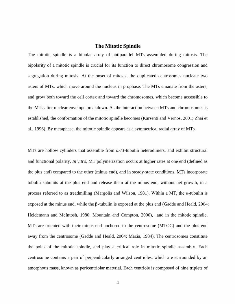
Figure 1.2. Diagrammatic representation of the mitotic spindle and
the three classes of MTs. MTs extend from the centrosome (minus
end) and grow towards cell cortex (plus end) or chromosomes (plus
end). Astral MTs extend away from the spindle poles. Interpolar
MTs can overlap with MTs from the opposite poles and are
important for maintaining spindle structure. Multiple kMTs bind
each KT to form a k-fiber, which connects the KT to the spindle
pole.
MTs. A third type of tubulin, -tubulin, is exclusively found at centrosomes. By binding one or
more proteins, it can form -tubulin ring complex (-TuRC), a ring-like structure which is
involved in nucleating MTs at the centrosome (Wiese and Zheng, 2006; Zheng et al., 1995).
Three classes of MTs are defined within a mitotic spindle based on their distinct structures and
roles in mitotic cells. These
are astral, interpolar, and
kMTs (Figure 1.2) (Karsenti
and Vernos, 2001). Astral
MTs are oriented with their
plus end away from the
chromosomes and toward
the cell cortex, with which
they can interact via motor
proteins. This interaction
with the cell cortex plays a role in orienting the spindle within the cell (Gadde and Heald, 2004;
Mountain and Compton, 2000). Interpolar MTs overlap in an antiparallel way in the spindle
midzone. The interaction of interpolar MTs with minus and plus end-directed motor proteins
contributes to maintaining mitotic spindle structure (Odde, 2005). Finally, kMTs are those MTs
whose plus end interacts with a KT. In most cell types, each KT can bind multiple MTs, which
form a MT bundle named kinetochore-fiber (K-fiber) (Figure 1.2). kMTs are mainly responsible
for directing chromosome movement during mitosis.
6
MTs switch rapidly between phases of growth and shrinkage, a process termed dynamic
instability (Kirschner and Mitchison, 1986; O'Connell and Khodjakov, 2007; van der Vaart et al.,
2009). At the onset of mitosis, the half-life of MTs decreases compared with that of interphase
MTs. Many studies in the field of mitosis have focused on how the dynamic assembly and
disassembly of MTs leads to the formation of a stable bipolar mitotic spindle. The “search and
capture” model proposes that MTs grow from the centrosomes in all directions. KT-MT
attachment occurs by random encounter, and spindle assembly is complete when all KTs have
established MT attachments (Hyman and Mitchison, 1990; Mitchison and Kirschner, 1985). The
“search and capture” model has been validated in experimental systems in various organisms
(Gadde and Heald, 2004; Mitchison and Kirschner, 1985). However, it presents an inefficient
mechanism due to the passive and random capture (Wollman et al., 2005), and mathematical
modeling has shown that spindle assembly could not be completed within physiological times if
“search and capture” were the only mechanism in action. An alternative model proposes that
MTs can nucleate from KTs and/or chromatin (Wadsworth and Khodjakov, 2004). These
chromatin-nucleated MTs become stabilized thanks to a RanGTP concentration gradient (Kalab
and Heald, 2008; Nachury et al., 2001; Wiese et al., 2001). Motor proteins associated with
chromosome arms take part in sorting these MTs into a bipolar array that surrounds
chromosomes (Cai et al., 2009). It is reasonable to assume that both “search and capture” and
RanGTP stabilization of chromatin-nucleated MTs contribute to spindle assembly in mitotic cells
(Wollman et al., 2005; O'Connell and Khodjakov, 2007; Kalab and Heald, 2008).
The Kinetochore
7
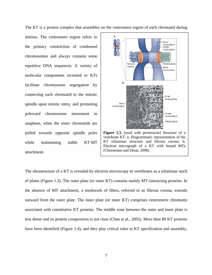
Figure 1.3. [used with permission] Structure of a
vertebrate KT. a. Diagrammatic representation of the
KT trilaminar structure and fibrous corona; b.
Electron micrograph of a KT with bound MTs
(Cheeseman and Desai, 2008).
The KT is a protein complex that assembles on the centromere region of each chromatid during
mitosis. The centromere region refers to
the primary constriction of condensed
chromosomes and always contains some
repetitive DNA sequences. A variety of
molecular components recruited to KTs
facilitate chromosome segregation by
connecting each chromatid to the mitotic
spindle upon mitotic entry, and promoting
poleward chromosome movement in
anaphase, when the sister chromatids are
pulled towards opposite spindle poles
while maintaining stable KT-MT
attachment.
The ultrastructure of a KT is revealed by electron microscopy in vertebrates as a trilaminar stack
of plates (Figure 1.3). The outer plate (or outer KT) contains mainly MT-interacting proteins. In
the absence of MT attachment, a meshwork of fibers, referred to as fibrous corona, extends
outward from the outer plate. The inner plate (or inner KT) comprises centromeric chromatin
associated with constitutive KT proteins. The middle zone between the outer and inner plate is
less dense and its protein composition is not clear (Chan et al., 2005). More than 80 KT proteins
have been identified (Figure 1.4), and they play critical roles in KT specification and assembly,
8
binding with spindle MTs, monitoring KT attachment and tension, and providing the driving
forces for chromosome movement (Cheeseman and Desai, 2008).
Molecular composition and function of the vertebrate inner KT
The inner KT serves as a platform for the assembly of the outer portions of the KT. CENP-A
(Centromeric Protein A), CENP-B, and CENP-C, which constitutively localize at the inner KT,
were the first three KT proteins to be identified using human autoantibodies (Earnshaw and
Rothfield, 1985), and they (particularly CENP-A) are believed to specify the site of KT assembly.
In humans, CENP-B protein appears to bind the CENP-B box in a sequence-specific manner.
CENP-B box is a 17-bp motif within the 171-bp tandem repeat sequence, known as -satellite
DNA, and was found at the centromere of human chromosomes (Earnshaw and Rothfield, 1985).
Some studies have suggested that both -satellite DNA and CENP-B boxes are required for de
novo centromere formation (Ohzeki et al., 2002). However, in most eukaryotes centromeric loci
can be stably maintained in the absence of CENP-B, CENP-B box, or –satellite DNA,
indicating that the site of KT assembly and maintenance are primarily controlled by epigenetic
rather than sequence-based mechanisms (Allshire and Karpen, 2008; Black and Bassett, 2008;
Karpen and Allshire, 1997). CENP-A is a histone H3 variant only found at the centromere. It
serves as the fundamental determinant of KT identity and is targeted to the centromere through a
15-residue sequence known as CATD (CENP-A targeting domain) (Sullivan et al., 1994). The
H3CATD
chimera not only specifies the centromere localization but also mediates the recruitment
of additional KT proteins (Black et al., 2007; Black et al., 2004). Recent studies have also shown
a key role of CENP-C in directing KT assembly (Cheeseman et al., 2004; Desai et al., 2003).
Indicative of such a role is the fact that CENP-C interacts with 13 other proteins, including
9
CENP-H, CENP-I, CENP-K-U, to constitute a network of proteins proximal to the CENP-A
nucleosome. Although these proteins, referred to as constitutive centromere-associated network
(CCAN), do not affect the association of CENP-A with the centromere after CENP-A deposition,
the CENP-H/I/K subclass, recruited by the CENP T/W sub-complex, and CENP-M/N may help
target and/or stabilize new CENP-A (Foltz et al., 2006; Izuta et al., 2006; Okada et al., 2006).
Indeed, a recent study establishes CENP-N as the promoter and stabilizer of centromere
assembly due to its direct recognition of CENP-A nucleosomes (Carroll et al., 2009). Another
group of proteins localize in proximity of the inner KT, but are not integral part of the KT itself.
Rather, they are believed to localize at the inner centromere, in the inter-KT region (i.e., between
the two sister KTs). These proteins comprise the chromosomal passenger complex (CPC), which
includes Borealin, Bir1/Survivin, Aurora B kinase (AurB), Sli15/INCENP (inner centromere
protein), and mitotic centromere-associated kinesin (MCAK), and are mainly responsible for
regulating the stability of KT-MT attachments (Musacchio and Salmon, 2007; Sandall et al.,
2006; Vader et al., 2006). Sli15/INCENP-Bir1/Survivin may also couple KTs with MTs, given
that INCENP contains a MT-binding site in its C-terminal region (Sandall et al., 2006). The
localization and activity of AurB require the three CPC regulatory subunits (Carmena et al.,
2009). For example, Borealin is suggested to promote local clustering that leads to AurB auto-
activation at the centromere (Kelly et al., 2007; Sessa et al., 2005). INCENP binds to AurB and
increases its basal activation (Bishop and Schumacher, 2002; Sessa et al., 2005). Besides,
INCENP functions as a tension sensor through its interaction with MTs and relays the
mechanical state of KT-MT attachments into local control of Ipl1 kinase (yeast homolog of AurB)
activity (Sandall et al., 2006). By phosphorylating its substrates such as MCAK (a MT
depolymerase at the plus end), the Ndc80 complex (a core protein complex localized at the outer
10
KT), and the Dam1 complex (a ten-subunit MT-binding protein complex necessary for end-on
attachment in yeast) (Cheeseman et al., 2002), AurB contributes to reducing the binding affinity
of KTs for MTs in vitro (Cheeseman et al., 2006) and serves to correct KT-MT interactions until
bipolar attachment is achieved(Cimini et al., 2006; DeLuca et al., 2006). In addition, cells
expressing a CPC mutant compromised the mitotic arrest but were still able to create unattached
KTs, a characteristic of AurB to correct non-bipolar attachments (Vader et al., 2007). Therefore,
AurB was suggested to influence the efficiency of anaphase progression independent from its
MT destabilizing activity because the AurB-dependent destabilization of non-bipolar
attachments could be uncoupled from its ability to induce mitotic arrest (Vader et al., 2008).
MCAK, a member of the kinesin-13 family, is also important in coupling MT dynamics with
sister KT motility. Indeed, recruitment of additional MCAK increased the chromosome speed,
the oscillation amplitude, and the coordination between sister KTs (Joglekar et al., 2010). To
conclude, the CPC complex plays a critical role in ensuring correct chromosome orientation and
alignment. Another inner KT protein, Shugoshin (Sgo/MEI-S332), is responsible for maintaining
centromeric cohesion until metaphase. For example, mammalian shugoshin (Sgo1) is a
centromeric protein that is localized at the inner KT from G2 phase to metaphase (Wang et al.,
2006). Interestingly, a truncated version of Sgo1 (sSgo1) has been shown to localize to
centrosomes and spindle poles, and has a role in the maintenance of spindle integrity (Dai, 2009;
Macy et al., 2009; Wang et al., 2008). Sgo2, another shugoshin-like protein, is also required for
the centromeric protection of cohesion, but in germ cells. AurB-phosphorylated Sgo2 can recruit
MCAK and PP2A to centromeres, thus contributing to both centromeric protection and
attachment correction (Tanno et al., 2010).
11
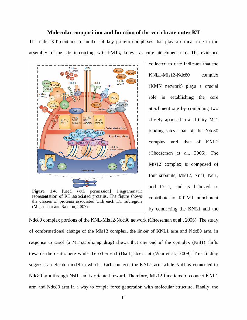
Figure 1.4. [used with permission] Diagrammatic
representation of KT associated proteins. The figure shows
the classes of proteins associated with each KT subregion
(Musacchio and Salmon, 2007).
Molecular composition and function of the vertebrate outer KT
The outer KT contains a number of key protein complexes that play a critical role in the
assembly of the site interacting with kMTs, known as core attachment site. The evidence
collected to date indicates that the
KNL1-Mis12-Ndc80 complex
(KMN network) plays a crucial
role in establishing the core
attachment site by combining two
closely apposed low-affinity MT-
binding sites, that of the Ndc80
complex and that of KNL1
(Cheeseman et al., 2006). The
Mis12 complex is composed of
four subunits, Mis12, Nnf1, Nsl1,
and Dsn1, and is believed to
contribute to KT-MT attachment
by connecting the KNL1 and the
Ndc80 complex portions of the KNL-Mis12-Ndc80 network (Cheeseman et al., 2006). The study
of conformational change of the Mis12 complex, the linker of KNL1 arm and Ndc80 arm, in
response to taxol (a MT-stabilizing drug) shows that one end of the complex (Nnf1) shifts
towards the centromere while the other end (Dsn1) does not (Wan et al., 2009). This finding
suggests a delicate model in which Dsn1 connects the KNL1 arm while Nnf1 is connected to
Ndc80 arm through Nsl1 and is oriented inward. Therefore, Mis12 functions to connect KNL1
arm and Ndc80 arm in a way to couple force generation with molecular structure. Finally, the
12
Mis12 complex has been proposed to serve as a protein interaction hub for outer KT assembly
(Petrovic et al., 2010). The rod-like Ndc80 complex is composed of four protein subunits, Ndc80,
Nuf2, Spc24 and Spc25 (Wilson-Kubalek et al., 2008). The globular regions of Spc24 and Spc25
form one end and those of Nuf2 and Ndc80 form the other (Miller et al., 2008). In C. elegans,
Spc24 and Spc25 associate with Mis12, and the interaction of KNL1 and Mis12 complex
generates a binding site for the Ndc80 complex (Cheeseman et al., 2006). The Ndc80 and Nuf2
subunits contain a pair of globular, calponin-homology (CH) domains that contribute to high-
affinity MT binding (Wei et al., 2007). Besides, KNL1 may also contain a MT-binding region,
but the boundaries of the region are still unknown (Cheeseman et al., 2006). A high-resolution
map of the KT reveals that the Ndc80 arm moves inward toward the inner KT component CENP-
I relative to the KNL1 arm upon treatment with taxol, as the KNL1 arm maintains a relatively
constant distance from CENP-I (Wan et al., 2009). In this way, the intra-KT stretching (distance
between inner KT and outer KT within a KT) is reduced. In conclusion, connecting the low-
affinity binding sites of Nuf2/Ndc80 and KNL1 within the KMN network synergizes the overall
MT binding activity. However, Ndc80 appears to be the main substrate for AurB-regulated kMT
dynamics (DeLuca et al., 2006). In addition, the outer KT-localized Hec1 (homologue of Ndc80
in vertebrates) also plays a critical rule in controlling dynamic behavior of kMTs through the
AurB-dependent phosphorylation of its N terminus (DeLuca et al., 2005; DeLuca et al., 2006).
Molecular composition and function of the fibrous corona
The outermost portion of the KT is the “fibrous corona” (Figure 1.3). This region, which
constitutes the KT-MT interface, is occupied by a number of proteins with different functions.
These include motor proteins like dynein and CENP-E, non-motor proteins like CENP-F and
13
dynein-interacting proteins (NDE1 and NDEL1), and many proteins (such as Mad1, Mad2, Bub1,
BubR1, Bub3, Cdc20, and the RZZ complex) involved in mitotic checkpoint signaling. The
motor proteins found at the fibrous corona are thought to play a key role in the initial steps of KT
attachments and chromosome congression. Indeed, both CENP-E (Cai et al., 2009; Kapoor et al.,
2006) and dynein (Gassmann et al., 2008; Varma et al., 2008) play an important role in
establishment of the initial KT-MT lateral interaction, which precedes the formation of stable
end-on attachments. The Rod-ZW10-Zwilch (RZZ) complex functions to recruit the
dynein/dynactin complex to the KT (Karess, 2005). However, this pathway seems to be more
important for mitotic checkpoint signaling (discussed in next section) than for KT attachment
(Gassmann et al., 2008). Finally, a number of MT-binding proteins are also found at the fibrous
corona. For example, cytoplasmic linker protein (CLIP)-associating protein (CLASP), MAP215
(chTOG), and CLIP170 are three major MT-associated non-motor proteins that promote the
polymerization of MTs at the KT interface (Maiato et al., 2003; Maiato et al., 2005; Tanenbaum
et al., 2006). EB1, a MT-plus-end tracking protein, is also found in the corona region, where it is
believed to bind to the MT lattice to stabilize MTs (Sandblad et al., 2006; Tanaka and Desai,
2008). APC (Adenomatous Polyposis Coli) is another protein that is associated with MT plus
ends, thus regulating MT dynamics. Studies of APC/EB1 found APC to be a substrate for
Bub1/BubR1 kinases in vitro. By directly interacting with APC/EB1, BubR1 is able to regulate
the position of chromosomes at the metaphase plate and the establishment of stable KT-MT
attachments (Logarinho and Bousbaa, 2008). Recent studies also reveal the possible association
of the Ska (Ska1, Ska2 and Ska3) complex (Hanisch et al., 2006) with MTs at the KT interface in
metazoan (Daum et al., 2009; Gaitanos et al., 2009; Theis et al., 2009; Welburn et al., 2009).
Indeed, the Ska complex has been proposed as a functional homolog of the yeast Dam1 ring
14
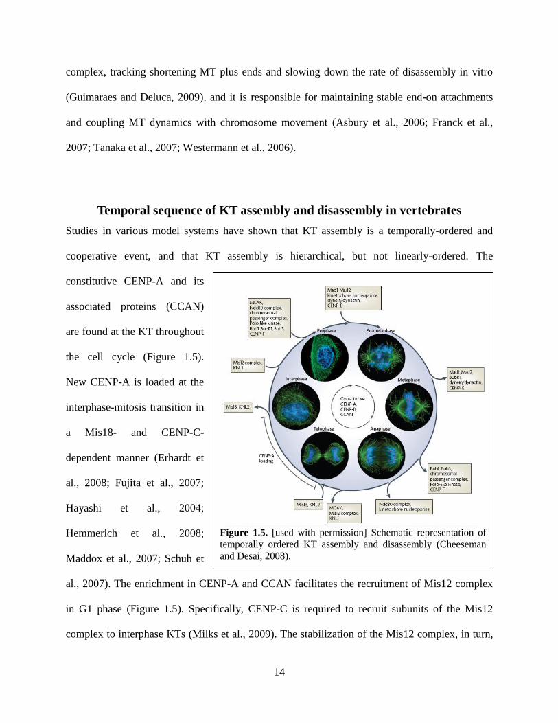
Figure 1.5. [used with permission] Schematic representation of
temporally ordered KT assembly and disassembly (Cheeseman
and Desai, 2008).
complex, tracking shortening MT plus ends and slowing down the rate of disassembly in vitro
(Guimaraes and Deluca, 2009), and it is responsible for maintaining stable end-on attachments
and coupling MT dynamics with chromosome movement (Asbury et al., 2006; Franck et al.,
2007; Tanaka et al., 2007; Westermann et al., 2006).
Temporal sequence of KT assembly and disassembly in vertebrates
Studies in various model systems have shown that KT assembly is a temporally-ordered and
cooperative event, and that KT assembly is hierarchical, but not linearly-ordered. The
constitutive CENP-A and its
associated proteins (CCAN)
are found at the KT throughout
the cell cycle (Figure 1.5).
New CENP-A is loaded at the
interphase-mitosis transition in
a Mis18- and CENP-C-
dependent manner (Erhardt et
al., 2008; Fujita et al., 2007;
Hayashi et al., 2004;
Hemmerich et al., 2008;
Maddox et al., 2007; Schuh et
al., 2007). The enrichment in CENP-A and CCAN facilitates the recruitment of Mis12 complex
in G1 phase (Figure 1.5). Specifically, CENP-C is required to recruit subunits of the Mis12
complex to interphase KTs (Milks et al., 2009). The stabilization of the Mis12 complex, in turn,
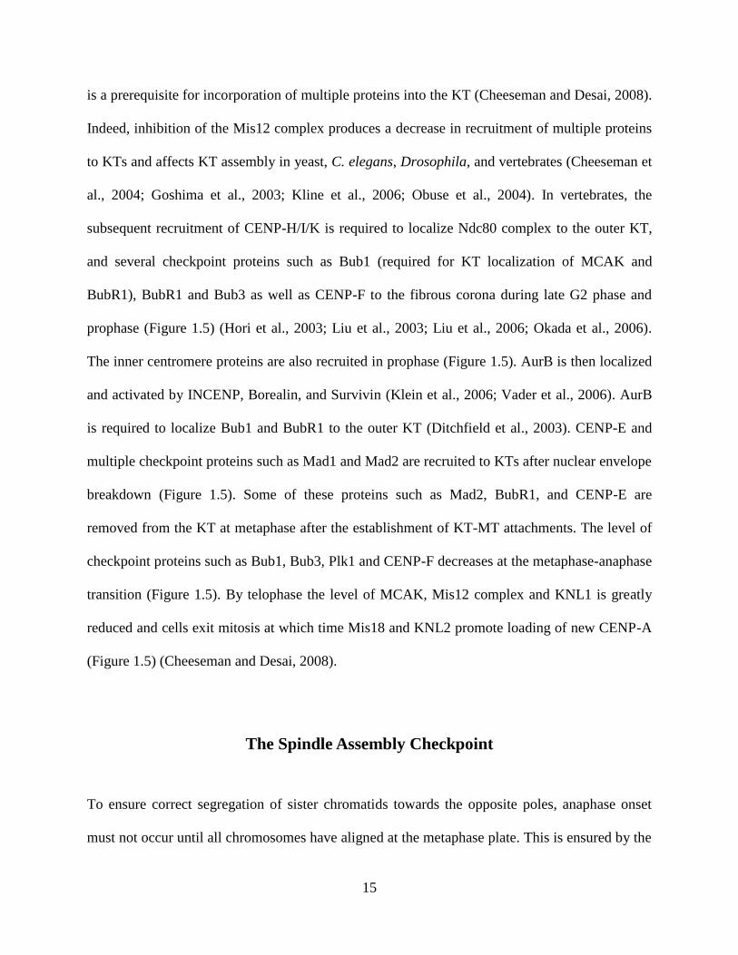
15
is a prerequisite for incorporation of multiple proteins into the KT (Cheeseman and Desai, 2008).
Indeed, inhibition of the Mis12 complex produces a decrease in recruitment of multiple proteins
to KTs and affects KT assembly in yeast, C. elegans, Drosophila, and vertebrates (Cheeseman et
al., 2004; Goshima et al., 2003; Kline et al., 2006; Obuse et al., 2004). In vertebrates, the
subsequent recruitment of CENP-H/I/K is required to localize Ndc80 complex to the outer KT,
and several checkpoint proteins such as Bub1 (required for KT localization of MCAK and
BubR1), BubR1 and Bub3 as well as CENP-F to the fibrous corona during late G2 phase and
prophase (Figure 1.5) (Hori et al., 2003; Liu et al., 2003; Liu et al., 2006; Okada et al., 2006).
The inner centromere proteins are also recruited in prophase (Figure 1.5). AurB is then localized
and activated by INCENP, Borealin, and Survivin (Klein et al., 2006; Vader et al., 2006). AurB
is required to localize Bub1 and BubR1 to the outer KT (Ditchfield et al., 2003). CENP-E and
multiple checkpoint proteins such as Mad1 and Mad2 are recruited to KTs after nuclear envelope
breakdown (Figure 1.5). Some of these proteins such as Mad2, BubR1, and CENP-E are
removed from the KT at metaphase after the establishment of KT-MT attachments. The level of
checkpoint proteins such as Bub1, Bub3, Plk1 and CENP-F decreases at the metaphase-anaphase
transition (Figure 1.5). By telophase the level of MCAK, Mis12 complex and KNL1 is greatly
reduced and cells exit mitosis at which time Mis18 and KNL2 promote loading of new CENP-A
(Figure 1.5) (Cheeseman and Desai, 2008).
The Spindle Assembly Checkpoint
To ensure correct segregation of sister chromatids towards the opposite poles, anaphase onset
must not occur until all chromosomes have aligned at the metaphase plate. This is ensured by the
16
mitotic checkpoint, or spindle assembly checkpoint (SAC), a biochemical pathway involving a
myriad of proteins. The SAC monitors KT-MT attachment and/or tension and generates a “wait-
anaphase” signal if these requirements (attachment and tension) are not satisfied. In a study
published in 1994, Rieder et al. found that PtK1 cells entered anaphase about 23 minutes after
the last KT attached to the spindle, suggesting that vertebrate cells possess a metaphase-anaphase
checkpoint control that monitors not only sister KT attachment to the spindle but also the
increase in tension between sister KTs or between KTs and their associated MTs (Rieder et al.,
1994). In a subsequent study, Rieder et al. used laser ablation to destroy the unattached KT of the
last mono-oriented (one sister KT bound to MTs and one unattached) chromosome, and
concluded that the inhibitory signal for metaphase-anaphase transition was generated at or near
the unattached KT (Rieder et al., 1995). Thus, by detecting single unattached KTs or some kinds
of improper attachments, the SAC can help prevent unequal distribution of genetic material
during cell division.
Upon mitotic entry, sister chromatids are bound along their length by a multi-subunit protein
complex called cohesin. Most cohesin is removed by metaphase, except for that at the
centromere, which maintains cohesion between sister chromatids until anaphase onset
(Waizenegger et al., 2000). The removal of these last cohesin molecules depends on degradation
by the protease separase. Prior to anaphase, however, separase, is maintained inactive by binding
of its inhibitory subunit, securin. Once all KTs are bi-oriented and attached to kMTs, the
anaphase promoting complex/cyclosome (APC/C), activated by Cdc20, polyubiquitinates both
cyclin B and securin. The polyubiquitination of these two proteins leads to their degradation by
the 26S proteasome (Musacchio and Salmon, 2007). As a result of securin degradation, separase
17
is released and proteolyzes centromeric cohesin, thus leading to sister chromatid separation.
Therefore, anaphase onset is triggered by the interaction of APC/C with Cdc20. The SAC
functions by inhibiting formation of the Cdc20-APC/C complex, thus preventing precocious
degradation of cohesin until all KTs establish bipolar attachment with spindle MTs and all
chromosomes are aligned at the metaphase plate (Fang et al., 1998).
The role of the SAC in determining anaphase onset relies on the ability of numerous SAC
proteins to interact with unattached KTs. Core components of the SAC include six evolutionary
conserved proteins, Bub1, Bub3, Mad1, Mad2, BubR1 (Mad3 in yeast), and Mps1 (multipolar
spindle-1) (Hardwick et al., 1996; Musacchio and Salmon, 2007). Additional proteins, including
the plus-end directed kinesin motor CENP-E (Mao et al., 2005), the minus-end directed motor
dynein, dynein-interacting proteins such as dynactin (Howell et al., 2001; Tai et al., 2002),
CLIP170 and LIS1 (Tai et al., 2002), the RZZ (ROD-ZW10-ZWILCH) complex (Karess, 2005;
Lu et al., 2009), p31comet
(Mapelli et al., 2006; Xia et al., 2004). CDK1-cyclin B (D'Angiolella et
al., 2003), Plk1 (polo-like kinase 1) (van Vugt and Medema, 2005), AurB (Hauf et al., 2003;
Murata-Hori et al., 2002), and MAPK (mitogen-activated protein kinase), can indirectly affect
the SAC, but do not participate to SAC signaling.
Recent studies suggested that the SAC might be maintained active by the sequestration of Cdc20
into a mitotic checkpoint complex (MCC) containing three core SAC proteins, Mad2,
BubR1/Mad3 and Bub3, as well as Cdc20 (Musacchio and Salmon, 2007). Studies about the
formation of MCC proposed a Mad2 template model. In this model, Mad2 is present in two
conformations, closed-Mad2 (C-Mad2) and open-Mad2 (O-Mad2). Mad1-C-Mad2 complexes
18
are deposited at the KT by p31comet
(Vink et al., 2006; Xia et al., 2004). O-Mad2 is then recruited
at the KT by this Mad1-C-Mad2 complex, and converted into C-Mad2, which can bind Cdc20,
generating C-Mad2-Cdc20 complex, and possibly MCC by binding of other SAC proteins. The
C-Mad2 within the C-Mad2-Cdc20 complex was also proposed to function as a cytoplasmic
template for conversion of cytoplasmic O-Mad2 into Cdc20-bound C-Mad2 (De Antoni et al.,
2005). This would lead to amplification of the SAC signal, thus explaining how even a single
unattached KT can sustain mitotic arrest. All SAC proteins concentrate at KTs during
prometaphase, indicating that KTs act as a catalytic platform to accelerate the production of the
MCC (Howell et al., 2000; Howell et al., 2001; Kallio et al., 2002; Shah et al., 2004; Vink et al.,
2006). However, the contribution of KTs to MCC formation remains controversial based on a
number of studies. For example, mature KTs only exist in mitosis, but the MCC might exist
throughout the cell cycle (Sudakin et al., 2001). Consistent with the finding above, the MCC
persisted in SAC-inactive S. cerevisiae Ndc10 mutants whose KT assembly was defective
(Gillett et al., 2004).
Studies aimed at determining the exact nature of the defect(s) detected by the SAC have
indicated that both lack of attachment (Rieder et al., 1995) (Figure 1.6A) and lack of tension
(Jang et al., 1995; Li and Nicklas, 1995) at the KT can be detected, although the role of tension
in checkpoint signaling is still controversial (Khodjakov and Rieder, 2009; King and Nicklas,
2000; Nicklas and Ward, 1994). The attachment hypothesis is supported by the evidence that
some SAC proteins are immediately removed from KTs once MT attachment is established. For
example, Mad2 localizes to unattached KTs in prometaphase but its level is highly reduced at
metaphase KTs (Chen et al., 1996; Li and Benezra, 1996), when their multiple MT-binding sites
19
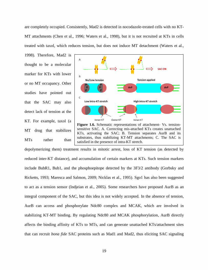
Figure 1.6. Schematic representations of attachment- Vs. tension-
sensitive SAC. A. Correcting mis-attached KTs creates unattached
KTs, activating the SAC; B. Tension separates AurB and its
substrates, thus stabilizing KT-MT attachments; C. The SAC is
satisfied in the presence of intra-KT stretch.
are completely occupied. Consistently, Mad2 is detected in nocodazole-treated cells with no KT-
MT attachments (Chen et al., 1996; Waters et al., 1998), but it is not recruited at KTs in cells
treated with taxol, which reduces tension, but does not induce MT detachment (Waters et al.,
1998). Therefore, Mad2 is
thought to be a molecular
marker for KTs with lower
or no MT occupancy. Other
studies have pointed out
that the SAC may also
detect lack of tension at the
KT. For example, taxol (a
MT drug that stabilizes
MTs rather than
depolymerizing them) treatment results in mitotic arrest, loss of KT tension (as detected by
reduced inter-KT distance), and accumulation of certain markers at KTs. Such tension markers
include BubR1, Bub1, and the phosphoepitope detected by the 3F3/2 antibody (Gorbsky and
Ricketts, 1993; Maresca and Salmon, 2009; Nicklas et al., 1995). Sgo1 has also been suggested
to act as a tension sensor (Indjeian et al., 2005). Some researchers have proposed AurB as an
integral component of the SAC, but this idea is not widely accepted. In the absence of tension,
AurB can access and phosphorylate Ndc80 complex and MCAK, which are involved in
stabilizing KT-MT binding. By regulating Ndc80 and MCAK phosphorylation, AurB directly
affects the binding affinity of KTs to MTs, and can generate unattached KTs/attachment sites
that can recruit bona fide SAC proteins such as Mad1 and Mad2, thus eliciting SAC signaling

20
(Pinsky and Biggins, 2005; Pinsky et al., 2006). Moreover, AurB activity appears to be high at
unattached/ tensionless KTs (Liu et al., 2009). These observations have led to the idea that AurB
is directly involved in SAC control (DeLuca et al., 2006; Knowlton et al., 2006; Santaguida and
Musacchio, 2009). As shown in figure 1.6B, when KTs are stretched (i.e., under tension), AurB
is unable to access its substrates, and this results in dephosphorylation of its substrates, which
stabilizes KT-MT attachments. Studies in yeast suggested that that the CPC components Bir1-
Sli15 link centromeres to MTs in a manner that allows Sli15 to locally activate Ipl1 when core
attachment sites are not under tension (Sandall et al., 2006). Whereas KT tension has been
traditionally measured as separation between sister KTs (inter-KT stretch), two recent studies
(Maresca and Salmon, 2009; Uchida et al., 2009) suggested that intra-, rather than inter-, KT
stretching might be the actual tension-signaling mechanism (Figure 1.6C). However, this would
not explain how the inner centromere CPC components may sense the reduced tension.
Some investigators have suggested the separation of the SAC into two signaling branches, one
depending on the attachment and the other on the tension status of the KT (Zhou et al., 2002).
However, previous studies have also pointed out a possible interdependence between tension and
attachment (Nicklas et al., 2001). Indeed, whereas tension is known to stabilize KT-MT
attachment, loss of tension could lead to destabilization and detachment of kMTs, thus making it
difficult to separate tension from attachment. On the other hand, studies in budding yeast have
attempted to resolve this controversy. Deletion of a cohesin subunit results in mitotic cells
possessing only single chromatids, and each yeast chromatid possess a single MT attachment
site (Shonn et al., 2000). Thus, these KTs exhibit full occupancy, yet they lack tension because
they lack a sister KT (Pinsky and Biggins, 2005; Shonn et al., 2000). Interestingly, these cells
21
exhibit a SAC-dependent mitotic arrest (Pinsky and Biggins, 2005). Maresca and Salmon have
recently proposed a model that explains how tension defects could be translated into a wait-
anaphase signal. They proposed that low intra-KT stretching promotes Mad1-Mad2 binding at
the KT by positioning multiple low-affinity MT binding sites near each other. In this case,
Mad1-Mad2 is phosphorylated by KT- and centromere-localized kinases, and this increases its
binding affinity to the KT, amplifying the SAC signal. However, the affinity of Mad1-Mad2 for
the KT is reduced when the KT is stretched because the binding sites are repositioned and Mad1-
Mad2 is dephosphorylated. As a result, the SAC is satisfied upon the increase in the intra-KT
stretching (Maresca and Salmon, 2010; Wan et al., 2009). In conclusion, although this topic is
still controversial, both attachment and tension seem to be important for SAC signaling.
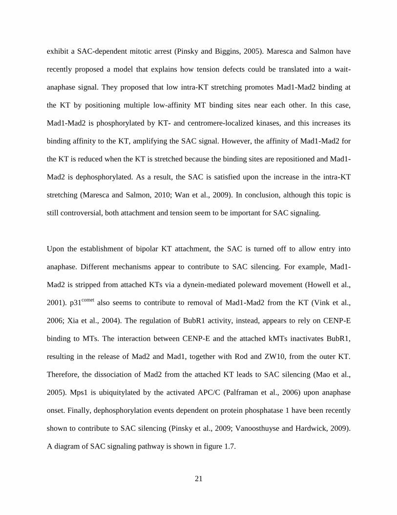
Upon the establishment of bipolar KT attachment, the SAC is turned off to allow entry into
anaphase. Different mechanisms appear to contribute to SAC silencing. For example, Mad1-
Mad2 is stripped from attached KTs via a dynein-mediated poleward movement (Howell et al.,
2001). p31comet
also seems to contribute to removal of Mad1-Mad2 from the KT (Vink et al.,
2006; Xia et al., 2004). The regulation of BubR1 activity, instead, appears to rely on CENP-E
binding to MTs. The interaction between CENP-E and the attached kMTs inactivates BubR1,
resulting in the release of Mad2 and Mad1, together with Rod and ZW10, from the outer KT.
Therefore, the dissociation of Mad2 from the attached KT leads to SAC silencing (Mao et al.,
2005). Mps1 is ubiquitylated by the activated APC/C (Palframan et al., 2006) upon anaphase
onset. Finally, dephosphorylation events dependent on protein phosphatase 1 have been recently
shown to contribute to SAC silencing (Pinsky et al., 2009; Vanoosthuyse and Hardwick, 2009).
A diagram of SAC signaling pathway is shown in figure 1.7.
22
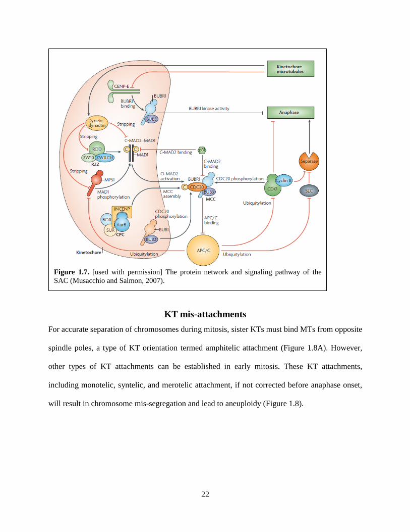
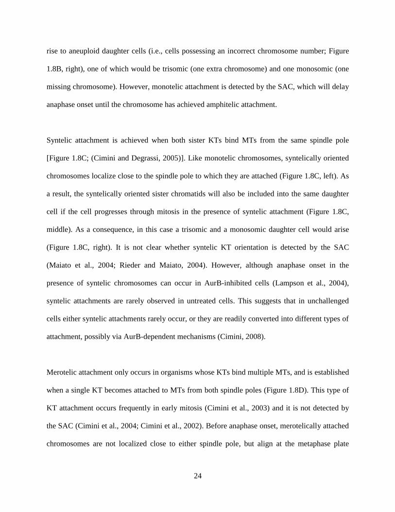
KT mis-attachments
For accurate separation of chromosomes during mitosis, sister KTs must bind MTs from opposite
spindle poles, a type of KT orientation termed amphitelic attachment (Figure 1.8A). However,
other types of KT attachments can be established in early mitosis. These KT attachments,
including monotelic, syntelic, and merotelic attachment, if not corrected before anaphase onset,
will result in chromosome mis-segregation and lead to aneuploidy (Figure 1.8).
Figure 1.7. [used with permission] The protein network and signaling pathway of the
SAC (Musacchio and Salmon, 2007).

23
Monotelic attachment occurs when one sister KT becomes attached to MTs from one spindle
pole but the other remains unattached [Figure 1.8B; (Cimini, 2008)]. Monotelic orientation is a
common event during the early stages of mitosis. In fact, each chromosome initially establishes
monotelic attachment, until the sister KT binds MTs from the opposite spindle pole, which will
lead to amphitelic attachment and chromosome congression to the metaphase plate.
Monotelically oriented chromosomes are positioned close to the pole to which they are attached
(Figure 1.8B, left). If cells progress into anaphase in the presence of monotelic attachment, the
sister chromatids separate, but will be inevitably included into the same daughter cell due to their
proximity to one spindle pole (Figure 1.8B, middle). Therefore, monotelic attachment could give
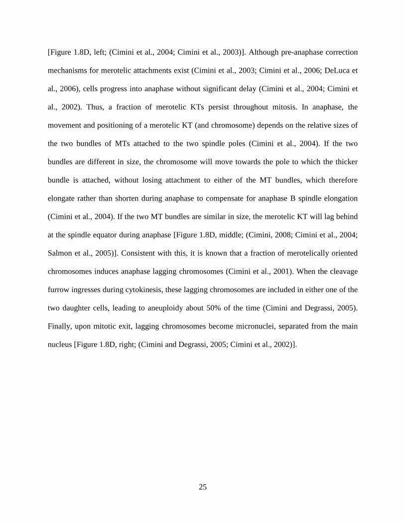
Figure 1.8. [used with permission] Types of KT attachments. Left column, KT attachment in
metaphase. Middle column, anaphase in the presence of different types of KT attachment.
Right column, daughter cells (Cimini, 2008).
24
rise to aneuploid daughter cells (i.e., cells possessing an incorrect chromosome number; Figure
1.8B, right), one of which would be trisomic (one extra chromosome) and one monosomic (one
missing chromosome). However, monotelic attachment is detected by the SAC, which will delay
anaphase onset until the chromosome has achieved amphitelic attachment.
Syntelic attachment is achieved when both sister KTs bind MTs from the same spindle pole
[Figure 1.8C; (Cimini and Degrassi, 2005)]. Like monotelic chromosomes, syntelically oriented
chromosomes localize close to the spindle pole to which they are attached (Figure 1.8C, left). As
a result, the syntelically oriented sister chromatids will also be included into the same daughter
cell if the cell progresses through mitosis in the presence of syntelic attachment (Figure 1.8C,
middle). As a consequence, in this case a trisomic and a monosomic daughter cell would arise
(Figure 1.8C, right). It is not clear whether syntelic KT orientation is detected by the SAC
(Maiato et al., 2004; Rieder and Maiato, 2004). However, although anaphase onset in the
presence of syntelic chromosomes can occur in AurB-inhibited cells (Lampson et al., 2004),
syntelic attachments are rarely observed in untreated cells. This suggests that in unchallenged
cells either syntelic attachments rarely occur, or they are readily converted into different types of
attachment, possibly via AurB-dependent mechanisms (Cimini, 2008).
Merotelic attachment only occurs in organisms whose KTs bind multiple MTs, and is established
when a single KT becomes attached to MTs from both spindle poles (Figure 1.8D). This type of
KT attachment occurs frequently in early mitosis (Cimini et al., 2003) and it is not detected by
the SAC (Cimini et al., 2004; Cimini et al., 2002). Before anaphase onset, merotelically attached
chromosomes are not localized close to either spindle pole, but align at the metaphase plate
25
[Figure 1.8D, left; (Cimini et al., 2004; Cimini et al., 2003)]. Although pre-anaphase correction
mechanisms for merotelic attachments exist (Cimini et al., 2003; Cimini et al., 2006; DeLuca et
al., 2006), cells progress into anaphase without significant delay (Cimini et al., 2004; Cimini et
al., 2002). Thus, a fraction of merotelic KTs persist throughout mitosis. In anaphase, the
movement and positioning of a merotelic KT (and chromosome) depends on the relative sizes of
the two bundles of MTs attached to the two spindle poles (Cimini et al., 2004). If the two
bundles are different in size, the chromosome will move towards the pole to which the thicker
bundle is attached, without losing attachment to either of the MT bundles, which therefore
elongate rather than shorten during anaphase to compensate for anaphase B spindle elongation
(Cimini et al., 2004). If the two MT bundles are similar in size, the merotelic KT will lag behind
at the spindle equator during anaphase [Figure 1.8D, middle; (Cimini, 2008; Cimini et al., 2004;
Salmon et al., 2005)]. Consistent with this, it is known that a fraction of merotelically oriented
chromosomes induces anaphase lagging chromosomes (Cimini et al., 2001). When the cleavage
furrow ingresses during cytokinesis, these lagging chromosomes are included in either one of the
two daughter cells, leading to aneuploidy about 50% of the time (Cimini and Degrassi, 2005).
Finally, upon mitotic exit, lagging chromosomes become micronuclei, separated from the main
nucleus [Figure 1.8D, right; (Cimini and Degrassi, 2005; Cimini et al., 2002)].
26
Chapter 2: Introduction
The behavior of the SAC in response to KT-MT mis-attachments
Accurate chromosome segregation during mitosis is critical to organism development and
maintenance. The sister chromatids of each replicated chromosome must be segregated to the
two daughter cells during mitosis, and this is achieved thanks to the interaction of the two sister
KTs with MTs from opposite poles of the mitotic spindle. This type of KT-MT interaction is
referred to as amphitelic attachment, and it is the only one that ensures accurate chromosome
segregation. Other types of attachments can occur in early mitosis and cause chromosome mis-
segregation and aneuploidy if not corrected before anaphase onset (Cimini and Degrassi, 2005).
These mis-attachments include (i) monotelic attachments, in which one sister KT is bound to
MTs and the other is unattached; (ii) syntelic attachments, in which the two sister KTs are
attached to MTs from the same spindle pole; and (iii) merotelic attachments, in which a single
KT is attached to MTs from both spindle poles. A biochemical pathway named SAC is
responsible for detecting mis-attached chromosomes and delay anaphase onset until those mis-
attachments have been corrected (Musacchio and Salmon, 2007). However, only monotelic
attachments have been clearly shown to sustain SAC signaling (Rieder et al., 1995). This is
believed to occur thanks to the accumulation of SAC proteins (e.g., Mad1, Mad2, etc.) at the
unattached KT of monotelic chromosomes (Cheeseman and Desai, 2008; Chen et al., 1996; Li
and Benezra, 1996; Waters et al., 1998). Conversely, all the evidence available to date suggests
that merotelic attachments are not detected by the SAC (Cimini et al., 2004; Cimini et al., 2002;
Khodjakov et al., 1997; Wise and Brinkley, 1997), although a previous study in Xenopus leavis
cells showed accumulation of certain proteins involved in MT turnover (and hence mis-
attachment correction), such as AurB and MCAK, at the centromere of chromosomes with one
27
merotelic KT (Knowlton et al., 2006). Finally, syntelic attachments have not been extensively
studied and whether the SAC can detect such mis-attachments is currently a matter of debate
(Maiato et al., 2004; Rieder and Maiato, 2004).
Attachment Vs. Tension in SAC signaling
An unanswered question about SAC signaling is whether the SAC can only detect lack of
attachment or also lack of tension (Khodjakov and Rieder, 2009; King and Nicklas, 2000;
Nicklas et al., 2001; Pinsky and Biggins, 2005). Several studies suggested that lack of tension at
the KT might be detected by the SAC (Jang et al., 1995; Li and Nicklas, 1995). If that is the case,
then syntelic KTs should maintain the SAC active, because they do not experience the inter-KT
tension that normally develops upon amphitelic attachment. However, many researchers believe
that the SAC can only detect lack of attachment and not lack of tension (Khodjakov and Rieder,
2009; Nezi and Musacchio, 2009). Thus, syntelic attachments would not be detetcted by the SAC.
However, the fact that syntelic attachments are rarely seen in cells progressing through mitosis
(Hauf et al., 2003) suggests that they might be efficiently corrected. A new hypothesis has
recently emerged (Liu et al., 2009; Nezi and Musacchio, 2009) to explain how certain KT mis-
attachments (merotelic and syntelic) can be corrected despite the inability of the SAC to detect
them. This hypothesis states that, although the SAC cannot detect the lack of tension at syntelic
(or merotelic) KTs, this lack of tension causes the MT attachment sites on these KTs to be
proximal to the inner centromere, where AurB is enriched, and this will promote AurB-
dependent detachment of kMTs. This hypothesis is based on recent work showing that proximity
of Hec1 to the inner centromere will lead to its phosphorylation by AurB (Liu et al., 2009), and
28
AurB-dependent Hec1 phosphorylation is known to result in increased kMT turnover (DeLuca et
al., 2006).
But how can tension be measured? It is widely documented that unattached chromosomes exhibit
minimal inter-KT stretching, but as chromosomes bind MTs and establish amphitelic attachment
the inter-KT stretching increases, reaching a maximum for chromosomes aligned at the
metaphase plate (Musacchio and Hardwick, 2002). Based on this, one could argue that syntelic
KTs may be detected by the SAC because of a reduced inter-KT stretching. Although
traditionally the inter-KT stretching (distance between sister KTs) has been used as a measure of
KT tension, two recent studies (Maresca and Salmon, 2009; Uchida et al., 2009) suggested that
what may be important for SAC signaling is the intra-KT stretching (measured as the distance
between an inner and an outer KT marker) rather than inter-KT stretching (the distance between
two sister KTs). This raises the possibility that syntelic attachments may be detected by the SAC
due to a reduction of intra-KT stretching in the syntelic KTs, leading some to propose that
reduced intra-KT stretching in one or both of the syntelic KTs may result in both attachment
correction and wait-anaphase (SAC) signaling (Maresca and Salmon, 2010).
Rationale and Hypothesis
Many SAC proteins are known to localize at the KT in early mitosis (Logarinho et al., 2004;
Vigneron et al., 2004), and this localization is believed to play a key role in SAC signaling (Zhou
et al., 2002). Most studies, however, have focused exclusively on the unattached KT of
monotelic chromosomes. To gain a better understanding on whether and how the SAC may
detect various types of mis-attachments (in particular syntelic), we systematically studied how
29
the molecular composition and structure of the KT changes in response to different types of
attachments. Our hypothesis is that if syntelic attachments are detected by the SAC, then syntelic
KTs should exhibit the same molecular composition (localization of SAC proteins) and structure
(inter- and intra-KT stretching) displayed by unattached KTs, which are known to trigger a SAC
response. Alternatively, syntelic attachments may not be detected by the SAC, but may be
efficiently corrected, in which case syntelic KTs would be expected to exhibit features similar to
those of merotelic KTs, many of which are formed in early mitosis, but corrected before
anaphase onset without triggering a SAC response. Specifically, syntelic KTs may exhibit
similar stretching and/or accumulation of proteins involved in mis-attachment correction, as
previously shown for merotelic attachments (Knowlton et al., 2006). To test these different
possibilities, the following markers were quantified at KTs with different types of attachments in
PtK1 cells: Mad2 (marker for partial or lack of KT-MT attachment) (Waters et al., 1998),
BubR1 (previously proposed to participate in the tension-sensing portion of SAC signaling)
(Logarinho and Bousbaa, 2008; Logarinho et al., 2004), 3F3/2 phosphoepitope (known to
accumulate as a result of low KT tension) (Logarinho et al., 2004; Nicklas et al., 1995), MCAK
(involved in correction of mis-attachments) (Knowlton et al., 2006), and AurB (involved in
correction of mis-attachments) (Biggins and Murray, 2001; Cimini et al., 2006; Tanaka et al.,
2002). Furthermore, we measured and compared intra- and inter-KT stretching of
KTs/chromosomes with different types of attachment.
30
Chapter 3: Materials and Methods
Cell culture and drug treatment
PtK1 cells (American Type Culture Collection, Rockville, MD) were cultured in HAM F-12
media (Invitrogen, Carlsbad, CA) supplemented with 0.5% sodium pyruvate (Fisher Scientific,
Pittsburg, PA), 1% antibiotic-antimycotic (Invitrogen, Carlsbad, CA), and 10% fetal bovine
serum (Invitrogen, Carlsbad, CA) in a 37℃, 5% CO2, humidified incubator. For experiments,
cells were grown on coverslips up to ~70% confluency, and then treated for 2 hours with 2 M
of the Eg5 inhibitor S-trityl-L-cysteine (STLC) to increase the number of syntelic and monotelic
attachments (Kapoor et al., 2000). To increase the number of merotelic attachments, cells were
arrested in 2 M STLC for 2 hours, washed out of the drug, and then fixed after a 40-minute
incubation in drug-free media (Silkworth, Nardi, and Cimini, unpublished). DMSO (Fisher
Scientific, Pittsburg, PA) was added in control cultures used to image amphitelic attachments.
Antibodies
Antibodies used in this study included: rabbit anti-Mad2 (from Dr. E. D. Salmon, The University
of North Carolina at Chapel Hill); mouse anti-3F3/2 (Boston Biologicals, Wellesley, MA); rabbit
anti-BubR1 (from Dr. T. Yen, University of Pennsylvania); rabbit anti-AurB (Abcam,
Cambridge, MA); rabbit anti-MCAK (from Dr. R. Ohi, Vanderbilt University); rabbit anti-
INCENP (from Dr. Aaron Straight, Stanford University); mouse anti-Hec1 (Abcam, Cambridge,
MA); human anti-ACA (anti-centromere antigen) (Antibodies Inc., Davis, CA); mouse anti-
DM1anti--tubulin) (Sigma-Aldrich Corp, St. Louis, MO); Alexa-488 goat anti-mouse
(Invitrogen, Carlsbad, CA); Alexa-488 goat anti-rabbit (Invitrogen, Carlsbad, CA); X-
31
Rhodamine goat anti-human (Jackson Immunoresearch Laboratories Inc., West Grove, PA); Cy5
goat anti-mouse (Abcam, Cambridge, MA); Cy5 goat anti-rabbit (Invitrogen, Carlsbad, CA).
The antibodies were diluted for immunostaining as follows: anti-Mad2, 1:200; 3F3/2, 1:250;
anti-BubR1, 1:1000; anti-AurB, 1:200; anti-MCAK, 1:200; anti-INCENP, 1:1250; anti-Hec1,
1:500; ACA, 1:100; DM1, 1:500; Alexa-488 goat anti-mouse, 1:400; Alexa-488 goat anti-
rabbit, 1:400; X-Rhodamine goat anti-human, 1:100; Cy5 goat anti-mouse, 1:100; Cy5 goat anti-
rabbit, 1:100.
Immunostaining
For BubR1, INCENP, and AurB staining, cells were first fixed in freshly prepared 4%
formaldehyde (Fisher Scientific, Pittsburg, PA) for 20 minutes and then permeabilized in 0.5%
Triton X-100 (Fisher Scientific, Pittsburg, PA Fisher) in 1XPHEM buffer [60mM Pipes (Fisher
Scientific, Pittsburg, PA), 25mM HEPES (Fisher Scientific, Pittsburg, PA), 10mM EGTA, 2mM
MgSO4 (Fisher Scientific, Pittsburg, PA), pH 7.0] at room temperature for 10 minutes. For
MCAK staining, cells were fixed as described above, but permeabilized in 0.1%Triton X-100 in
1XPHEM buffer. For Mad2 staining, cells were first lysed in freshly prepared 0.5% Triton X-
100 in 1XPHEM buffer for 5 minutes and then fixed in freshly prepared 4% formaldehyde in
1XPHEM for 20 minutes at room temperature. For Hec1 staining, cells were prefixed in freshly
prepared 4% formaldehyde for 5 seconds before a 5-minute lysis in 0.5% Triton-X and 20-
minute fixation in 4% formaldehyde. For 3F3/2 staining, cells were processed as for Hec1
staining, but the lysis buffer consisted of 0.5% Chaps (VWR, Radnor, PA) in 1XPHEM to which
100nM microcystin LR (VWR, Radnor, PA) was added. Cells were then rinsed three times for 5

32
minutes in 1XPBS with 0.05% Tween-20 (PBST) (Fisher Scientific, Pittsburg, PA) and blocked
for 1 hour at room temperature in 10% boiled goat serum (BGS) (Jackson Immunoresearch
Laboratories, West Grove, PA). Primary antibodies were diluted into 1XPHEM with 5% BGS,
and cells were incubated overnight at 4℃ . After four 5-minute PBST washes, cells were
incubated with secondary antibodies in 5% BGS for 45 minutes at room temperature. After four
5-minute PBST washes, cells were counterstained with DAPI (Sigma-Aldrich Corp, St. Louis,
MO) for 5 minutes, then rinsed three times with PBST for 5 minutes, and mounted on
microscope slides with an anti-fading solution containing 90% glycerol, 10% Tris buffer, and
0.5–1% n-propyl galate.
Confocal microscopy and image acquisition
Immunofluorescently labeled cells were imaged with a swept field confocal unit (Prairie
Technologies) attached to a Nikon Eclipse TE2000-U microscope. Images were obtained with a
cooled CCD digital camera, using a 100X / 1.4NA Plan-Apochromatic phase-contrast objective
lens. Digital images were acquired by Elements image processing software. Z-series optical
sections through each cell were obtained at 0.6-m steps.
Data analysis
Identification of different types of attachments. Merged images of the -tubulin and ACA
fluorescence were used to identify the different types of KT attachments. First, maximum
intensity projections of the two fluorescence channels were generated and merged. The merged
image was viewed for identification of possible examples of various attachments. To ascertain
the nature of the attachment, the original Z-stacks were then merged and a few selected focal
33
planes above and below the KT/KT pair of interest were viewed by scrolling up and down. This
was done to make sure that a KT that appeared unattached was not attached to a MT bundle
going off the focal plane at an angle or that a KT that appeared attached was not simply
overlapping with a MT bundle running past it on an adjacent focal plane. Once the KTs of
interest were identified, the original single channel images were used for measurements and
quantifications.
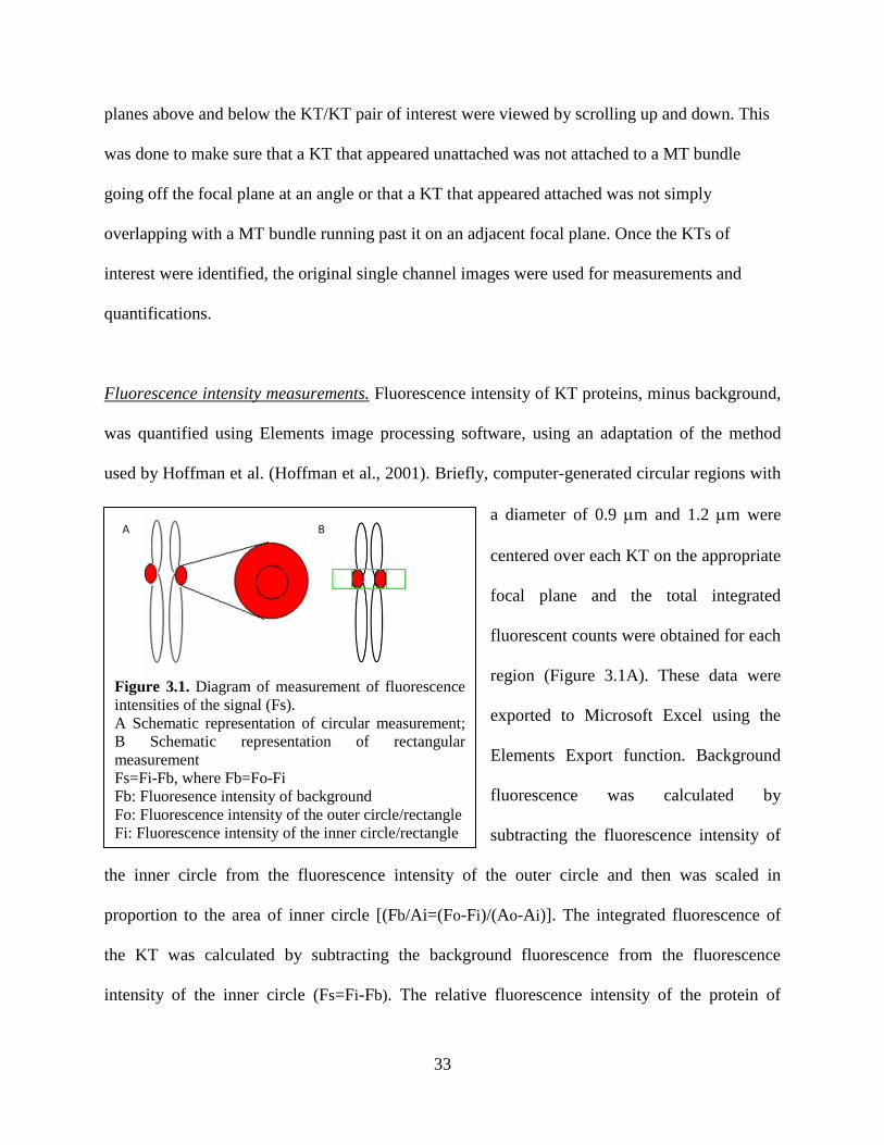
Fluorescence intensity measurements. Fluorescence intensity of KT proteins, minus background,
was quantified using Elements image processing software, using an adaptation of the method
used by Hoffman et al. (Hoffman et al., 2001). Briefly, computer-generated circular regions with
a diameter of 0.9 m and 1.2 m were
centered over each KT on the appropriate
focal plane and the total integrated
fluorescent counts were obtained for each
region (Figure 3.1A). These data were
exported to Microsoft Excel using the
Elements Export function. Background
fluorescence was calculated by
subtracting the fluorescence intensity of
the inner circle from the fluorescence intensity of the outer circle and then was scaled in
proportion to the area of inner circle [(Fb/Ai=(Fo-Fi)/(Ao-Ai)]. The integrated fluorescence of
the KT was calculated by subtracting the background fluorescence from the fluorescence
intensity of the inner circle (Fs=Fi-Fb). The relative fluorescence intensity of the protein of
Figure 3.1. Diagram of measurement of fluorescence
intensities of the signal (Fs).
A Schematic representation of circular measurement;
B Schematic representation of rectangular
measurement
Fs=Fi-Fb, where Fb=Fo-Fi
Fb: Fluoresence intensity of background Fo: Fluorescence intensity of the outer circle/rectangle
Fi: Fluorescence intensity of the inner circle/rectangle
34
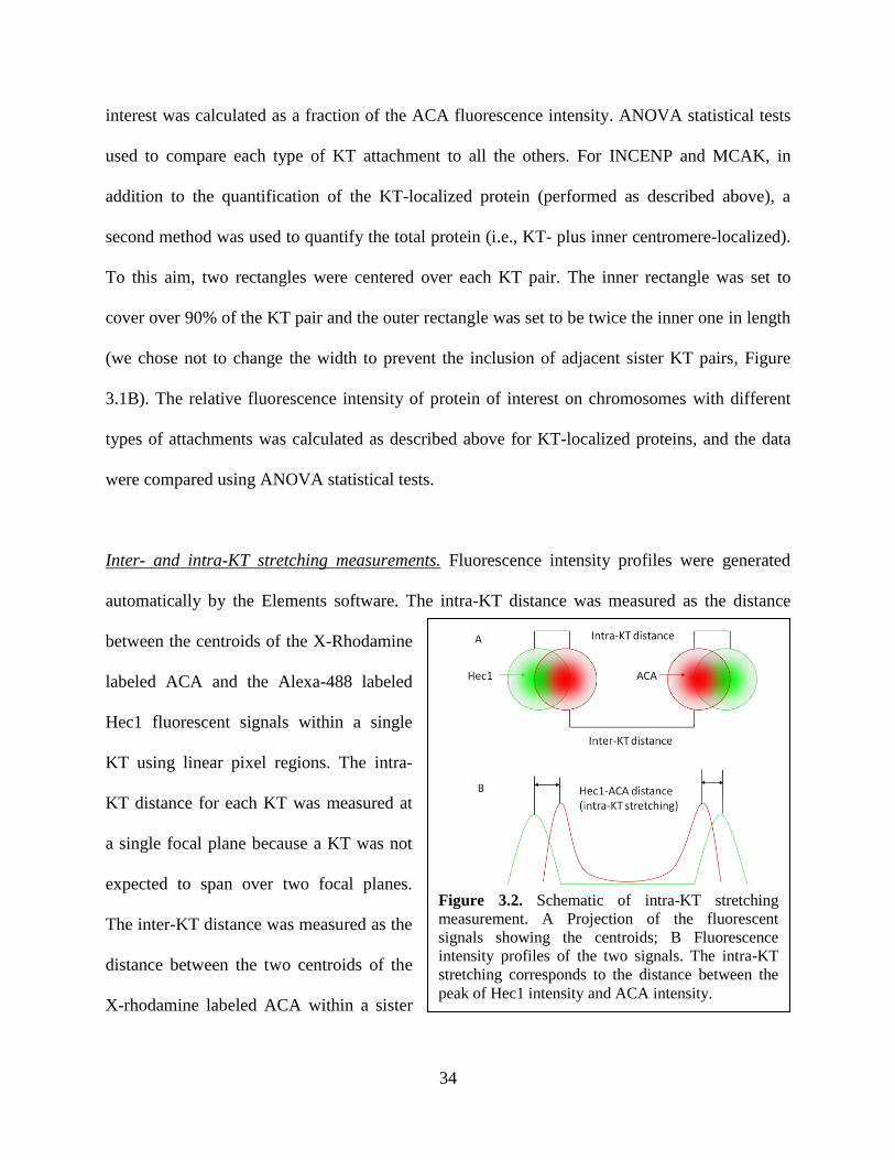
Figure 3.2. Schematic of intra-KT stretching
measurement. A Projection of the fluorescent
signals showing the centroids; B Fluorescence
intensity profiles of the two signals. The intra-KT
stretching corresponds to the distance between the
peak of Hec1 intensity and ACA intensity.
interest was calculated as a fraction of the ACA fluorescence intensity. ANOVA statistical tests
used to compare each type of KT attachment to all the others. For INCENP and MCAK, in
addition to the quantification of the KT-localized protein (performed as described above), a
second method was used to quantify the total protein (i.e., KT- plus inner centromere-localized).
To this aim, two rectangles were centered over each KT pair. The inner rectangle was set to
cover over 90% of the KT pair and the outer rectangle was set to be twice the inner one in length
(we chose not to change the width to prevent the inclusion of adjacent sister KT pairs, Figure
3.1B). The relative fluorescence intensity of protein of interest on chromosomes with different
types of attachments was calculated as described above for KT-localized proteins, and the data
were compared using ANOVA statistical tests.
Inter- and intra-KT stretching measurements. Fluorescence intensity profiles were generated
automatically by the Elements software. The intra-KT distance was measured as the distance
between the centroids of the X-Rhodamine
labeled ACA and the Alexa-488 labeled
Hec1 fluorescent signals within a single
KT using linear pixel regions. The intra-
KT distance for each KT was measured at
a single focal plane because a KT was not
expected to span over two focal planes.
The inter-KT distance was measured as the
distance between the two centroids of the
X-rhodamine labeled ACA within a sister
35
KT pair (Figure 3.2). The measurements were exported and recorded into Microsoft Excel
spreadsheets and ANOVA statistical tests were used to compare each type of KT/chromosome
attachment to all the others.
36
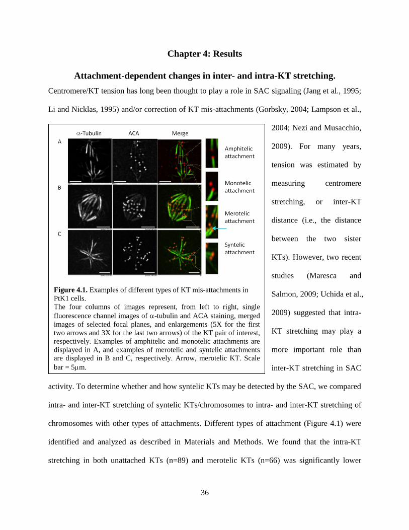
Figure 4.1. Examples of different types of KT mis-attachments in
PtK1 cells.
The four columns of images represent, from left to right, single
fluorescence channel images of -tubulin and ACA staining, merged
images of selected focal planes, and enlargements (5X for the first
two arrows and 3X for the last two arrows) of the KT pair of interest,
respectively. Examples of amphitelic and monotelic attachments are
displayed in A, and examples of merotelic and syntelic attachments
are displayed in B and C, respectively. Arrow, merotelic KT. Scale
bar = 5m.
Chapter 4: Results
Attachment-dependent changes in inter- and intra-KT stretching.
Centromere/KT tension has long been thought to play a role in SAC signaling (Jang et al., 1995;
Li and Nicklas, 1995) and/or correction of KT mis-attachments (Gorbsky, 2004; Lampson et al.,
2004; Nezi and Musacchio,
2009). For many years,
tension was estimated by
measuring centromere
stretching, or inter-KT
distance (i.e., the distance
between the two sister
KTs). However, two recent
studies (Maresca and
Salmon, 2009; Uchida et al.,
2009) suggested that intra-
KT stretching may play a
more important role than
inter-KT stretching in SAC
activity. To determine whether and how syntelic KTs may be detected by the SAC, we compared
intra- and inter-KT stretching of syntelic KTs/chromosomes to intra- and inter-KT stretching of
chromosomes with other types of attachments. Different types of attachment (Figure 4.1) were
identified and analyzed as described in Materials and Methods. We found that the intra-KT
stretching in both unattached KTs (n=89) and merotelic KTs (n=66) was significantly lower
37
compared with that in amphitelic KTs (n=237) (Figure 4.2E). This is not surprising because the
force exerted on these KTs is either eliminated (unattached) or exerted in a direction that is at an
angle rather than perpendicular to the KT (merotelically attached). We also found that both intra-
KT and inter-KT stretching in syntelic KTs or chromosomes (n=69) were lower (ANOVA,
P<0.05) compared to those in amphitelic KTs (n=474) or chromosomes (n=237). Finally, there
was no difference in inter-KT stretching between amphitelic (n=237) and merotelic (n=66)
chromosomes, whereas monotelic chromosomes (n=89) exhibited a slight decrease in inter-KT
stretching (ANOVA, P<0.05) compared to amphitelic chromosomes (Figure 4.2D). We then
determined the difference in KT stretching between mis-attached and amphitelically attached
chromosomes. There was a substantial decrease in intra-KT stretching in response to monotelic
(61.5%) and merotelic (54.7%) attachments. But the decrease in inter-KT stretching was much
Figure 4.2. Changes in intra- and inter-KT stretching in response to different types of attachments.
A. Representative fluorescent images of single focal plane of a PtK1 cell immunostained for inner KT
proteins (ACA, red), Hec1 (green), and -tubulin (purple). The blue arrow was traced so that it
overlapped with the inter-KT axis of an amphitelic KT pair. The intensity profiles for the three
fluorescence channels are shown in B and C. The yellow marks in B illustrate how the inter-KT
stretching was measured, whereas those in C show how intra-KT stretching measurements were
performed. Quantification of inter- and intra-KT stretching are shown in D and E, respectively. Error
bars represent standard errors of the mean. *: ANOVA, P<0.05; **: ANOVA, P<0.01
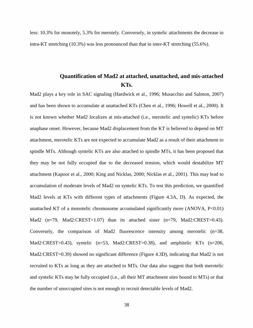
38
less: 10.3% for monotely, 5.3% for merotely. Conversely, in syntelic attachments the decrease in
intra-KT stretching (10.3%) was less pronounced than that in inter-KT stretching (55.6%).
Quantification of Mad2 at attached, unattached, and mis-attached
KTs.
Mad2 plays a key role in SAC signaling (Hardwick et al., 1996; Musacchio and Salmon, 2007)
and has been shown to accumulate at unattached KTs (Chen et al., 1996; Howell et al., 2000). It
is not known whether Mad2 localizes at mis-attached (i.e., merotelic and syntelic) KTs before
anaphase onset. However, because Mad2 displacement from the KT is believed to depend on MT
attachment, merotelic KTs are not expected to accumulate Mad2 as a result of their attachment to
spindle MTs. Although syntelic KTs are also attached to spindle MTs, it has been proposed that
they may be not fully occupied due to the decreased tension, which would destabilize MT
attachment (Kapoor et al., 2000; King and Nicklas, 2000; Nicklas et al., 2001). This may lead to
accumulation of moderate levels of Mad2 on syntelic KTs. To test this prediction, we quantified
Mad2 levels at KTs with different types of attachments (Figure 4.3A, D). As expected, the
unattached KT of a monotelic chromosome accumulated significantly more (ANOVA, P<0.01)
Mad2 (n=79, Mad2:CREST=1.07) than its attached sister (n=79, Mad2:CREST=0.43).
Conversely, the comparison of Mad2 fluorescence intensity among merotelic (n=38,
Mad2:CREST=0.43), syntelic (n=53, Mad2:CREST=0.38), and amphitelic KTs (n=206,
Mad2:CREST=0.39) showed no significant difference (Figure 4.3D), indicating that Mad2 is not
recruited to KTs as long as they are attached to MTs. Our data also suggest that both merotelic
and syntelic KTs may be fully occupied (i.e., all their MT attachment sites bound to MTs) or that
the number of unoccupied sites is not enough to recruit detectable levels of Mad2.
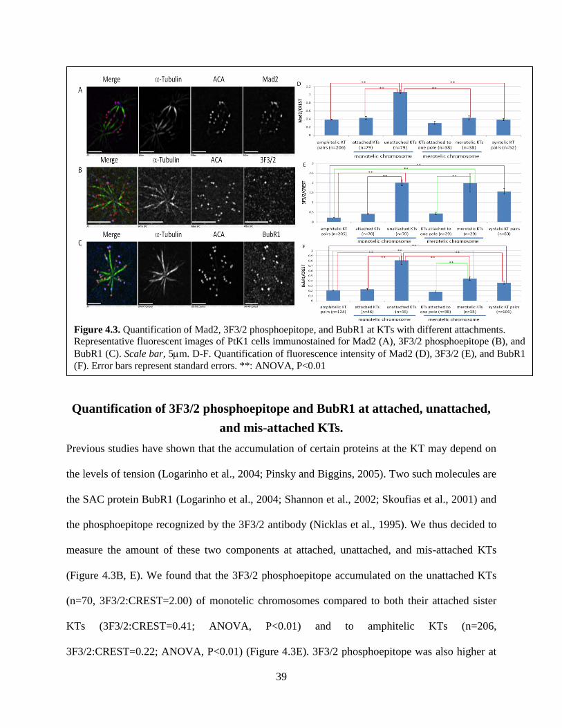
39
Quantification of 3F3/2 phosphoepitope and BubR1 at attached, unattached,
and mis-attached KTs.
Previous studies have shown that the accumulation of certain proteins at the KT may depend on
the levels of tension (Logarinho et al., 2004; Pinsky and Biggins, 2005). Two such molecules are
the SAC protein BubR1 (Logarinho et al., 2004; Shannon et al., 2002; Skoufias et al., 2001) and
the phosphoepitope recognized by the 3F3/2 antibody (Nicklas et al., 1995). We thus decided to
measure the amount of these two components at attached, unattached, and mis-attached KTs
(Figure 4.3B, E). We found that the 3F3/2 phosphoepitope accumulated on the unattached KTs
(n=70, 3F3/2:CREST=2.00) of monotelic chromosomes compared to both their attached sister
KTs (3F3/2:CREST=0.41; ANOVA, P<0.01) and to amphitelic KTs (n=206,
3F3/2:CREST=0.22; ANOVA, P<0.01) (Figure 4.3E). 3F3/2 phosphoepitope was also higher at
Figure 4.3. Quantification of Mad2, 3F3/2 phosphoepitope, and BubR1 at KTs with different attachments.
Representative fluorescent images of PtK1 cells immunostained for Mad2 (A), 3F3/2 phosphoepitope (B), and
BubR1 (C). Scale bar, 5m. D-F. Quantification of fluorescence intensity of Mad2 (D), 3F3/2 (E), and BubR1
(F). Error bars represent standard errors. **: ANOVA, P<0.01
40
merotelic KTs (n=29, 3F3/2:CREST=1.98), compared to both their sister KTs attached to one
spindle pole (3F3/2:CREST=0.43; ANOVA, P<0.01) and to amphitelic KTs (ANOVA, P<0.01).
Finally, syntelic KTs showed significantly higher levels of 3F3/2 phosphoepitope (n=83,
3F3/2:CREST=1.56) compared to amphitelic KTs (ANOVA, P<0.01). These observations
suggest that 3F3/2 phosphoepitope is accumulated on unattached and erroneously attached (i.e.,
merotelic and syntelic) KTs. The enrichment of 3F3/2 phosphoepitope on mis-attached KTs
indicates that these KTs are under low tension.
The levels of BubR1 exhibited a pattern that somewhat resembled that of 3F3/2 phosphoepitope
(Figure 4.3C, F). Indeed, unattached KTs of monotelic chromosomes displayed higher levels of
BubR1 (n=46, BubR1:CREST=0.81) than both their attached sister KTs (BubR1:CREST=0.23,
ANOVA, P<0.01) and amphitelic KTs (n=124, BubR1:CREST=0.21; ANOVA, P<0.01).
Similarly, BubR1 was enriched on merotelic KTs (n=38, BubR1:CREST=0.45) compared to
both their sister KTs attached to one spindle pole (BubR1:CREST=0.18; ANOVA, P<0.01) and
amphitelic KTs (ANOVA, P<0.01) (Figure 4.3F). Finally, syntelic KTs (n=107,
BubR1:CREST=0.36) recruited more BubR1 than did amphitelic KTs (ANOVA, P<0.01). These
findings revealed that BubR1 was enriched on unattached, merotelic, and syntelic KTs. In
summary, both the 3F3/2 phosphoepitope and BubR1 accumulated at unattached and mis-
attached (merotelic and syntelic) compared to normally attached KTs. However, BubR1
accumulated at significantly higher levels on unattached KTs compared to both merotelic and
syntelic KTs.
41
Quantification of AurB, INCENP, and MCAK at attached, unattached, and
mis-attached KTs.
Correction of KT mis-attachments is believed to largely depend on AurB activity (Hauf et al.,
2003; Lampson et al., 2004; Tanaka et al., 2002). AurB promotes correction of KT mis-
attachment by inducing kMT turnover (Cimini et al., 2006), which occurs as a result of AurB-
dependent phosphorylation of the outer KT protein Hec1 (DeLuca et al., 2006). AurB and its
binding partner INCENP localize along the chromosome arms in early mitosis, but relocalize to
the inner centromere during prometaphase. Substantial experimental evidence supports a model
for AurB-dependent correction of KT mis-attachments in which the mis-attached (e.g., merotelic
or syntelic) KTs are under reduced tension (thus exhibiting modest stretching), and therefore
more proximal to the region (inner centromere) of high AurB concentration (Cimini et al., 2003;
Liu et al., 2009). This would promote rapid turnover of kMTs, and thus correction of mis-
attachments. INCENP is a binding partner of AurB that is important for AurB localization at the
centromere (Bishop and Schumacher, 2002). Because none of the currently available antibodies
against AurB recognizes the centromere-bound AurB in PtK1 cells, we quantified INCENP as an
indirect measure of centromeric AurB. Another protein that is believed to contribute to
correction of KT mis-attachments is MCAK (Mitotic Centromere-Associated Kinesin). Reduced
levels of MCAK result in a dramatic increase in KT mis-attachments and anaphase lagging
chromosomes (Kline-Smith et al., 2004). One study in Xenopus tissue culture cells found that
both AurB and MCAK were increased at the centromeres of chromosomes with merotelic
attachment (Knowlton et al., 2006). Thus, it appears that correction of KT mis-attachments may
be promoted in two different ways: (i) by the proximity of AurB substrates (e.g., Hec1) to the
AurB-enriched centromeric region (spatial component); (ii) by a preferential localization of
AurB and MCAK at the centromere of chromosomes with mis-attached KTs (concentration

42
component). To account for both the spatial and the concentration component, we quantified the
fluorescence intensity of AurB, INCENP, and MCAK in two ways: 1. By quantifying the amount
overlapping with the KT (spatial component) as detected by CREST staining (same
quantification method as that used for quantification of Mad2, 3F3/2 phosphoepitope, and
BubR1). 2. By quantifying the amount of protein in a rectangular region including the sister KTs
and the centromere (concentration component).
Our data showed that AurB was enriched at unattached KTs (n=60, AurB: CREST=0.26)
compared to their attached sister KTs (AurB:CREST=0.08) (ANOVA, P<0.01) (Figure 4.4A, D).
AurB was also recruited to merotelic KTs (n=22, AurB: CREST=0.24), but not to their sister
KTs attached to one spindle pole (AurB: CREST=0.10) (ANOVA, P<0.01) (Figure 4.4D).
Besides, AurB was more enriched at syntelic KTs (n=76, AurB: CREST=0.16) than at
amphitelic KTs (n=134, AurB: CREST=0.10) (ANOVA, P<0.01) (Figure 4.4D). Thus, high
levels of AurB are present at unattached, merotelic, and syntelic KTs. Quantification of KT-
localized (spatial component) INCENP showed that it was present at higher levels on unattached
KTs (n=51, INCENP/CREST=0.11) compared to their attached sister KTs
(INCENP:CREST=0.07, ANOVA, P<0.05) (Figure 4.4B, E). INCENP was also enriched on
syntelic KTs (n=40, INCENP:CREST=0.08) compared to amphitelic KTs (n=45,
INCENP:CREST=0.04, ANOVA, P<0.01) (Figure 4.4E). However, INCENP showed similar
levels on merotelic KTs (n=31, INCENP:CREST =0.06) compared to their sister KTs
(INCENP:CREST =0.06) and to amphitelic KTs (ANOVA, P>0.05) (Figure 4.4E).
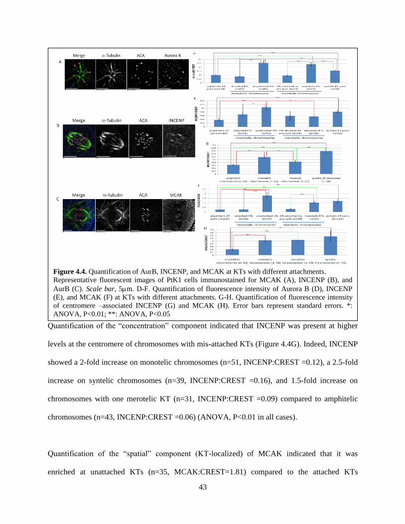
43
Quantification of the “concentration” component indicated that INCENP was present at higher
levels at the centromere of chromosomes with mis-attached KTs (Figure 4.4G). Indeed, INCENP
showed a 2-fold increase on monotelic chromosomes (n=51, INCENP:CREST =0.12), a 2.5-fold
increase on syntelic chromosomes (n=39, INCENP:CREST =0.16), and 1.5-fold increase on
chromosomes with one merotelic KT (n=31, INCENP:CREST =0.09) compared to amphitelic
chromosomes (n=43, INCENP:CREST =0.06) (ANOVA, P<0.01 in all cases).
Quantification of the “spatial” component (KT-localized) of MCAK indicated that it was
enriched at unattached KTs (n=35, MCAK:CREST=1.81) compared to the attached KTs
Figure 4.4. Quantification of AurB, INCENP, and MCAK at KTs with different attachments.
Representative fluorescent images of PtK1 cells immunostained for MCAK (A), INCENP (B), and
AurB (C). Scale bar, 5m. D-F. Quantification of fluorescence intensity of Aurora B (D), INCENP
(E), and MCAK (F) at KTs with different attachments. G-H. Quantification of fluorescence intensity
of centromere –associated INCENP (G) and MCAK (H). Error bars represent standard errors. *:
ANOVA, P<0.01; **: ANOVA, P<0.05

44
(MCAK:CREST=0.29) (ANOVA, P<0.01) of monotelic chromosomes (Figure 4.4C, F). MCAK
was also enriched at merotelic KTs (n=29, MCAK:CREST=1.04) compared to their sister KTs
(MCAK:CREST=0.32, ANOVA, P<0.01) attached to only one spindle pole (Figure 4.4F).
Finally, MCAK was present at significantly higher levels on syntelic KTs (n=104,
MCAK:CREST=1.21) than on amphitelic KTs (n=109, MCAK:CREST=0.26, ANOVA, P<0.01)
(Figure 4.4F). Quantification of the “concentration” component (centromere-associated) showed
that MCAK was present at high levels at the centromere of mis-attached chromosomes (Figure
4.4H). Indeed, the amount of MCAK at the centromere of both monotelic (n=28,
MCAK:CREST=1.07) and merotelic chromosomes (n=29, MCAK:CREST=1.08) was 2.5-fold
that found at the centromere of amphitelic chromosomes (n=44, MCAK:CREST=0.41). MCAK
was present at even higher levels (3.27-fold compared to amphitelic) at the centromeres of
syntelic chromosomes (n=59, MCAK:CREST=1.34) (ANOVA, P<0.01 in all cases).
Intra-KT stretching Mad2 3F3/2 BubR1 INCENP MCAK AurB
Unattached KTs ↓↓ ↑ ↑↑ ↑↑ ↑ ↑↑ ↑↑
Syntelic KTs ↓ = ↑ ↑ ↑↑ ↑↑ ↑
Merotelic KTs ↓↓ = ↑↑ ↑↑ = ↑↑ ↑
Table 4.1. Summary of the changes in KT structure and molecular composition in response to
mis-attachments. Amphitelic KTs are set as control.
Inter-KT stretching INCENP MCAK
Monotelic attachments ↓ ↑↑ ↑↑
Syntelic attachments ↓↓ ↑↑ ↑↑
Merotelic attachments = ↑↑ ↑↑
Table 4.2. Summary of the changes in KT structure and molecular composition in response to
mis-attachments. Amphitelic attachment is set as control.
45
Chapter 5: Discussion
Accurate mitotic chromosome segregation is ensured by amphitelic KT attachment. However,
different types of attachments, including monotelic, merotelic, and syntelic, can and do occur in
early mitosis. All these attachments are efficiently, although not always completely, converted
into amphitelic attachments before anaphase onset. In some cases, the SAC induces a delay in
anaphase onset to allow for correction. The SAC is known to respond to a signal generated by
unattached KTs (Rieder et al., 1995; Rieder et al., 1994). It has also been suggested that the SAC
may respond to lack of tension (Jang et al., 1995; Li and Nicklas, 1995), which for many years
was estimated by measuring the distance between the two sister KTs (inter-KT distance) (Waters
et al., 1996). Two recent studies (Maresca and Salmon, 2009; Uchida et al., 2009) suggested that
intra- rather than inter-KT distance may play a major role in SAC signaling and in the
consequent mitotic delay (Maresca and Salmon, 2010). Here, we investigated what factors may
be important for detection and/or correction of syntelic attachments. In order to do this, we also
characterized KTs with other types of attachments for which the SAC response and/or the
efficiency of correction are well characterized.
Intra-KT stretching per se does not appear to be the key factor in the response
to syntelic attachment.
We found that the intra-KT distance of syntelic KTs is reduced compared to amphitelic KTs.
However, the intra-KT distance is also reduced, and even more so, in merotelic KTs and in the
unattached KT of monotelic chromosomes. Whereas there is substantial evidence that unattached
KTs induce a SAC-dependent mitotic delay (Rieder et al., 1995; Rieder et al., 1994; Waters et al.,
1998), merotelic attachments do not appear to cause a mitotic delay (Cimini et al., 2004; Cimini
46
et al., 2002; Khodjakov et al., 1997), and they can persist throughout mitosis (Cimini et al.,
2001). Thus, it appears that the association between intra-KT stretching and SAC response is not
causal. This is not surprising if one considers the finding of Uchida et al. (Uchida et al., 2009)
that amphitelic KTs oscillate between high and low intra-KT stretching. One would imagine that
only a permanent structural change could cause a biochemical response. Given that intra-KT
stretching cycles between high and low, it seems unlikely that this alone may cause the SAC-
dependent mitotic delay in response of monotelic attachment.
We also found that inter-KT stretching is reduced for both monotelic and syntelic chromosomes
compared to amphitelic ones, but not for chromosomes in which one sister is merotelically
attached. This may suggest that the inter-KT stretching is important for the mitotic delay induced
by unattached KTs. However, Maresca and Salmon (Maresca and Salmon, 2009) could, under
certain conditions, reduce the inter-KT stretching without delaying anaphase onset. Thus, we
conclude that neither intra- nor inter-KT stretching is a direct cause of mitotic delay in response
to KT mis-attachments. However, the reduced inter-KT stretching may play a key role in rapid
correction of syntelic attachments (see below).
What causes the reduced intra-KT distance in unattached, merotelic, and
syntelic KTs?
We propose that intra-KT stretching is due to opposing forces applied on individual KTs. These
forces are the pulling force from the attached MTs and an opposing force dependent on the
anchoring of the KT to the underlying centromeric chromatin (Figure 5.1A). Based on this
assumption, it is not surprising that unattached KTs exhibit reduced intra-KT stretching. Indeed,
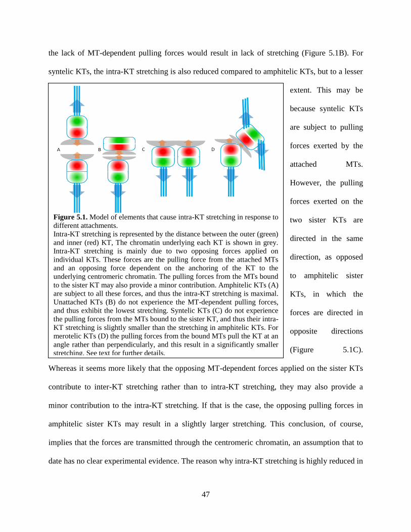
47
Figure 5.1. Model of elements that cause intra-KT stretching in response to
different attachments.
Intra-KT stretching is represented by the distance between the outer (green)
and inner (red) KT, The chromatin underlying each KT is shown in grey.
Intra-KT stretching is mainly due to two opposing forces applied on
individual KTs. These forces are the pulling force from the attached MTs
and an opposing force dependent on the anchoring of the KT to the
underlying centromeric chromatin. The pulling forces from the MTs bound
to the sister KT may also provide a minor contribution. Amphitelic KTs (A)
are subject to all these forces, and thus the intra-KT stretching is maximal.
Unattached KTs (B) do not experience the MT-dependent pulling forces,
and thus exhibit the lowest stretching. Syntelic KTs (C) do not experience
the pulling forces from the MTs bound to the sister KT, and thus their intra-
KT stretching is slightly smaller than the stretching in amphitelic KTs. For
merotelic KTs (D) the pulling forces from the bound MTs pull the KT at an
angle rather than perpendicularly, and this result in a significantly smaller
stretching. See text for further details.
the lack of MT-dependent pulling forces would result in lack of stretching (Figure 5.1B). For
syntelic KTs, the intra-KT stretching is also reduced compared to amphitelic KTs, but to a lesser
extent. This may be
because syntelic KTs
are subject to pulling
forces exerted by the
attached MTs.
However, the pulling
forces exerted on the
two sister KTs are
directed in the same
direction, as opposed
to amphitelic sister
KTs, in which the
forces are directed in
opposite directions
(Figure 5.1C).
Whereas it seems more likely that the opposing MT-dependent forces applied on the sister KTs
contribute to inter-KT stretching rather than to intra-KT stretching, they may also provide a
minor contribution to the intra-KT stretching. If that is the case, the opposing pulling forces in
amphitelic sister KTs may result in a slightly larger stretching. This conclusion, of course,
implies that the forces are transmitted through the centromeric chromatin, an assumption that to
date has no clear experimental evidence. The reason why intra-KT stretching is highly reduced in
48
merotelic KTs is less obvious. However, it may be explained by the fact that for merotelic
attachments the bound MTs pull the KT at an angle rather than perpendicularly. Thus, merotelic
KTs lack the MT-dependent pulling forces oriented perpendicularly to the KT that would cause a
significant increase in intra-KT stretching (Figure 5.1D).
Kinetochore attachment and accumulation of Mad2, 3F3/2, and BubR1.
We found high levels of Mad2 only at unattached KTs, whereas mis-attached (merotelic or
syntelic) KTs possessed levels of Mad2 comparable to those found at amphitelic KTs. This
finding indicates, in agreement with previous studies (Waters et al., 1998; Yang et al., 2009), that
the Mad2-dependent SAC response is only triggered by lack of attachment. Our finding further
suggests that merotelic and syntelic KTs are fully attached.
Conversely, we found that both the 3F3/2 phosphoepitope and BubR1 were present at high levels
at unattached as well as at mis-attached (merotelic and syntelic) KTs compared to amphitelic
KTs. Because cells with merotelic attachments do not exhibit a delay of anaphase onset (Cimini
et al., 2001; Kline-Smith et al., 2004), this finding may indicate that the 3F3/2 phosphoepitope
and BubR1 do not play a significant role in the SAC response. Nevertheless, most cells are
believed to establish at least one merotelic attachment in early mitosis (Cimini et al., 2003;
Silkworth et al., 2009), so it is possible that the average duration of mitosis includes a 3F3/2-
and/or BubR1-dependent delay. But because nearly all cells would exhibit this kind of delay, it is
impossible to separate the contribution of merotelic and syntelic attachments and the contribution
of 3F3/2 and BubR1 to the mitotic delay. It is also worth noting that although BubR1 levels are
high at both unattached and mis-attached KTs, the levels at unattached KTs are the highest when
49
compared to merotelic and syntelic KTs. Thus, one possibility is that there is a threshold for the
amount of BubR1 necessary to cause a significant mitotic delay.
Aurora B, INCENP, and MCAK at unattached and mis-attached KTs.
We found high levels of Aurora B kinase (AurB) at unattached, merotelic, and syntelic KTs.
However, we were only able to detect AurB at the outer KT and could not detect centromeric
AurB. A recent study showed that there are, indeed, two pools of AurB, one at the centromere
and one at the outer KT, and the outer KT pool appears to be the active, phosphorylated version
of the protein (DeLuca et al., 2011). It has been suggested that the distance between the AurB
substrates at the outer KT and the centromeric AurB may play a key role in correction of KT
mis-attachments. However, the antibody used in this study and many others commercially
available do not recognize the centromeric AurB in PtK1 cells (our tests and DeLuca, personal
communication), and thus we estimated the amount of centromere-bound AurB by quantifying
its binding partner INCENP. We found that INCENP is high at unattached, merotelic, and
syntelic KTs. We also observed high levels of the centromeric INCENP at monotelic, merotelic,
and syntelic chromosomes. The same pattern, more at the KT and more at the centromere, was
observed for MCAK. However, for INCENP and MCAK the KT-associated population did not
appear as a separate pool. Instead, it simply appeared to be portion of the centromeric pool
overlapping with the KT, whose amount varied depending on the inter-KT stretch. Indeed, the
levels of KT-associated MCAK and INCENP were highest for monotelic and syntelic
chromosomes, which exhibited the lowest inter-KT stretching. However, there was also a
difference between the unattached and the attached sister of monotelic chromosomes and
between merotelic KTs and their sisters. This observation suggests that intra-KT stretching may
50
play a role in removal of these proteins from specific sub-regions of the centromere. In other
words, upon attachment, the protein would leave the centromeric region closest to the attached
KT, but not the region closest to the unattached or merotelically attached KT.
How do cells respond to different types of attachments?
Taken together, our data suggest that monotelic attachments are the only ones that can trigger a
SAC-dependent mitotic delay, and that Mad2 is the major molecular player in SAC signaling.
Whereas intra-KT stretching is significantly reduced in unattached KTs, the same levels of intra-
KT stretching are observed for merotelic KTs, which do not induce a SAC-dependent mitotic
delay, thus suggesting the intra-KT stretching per se is not a key factor in SAC signaling. Inter-
KT stretching does not appear to play a key role in SAC signaling either (Maresca and Salmon,
2009; Uchida et al., 2009). However, it may play a role in promoting correction of KT mis-
attachment. Indeed, syntelic chromosomes exhibit the lowest inter-KT stretching and the largest
KT-associated amounts of those proteins (AurB, MCAK) that have been shown to play a role in
correction of KT mis-attachments. This may lead to rapid and efficient correction of syntelic
attachments. That syntelic attachments are not detected by the SAC is not surprising given that
conditions that induce an increase in syntelic attachments do not necessarily induce a mitotic
arrest (Yang et al., 2009).
In figure 5.2, we illustrate how inter-KT stretching contributes to AurB-dependent error
correction. Upon amphitelic attachment (Figure 5.2A), the sister KTs are pulled away from the
region of high AurB concentration. Although there appears to be an outer KT-localized
subpopulation of AurB, amphitelic attachment is also believed to lead to Protein Phosphatase 1
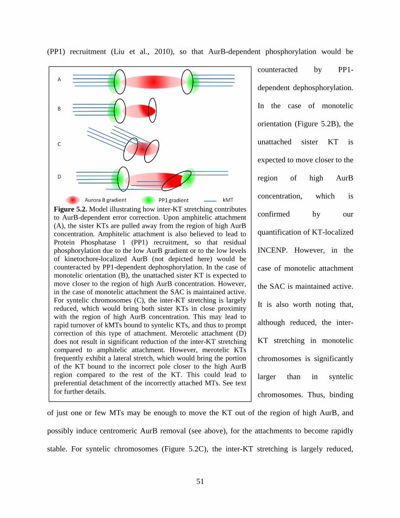
51
Figure 5.2. Model illustrating how inter-KT stretching contributes
to AurB-dependent error correction. Upon amphitelic attachment
(A), the sister KTs are pulled away from the region of high AurB
concentration. Amphitelic attachment is also believed to lead to
Protein Phosphatase 1 (PP1) recruitment, so that residual
phosphorylation due to the low AurB gradient or to the low levels
of kinetochore-localized AurB (not depicted here) would be
counteracted by PP1-dependent dephosphorylation. In the case of
monotelic orientation (B), the unattached sister KT is expected to
move closer to the region of high AurB concentration. However,
in the case of monotelic attachment the SAC is maintained active.
For syntelic chromosomes (C), the inter-KT stretching is largely
reduced, which would bring both sister KTs in close proximity
with the region of high AurB concentration. This may lead to
rapid turnover of kMTs bound to syntelic KTs, and thus to prompt
correction of this type of attachment. Merotelic attachment (D)
does not result in significant reduction of the inter-KT stretching
compared to amphitelic attachment. However, merotelic KTs
frequently exhibit a lateral stretch, which would bring the portion
of the KT bound to the incorrect pole closer to the high AurB
region compared to the rest of the KT. This could lead to
preferential detachment of the incorrectly attached MTs. See text
for further details.
(PP1) recruitment (Liu et al., 2010), so that AurB-dependent phosphorylation would be
counteracted by PP1-
dependent dephosphorylation.
In the case of monotelic
orientation (Figure 5.2B), the
unattached sister KT is
expected to move closer to the
region of high AurB
concentration, which is
confirmed by our
quantification of KT-localized
INCENP. However, in the
case of monotelic attachment
the SAC is maintained active.
It is also worth noting that,
although reduced, the inter-
KT stretching in monotelic
chromosomes is significantly
larger than in syntelic
chromosomes. Thus, binding
of just one or few MTs may be enough to move the KT out of the region of high AurB, and
possibly induce centromeric AurB removal (see above), for the attachments to become rapidly
stable. For syntelic chromosomes (Figure 5.2C), the inter-KT stretching is largely reduced,
52
which would bring both sister KTs in close proximity with the region of high AurB
concentration. This may lead to rapid turnover of kMTs bound to syntelic KTs, and thus to
prompt correction of this type of attachment. Merotelic attachment (Figure 5.2D) does not result
in significant reduction of the inter-KT stretching compared to amphitelic attachment. As a result,
the merotelic KT as a whole is not expected to move close to the region of high AurB. However,
merotelic KTs frequently exhibit a lateral stretch, which would bring the portion of the KT
bound to the incorrect pole closer to the high AurB region compared to the rest of the KT. This
could lead to preferential detachment of the incorrectly attached MTs. It will be interesting to
determine whether PP1 localizes at syntelic and merotelic KTs to determine its possible
contribution to kMT turnover versus stabilization.
53
Chapter 6: Summary and future direction
In summary, the data shown here indicate that monotelic attachment is the only type of
attachment that can induce a SAC response. The unattached KT of monotelic chromosomes
retains significantly higher level of SAC protein, Mad2, than its sister KT or amphitelic KTs.
Neither merotelic KTs nor syntelic KTs accumulate Mad2, strongly suggesting that these KTs
are fully occupied by MTs and cannot induce a SAC response. However, our data indicate that
both syntelic and merotelic KTs are under less tension because they show higher level of 3F3/2
phosphoepitope and BubR1. Our data also suggest that neither intra- nor inter-KT stretching is a
direct cause of SAC activation. But together with the MCAK, INCENP, and AurB data, our
findings suggest that the decrease in KT stretching may contribute to bringing specific KT
substrates closer to AurB for rapid and efficient correction of KT mis-attachments. Furthermore,
decreased stretching also appears to result in retention of the error correction complex at the
centromere, which would thus result in efficient correction of merotelic and syntelic attachments.
A question that remains to be addressed is how tension regulates preferential turnover of
incorrectly attached MTs. It is possible, for instance, that the phosphorylated form of certain
proteins (e.g., AurB substrates) may not persist at the same location as the unphosphorylated
form, as is the case for MCAK (Knowlton et al., 2006). Therefore, it will be interesting to map
the localization of active AurB, its substrates, its regulators [e.g., ICIS (Ohi et al., 2003)], and its
binding partners (e.g., INCENP) within a KT on a nanometer scale under changing KT
attachment conditions. Other unanswered questions include if/how the AurB regulators/partners
directly respond to tension. For example, it was recently proposed that INCENP may function as
a tension-sensor (Sandall et al., 2006), but it is still largely unknown how INCENP and MTs
interact and how this interaction can be translated into an increase in AurB activity. Structural
54
studies may be necessary to gain insight into this issue. Finally, because of its proposed role in
counteracting AurB activity, it will be important to determine whether and how PP1 localizes at
syntelic and merotelic KTs, and how its localization is regulated by tension at the level of
individual KTs. The combination of all these pieces of knowledge will be critical to gain
complete understanding of how the equilibrium between kMT turnover and stabilization is
achieved. Because cancer cells are prone to form KT mis-attachments (Ganem et al., 2009;
Silkworth et al., 2009; Thompson and Compton, 2008) and exhibit defective error correction
(Bakhoum et al., 2009), understanding the mechanisms behind SAC activation and error
correction might yield new insights into the mechanisms of chromosomal instability in cancer
cells.
55
References
Allshire, R.C., and Karpen, G.H. (2008). Epigenetic regulation of centromeric chromatin: old
dogs, new tricks? Nat Rev Genet 9, 923-937.
Asbury, C.L., Gestaut, D.R., Powers, A.F., Franck, A.D., and Davis, T.N. (2006). The Dam1
kinetochore complex harnesses microtubule dynamics to produce force and movement. Proc Natl
Acad Sci U S A 103, 9873-9878.
Bakhoum, S.F., Genovese, G., and Compton, D.A. (2009). Deviant Kinetochore Microtubule
Dynamics Underlie Chromosomal Instability. Current Biology 19, 1937-1942.
Biggins, S., and Murray, A.W. (2001). The budding yeast protein kinase Ipl1/Aurora allows the
absence of tension to activate the spindle checkpoint. Genes Dev 15, 3118-3129.
Bishop, J.D., and Schumacher, J.M. (2002). Phosphorylation of the carboxyl terminus of inner
centromere protein (INCENP) by the Aurora B Kinase stimulates Aurora B kinase activity. J
Biol Chem 277, 27577-27580.
Black, B.E., and Bassett, E.A. (2008). The histone variant CENP-A and centromere specification.
Curr Opin Cell Biol 20, 91-100.
Black, B.E., Brock, M.A., Bedard, S., Woods, V.L., Jr., and Cleveland, D.W. (2007). An
epigenetic mark generated by the incorporation of CENP-A into centromeric nucleosomes. Proc
Natl Acad Sci U S A 104, 5008-5013.
Black, B.E., Foltz, D.R., Chakravarthy, S., Luger, K., Woods, V.L., Jr., and Cleveland, D.W.
(2004). Structural determinants for generating centromeric chromatin. Nature 430, 578-582.
Blow, J.J., and Tanaka, T.U. (2005). The chromosome cycle: coordinating replication and
segregation. Second in the cycles review series. EMBO Rep 6, 1028-1034.
Cai, S., Weaver, L.N., Ems-McClung, S.C., and Walczak, C.E. (2009). Kinesin-14 family
proteins HSET/XCTK2 control spindle length by cross-linking and sliding microtubules.
Molecular biology of the cell 20, 1348-1359.
Carmena, M., Ruchaud, S., and Earnshaw, W.C. (2009). Making the Auroras glow: regulation of
Aurora A and B kinase function by interacting proteins. Curr Opin Cell Biol 21, 796-805.
Carroll, C.W., Silva, M.C., Godek, K.M., Jansen, L.E., and Straight, A.F. (2009). Centromere
assembly requires the direct recognition of CENP-A nucleosomes by CENP-N. Nat Cell Biol 11,
896-902.
Chan, G.K., Liu, S.T., and Yen, T.J. (2005). Kinetochore structure and function. Trends Cell
Biol 15, 589-598.
Cheeseman, I.M., Anderson, S., Jwa, M., Green, E.M., Kang, J., Yates, J.R., 3rd, Chan, C.S.,
Drubin, D.G., and Barnes, G. (2002). Phospho-regulation of kinetochore-microtubule
attachments by the Aurora kinase Ipl1p. Cell 111, 163-172.
Cheeseman, I.M., Chappie, J.S., Wilson-Kubalek, E.M., and Desai, A. (2006). The conserved
KMN network constitutes the core microtubule-binding site of the kinetochore. Cell 127, 983-
997.
Cheeseman, I.M., and Desai, A. (2008). Molecular architecture of the kinetochore-microtubule
interface. Nat Rev Mol Cell Bio 9, 33-46.
Cheeseman, I.M., Niessen, S., Anderson, S., Hyndman, F., Yates, J.R., 3rd, Oegema, K., and
Desai, A. (2004). A conserved protein network controls assembly of the outer kinetochore and its
ability to sustain tension. Genes Dev 18, 2255-2268.
Chen, R.H., Waters, J.C., Salmon, E.D., and Murray, A.W. (1996). Association of spindle
assembly checkpoint component XMAD2 with unattached kinetochores. Science 274, 242-246.
56
Cimini, D. (2008). Merotelic kinetochore orientation, aneuploidy, and cancer. Biochim Biophys
Acta 1786, 32-40.
Cimini, D., Cameron, L.A., and Salmon, E.D. (2004). Anaphase spindle mechanics prevent mis-
segregation of merotelically oriented chromosomes. Curr Biol 14, 2149-2155.
Cimini, D., and Degrassi, F. (2005). Aneuploidy: a matter of bad connections. Trends Cell Biol
15, 442-451.
Cimini, D., Fioravanti, D., Salmon, E.D., and Degrassi, F. (2002). Merotelic kinetochore
orientation versus chromosome mono-orientation in the origin of lagging chromosomes in
human primary cells. J Cell Sci 115, 507-515.
Cimini, D., Howell, B., Maddox, P., Khodjakov, A., Degrassi, F., and Salmon, E.D. (2001).
Merotelic kinetochore orientation is a major mechanism of aneuploidy in mitotic mammalian
tissue cells. J Cell Biol 153, 517-527.
Cimini, D., Moree, B., Canman, J.C., and Salmon, E.D. (2003). Merotelic kinetochore
orientation occurs frequently during early mitosis in mammalian tissue cells and error correction
is achieved by two different mechanisms. J Cell Sci 116, 4213-4225.
Cimini, D., Wan, X., Hirel, C.B., and Salmon, E.D. (2006). Aurora kinase promotes turnover of
kinetochore microtubules to reduce chromosome segregation errors. Curr Biol 16, 1711-1718.
D'Angiolella, V., Mari, C., Nocera, D., Rametti, L., and Grieco, D. (2003). The spindle
checkpoint requires cyclin-dependent kinase activity. Genes & development 17, 2520-2525.
Dai, W. (2009). Suppression of genomic instabilities caused by chromosome mis-segregation: a
perspective from studying BubR1 and Sgo1. J Formos Med Assoc 108, 904-911.
Daum, J.R., Wren, J.D., Daniel, J.J., Sivakumar, S., McAvoy, J.N., Potapova, T.A., and Gorbsky,
G.J. (2009). Ska3 is required for spindle checkpoint silencing and the maintenance of
chromosome cohesion in mitosis. Curr Biol 19, 1467-1472.
De Antoni, A., Pearson, C.G., Cimini, D., Canman, J.C., Sala, V., Nezi, L., Mapelli, M., Sironi,
L., Faretta, M., Salmon, E.D., et al. (2005). The Mad1/Mad2 complex as a template for Mad2
activation in the spindle assembly checkpoint. Curr Biol 15, 214-225.
DeLuca, J.G., Dong, Y., Hergert, P., Strauss, J., Hickey, J.M., Salmon, E.D., and McEwen, B.F.
(2005). Hec1 and nuf2 are core components of the kinetochore outer plate essential for
organizing microtubule attachment sites. Mol Biol Cell 16, 519-531.
DeLuca, J.G., Gall, W.E., Ciferri, C., Cimini, D., Musacchio, A., and Salmon, E.D. (2006).
Kinetochore microtubule dynamics and attachment stability are regulated by Hec1. Cell 127,
969-982.
DeLuca, K.F., Lens, S.M., and DeLuca, J.G. (2011). Temporal changes in Hec1 phosphorylation
control kinetochore-microtubule attachment stability during mitosis. J Cell Sci 124, 622-634.
Desai, A., Rybina, S., Muller-Reichert, T., Shevchenko, A., Hyman, A., and Oegema, K. (2003).
KNL-1 directs assembly of the microtubule-binding interface of the kinetochore in C. elegans.
Genes Dev 17, 2421-2435.
Ditchfield, C., Johnson, V.L., Tighe, A., Ellston, R., Haworth, C., Johnson, T., Mortlock, A.,
Keen, N., and Taylor, S.S. (2003). Aurora B couples chromosome alignment with anaphase by
targeting BubR1, Mad2, and Cenp-E to kinetochores. J Cell Biol 161, 267-280.
Earnshaw, W.C., and Rothfield, N. (1985). Identification of a family of human centromere
proteins using autoimmune sera from patients with scleroderma. Chromosoma 91, 313-321.
Erhardt, S., Mellone, B.G., Betts, C.M., Zhang, W., Karpen, G.H., and Straight, A.F. (2008).
Genome-wide analysis reveals a cell cycle-dependent mechanism controlling centromere
propagation. J Cell Biol 183, 805-818.
57
Fang, G., Yu, H., and Kirschner, M.W. (1998). The checkpoint protein MAD2 and the mitotic
regulator CDC20 form a ternary complex with the anaphase-promoting complex to control
anaphase initiation. Genes Dev 12, 1871-1883.
Foltz, D.R., Jansen, L.E., Black, B.E., Bailey, A.O., Yates, J.R., 3rd, and Cleveland, D.W. (2006).
The human CENP-A centromeric nucleosome-associated complex. Nat Cell Biol 8, 458-469.
Franck, A.D., Powers, A.F., Gestaut, D.R., Gonen, T., Davis, T.N., and Asbury, C.L. (2007).
Tension applied through the Dam1 complex promotes microtubule elongation providing a direct
mechanism for length control in mitosis. Nature Cell Biology 9, 832-U171.
Fujita, Y., Hayashi, T., Kiyomitsu, T., Toyoda, Y., Kokubu, A., Obuse, C., and Yanagida, M.
(2007). Priming of centromere for CENP-A recruitment by human hMis18alpha, hMis18beta,
and M18BP1. Dev Cell 12, 17-30.
Gadde, S., and Heald, R. (2004). Mechanisms and molecules of the mitotic spindle. Curr Biol 14,
R797-805.
Gaitanos, T.N., Santamaria, A., Jeyaprakash, A.A., Wang, B., Conti, E., and Nigg, E.A. (2009).
Stable kinetochore-microtubule interactions depend on the Ska complex and its new component
Ska3/C13Orf3. Embo Journal 28, 1442-1452.
Ganem, N.J., Godinho, S.A., and Pellman, D. (2009). A mechanism linking extra centrosomes to
chromosomal instability. Nature 460, 278-282.
Gassmann, R., Essex, A., Hu, J.S., Maddox, P.S., Motegi, F., Sugimoto, A., O'Rourke, S.M.,
Bowerman, B., McLeod, I., Yates, J.R., 3rd, et al. (2008). A new mechanism controlling
kinetochore-microtubule interactions revealed by comparison of two dynein-targeting
components: SPDL-1 and the Rod/Zwilch/Zw10 complex. Genes & development 22, 2385-2399.
Gillett, E.S., Espelin, C.W., and Sorger, P.K. (2004). Spindle checkpoint proteins and
chromosome-microtubule attachment in budding yeast. The Journal of cell biology 164, 535-546.
Gorbsky, G.J. (2004). Mitosis: MCAK under the aura of Aurora B. Curr Biol 14, R346-348.
Gorbsky, G.J., and Ricketts, W.A. (1993). Differential Expression of a Phosphoepitope at the
Kinetochores of Moving Chromosomes. Journal of Cell Biology 122, 1311-1321.
Goshima, G., Kiyomitsu, T., Yoda, K., and Yanagida, M. (2003). Human centromere chromatin
protein hMis12, essential for equal segregation, is independent of CENP-A loading pathway. The
Journal of cell biology 160, 25-39.
Guimaraes, G.J., and Deluca, J.G. (2009). Connecting with Ska, a key complex at the
kinetochore-microtubule interface. EMBO J 28, 1375-1377.
Hanisch, A., Sillje, H.H., and Nigg, E.A. (2006). Timely anaphase onset requires a novel spindle
and kinetochore complex comprising Ska1 and Ska2. The EMBO journal 25, 5504-5515.
Hardwick, K.G., Weiss, E., Luca, F.C., Winey, M., and Murray, A.W. (1996). Activation of the
budding yeast spindle assembly checkpoint without mitotic spindle disruption. Science 273, 953-
956.
Hauf, S., Cole, R.W., LaTerra, S., Zimmer, C., Schnapp, G., Walter, R., Heckel, A., van Meel, J.,
Rieder, C.L., and Peters, J.M. (2003). The small molecule Hesperadin reveals a role for Aurora B
in correcting kinetochore-microtubule attachment and in maintaining the spindle assembly
checkpoint. J Cell Biol 161, 281-294.
Hayashi, T., Fujita, Y., Iwasaki, O., Adachi, Y., Takahashi, K., and Yanagida, M. (2004). Mis16
and Mis18 are required for CENP-A loading and histone deacetylation at centromeres. Cell 118,
715-729.

58
Hayden, J.H., Bowser, S.S., and Rieder, C.L. (1990). Kinetochores capture astral microtubules
during chromosome attachment to the mitotic spindle: direct visualization in live newt lung cells.
J Cell Biol 111, 1039-1045.
Heidemann, S.R., and McIntosh, J.R. (1980). Visualization of the structural polarity of
microtubules. Nature 286, 517-519.
Hemmerich, P., Weidtkamp-Peters, S., Hoischen, C., Schmiedeberg, L., Erliandri, I., and
Diekmann, S. (2008). Dynamics of inner kinetochore assembly and maintenance in living cells. J
Cell Biol 180, 1101-1114.
Hirano, M., Anderson, D.E., Erickson, H.P., and Hirano, T. (2001). Bimodal activation of SMC
ATPase by intra- and inter-molecular interactions. EMBO J 20, 3238-3250.
Hoffman, D.B., Pearson, C.G., Yen, T.J., Howell, B.J., and Salmon, E.D. (2001). Microtubule-
dependent changes in assembly of microtubule motor proteins and mitotic spindle checkpoint
proteins at PtK1 kinetochores. Mol Biol Cell 12, 1995-2009.
Hori, T., Haraguchi, T., Hiraoka, Y., Kimura, H., and Fukagawa, T. (2003). Dynamic behavior
of Nuf2-Hec1 complex that localizes to the centrosome and centromere and is essential for
mitotic progression in vertebrate cells. Journal of cell science 116, 3347-3362.
Howard, A.a.P.S.R. (1951). Nuclear incorporation of P32
as demonstrated by autoradiographs.
Exp Cell Res 2, 178-187.
Howell, B.J., Hoffman, D.B., Fang, G., Murray, A.W., and Salmon, E.D. (2000). Visualization
of Mad2 dynamics at kinetochores, along spindle fibers, and at spindle poles in living cells. J
Cell Biol 150, 1233-1250.
Howell, B.J., McEwen, B.F., Canman, J.C., Hoffman, D.B., Farrar, E.M., Rieder, C.L., and
Salmon, E.D. (2001). Cytoplasmic dynein/dynactin drives kinetochore protein transport to the
spindle poles and has a role in mitotic spindle checkpoint inactivation. J Cell Biol 155, 1159-
1172.
Hyman, A.A., and Mitchison, T.J. (1990). Modulation of microtubule stability by kinetochores in
vitro. J Cell Biol 110, 1607-1616.
Indjeian, V.B., Stern, B.M., and Murray, A.W. (2005). The centromeric protein Sgo1 is required
to sense lack of tension on mitotic chromosomes. Science 307, 130-133.
Izuta, H., Ikeno, M., Suzuki, N., Tomonaga, T., Nozaki, N., Obuse, C., Kisu, Y., Goshima, N.,
Nomura, F., Nomura, N., et al. (2006). Comprehensive analysis of the ICEN (Interphase
Centromere Complex) components enriched in the CENP-A chromatin of human cells. Genes
Cells 11, 673-684.
Jang, J.K., Messina, L., Erdman, M.B., Arbel, T., and Hawley, R.S. (1995). Induction of
metaphase arrest in Drosophila oocytes by chiasma-based kinetochore tension. Science 268,
1917-1919.
Joglekar, A.P., Bloom, K.S., and Salmon, E.D. (2010). Mechanisms of force generation by end-
on kinetochore-microtubule attachments. Curr Opin Cell Biol 22, 57-67.
Kalab, P., and Heald, R. (2008). The RanGTP gradient - a GPS for the mitotic spindle. Journal of
cell science 121, 1577-1586.
Kallio, M.J., Beardmore, V.A., Weinstein, J., and Gorbsky, G.J. (2002). Rapid microtubule-
independent dynamics of Cdc20 at kinetochores and centrosomes in mammalian cells. Journal of
Cell Biology 158, 841-847.
Kapoor, T.M., Lampson, M.A., Hergert, P., Cameron, L., Cimini, D., Salmon, E.D., McEwen,
B.F., and Khodjakov, A. (2006). Chromosomes can congress to the metaphase plate before
biorientation. Science 311, 388-391.
59
Kapoor, T.M., Mayer, T.U., Coughlin, M.L., and Mitchison, T.J. (2000). Probing spindle
assembly mechanisms with monastrol, a small molecule inhibitor of the mitotic kinesin, Eg5.
Journal of Cell Biology 150, 975-988.
Karess, R. (2005). Rod-Zw10-Zwilch: a key player in the spindle checkpoint. Trends in cell
biology 15, 386-392.
Karpen, G.H., and Allshire, R.C. (1997). The case for epigenetic effects on centromere identity
and function. Trends Genet 13, 489-496.
Karsenti, E., and Vernos, I. (2001). The mitotic spindle: a self-made machine. Science 294, 543-
547.
Kelly, A.E., Sampath, S.C., Maniar, T.A., Woo, E.M., Chait, B.T., and Funabiki, H. (2007).
Chromosomal enrichment and activation of the aurora B pathway are coupled to spatially
regulate spindle assembly. Dev Cell 12, 31-43.
Khodjakov, A., Cole, R.W., McEwen, B.F., Buttle, K.F., and Rieder, C.L. (1997). Chromosome
fragments possessing only one kinetochore can congress to the spindle equator. J Cell Biol 136,
229-240.
Khodjakov, A., and Rieder, C.L. (2009). The nature of cell-cycle checkpoints: facts and fallacies.
J Biol 8, 88.
King, J.M., and Nicklas, R.B. (2000). Tension on chromosomes increases the number of
kinetochore microtubules but only within limits. J Cell Sci 113 Pt 21, 3815-3823.
Kirschner, M.W., and Mitchison, T. (1986). Microtubule dynamics. Nature 324, 621.
Klein, U.R., Nigg, E.A., and Gruneberg, U. (2006). Centromere targeting of the chromosomal
passenger complex requires a ternary subcomplex of Borealin, Survivin, and the N-terminal
domain of INCENP. Mol Biol Cell 17, 2547-2558.
Kline-Smith, S.L., Khodjakov, A., Hergert, P., and Walczak, C.E. (2004). Depletion of
centromeric MCAK leads to chromosome congression and segregation defects due to improper
kinetochore attachments. Mol Biol Cell 15, 1146-1159.
Kline, S.L., Cheeseman, I.M., Hori, T., Fukagawa, T., and Desai, A. (2006). The human Mis12
complex is required for kinetochore assembly and proper chromosome segregation. The Journal
of cell biology 173, 9-17.
Knowlton, A.L., Lan, W., and Stukenberg, P.T. (2006). Aurora B is enriched at merotelic
attachment sites, where it regulates MCAK. Curr Biol 16, 1705-1710.
Lampson, M.A., Renduchitala, K., Khodjakov, A., and Kapoor, T.M. (2004). Correcting
improper chromosome-spindle attachments during cell division. Nat Cell Biol 6, 232-237.
Li, X.T., and Nicklas, R.B. (1995). Mitotic Forces Control a Cell-Cycle Checkpoint. Nature 373,
630-632.
Li, Y., and Benezra, R. (1996). Identification of a human mitotic checkpoint gene: hsMAD2.
Science 274, 246-248.
Liu, D., Vader, G., Vromans, M.J., Lampson, M.A., and Lens, S.M. (2009). Sensing
chromosome bi-orientation by spatial separation of aurora B kinase from kinetochore substrates.
Science 323, 1350-1353.
Liu, D., Vleugel, M., Backer, C.B., Hori, T., Fukagawa, T., Cheeseman, I.M., and Lampson,
M.A. (2010). Regulated targeting of protein phosphatase 1 to the outer kinetochore by KNL1
opposes Aurora B kinase. Journal of Cell Biology 188, 809-820.
Liu, S.T., Hittle, J.C., Jablonski, S.A., Campbell, M.S., Yoda, K., and Yen, T.J. (2003). Human
CENP-I specifies localization of CENP-F, MAD1 and MAD2 to kinetochores and is essential for
mitosis. Nature cell biology 5, 341-345.
60
Liu, S.T., Rattner, J.B., Jablonski, S.A., and Yen, T.J. (2006). Mapping the assembly pathways
that specify formation of the trilaminar kinetochore plates in human cells. The Journal of cell
biology 175, 41-53.
Logarinho, E., and Bousbaa, H. (2008). Kinetochore-microtubule interactions "in check" by
Bub1, Bub3 and BubR1: The dual task of attaching and signalling. Cell Cycle 7, 1763-1768.
Logarinho, E., Bousbaa, H., Dias, J.M., Lopes, C., Amorim, I., Antunes-Martins, A., and Sunkel,
C.E. (2004). Different spindle checkpoint proteins monitor microtubule attachment and tension
at kinetochores in Drosophila cells. J Cell Sci 117, 1757-1771.
Lu, Y., Wang, Z., Ge, L., Chen, N., and Liu, H. (2009). The RZZ complex and the spindle
assembly checkpoint. Cell structure and function 34, 31-45.
Macy, B., Wang, M., and Yu, H.G. (2009). The many faces of shugoshin, the "guardian spirit,"
in chromosome segregation. Cell Cycle 8, 35-37.
Maddox, P., Desai, A., Oegema, K., Mitchison, T.J., and Salmon, E.D. (2002). Poleward
microtubule flux is a major component of spindle dynamics and anaphase a in mitotic Drosophila
embryos. Curr Biol 12, 1670-1674.
Maddox, P.S., Hyndman, F., Monen, J., Oegema, K., and Desai, A. (2007). Functional genomics
identifies a Myb domain-containing protein family required for assembly of CENP-A chromatin.
J Cell Biol 176, 757-763.
Maiato, H., DeLuca, J., Salmon, E.D., and Earnshaw, W.C. (2004). The dynamic kinetochore-
microtubule interface. J Cell Sci 117, 5461-5477.
Maiato, H., Fairley, E.A., Rieder, C.L., Swedlow, J.R., Sunkel, C.E., and Earnshaw, W.C. (2003).
Human CLASP1 is an outer kinetochore component that regulates spindle microtubule dynamics.
Cell 113, 891-904.
Maiato, H., Khodjakov, A., and Rieder, C.L. (2005). Drosophila CLASP is required for the
incorporation of microtubule subunits into fluxing kinetochore fibres. Nat Cell Biol 7, 42-47.
Mao, Y., Desai, A., and Cleveland, D.W. (2005). Microtubule capture by CENP-E silences
BubR1-dependent mitotic checkpoint signaling. The Journal of cell biology 170, 873-880.
Mapelli, M., Filipp, F.V., Rancati, G., Massimiliano, L., Nezi, L., Stier, G., Hagan, R.S.,
Confalonieri, S., Piatti, S., Sattler, M., et al. (2006). Determinants of conformational
dimerization of Mad2 and its inhibition by p31(comet). Embo Journal 25, 1273-1284.
Maresca, T.J., and Salmon, E.D. (2009). Intrakinetochore stretch is associated with changes in
kinetochore phosphorylation and spindle assembly checkpoint activity. Journal of Cell Biology
184, 373-381.
Maresca, T.J., and Salmon, E.D. (2010). Welcome to a new kind of tension: translating
kinetochore mechanics into a wait-anaphase signal. Journal of Cell Science 123, 825-835.
Margolis, R.L., and Wilson, L. (1981). Microtubule treadmills--possible molecular machinery.
Nature 293, 705-711.
Matos, I., Pereira, A.J., Lince-Faria, M., Cameron, L.A., Salmon, E.D., and Maiato, H. (2009).
Synchronizing chromosome segregation by flux-dependent force equalization at kinetochores. J
Cell Biol 186, 11-26.
Mazia, D. (1984). Centrosomes and mitotic poles. Exp Cell Res 153, 1-15.
Milks, K.J., Moree, B., and Straight, A.F. (2009). Dissection of CENP-C-directed centromere
and kinetochore assembly. Mol Biol Cell 20, 4246-4255.
Miller, S.A., Johnson, M.L., and Stukenberg, P.T. (2008). Kinetochore attachments require an
interaction between unstructured tails on microtubules and Ndc80(Hec1). Curr Biol 18, 1785-
1791.
61
Mitchison, T.J., and Kirschner, M.W. (1985). Properties of the kinetochore in vitro. II.
Microtubule capture and ATP-dependent translocation. J Cell Biol 101, 766-777.
Mountain, V., and Compton, D.A. (2000). Dissecting the role of molecular motors in the mitotic
spindle. Anat Rec 261, 14-24.
Murata-Hori, M., Tatsuka, M., and Wang, Y.L. (2002). Probing the dynamics and functions of
aurora B kinase in living cells during mitosis and cytokinesis. Mol Biol Cell 13, 1099-1108.
Musacchio, A., and Hardwick, K.G. (2002). The spindle checkpoint: structural insights into
dynamic signalling. Nature reviews 3, 731-741.
Musacchio, A., and Salmon, E.D. (2007). The spindle-assembly checkpoint in space and time.
Nat Rev Mol Cell Bio 8, 379-393.
Nachury, M.V., Maresca, T.J., Salmon, W.C., Waterman-Storer, C.M., Heald, R., and Weis, K.
(2001). Importin beta is a mitotic target of the small GTPase Ran in spindle assembly. Cell 104,
95-106.
Nezi, L., and Musacchio, A. (2009). Sister chromatid tension and the spindle assembly
checkpoint. Curr Opin Cell Biol 21, 785-795.
Nicklas, R.B., and Ward, S.C. (1994). Elements of error correction in mitosis: microtubule
capture, release, and tension. J Cell Biol 126, 1241-1253.
Nicklas, R.B., Ward, S.C., and Gorbsky, G.J. (1995). Kinetochore chemistry is sensitive to
tension and may link mitotic forces to a cell cycle checkpoint. J Cell Biol 130, 929-939.
Nicklas, R.B., Waters, J.C., Salmon, E.D., and Ward, S.C. (2001). Checkpoint signals in
grasshopper meiosis are sensitive to microtubule attachment, but tension is still essential. J Cell
Sci 114, 4173-4183.
O'Connell, C.B., and Khodjakov, A.L. (2007). Cooperative mechanisms of mitotic spindle
formation. Journal of cell science 120, 1717-1722.
Obuse, C., Iwasaki, O., Kiyomitsu, T., Goshima, G., Toyoda, Y., and Yanagida, M. (2004). A
conserved Mis12 centromere complex is linked to heterochromatic HP1 and outer kinetochore
protein Zwint-1. Nature cell biology 6, 1135-1141.
Odde, D.J. (2005). Mitotic spindle: disturbing a subtle balance. Curr Biol 15, R956-959.
Ohi, R., Coughlin, M.L., Lane, W.S., and Mitchison, T.J. (2003). An inner centromere protein
that stimulates the microtubule depolymerizing activity of a KinI kinesin. Dev Cell 5, 309-321.
Ohzeki, J., Nakano, M., Okada, T., and Masumoto, H. (2002). CENP-B box is required for de
novo centromere chromatin assembly on human alphoid DNA. J Cell Biol 159, 765-775.
Okada, M., Cheeseman, I.M., Hori, T., Okawa, K., McLeod, I.X., Yates, J.R., 3rd, Desai, A., and
Fukagawa, T. (2006). The CENP-H-I complex is required for the efficient incorporation of
newly synthesized CENP-A into centromeres. Nat Cell Biol 8, 446-457.
Palframan, W.J., Meehl, J.B., Jaspersen, S.L., Winey, M., and Murray, A.W. (2006). Anaphase
inactivation of the spindle checkpoint. Science (New York, NY 313, 680-684.
Petrovic, A., Pasqualato, S., Dube, P., Krenn, V., Santaguida, S., Cittaro, D., Monzani, S.,
Massimiliano, L., Keller, J., Tarricone, A., et al. (2010). The MIS12 complex is a protein
interaction hub for outer kinetochore assembly. Journal of Cell Biology 190, 835-852.
Pinsky, B.A., and Biggins, S. (2005). The spindle checkpoint: tension versus attachment. Trends
in Cell Biology 15, 486-493.
Pinsky, B.A., Kung, C., Shokat, K.M., and Biggins, S. (2006). The Ipl1-Aurora protein kinase
activates the spindle checkpoint by creating unattached kinetochores. Nat Cell Biol 8, 78-83.
Pinsky, B.A., Nelson, C.R., and Biggins, S. (2009). Protein phosphatase 1 regulates exit from the
spindle checkpoint in budding yeast. Curr Biol 19, 1182-1187.
62
Rieder, C.L., and Alexander, S.P. (1990). Kinetochores are transported poleward along a single
astral microtubule during chromosome attachment to the spindle in newt lung cells. J Cell Biol
110, 81-95.
Rieder, C.L., Cole, R.W., Khodjakov, A., and Sluder, G. (1995). The checkpoint delaying
anaphase in response to chromosome monoorientation is mediated by an inhibitory signal
produced by unattached kinetochores. J Cell Biol 130, 941-948.
Rieder, C.L., and Khodjakov, A. (2003). Mitosis through the microscope: advances in seeing
inside live dividing cells. Science 300, 91-96.
Rieder, C.L., and Maiato, H. (2004). Stuck in division or passing through: What happens when
cells cannot satisfy the spindle assembly checkpoint. Developmental Cell 7, 637-651.
Rieder, C.L., and Salmon, E.D. (1998). The vertebrate cell kinetochore and its roles during
mitosis. Trends Cell Biol 8, 310-318.
Rieder, C.L., Schultz, A., Cole, R., and Sluder, G. (1994). Anaphase onset in vertebrate somatic
cells is controlled by a checkpoint that monitors sister kinetochore attachment to the spindle. J
Cell Biol 127, 1301-1310.
Salmon, E.D., Cimini, D., Cameron, L.A., and DeLuca, J.G. (2005). Merotelic kinetochores in
mammalian tissue cells. Philos Trans R Soc Lond B Biol Sci 360, 553-568.
Sandall, S., Severin, F., McLeod, I.X., Yates, J.R., 3rd, Oegema, K., Hyman, A., and Desai, A.
(2006). A Bir1-Sli15 complex connects centromeres to microtubules and is required to sense
kinetochore tension. Cell 127, 1179-1191.
Sandblad, L., Busch, K.E., Tittmann, P., Gross, H., Brunner, D., and Hoenger, A. (2006). The
Schizosaccharomyces pombe EB1 homolog Mal3p binds and stabilizes the microtubule lattice
seam. Cell 127, 1415-1424.
Santaguida, S., and Musacchio, A. (2009). The life and miracles of kinetochores. EMBO J 28,
2511-2531.
Schuh, M., Lehner, C.F., and Heidmann, S. (2007). Incorporation of Drosophila CID/CENP-A
and CENP-C into centromeres during early embryonic anaphase. Curr Biol 17, 237-243.
Sessa, F., Mapelli, M., Ciferri, C., Tarricone, C., Areces, L.B., Schneider, T.R., Stukenberg, P.T.,
and Musacchio, A. (2005). Mechanism of Aurora B activation by INCENP and inhibition by
hesperadin. Mol Cell 18, 379-391.
Shah, J.V., Botvinick, E., Bonday, Z., Furnari, F., Berns, M., and Cleveland, D.W. (2004).
Dynamics of centromere and kinetochore proteins; implications for checkpoint signaling and
silencing. Curr Biol 14, 942-952.
Shannon, K.B., Canman, J.C., and Salmon, E.D. (2002). Mad2 and BubR1 function in a single
checkpoint pathway that responds to a loss of tension. Mol Biol Cell 13, 3706-3719.
Shonn, M.A., McCarroll, R., and Murray, A.W. (2000). Requirement of the spindle checkpoint
for proper chromosome segregation in budding yeast meiosis. Science 289, 300-303.
Silkworth, W.T., Nardi, I.K., Scholl, L.M., and Cimini, D. (2009). Multipolar Spindle Pole
Coalescence Is a Major Source of Kinetochore Mis-Attachment and Chromosome Mis-
Segregation in Cancer Cells. PLoS ONE 4, -.
Skibbens, R.V., Skeen, V.P., and Salmon, E.D. (1993). Directional instability of kinetochore
motility during chromosome congression and segregation in mitotic newt lung cells: a push-pull
mechanism. J Cell Biol 122, 859-875.
Skoufias, D.A., Andreassen, P.R., Lacroix, F.B., Wilson, L., and Margolis, R.L. (2001).
Mammalian mad2 and bub1/bubR1 recognize distinct spindle-attachment and kinetochore-
tension checkpoints. Proc Natl Acad Sci U S A 98, 4492-4497.
63
Smith, J.A., and Martin, L. (1973). Do cells cycle? Proc Natl Acad Sci U S A 70, 1263-1267.
Sudakin, V., Chan, G.K., and Yen, T.J. (2001). Checkpoint inhibition of the APC/C in HeLa
cells is mediated by a complex of BUBR1, BUB3, CDC20, and MAD2. J Cell Biol 154, 925-936.
Sullivan, K.F., Hechenberger, M., and Masri, K. (1994). Human CENP-A contains a histone H3
related histone fold domain that is required for targeting to the centromere. J Cell Biol 127, 581-
592.
Tai, C.Y., Dujardin, D.L., Faulkner, N.E., and Vallee, R.B. (2002). Role of dynein, dynactin, and
CLIP-170 interactions in LIS1 kinetochore function. J Cell Biol 156, 959-968.
Tanaka, K., Kitamura, E., Kitamura, Y., and Tanaka, T.U. (2007). Molecular mechanisms of
microtubule-dependent kinetochore transport toward spindle poles. Journal of Cell Biology 178,
269-281.
Tanaka, K., Mukae, N., Dewar, H., van Breugel, M., James, E.K., Prescott, A.R., Antony, C.,
and Tanaka, T.U. (2005). Molecular mechanisms of kinetochore capture by spindle microtubules.
Nature 434, 987-994.
Tanaka, T.U., and Desai, A. (2008). Kinetochore-microtubule interactions: the means to the end.
Curr Opin Cell Biol 20, 53-63.
Tanaka, T.U., Rachidi, N., Janke, C., Pereira, G., Galova, M., Schiebel, E., Stark, M.J., and
Nasmyth, K. (2002). Evidence that the Ipl1-Sli15 (Aurora kinase-INCENP) complex promotes
chromosome bi-orientation by altering kinetochore-spindle pole connections. Cell 108, 317-329.
Tanenbaum, M.E., Galjart, N., van Vugt, M.A., and Medema, R.H. (2006). CLIP-170 facilitates
the formation of kinetochore-microtubule attachments. EMBO J 25, 45-57.
Tanno, Y., Kitajima, T.S., Honda, T., Ando, Y., Ishiguro, K., and Watanabe, Y. (2010).
Phosphorylation of mammalian Sgo2 by Aurora B recruits PP2A and MCAK to centromeres.
Genes & Development 24, 2169-2179.
Theis, M., Slabicki, M., Junqueira, M., Paszkowski-Rogacz, M., Sontheimer, J., Kittler, R.,
Heninger, A.K., Glatter, T., Kruusmaa, K., Poser, I., et al. (2009). Comparative profiling
identifies C13orf3 as a component of the Ska complex required for mammalian cell division.
Embo Journal 28, 1453-1465.
Thompson, S.L., and Compton, D.A. (2008). Examining the link between chromosomal
instability and aneuploidy in human cells. J Cell Biol 180, 665-672.
Uchida, K.S., Takagaki, K., Kumada, K., Hirayama, Y., Noda, T., and Hirota, T. (2009).
Kinetochore stretching inactivates the spindle assembly checkpoint. J Cell Biol 184, 383-390.
Uhlmann, F. (2003). Chromosome cohesion and separation: from men and molecules. Curr Biol
13, R104-114.
Vader, G., Cruijsen, C.W., van Harn, T., Vromans, M.J., Medema, R.H., and Lens, S.M. (2007).
The chromosomal passenger complex controls spindle checkpoint function independent from its
role in correcting microtubule kinetochore interactions. Mol Biol Cell 18, 4553-4564.
Vader, G., Maia, A.F., and Lens, S.M.A. (2008). The chromosomal passenger complex and the
spindle assembly checkpoint: kinetochore-microtubule error correction and beyond. Cell
Division 3, -.
Vader, G., Medema, R.H., and Lens, S.M. (2006). The chromosomal passenger complex:
guiding Aurora-B through mitosis. J Cell Biol 173, 833-837.
van der Vaart, B., Akhmanova, A., and Straube, A. (2009). Regulation of microtubule dynamic
instability. Biochem Soc Trans 37, 1007-1013.
van Vugt, M.A., and Medema, R.H. (2005). Getting in and out of mitosis with Polo-like kinase-1.
Oncogene 24, 2844-2859.
64
Vanoosthuyse, V., and Hardwick, K.G. (2009). A novel protein phosphatase 1-dependent spindle
checkpoint silencing mechanism. Curr Biol 19, 1176-1181.
Varma, D., Monzo, P., Stehman, S.A., and Vallee, R.B. (2008). Direct role of dynein motor in
stable kinetochore-microtubule attachment, orientation, and alignment. The Journal of cell
biology 182, 1045-1054.
Vigneron, S., Prieto, S., Bernis, C., Labbe, J.C., Castro, A., and Lorca, T. (2004). Kinetochore
localization of spindle checkpoint proteins: Who controls whom? Molecular Biology of the Cell
15, 4584-4596.
Vink, M., Simonetta, M., Transidico, P., Ferrari, K., Mapelli, M., De Antoni, A., Massimiliano,
L., Ciliberto, A., Faretta, M., Salmon, E.D., et al. (2006). In vitro FRAP identifies the minimal
requirements for Mad2 kinetochore dynamics. Curr Biol 16, 755-766.
Wadsworth, P., and Khodjakov, A. (2004). E pluribus unum: towards a universal mechanism for
spindle assembly. Trends Cell Biol 14, 413-419.
Waizenegger, I.C., Hauf, S., Meinke, A., and Peters, J.M. (2000). Two distinct pathways remove
mammalian cohesin from chromosome arms in prophase and from centromeres in anaphase. Cell
103, 399-410.
Wan, X., O'Quinn, R.P., Pierce, H.L., Joglekar, A.P., Gall, W.E., DeLuca, J.G., Carroll, C.W.,
Liu, S.T., Yen, T.J., McEwen, B.F., et al. (2009). Protein architecture of the human kinetochore
microtubule attachment site. Cell 137, 672-684.
Wang, X., Yang, Y., and Dai, W. (2006). Differential subcellular localizations of two human
Sgo1 isoforms: implications in regulation of sister chromatid cohesion and microtubule
dynamics. Cell Cycle 5, 635-640.
Wang, X., Yang, Y., Duan, Q., Jiang, N., Huang, Y., Darzynkiewicz, Z., and Dai, W. (2008).
sSgo1, a major splice variant of Sgo1, functions in centriole cohesion where it is regulated by
Plk1. Dev Cell 14, 331-341.
Waters, J.C., Chen, R.H., Murray, A.W., and Salmon, E.D. (1998). Localization of Mad2 to
kinetochores depends on microtubule attachment, not tension. Journal of Cell Biology 141,
1181-1191.
Waters, J.C., Skibbens, R.V., and Salmon, E.D. (1996). Oscillating mitotic newt lung cell
kinetochores are, on average, under tension and rarely push. J Cell Sci 109 ( Pt 12), 2823-2831.
Wei, R.R., Al-Bassam, J., and Harrison, S.C. (2007). The Ndc80/HEC1 complex is a contact
point for kinetochore-microtubule attachment. Nat Struct Mol Biol 14, 54-59.
Welburn, J.P., Grishchuk, E.L., Backer, C.B., Wilson-Kubalek, E.M., Yates, J.R., 3rd, and
Cheeseman, I.M. (2009). The human kinetochore Ska1 complex facilitates microtubule
depolymerization-coupled motility. Dev Cell 16, 374-385.
Westermann, S., Wang, H.W., Avila-Sakar, A., Drubin, D.G., Nogales, E., and Barnes, G. (2006).
The Dam1 kinetochore ring complex moves processively on depolymerizing microtubule ends.
Nature 440, 565-569.
Wiese, C., Wilde, A., Moore, M.S., Adam, S.A., Merdes, A., and Zheng, Y. (2001). Role of
importin-beta in coupling Ran to downstream targets in microtubule assembly. Science 291, 653-
656.
Wiese, C., and Zheng, Y. (2006). Microtubule nucleation: gamma-tubulin and beyond. J Cell Sci
119, 4143-4153.
Wilson-Kubalek, E.M., Cheeseman, I.M., Yoshioka, C., Desai, A., and Milligan, R.A. (2008).
Orientation and structure of the Ndc80 complex on the microtubule lattice. The Journal of cell
biology 182, 1055-1061.
65
Wise, D.A., and Brinkley, B.R. (1997). Mitosis in cells with unreplicated genomes (MUGs):
spindle assembly and behavior of centromere fragments. Cell Motil Cytoskeleton 36, 291-302.
Wittmann, T., Hyman, A., and Desai, A. (2001). The spindle: a dynamic assembly of
microtubules and motors. Nat Cell Biol 3, E28-34.
Wollman, R., Cytrynbaum, E.N., Jones, J.T., Meyer, T., Scholey, J.M., and Mogilner, A. (2005).
Efficient chromosome capture requires a bias in the 'search-and-capture' process during mitotic-
spindle assembly. Curr Biol 15, 828-832.
Xia, G., Luo, X., Habu, T., Rizo, J., Matsumoto, T., and Yu, H. (2004). Conformation-specific
binding of p31(comet) antagonizes the function of Mad2 in the spindle checkpoint. EMBO J 23,
3133-3143.
Yang, Z.Y., Kenny, A.E., Brito, D.A., and Rieder, C.L. (2009). Cells satisfy the mitotic
checkpoint in Taxol, and do so faster in concentrations that stabilize syntelic attachments.
Journal of Cell Biology 186, 675-684.
Zhai, Y., Kronebusch, P.J., Simon, P.M., and Borisy, G.G. (1996). Microtubule dynamics at the
G2/M transition: abrupt breakdown of cytoplasmic microtubules at nuclear envelope breakdown
and implications for spindle morphogenesis. J Cell Biol 135, 201-214.
Zheng, Y., Wong, M.L., Alberts, B., and Mitchison, T. (1995). Nucleation of microtubule
assembly by a gamma-tubulin-containing ring complex. Nature 378, 578-583.
Zhou, J., Yao, J., and Joshi, H.C. (2002). Attachment and tension in the spindle assembly
checkpoint. Journal of cell science 115, 3547-3555.