Embed Size (px)
Citation preview

5/8/2015
1
Challenging Complex
CardiacPulmonary
Case Studies
www.cherylherrmann.com UnityPoint Health- Methodist, PeoriaHeart of IL AACN – President
Class M180M607
▪ Please only use these slides for your personal review Do not share with others
▪ Thank you!
Challenging Complex
CardiacPulmonary
Case Studies
www.cherylherrmann.com
Put on your critical thinking hat!
17 Case
Studies to
conquer!
Case Study # 1STEMI
1
49 y/o male with crushing chest pain is
enroute to your facility via ambulance

5/8/2015
2
Time Is MuscleDoor to PCI time = 49 minutes
Ambulance EKG to PCI time = 66 minutes
▪ Occluded RCA ▪ RCA post stent
Case Study # 2TAKOTSUBO CARDIOMYOPATHY
Case Study #2
▪ 69 y/o female comes to ED with c/o of severe chest discomfort
▪ PMH: mild HTN and hyperlipidemia
▪ B/P 173/89, HR 91, RR 21 SpO2 98% on 2 l/np
1401
EKG on admission
▪ Rural hospital with no cath lab
▪ NTG 0.4 mg SL x 3 in 30 minutes
▪ ASA 81 mg po
▪ Metoprolol 25 mg po
▪ Retavase
More history….
▪ A few hours earlier in the same ED, her husband came in full arrest and was not able to be resuscitated
5/8/2015
3
No relief of symptoms… Repeat EKGNo improvement
Transported via helicopter to hospital with cardiac cath
Labs on admission
▪ CK = 156
▪ CKMB = 10.7 ↑
▪ Myoglobin = 298 ↑
▪ Troponin I = 2.91 ↑
▪ BNP = 35
Cardiac Cath findings
Normal coronary anatomy – No CAD
Cardiac Cath findings
▪ Markedly depressed LV function with ejection fraction = 5 – 10%
▪ Severe hypokinesis to akinesis of the distal 2/3 anterolateral, apical, and inferior walls.
▪ The basal segments contract vigorously giving it very Japanese amphora shape suggestive of Takotsubo Cardiomyopathy
Management
▪ Transferred to CVICU
▪ No IABP due to hemodynamically stable and recent Retavase
▪ Diagnosis: Broken Heart Syndrome or TakatsuboCardiomyopathy
Discharged the next day so she could attend her husband’s funeral
▪ Discharge medications
▪ Aldactone 25 mg every day
▪ Alprazolam 0.5 mg prn
▪ Altace 2.5 mg every day
▪ ASA 81 mg every day
▪ Coreg 6.35 mg every 12 hours
▪ Coumadin 5 mg po every day
▪ Lasix 20 mg every other day
▪ Lipitor 40 mg po at hs
5/8/2015
4
6 weeks later
▪ EF 60%
▪ Patient doing well
Broken Heart Syndrome
▪ A specific syndrome of stress-related reversible cardiomyopathy
▪ Mimics acute myocardial infarction without obstructive disease
Precipitating factors
Marked psychosocial
or physical stress
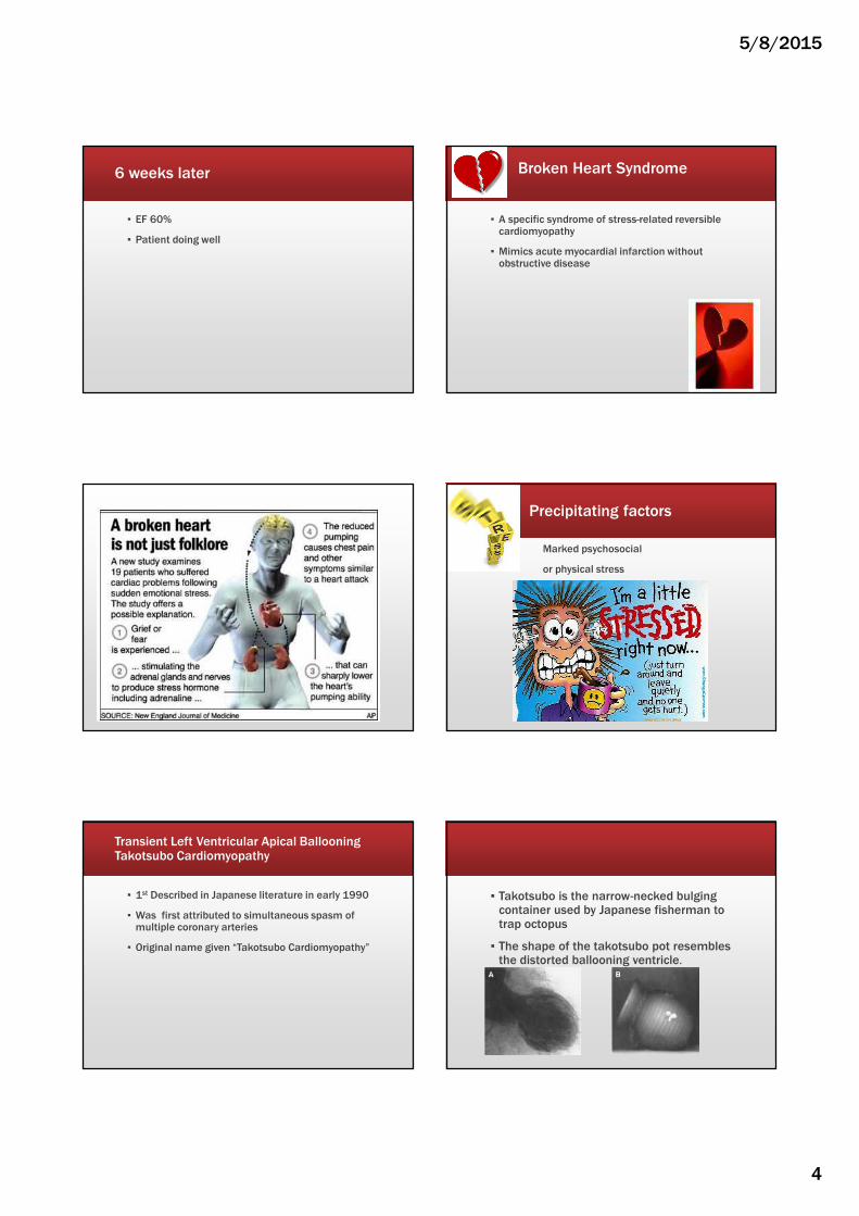
Transient Left Ventricular Apical BallooningTakotsubo Cardiomyopathy
▪ 1st Described in Japanese literature in early 1990
▪ Was first attributed to simultaneous spasm of multiple coronary arteries
▪ Original name given “Takotsubo Cardiomyopathy”
▪ Takotsubo is the narrow-necked bulging container used by Japanese fisherman to trap octopus
▪ The shape of the takotsubo pot resembles the distorted ballooning ventricle.

5/8/2015
5
Etiology
▪ Unclear etiology
▪ 1 – 2% of patients who have S/S AMI have apical ballooning (Japan & USA)
▪ 6-9 times more common in women
▪ 6% of women with AMI have apical ballooning
▪ Most often in postmenopausal women
Pathophysiology
▪ Marked systolic ballooning of the ventricular apex
▪ Hypercontractility of the base of the heart
▪ Most common in LV ---can occur in RV
▪ Initial reports thought it was due to spasm
▪ Now thought to be related to stunning of the myocardium related to excessive catecholamines
▪ Since preceded by increased psychosocial or physical stress suggest an association with ↑SNS activity
▪ Catecholamines have a toxic effect on the myocardium
▪ Catecholamine levels reported to be 7 – 34 times as high as the normal 2 – 3 elevation in classic AMI patients
Signs & Symptoms (not consistent)
▪ Chest pain
▪ ST segment changes
▪ Release cardiac biomarkers
▪ Syncope or near syncope
▪ Fatigue/malaise
▪ Palpitations
▪ Dyspnea
▪ Hypotension
▪ Pulmonary edema
▪ Cardiogenic shock
▪ Lethal ventricular arrhythmias
12 Lead EKG
▪ Variable findings
▪ ST segment elevation or depression usually in the precordial leads (V2 – V5)
▪ Reciprocal changes in the inferior leads may not occur
▪ Q waves usually do not develop
or Q waves V3 – V6
▪ Deeply inverted T waves are common in the recovery period
▪ Markedly prolonged QT interval
Cardiac biomarkers
▪ Only moderately elevated
▪ Do not follow the typical rise-fall-pattern seen with AMI
5/8/2015
6
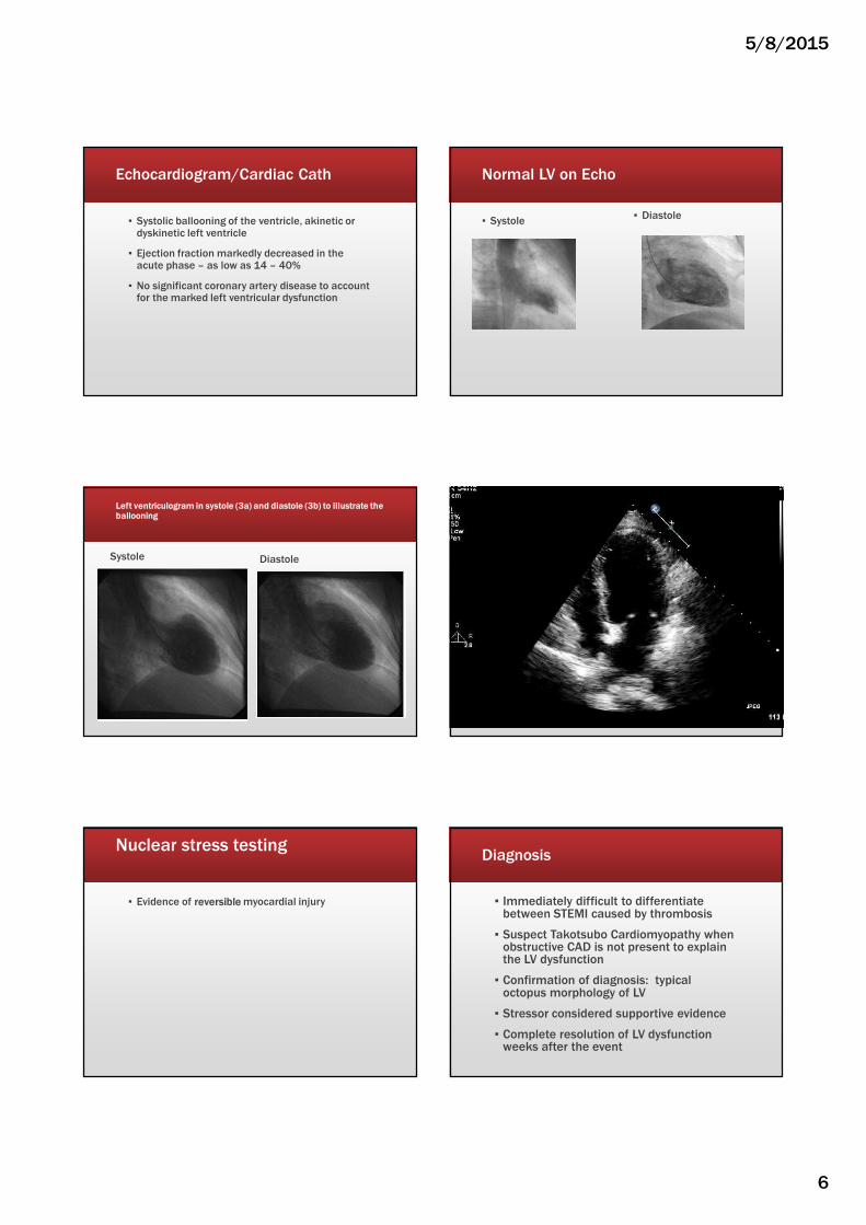
Echocardiogram/Cardiac Cath
▪ Systolic ballooning of the ventricle, akinetic or dyskinetic left ventricle
▪ Ejection fraction markedly decreased in the acute phase – as low as 14 – 40%
▪ No significant coronary artery disease to account for the marked left ventricular dysfunction
Normal LV on Echo
▪ Systole▪ Diastole
Left ventriculogram in systole (3a) and diastole (3b) to illustrate the ballooning
Systole Diastole
Nuclear stress testing
▪ Evidence of reversible myocardial injury
Diagnosis
▪ Immediately difficult to differentiate between STEMI caused by thrombosis
▪ Suspect Takotsubo Cardiomyopathy when obstructive CAD is not present to explain the LV dysfunction
▪ Confirmation of diagnosis: typical octopus morphology of LV
▪ Stressor considered supportive evidence
▪ Complete resolution of LV dysfunction weeks after the event
5/8/2015
7
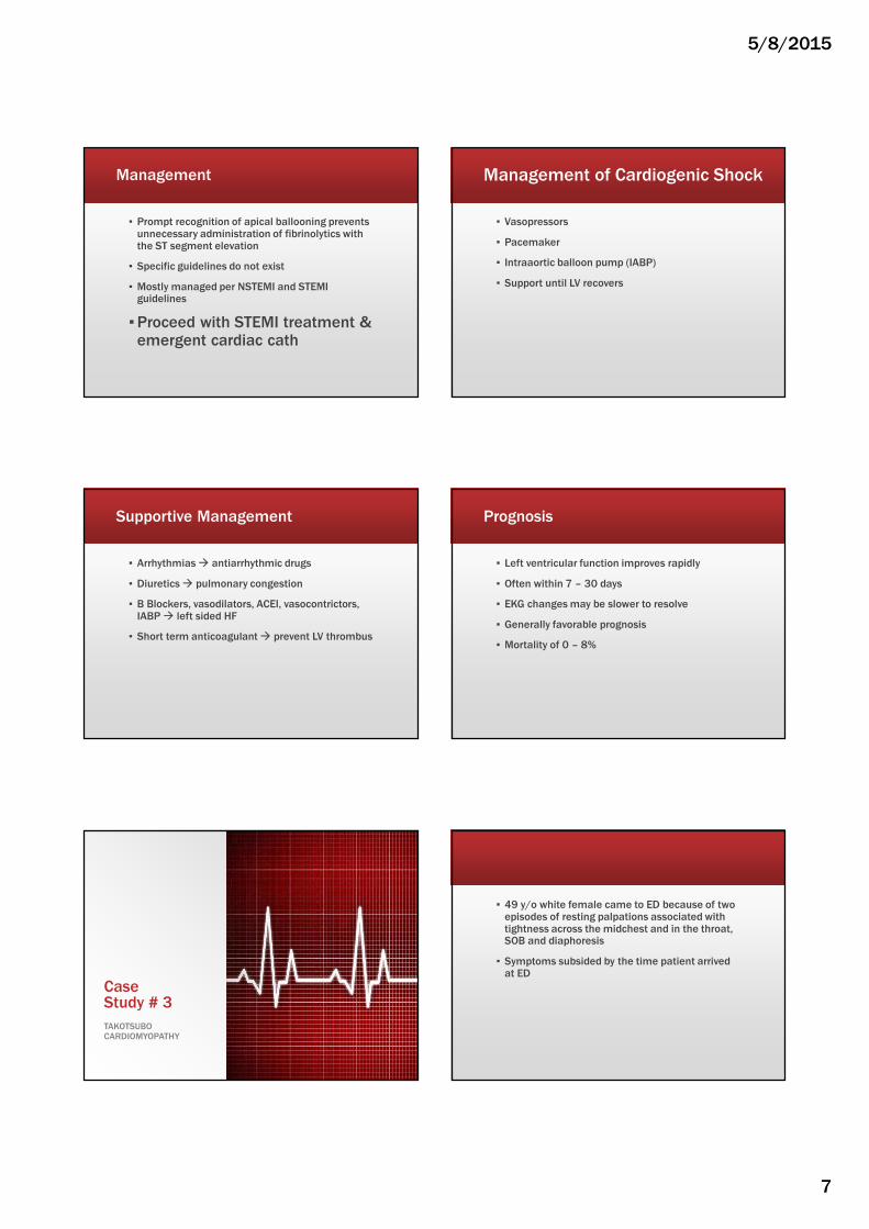
Management
▪ Prompt recognition of apical ballooning prevents unnecessary administration of fibrinolytics with the ST segment elevation
▪ Specific guidelines do not exist
▪ Mostly managed per NSTEMI and STEMI guidelines
▪Proceed with STEMI treatment & emergent cardiac cath
Management of Cardiogenic Shock
▪ Vasopressors
▪ Pacemaker
▪ Intraaortic balloon pump (IABP)
▪ Support until LV recovers
Supportive Management
▪ Arrhythmias � antiarrhythmic drugs
▪ Diuretics � pulmonary congestion
▪ B Blockers, vasodilators, ACEI, vasocontrictors, IABP � left sided HF
▪ Short term anticoagulant � prevent LV thrombus
Prognosis
▪ Left ventricular function improves rapidly
▪ Often within 7 – 30 days
▪ EKG changes may be slower to resolve
▪ Generally favorable prognosis
▪ Mortality of 0 – 8%
Case Study # 3
TAKOTSUBO CARDIOMYOPATHY
▪ 49 y/o white female came to ED because of two episodes of resting palpations associated with tightness across the midchest and in the throat, SOB and diaphoresis
▪ Symptoms subsided by the time patient arrived at ED
5/8/2015
8
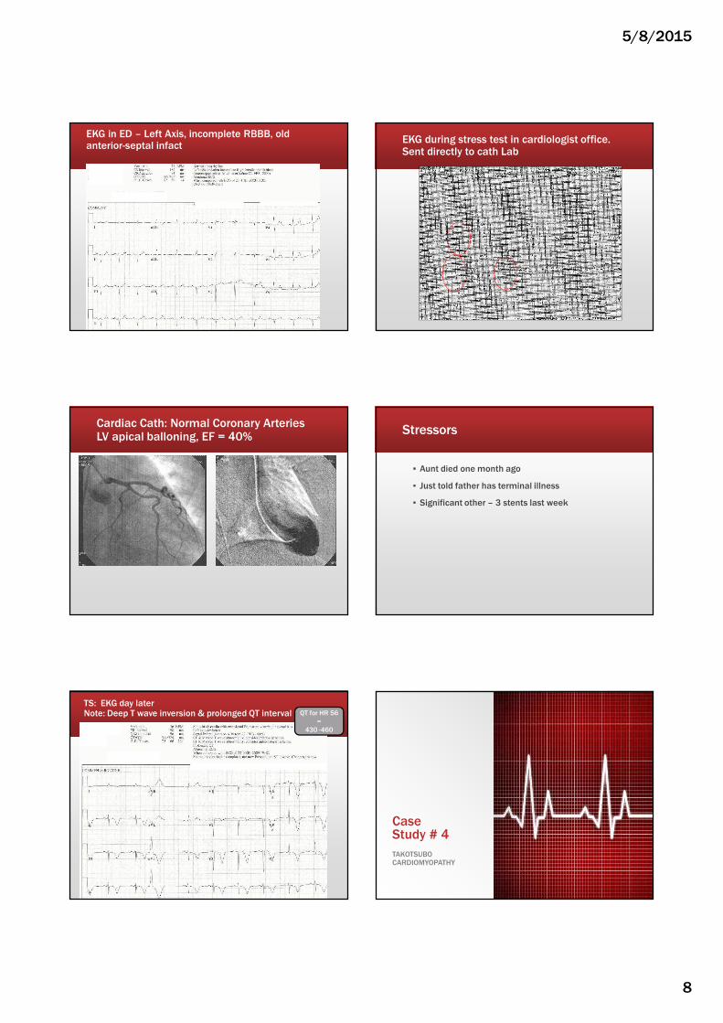
EKG in ED – Left Axis, incomplete RBBB, old anterior-septal infact
EKG during stress test in cardiologist office.Sent directly to cath Lab
Cardiac Cath: Normal Coronary ArteriesLV apical balloning, EF = 40%
Stressors
▪ Aunt died one month ago
▪ Just told father has terminal illness
▪ Significant other – 3 stents last week
TS: EKG day later Note: Deep T wave inversion & prolonged QT interval QT for HR 56
=
430 -460
Case Study # 4
TAKOTSUBO CARDIOMYOPATHY

5/8/2015
9
▪ 74 y/o female POD #2 rectal prolapse repair & cholecystectomy
▪ PMH
– 2 coronary stents three years ago & iliac stents.
– Quit smoking 4 years ago. Smoked 1 ½ packs x 50 years
▪ Clear lung sounds, uneventful post op course. SpO2 97% on room air
3/6 POD #2 1450
▪ Patient abruptly has respiratory distress.
▪ Respirations 36 labored
▪ SpO2 drops to 78% on 3 liters
▪ RRT called
RRT assessment
▪ O2 increased to 7 l/min. SpO2 81%
▪ BP 197/111, HR 139, Resp Rate 36 labored
▪ Lungs crackles throughout
▪ Color dusky
ABGs
pH 7.45
pC02 30
pO2 45
TCO2 21.8
O2% 83
BE -3.1
Lactic Acid 1.9
▪ O2 increased to 100% nonrebreather.
▪ SpO2 increased to 91%
▪ Transferred to ICU at 1505
EKG at 1509
5/8/2015
10
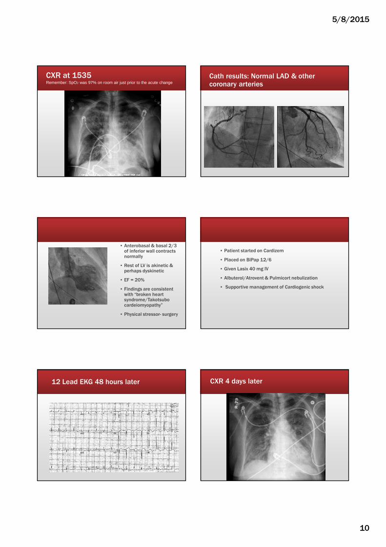
CXR at 1535Remember: SpO2 was 97% on room air just prior to the acute change
Cath results: Normal LAD & other coronary arteries
▪ Anterobasal & basal 2/3 of inferior wall contracts normally
▪ Rest of LV is akinetic & perhaps dyskinetic
▪ EF = 20%
▪ Findings are consistent with “broken heart syndrome/Takotsubo cardeiomyopathy”
▪ Physical stressor- surgery
▪ Patient started on Cardizem
▪ Placed on BiPap 12/6
▪ Given Lasix 40 mg IV
▪ Albuterol/Atrovent & Pulmicort nebulization
▪ Supportive management of Cardiogenic shock
12 Lead EKG 48 hours later CXR 4 days later
5/8/2015
11
10 days later 3/20
▪ Back on telemetry unit
▪ Patient abruptly goes into respiratory distress and is diaphoretic.
▪ BP 97/45, HR 131, RR 40 SpO2 92%
▪ Placed back on BiPAP 14/10
3/20 1600
▪ Started on Cardizem @ 10 mg/hour
▪ ABGs
ph 7.53
pCO2 23
pO2 60
TCO2 20
O2% 94
BE – 3.3
– Lactic Acid 4.7 (Abdomen tender)
CXR on 3/20
▪ Transferred back to ICU
▪ Supportive Care of Cardiogenic Shock
▪ Started having Ventricular Tachycardia --defibrillated several times over the next several hours. Then made DNR & expired shortly thereafter.
Broken Heart SyndromeSummary: Clinical features
▪ Onset of s/s often preceded by emotional/physical stressor
▪ Most common in postmenopausal women
▪ ST-segment abnormalities that mimic those of AMI
▪ Mild to moderate increase in levels of cardiac enzyme compared with the increase in AMI
▪ No significant coronary artery disease to account for the left ventricular dysfunction
▪ Left ventricular “ballooning” wall motion at the apex with hypercontractility at the base
▪ Transient and reversible left ventricular changes with favorable prognosis
Source: McCulloch B 2007: Critical Care Nurse 27(6): 20 - 27
5/8/2015
12
Broken Heart SyndromeTakotsubo Cardiomyopathy
▪ Avoid Fibrinolytics!
Case Study # 5 PERIPARTUM CARDIOMYOPATHY
37 y/o African American presents to ED with Shortness of Breath
▪ BP 152/102
▪ HR 100
▪ RR 28
▪ T 98.9 oral
▪ Sp02 88% room air
▪ Loose cough
▪ Coarse rhonchi and scattered wheezes
▪ 2+ pitting edema
PMH
▪ Asthma
▪ Pancreatitis
▪ Diabetes, type II (diet controlled)
▪ Smokes 4 cigarettes/day x 15 years
▪ Cocaine use in the past
▪ Sister and daughter � sickle cell anemia
Labs
▪ WBC 8.2
▪ Hbg 10.2
▪ HCT 32.7
▪ Glucose 79
▪ Potassium 3.2
▪ Creatinine 1.2
▪ Troponin 0.06
Symptoms
▪ C/o not breathing “normally” with increased effort and shortness' of breath
▪ Moderate SOB at rest
▪ Decreased exercise capacity with exertional SOB
▪ Started two weeks ago and getting worse
▪ Albuterol treatment taken 1 hour ago at home

5/8/2015
13
Admission EKG (37 y/o) Prolonged QT (K+ 3.2); ? Hypertrophy; PVCs
QT for HR 93 =
330 - 336
Admission CXR
A. Pneumonia
B. Pulmonary Edema
C. Cardiomyopathy
with Pulmonary
Edema
D. All of the above
Admission CXR ANSWER
D. All of the above
More history
▪ C-section two weeks ago for failure to progress
– 4th child
▪ SOB and wheezing have become progressively worse since delivery
▪ Low grade fever past few days
▪ Productive cough
Admission Diagnosis
▪ Bilateral pneumonia vs CHF
▪ ? Peripartum cardiomyopathy
▪ Treatments
– Albuterol/Atrovent nebs
– Prednisone 50 mg po
– Methylprednisolone 125 mg IV
– Lasix 60 mg IV
▪ Any concerns??
– Ceftriazone 1 gm IV
– Azithromycin 500 mg IV
– Echocardiogram
5/8/2015
14
Echo
▪ LV mildly dilated
▪ EF 35%
▪ Mild to moderate aortic regurgitation
▪ Moderate mitral regurgitation
▪ RV mildly enlarged
Discharged 2 days later
Discharge Diagnosis
▪ Community acquired pneumonia
▪ Asthma exacerbation
▪ Peripartum cardiomyopathy
▪ Anemia secondary to postpartum state
Discharge meds
▪ Lisinopril
▪ Metoprolol tartrate
▪ Prednisone
▪ Flovent Diskus
▪ Albuterol
▪ Augmentin
▪ Ferrous Sulfate
CXR three weeks later
Case Study # 6 PERIPARTUM CARDIOMYOPATHY
34 y/o African American status post normal vaginal delivery
▪ G4, P 4
▪ 38 weeks gestation
▪ Diabetes mellitus, type I
▪ Previous smoker
▪ Admission BP 140/91
▪ Pitocin for augmentation of labor
▪ Bilateral tubal ligation day after delivery
Admission BP 140/91
BP during Labor
▪ 151/99
▪ 154/95
▪ 182/104
▪ 166/81
▪ 175/89
BP post delivery
▪ 151/83
▪ 162/82
▪ 179/97 post BTL
▪ 205/95
▪ 184/94
▪ 169/87
▪ 177/97
▪ 181/99
▪ Started on Labetalol and PRN hydralazine
5/8/2015
15
Day 2
▪ 12;15 pm – Feels SOB today with intermittent cough
– Expiratory crackles, audible wheezing
– Accessory muscle usage
– 2+ pitting edema bilateral lower extremities
– BP 159/86
– Sp02 87% on 2 liters nasal cannula
▪ Plan– Increase Labetalol to 200 mg BID
– Continue prn Hydralazine
– CXR
– DuoNeb STAT
– CBC
CXR
▪ Cardiomegaly
▪ Bilateral consolidation in both lungs
▪ Acute pulmonary edema
▪ Plan
– Now on 4 liters oxygen
– Cardiology consult
– Lasix 40 mg IV
Cardiology Consult
▪ Flash Pulmonary Edema secondary to fluid overload and hypertension
▪ Hypertensive urgency and pregnancy induced hypertension
▪ Mild Peripartum cardiomyopathy
▪ Echo EF 40%
▪ Systolic murmur
▪ Plan
– Lasix 40 mg TID
– Nitroglycerin drip to keep SBP < 120
– Labetalol 200 mg BID po
Hypertensive
Urgency: severe
BP elevation
without evidence
of end-organ
dysfunction.
Discharged 2 days later
▪ Diuresis of 7 liters in 24 hours
▪ Clear lung sounds – no wheezing
▪ BP 140/91
▪ Heart Failure meds and protocol implemented
▪ Follow up with cardiology
Peripartum Cardiomyopathy (PPCM)ALSO CALLED PREGNANCY ASSOCIATED CARDIOMYOPATHY
Peripartum Cardiomyopathy (PPCM)
5th leading cause of mortality during the pregnancy period
Source: Moser & Riegel. 2008. Cardiac Nursing. And Tsang, W, Peripartum Cardiomyopathy:.Retrieved 2-9-15 from Up To Date

5/8/2015
16
Peripartum Cardiomyopathy (PPCM)
1. Cardiomyopathy in the last month or the first five months after pregnancy
2. Absence of another identifiable cause of HF
3. EF < 45%▪ LV may or may not be dilated
Source: Moser & Riegel. 2008. Cardiac Nursing. And Tsang, W, Peripartum Cardiomyopathy:.Retrieved 2-9-15 from Up To Date
Peripartum Cardiomyopathy (PPCM)
▪ Incidence per live births–1:4350 USA– 10 years ago
–1:2399 USA – 2011▪ ↑ maternal age, ↑ multifetal pg, ↑ recognition PPCM
–1:1000 South Africa
–1:300 Haiti
–1:100 Nigeria
▪ Cause – Unknown
–Usually occurs with first or second pregnancy
Source: Moser & Riegel. 2008. Cardiac Nursing: Tsang, W, Peripartum Cardiomyopathy:.Retrieved 2-9-15 from Up To Date: Sundin. C 2014. Peripartum Cardiomyopathy. MCN 39(4)
Pregnancy
▪ High output state
▪ 30% decrease in systemic vascular resistance
▪ 30-40% increase in cardiac output by 2nd and 3rd trimester
▪ Changes may not resolve completely until 12 weeks postpartum
Source: Garg, J et al. 2015. Peripartum Cardiomyopathy. Cardiology in Review; 23(2).
PPCM Risk Factors
▪ Advancing maternal age > 30 years– Extreme age (very young or advanced age)
▪ African descent
▪ Multi-fetal gestation
▪ History of preeclampsia, eclampsia, postpartum hypertension
▪ Long term (> 4 weeks) use of beta adrenergic agonists (terbutaline) for preterm labor suppression– Tocolytic agents used > 4 weeks have higher incidence of
pulmonary edema (terbutaline, salbutamol, ritodrine, and magnesium sulfate)
▪ Maternal cocaine abuse
Source: Moser & Riegel. 2008. Cardiac Nursing. And Tsang, W, Peripartum Cardiomyopathy:.Retrieved 2-9-15 from Up To DateGarg, J et al. 2015. Peripartum Cardiomyopathy. Cardiology in Review; 23(2).
PPCM signs and symptomsSimilar to other forms of systolic HF
▪ Dyspnea – most common
▪ Tachycardia – Early sign
▪ Cough
▪ Orthopnea
▪ Paroxysmal nocturnal dyspnea (PND)
▪ Pedal edema
▪ Nonspecific fatigue
▪ Hemoptysis
High clinical suspicion
▪ Elevated jugular venous pressure
▪ Displaced apical impulse
▪ S3
▪ Murmur from tricuspid or mitral regurgitation
Source: Tsang, W, Peripartum Cardiomyopathy:. Retrieved 2-9-15 from Up To DateGarg, J et al. 2015. Peripartum Cardiomyopathy. Cardiology in Review; 23(2).
Heart Sounds
▪ https://www.youtube.com/watch?v=L5DEqvgS_xs&index=15&list=PL3CE2BC4AF364AE80
▪ Mitral Regurgitation
http://www.blaufuss.org/tutorial/index2.html
▪ Aortic Regurgitation
http://www.blaufuss.org/tutorial/index2.html
5/8/2015
17
PPCM symptoms
▪ Often missed or delayed
▪ Similar signs and symptoms of normal pregnancy
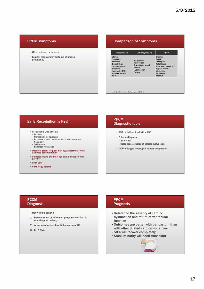
Comparison of Symptoms
Preeclampsia Similar Symptoms PPCM
Edema
Proteinuria
Headache
Blurred vision
Decreased renal
perfusion
Hyperactive DTRs
Impaired hepatic
function
Weight gain
Tachycardia
Adventitious breath
sounds
Hypertension
Fatigue
Dyspnea
Cough
Chest pain
Palpitations
Third heart sound S3
Jugular venous
distension
Tachypnea
Murmur
Sundin. C 2014. Peripartum Cardiomyopathy. MCN 39(4)
Early Recognition is Key!
▪ For patients who develop– Dyspnea– Increasing blood pressure– Increased edema or edema that doesn’t decrease– Chest pain– Tachycardia– Nonproductive cough
▪ Detailed, serial, frequent nursing assessments with accurate documentation
▪ Comprehensive and thorough communication with provider
▪ BNP, Echo
▪ Cardiology consult
PPCMDiagnostic tools
▪ BNP > 100 or ProBNP > 450
▪ Echocardiogram
– EF < 45%
– Helps assess degree of cardiac dysfunction
▪ CXR- enlarged heart, pulmonary congestion
PCCMDiagnosis
Three Clinical criteria
1. Development of HF end of pregnancy or first 5 months post delivery
2. Absence of other identifiable cause of HF
3. EF < 45%
PPCMPrognosis
▪ Related to the severity of cardiac dysfunction and return of ventricular function▪ Outcomes are better with peripartum than
with other dilated cardiomyopathies▪ 50% will recover completely▪ Small minority will need transplant
5/8/2015
18
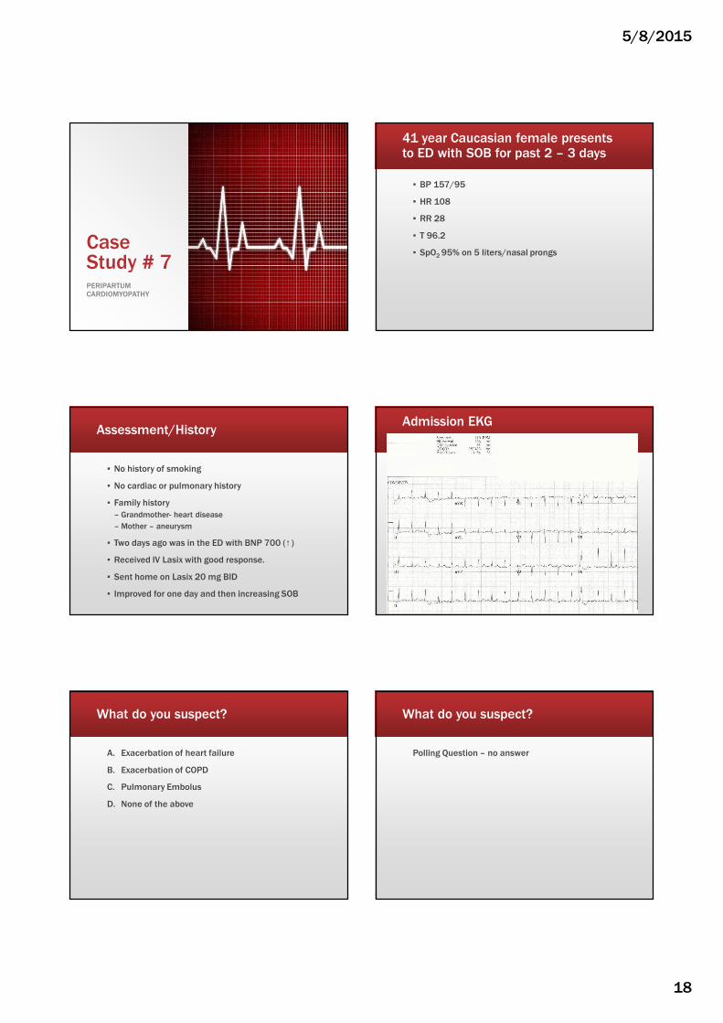
Case Study # 7 PERIPARTUM CARDIOMYOPATHY
41 year Caucasian female presents to ED with SOB for past 2 – 3 days
▪ BP 157/95
▪ HR 108
▪ RR 28
▪ T 96.2
▪ SpO2 95% on 5 liters/nasal prongs
Assessment/History
▪ No history of smoking
▪ No cardiac or pulmonary history
▪ Family history
– Grandmother- heart disease
– Mother – aneurysm
▪ Two days ago was in the ED with BNP 700 (↑)
▪ Received IV Lasix with good response.
▪ Sent home on Lasix 20 mg BID
▪ Improved for one day and then increasing SOB
Admission EKG
What do you suspect?
A. Exacerbation of heart failure
B. Exacerbation of COPD
C. Pulmonary Embolus
D. None of the above
What do you suspect?
Polling Question – no answer
5/8/2015
19
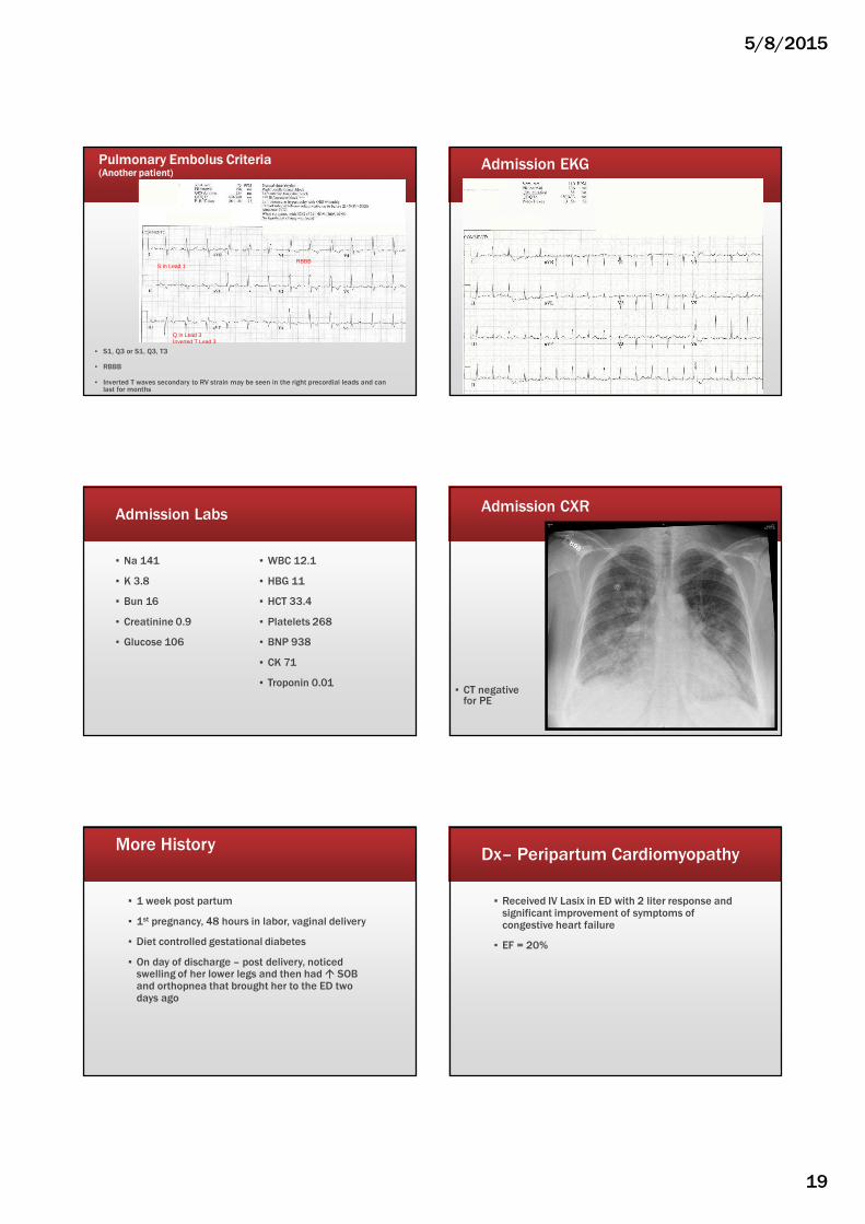
Pulmonary Embolus Criteria(Another patient)
▪ S1, Q3 or S1, Q3, T3
▪ RBBB
▪ Inverted T waves secondary to RV strain may be seen in the right precordial leads and can last for months
S in Lead 1
Q in Lead 3Inverted T Lead 3
RBBB
Admission EKG
Admission Labs
▪ Na 141
▪ K 3.8
▪ Bun 16
▪ Creatinine 0.9
▪ Glucose 106
▪ WBC 12.1
▪ HBG 11
▪ HCT 33.4
▪ Platelets 268
▪ BNP 938
▪ CK 71
▪ Troponin 0.01
Admission CXR
▪ CT negative for PE
More History
▪ 1 week post partum
▪ 1st pregnancy, 48 hours in labor, vaginal delivery
▪ Diet controlled gestational diabetes
▪ On day of discharge – post delivery, noticed swelling of her lower legs and then had � SOB and orthopnea that brought her to the ED two days ago
Dx– Peripartum Cardiomyopathy
▪ Received IV Lasix in ED with 2 liter response and significant improvement of symptoms of congestive heart failure
▪ EF = 20%
5/8/2015
20
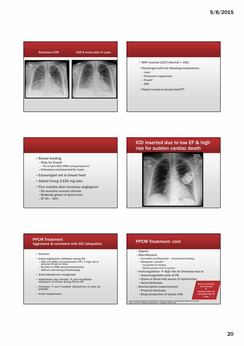
Admission CXR CXR 6 hours after IV Lasix
▪ BNP reached 1233 (Normal < 100)
▪ Discharged with the following medications:
– Lasix
– Potassium supplement
– Enapril
– ASA
▪ Patient wants to breast feed???
▪ Breast feeding – Okay for Enapril▪ Do not give ACEI/ARBs during pregnancy!
– Unknown/controversial for Lasix
▪ Encouraged not to breast feed
▪ Added Coreg 3.625 mg later
▪ Five months later Coronary angiogram – No occlusive coronary disease
– Moderate global LV dysfunction
– EF 20 – 30%
ICD inserted due to low EF & high risk for sudden cardiac death
PPCM TreatmentAggressive & consistent with IDC (idiopathic)
▪ Diuretics
▪ Avoid angiotensin inhibition during PG – ACEI and ARBs contraindicated in PG � high risk of
adverse effects on fetus– No data on ARBs during breastfeeding– ACEI are safe during breastfeeding
▪ Avoid Aldosterone Antagonists
▪ Hydralazine plus nitrates � oral vasodilator treatment of choice during PG for HF
▪ Inotropes � use if needed; discontinue as soon as possible
▪ Avoid vasopressors
PPCM Treatment- cont
– Digoxin
– Beta Blockers▪ Carvedilol and Bisoprolol – avoid breast feeding
▪ Metoprolol tartrate *
▫ Compatible for feeding
▫ Monitor growth curve in neonate
– Anticoagulation � High risk for thrombus due to:
▪ Hypercoagulable state of PG
▪ Stasis of blood with severe LV dysfunction
▪ Atrial fibrillation
– Bromocriptine (experimental)
▪ Prolactin blockade
▪ Stops production of breast milk
Some recommend
breast feeding
IF
clinically stable and
compatible with HF
meds
Source: Tsang, W, Peripartum Cardiomyopathy: Treatment and Prognosis. Retrieved Feb 9, 2015 from Up To Date
Garg, J et al. 2015. Peripartum Cardiomyopathy. Cardiology in Review; 23(2).
5/8/2015
21
Implantable Cardioverter Difibrillator (ICD)Cardiac Resynchronization Therapy (CRT)
▪ 40% PPCM – EF back to normal by 6 months
▪ Defer ICD and CRT placement at least 3 months; possibly 6 months
In SummaryEarly Recognition is Key!
▪ For patients who develop– Dyspnea– Increasing blood pressure– Increased edema or edema that doesn’t decrease– Chest pain– Tachycardia– Nonproductive cough
▪ Detailed, serial, frequent nursing assessments with accurate documentation
▪ Comprehensive and thorough communication with provider
▪ BNP, Echo
▪ Cardiology consult
Case Study # 8 HYPERTROPHIC OBSTRUCTIVE CARDIOMYOPATHY.
Case study
▪ 25 y/o female presents to ED complaining of significant chest pain that was continuous for several hours.
▪ What diagnostic test do you want?
Should you go to Cath lab?1 = Yes; 2 = No; 3 = Undecided
Let’s look at each lead
5/8/2015
22
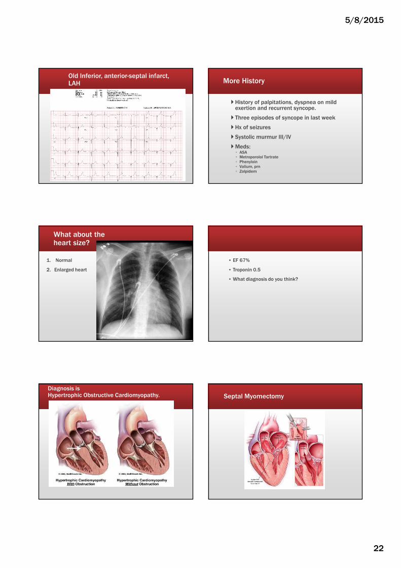
Old Inferior, anterior-septal infarct, LAH More History
�History of palpitations, dyspnea on mild exertion and recurrent syncope.
� Three episodes of syncope in last week
�Hx of seizures
� Systolic murmur III/IV
�Meds:◦ ASA◦ Metroporolol Tartrate◦ Phenyloin◦ Valium, prn◦ Zolpidem
What about the heart size?
1. Normal
2. Enlarged heart
▪ EF 67%
▪ Troponin 0.5
▪ What diagnosis do you think?
Diagnosis is Hypertrophic Obstructive Cardiomyopathy. Septal Myomectomy

5/8/2015
23
� Myocardial hypertrophy without the presence of associated hemodynamic stress (no ↑ in afterload)
� Hypertrophy of the heart muscle including the septum and ventricular free wall
� Previously called IHSS –idiopathic hypertrophic subaortic stenosis
� Leading cause of death in athletics < 35 y/o
Hypertrophic Cardiomyopathy (HCM)Hypertrophic Obstructive Cardiomyopathy (HOCM)
▪ Subgroup of patients with HCM develop obstruction
▪ Once obstruction occurs it is called: Hypertrophic obstructive cardiomyopathy (HOCM)
Hypertrophic Cardiomyopathy (HCM)
Pathophysiology
1. Hypertrophy of heart muscle including septum and ventricular free wall.
2. Rigid, noncompliant ventricles do not stretch
3. Causes diastolic dysfunction
4. ↓ preload and cardiac output
5. Left atrial dilatation from inability to empty LA
6. Mitral regurgitation occurs from papillary muscles and mitral valve pulled out of alignment
Hypertrophy of LV, septum and ventricular wall, LA enlargement, MR
Hypertrophic Obstructive Cardiomyopathy (HOCM)
Pathophysiology
1. With severe hypertrophy, left ventricular outflow tract becomes obstructed --- especially with ↑contractility from ↑ catecholamines (exercise)
2. Decrease in blood flow to coronary arteries (angina) and brain (syncope)
3. May result in sudden cardiac death
LV outflow tract obstructed – syncope, sudden death
Hypertrophic Cardiomyopathy (HCM)
Causes
▪ Probably genetic
▪ May occur as early as the 1st year of life
▪ Develops most commonly during adolescence
▪ Hypertrophy manifests after age 20
▪ Diagnosis is usually made by age 25
▪ Persons with normal echo and EKG after 25 y/o are unlikely to develop HCM
Hypertrophic Cardiomyopathy (HCM)
Clinical Presentation
Sudden cardiac death often the first presentation
▪ Often diagnosed incidentally as may be asymptomatic
▪ Dyspnea on exertion
▪ Chest pain on exertion – relieves with rest
▪ Syncope on exertion or rest
▪ Palpitations
� Jugular venous palpitation◦ Associated with prominent “a”
wave secondary to ↓ RV compliance
� Heart Sounds◦ Harsh systolic murmur LSB◦ Murmur increases with
movement◦ S4 from LVH
� EKG◦ Repolarization abnormalities◦ Atrial enlargement (large p
waves)◦ Pathological Q waves – inferior
leads
5/8/2015
24
Hypertrophic Cardiomyopathy (HCM)
Clinical Management
� Symptom relief
� Prevention of sudden cardiac death
� Beta blockers for chest pain and dyspnea with exertion in HOCM
� Disopyramide (Norpace and Rythmodan) – reduces obstruction by ↓ inotropic action
� Verapamil – used only for mild obstruction
� Atrial kick more essential than normal
� If symptoms persist◦ Ventricular Septal myectomy –
removal of muscle from septum.◦ Percutaneous septal alcohol ablation
– causes controlled septal MI
� ICD ◦ History of cardiac arrest or sustained
ventricular dysrhythmias◦ Multiple clinical risk factors
� Counseling & genetic testing ◦ Restrict from intense competitive
sports
� SBE prophylaxis for HOCM
Symptom relief & prevention of cardiac arrest
Hypertrophic Cardiomyopathy (HCM)
Medications
� Beta blockers◦ 1st choice◦ Increase exercise tolerance◦ ↓ heart rate◦ Improves LV relaxation◦ Control of arrhythmias
� Disopyramide (Norpace and Rythmodan) ◦ Negative inotrope ( ↓ contractility)◦ Used with BB to treat LV outflow
track obstruction◦ ↓ SAM◦ Assists with HR control◦ Monitor QT – may causes
arrhythmias◦ Class I antiarrhythmic
▪ Calcium Channel Blockers
– Verapamil or diltiazem
– Used only for mild obstruction
– Use if BB ineffective
– ↓ LV wall tension
– Negative inotrope
– ↓HR
▪ Antiarrhythmic medications
– Treat A fib and/or vent arrhythmias
– Amiodarone or sotolol
Disopyramide may cause uncomfortable anticholinergic side effects and may enhance the hypoglycemic effect of gliclazide, insulin, and metformin.
� Diuretics◦ Give with caution as volume
status is important
AVOID in HOCM
� Nitroglycerin
� Ace Inhibitors
� Positive inotropes
� Anything that ↑ contractility
� Nifedipine, amiodipine,felopine because of the vasodilatory effects
Hypertrophic Cardiomyopathy (HCM)
Medications CAUTION▪ Hypertrophic
▪ Dilated (ischemic and nonischemic)– Idiopathic Dilated Cardiomyopathy – Ischemic Dilated Cardiomyopathy– Hypertensive Dilated Cardiomyopathy– Valvular Dilated Cardiomyopathy– Anthracycline Dilated Cardiomyopathy– Peripartium Dilated Cardiomyopathy– Alcohol Dilated Cardiomyopathy
▪ Stressed Induced (Takotsubo)
▪ Restrictive
Studying for CCRN or CMC?Cardiomyopathies
Case Study # 9 FLASH PULMONARY EDEMA AND PICKERING SYNDROME
Direct admit from rural hospital for SOB
▪ Not been feeling well for last 6 months
– Dyspnea
– Joint pain
▪ Major complaint~ lower extremity swelling and SOB on exertion which he attributes to his COPD
▪ This morning – more SOB and chest discomfort
▪ Chest pressure 6/10, left sided, non radiating
▪ Initial EKG Sinus Rhythm with Nonspecific ST changes
▪ Troponin 0.5
▪ Creatinine 2.5, BUN 29

5/8/2015
25
Initial EKG – nonspecific EKG Changes
▪ Should he be admitted? 1.Yes
2.No
PMH
▪ CAD, stent to LAD 2 ½ years ago
▪ PAD, stent
▪ Hypertension
▪ Hyperlipidemia
▪ Former smoker
▪ COPD
▪ Sleep apnea
▪ Fractured some left ribs 6 – 7 months ago – off work for 3 weeks
▪ dyspnea – pleural effusion – thoracentesis x 3 six weeks apart =1 liter. Negative for any malignancies. Last thoracentesis 6 weeks ago
▪ Extremity edema started around the time of last thoracentesis (6 weeks ago)
Transferred for further evaluation
EKG on Admission from referring hospitalWhat do you see on the EKG?
1. Normal EKG
2. STEMI
3. ST depression
4. Hypertrophy
5. ST depression and hypertrophy
Admission EKG
▪ Left – persistent atelectasis or pneumonia or pleural effusion
▪ Mild right basilar atelectasis noted
Cardiology consult
▪ Acute Coronary Syndrome/NSTEMI
– ST depression V 4 – V6 worrisome for cardiac ischemia
– 2nd Troponin 5.0 (previous 0.5)
– Dual antiplatelet therapy
▪ ASA, Clopidogrel (Plavix)
– BetaBlocker Therapy
▪ Stop Atenolol due to worsening kidney function
▪ Metoprolol
▪ Acute Kidney Injury
– Stop Losartan due to kidney function.
– May consider ACE I later
Plan
▪ Invasive vs conservative NSTEMI strategy discussed in detail with patient
▪ Conservative strategy due to acute renal injury with creatinine 2.5
▪ Monitor renal function closely
▪ If kidney function improves, consider coronary angiography

5/8/2015
26
Diagnostic Testing
Renal Ultrasound
• Bilateral renal artery stenosis
• Mild renal pelvis dilation bilaterally
Echocardiogram
▪ EF 55 %
▪ Inferior vena cava normal – collapses greater than 50% with inspiration
▪ No pulmonary hypertension
What does “Inferior vena cava normal – collapses greater than 50% with inspiration” mean?
▪ Volume depletion. In these patients, the diameter of the IVC will be decreased and the percentage collapse will be greater than 50%. With complete collapse, the IVC may become difficult to visualize (Figure 6).
▪ Volume overload. Patients with increased intravascular volume will have a large IVC diameter and minimal collapse on inspiration (Figure 7).
Source: Goldflam. Focus on Inferior Vena Cava Ultrasound June 2011: http://www.acep.org/Content.aspx?id=80791
Course of Stay over next 10 days
▪ Acute anemia --- blood transfusion
▪ Fever and microscopic hematuria without evidence of infection
▪ Autoimmune workup: Elevated ERS CRP and ANA, positive MPO (Myeloperoxidase) antibody
▪ Osteoarthritis of the hands and hips and probably right elbow
▪ Tissue biopsy – Wegener’s Granulomatosis
– Given Cytoxan and high dose prednisone
Wegener's Granulomatosis
▪ An uncommon systemic disorder and serious disease
▪ Causes inflammation of the blood vessels; often affects kidneys, lungs and upper respiratory tract. The restricted blood flow to these organs can damage them.
▪ Also produces a type of inflammatory tissue known as a granuloma that's found around the blood vessels. Granulomas can destroy normal tissue.
▪ No known cause
▪ Early diagnosis and treatment may lead to a full recovery.
▪ Without treatment, it can be fatal within months. Most commonly from kidney failure.
▪ Treatment is directed toward stopping the inflammation process by suppressing the immune system.
http://www.mayoclinic.org/diseases-conditions/wegeners-granulomatosis/basics/definition/con-20028113
Back to the heart….. NSTEMI
▪ Patient stabilized
– BP 156 – 189/73-86, HR 75- 86
– H/H 9.0/29
▪ Kidney function
– Creatinine 1.2, BUN 23
– 24 hour Intake and output = 2632/2250 with + 363 net
– Net Intake and output since admission (11 days)
+ 1894
▪ Scheduled for coronary angiogram
CXR day before coronary angiogram
5/8/2015
27
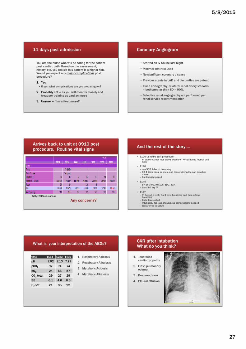
11 days post admission
You are the nurse who will be caring for the patient post cardiac cath. Based on the assessment, history, etc, you realize this patient is a higher risk. Would you expect any major complications post procedure?
1. Yes
• If yes, what complications are you preparing for?
2. Probably not – as you will monitor closely and treat per training as cardiac nurse
3. Unsure -- “I’m a float nurse!”
Coronary Angiogram
▪ Started on IV Saline last night
▪ Minimal contrast used
▪ No significant coronary disease
▪ Previous stents in LAD and circumflex are patent
▪ Flush aortography: Bilateral renal artery stenosis – both greater than 80 – 90%.
▪ Selective renal angiography not performed per renal service recommendation
Arrives back to unit at 0910 post procedure. Routine vital signs
Any concerns?SpO2 = 91% on room air
And the rest of the story….
▪ 1120 (2 hours post procedure)– Pt stable except high blood pressure. Respirations regular and
easy
▪ 1140 – c/o SOB, labored breathing
– 02 4 liters nasal cannula and then switched to non breather mask
– Cardiologist paged
▪ 1145– BP 150/92, HR 108, SpO2 51%
– Lasix 80 mg IV
▪ 1147– Pt having a really hard time breathing and then agonal
breathing
– Code blue called
– Intubated. No loss of pulse, no compressions needed
– Transferred to CVICU
What is your interpretation of the ABGs?
time 1154 1223 1405
pH 7.02 7.13 7.29
pCO2 97 74 74
pO2 24 66 57
C02 total 29 27 29
BE 6.1 4.6 0.6
O2 sat 21 85 92
1. Respiratory Acidosis
2. Respiratory Alkalosis
3. Metabolic Acidosis
4. Metabolic Alkalosis
CXR after intubation What do you think?
1. Takotsubo cardiomyopathy
2. Flash pulmonary edema
3. Pneumothorax
4. Pleural effusion
5/8/2015
28
VentAC 24, TV 550, PEEP 12
1154 1223 1405 2116 0600
pH 7.02 7.13 7.29 7.36 7.35
pCO2 97 74 74 46 47
pO2 24 66 57 130 111
C02 total 29 27 29 27 28
BE 6.1 4.6 0.6 0.4 1.6
O2 sat 21 85 92 99 98
CXR Day after code
CXR right after code
Diuresed
▪ Afternoon after code (about 24 hours later) right renal stent placed
▪ Extubated – sent to progressive unit
▪ Had some malignant hypertensive episodes
CXR 3 days after code7700 ml diuresis
CXR day after code
Discharged 5 days post cardiac cathLOS = 16 days
Discharge Diagnosis
▪ Malignant hypertension likely due to RAS
▪ NSTEMI
▪ Bilateral renal artery stenosis (RAS); s/p renal stent
▪ Recurrent Left pleural effusion – repeat TB gold in 4 weeks
▪ Wegener’s Granulomatosis
▪ CKD II
▪ Chronic iron Deficiency Anemia
▪ COPD
Discharge Medications
Albuterol neb
Amlodipine
Aspirin
Atenolol
Atorvastin
Clonidine
Clopidogrel
Cytoxan
Famotidine
Furosemide – Lasix
Hydralazine
Isorbide
Synthroid
Lisinopril
Potassium chloride
Prednisone
5/8/2015
29
Renal Consult
Flash Pulmonary Edema
▪ Classic clinical finding with renal artery stenosis (RAS)
– Hypertension related to RAS
Flash pulmonary edema
▪ General term used to describe a dramatic form of decompensated heart failure
▪ An acute increase of LV end diastolic pressure in the setting of hypertensive urgency, acute ischemia, new onset tachyarrhythmia, or obstructive valvular disease
▪ Flooding of the alveolar space can occur within minutes resulting in an acute life threatening emergency
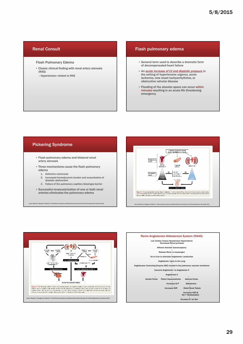
Pickering Syndrome
▪ Flash pulmonary edema and bilateral renal artery stenosis
▪ Three mechanisms cause the flash pulmonary edema
1. Defective natriuresis
2. Increased hemodynamic burden and exacerbation of diastolic dysfunction
3. Failure of the pulmonary capillary blood-gas barrier
▪ Successful revascularization of one or both renal arteries eliminates the pulmonary edema
Source: Messerli, F, Bangalore S, Makani H. Flash Pulmonary Oedema and Bilateral Renal Artery Stenosis: The Pickering Syndrome. Euroheartj. 2011Source: Messerli, F, Bangalore S, Makani H. Flash Pulmonary Oedema and Bilateral Renal Artery Stenosis: The Pickering Syndrome. Euroheartj. 2011
Source: Messerli, F, Bangalore S, Makani H. Flash Pulmonary Oedema and Bilateral Renal Artery Stenosis: The Pickering Syndrome. Euroheartj. 2011
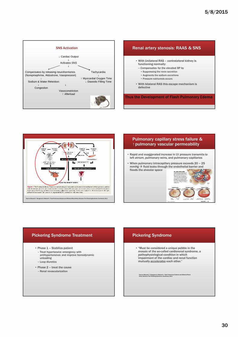
Renin-Angiotensin-Aldosterone System (RAAS)
Low Cardiac Output/Hypotension/HypovolemiaDecreased Renal perfusion
↓Afferent Arteriole (baroreceptors)
↓Release Renin (a messenger)
↓Go to Liver to stimulate Angiotensin I production
↓Angiotensin I goes to the Lung
↓Angiotension Converting Enzyme (ACE) located in the pulmonary vascular membrane
↓Converts Angiotensin I to Angiotensin II
↓Angiotensin II
↓ ↓ ↓Growth Factor Potent Vasoconstrictor Adrenal Cortex
↓ ↓Increases B/P Aldosterone
↓ ↓Increases SVR Distal Renal Tubule
↓Increases H2O &
Na++ Reabsorption↓
Excretes K+ for Na+
5/8/2015
30
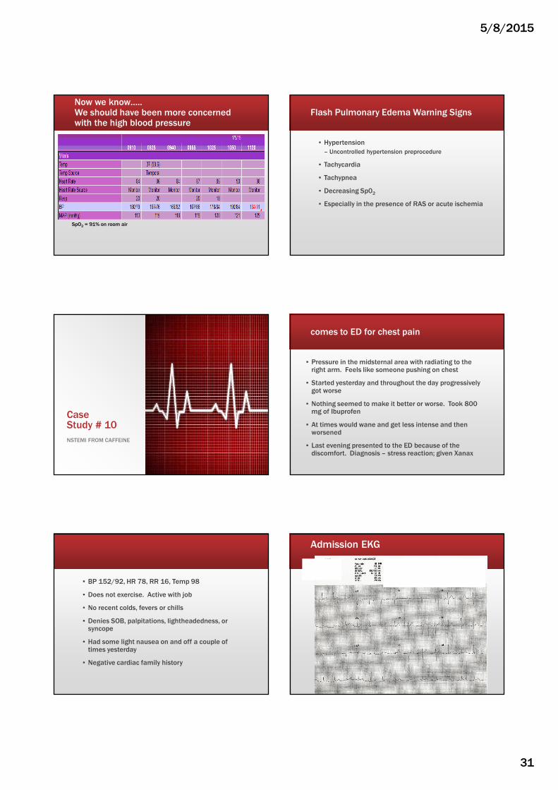
↓ Cardiac Output ↓
Activates SNS ↓
Compensates by releasing neurohormones Tachycardia (Norepinephrine, Aldostrone, Vasopression) ↓ ↓ ↑ Myocardial Oxygen Time Sodium & Water Retention ↓ Diastolic Filling Time
↓ ↓ Congestion
Vasoconstriction ↑ Afterload
SNS Activation Renal artery stenosis: RAAS & SNS
▪ With Unilateral RAS – contralateral kidney is functioning normally:
– Compensates for the elevated BP by
▪ Suppressing the renin secretion
▪ Augments the sodium excretions
▪ Pressure natriuresis occurs
▪ With bilateral RAS this escape mechanism is defective
Thus the Development of Flash Pulmonary Edema
Source: Messerli, F, Bangalore S, Makani H. Flash Pulmonary Oedema and Bilateral Renal Artery Stenosis: The Pickering Syndrome. Euroheartj. 2011
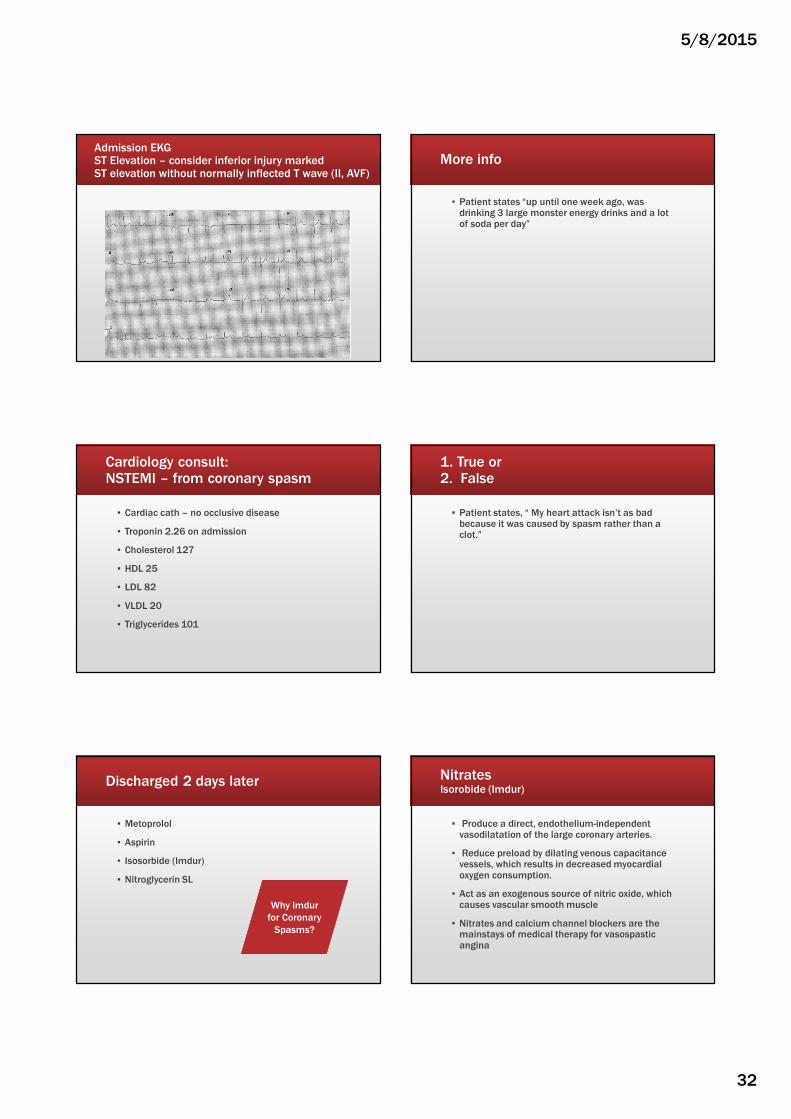
Pulmonary capillary stress failure & ↑pulmonary vascular permeability
▪ Rapid and exaggerated increase in LV pressure transmits to left atrium, pulmonary veins, and pulmonary capillaries
▪ When pulmonary intracapillary pressure exceeds 20 – 25 mmHg � fluid leaks through the endothelial barrier and floods the alveolar space
Pickering Syndrome Treatment
▪ Phase 1 – Stabilize patient
– Treat hypertensive emergency with antihypertensives and improve hemodynamic unloading
– Loop diuretics
▪ Phase 2 – treat the cause
– Renal revascularization
Pickering Syndrome
▪ “Must be considered a unique pebble in the mosaic of the so-called cardiorenal syndrome, a pathophysiological condition in which impairment of the cardiac and renal function mutually accelerates each other.”
Source: Messerli, F, Bangalore S, Makani H. Flash Pulmonary Oedema and Bilateral Renal
Artery Stenosis: The Pickering Syndrome. Euroheartj. 2011
5/8/2015
31
Now we know…..We should have been more concerned with the high blood pressure
SpO2 = 91% on room air
Flash Pulmonary Edema Warning Signs
▪ Hypertension
– Uncontrolled hypertension preprocedure
▪ Tachycardia
▪ Tachypnea
▪ Decreasing Sp02
▪ Especially in the presence of RAS or acute ischemia
Case Study # 10
NSTEMI FROM CAFFEINE
comes to ED for chest pain
▪ Pressure in the midsternal area with radiating to the right arm. Feels like someone pushing on chest
▪ Started yesterday and throughout the day progressively got worse
▪ Nothing seemed to make it better or worse. Took 800 mg of Ibuprofen
▪ At times would wane and get less intense and then worsened
▪ Last evening presented to the ED because of the discomfort. Diagnosis – stress reaction; given Xanax
▪ BP 152/92, HR 78, RR 16, Temp 98
▪ Does not exercise. Active with job
▪ No recent colds, fevers or chills
▪ Denies SOB, palpitations, lightheadedness, or syncope
▪ Had some light nausea on and off a couple of times yesterday
▪ Negative cardiac family history
Admission EKG
5/8/2015
32
Admission EKGST Elevation – consider inferior injury marked ST elevation without normally inflected T wave (II, AVF)
More info
▪ Patient states “up until one week ago, was drinking 3 large monster energy drinks and a lot of soda per day”
Cardiology consult:NSTEMI – from coronary spasm
▪ Cardiac cath – no occlusive disease
▪ Troponin 2.26 on admission
▪ Cholesterol 127
▪ HDL 25
▪ LDL 82
▪ VLDL 20
▪ Triglycerides 101
1. True or 2. False
▪ Patient states, “ My heart attack isn’t as bad because it was caused by spasm rather than a clot.”
Discharged 2 days later
▪ Metoprolol
▪ Aspirin
▪ Isosorbide (Imdur)
▪ Nitroglycerin SL
Why Imdur
for Coronary
Spasms?
NitratesIsorobide (Imdur)
▪ Produce a direct, endothelium-independent vasodilatation of the large coronary arteries.
▪ Reduce preload by dilating venous capacitance vessels, which results in decreased myocardial oxygen consumption.
▪ Act as an exogenous source of nitric oxide, which causes vascular smooth muscle
▪ Nitrates and calcium channel blockers are the mainstays of medical therapy for vasospastic angina
5/8/2015
33
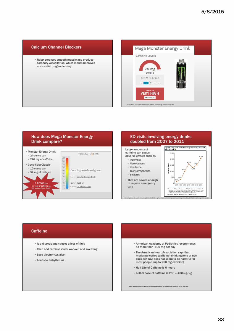
Calcium Channel Blockers
▪ Relax coronary smooth muscle and produce coronary vasodilation, which in turn improves myocardial oxygen delivery
Source: http://www.caffeineinformer.com/caffeine-content/mega-monster-energy-drink
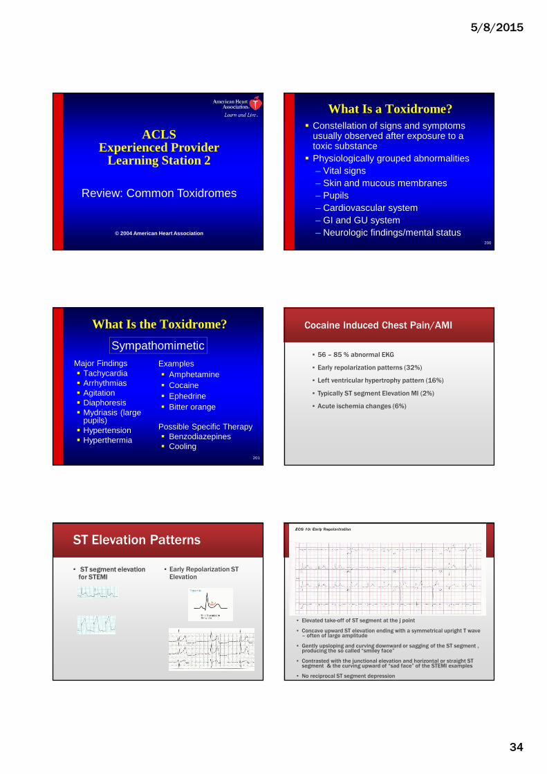
How does Mega Monster Energy Drink compare?
▪ Monster Energy Drink,
– 24-ounce can
– 240 mg of caffeine
▪ Coca-Cola Classic
– 12-ounce can
– 34 mg of caffeine
7 times the
amount of caffeine as
12 oz can Coca Cola!
ED visits involving energy drinks doubled from 2007 to 2011
Large amounts of caffeine can cause adverse effects such as:
• Insomnia
• Nervousness
• Headache
• Tachyarrhythmias
• Seizures
• That are severe enough to require emergency care
Source: Update on ED visits Involving Energy Drinks. Jan 2013. Drug Abuse Warning Networkhttp://archive.samhsa.gov/data/2k13/DAWN126/sr126-energy-drinks-use.htm
Caffeine
▪ Is a diuretic and causes a loss of fluid
▪ Then add cardiovascular workout and sweating
▪ Lose electrolytes also
▪ Leads to arrhythmias
▪ American Academy of Pediatrics recommends no more than 100 mg per day
▪ The American Heart Association says that moderate coffee (caffeine) drinking (one or two cups per day) does not seem to be harmful for most people. (up to 250 mg caffeine)
▪ Half Life of Caffeine is 6 hours
▪ Lethal dose of caffeine is 200 – 400mg/kg
Source: Sports drinks and energy drinks for children and adolescents: Are they appropriate? Pediatrics, 127(6), 1182-1189.
5/8/2015
34
ACLSExperienced Provider
Learning Station 2
Review: Common Toxidromes
© 2004 American Heart Association
200
What Is a Toxidrome?� Constellation of signs and symptoms
usually observed after exposure to a toxic substance
� Physiologically grouped abnormalities– Vital signs– Skin and mucous membranes– Pupils– Cardiovascular system– GI and GU system– Neurologic findings/mental status
201
What Is the Toxidrome?
Major Findings� Tachycardia� Arrhythmias� Agitation� Diaphoresis� Mydriasis (large
pupils)� Hypertension� Hyperthermia
Examples� Amphetamine� Cocaine� Ephedrine� Bitter orange
Possible Specific Therapy� Benzodiazepines� Cooling
Sympathomimetic
Cocaine Induced Chest Pain/AMI
▪ 56 – 85 % abnormal EKG
▪ Early repolarization patterns (32%)
▪ Left ventricular hypertrophy pattern (16%)
▪ Typically ST segment Elevation MI (2%)
▪ Acute ischemia changes (6%)
ST Elevation Patterns
▪ ST segment elevation for STEMI
▪ Early Repolarization ST Elevation
▪ Elevated take-off of ST segment at the j point
▪ Concave upward ST elevation ending with a symmetrical upright T wave – often of large amplitude
▪ Gently upsloping and curving downward or sagging of the ST segment , producing the so called “smiley face”
▪ Contrasted with the junctional elevation and horizontal or straight ST segment & the curving upward of “sad face” of the STEMI examples
▪ No reciprocal ST segment depression
5/8/2015
35
Cocaine induced chest painChest pain caused by coronary spasms
▪ RCA ▪ LAD & CX
Cocaine induced AMI Therapeutic Strategies Treat as ACS except…
▪ Avoid B-blockers acutely � due to the unopposed alpha-adrenergic effect, which may lead to
– worsening coronary vasoconstriction
– increased blood pressure
– risk of exacerbating coronary spasm. (Class III, C)
▪ IV NTG, Nitroprusside for persistent hypertension (phentolamine – alternative).
Cocaine induced AMI Therapeutic Strategies Treat as ACS except…
▪ IV Benzodiazepines to relieve chest pain & lead to beneficial effects on cardiac hemodynamics. Also relieves anxiety. (Class I, B)
▪ Calcium channel blockers should not be used as first-line therapy but may be considered in patients not responsive to benzodiazepines or NTG. (Class IIb/C)
▪ Phentolamine decreases coronary vascular resistance and blood pressure after cocaine ingestion, and may be considered in patients not respnsive to NTG or calcium channel blockers. (Class IIb,C)
Therapeutic StrategiesTreat as ACS except…
▪ In patients with chest pain of unclear origin, hypertension & tachycardia should be treated conservatively.
▪ Cautious use of fibrinolytic therapy for STEMI�higher rate of cranial hemorrhage with cocaine use.
210
Major Findings“SLUDGE”� Salivation� Lacrimation� Urination� Defecation� Gastrointestinal� Emesis
Examples� Organophosphates� Insecticides
Possible Specific Therapy� Atropine� Pralidoxime
Cholinergic
What Is the Toxidrome?
5/8/2015
36
211
Major Findings� Mental status ∆� Mydriasis (large
pupils)� Dry/flushed skin� Decreased bowel
sounds� Hyperthermia
“Mad as a bat, dry as a bone, hot as a poker”
Examples� Diphenhydramine� Scopolamine� Atropine� Jimson weed
Possible Specific Therapy� Physostigmine� Benzodiazepine� Cooling
Anticholinergic
What Is the Toxidrome?
212
Major Findings� CNS depression� Respiratory
depression� Miosis (small
pupils)
Examples� Heroin� Morphine� Fentanyl derivatives
(China White)
Possible Specific Therapy� Naloxone� Supportive care
Opioid
What Is the Toxidrome?
213
Major Findings� Weakness� Presyncope/
syncope� Bradycardia� Arrhythmias� Conduction
abnormalities� Hypotension
Examples� β-Adrenoreceptor blockers� Calcium channel blockers� Digitalis glycosides
Possible Specific Therapy� Vasopressors � Isuprel� Calcium� Glucagon� Digoxin-specific Fab
Cardiotoxic Agents
What Is the Toxidrome?
Case Study # 11
PRINZMETAL SPASM
42 y/o white male
▪ Came to ED due to c/o substernal burning pain that radiates up chest to both arms.
▪ Becomes SOB with chest pain
▪ Episodes last approx 10 minutes at a time.
▪ Episodes occur more when lying flat. This occurs several times during the night so he is not able to sleep
▪ Episodes have been occurring for last 4 months.
More History
▪ Had a negative stress test & normal GI workup.
▪ Denies any drug use of cocaine or other medications
▪ Quit Smoking 4 months ago. No other past medical history
▪ Father had some cardiac problems when he was in his 50s or 60s --- history unclear.

5/8/2015
37
▪ Pain free on arrival to ED
▪ Alert, Oriented
▪ Skin Warm/dry
Feb 24 at 1331
▪ When laid down for EKG developed chest pain
▪ BP 122/77, HR 87, RR 20 SpO2 99%
▪ Chest pain 7/10
▪ Weight: 70 kg
EKG in ED
▪ Chest pain resolved when sat up
▪ BP 118/56, HR 74, RR 20
▪ At 1339 on 2-24 (6 minutes later), the chest pain was gone. Pt was sitting up at the time.
▪ Troponin < 0.4 ng/ml
▪ CK = 71
▪ Total Cholesterol = 161
▪ Triglycerides = 66
▪ HDL = 35
▪ LDL = 113
5/8/2015
38
▪ Called cardiologist
▪ 1st EKG STEMI that resolved after a few minutes.
▪ Admit patient to CVICU. Started on ASA, plavix, heparin drip, nitroglycerin drip, and lopressor
▪ Hold cardiac cath for now as pain free with normal EKG
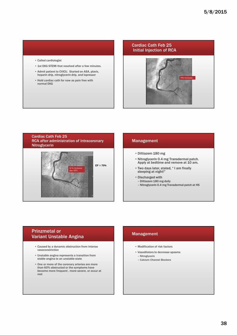
Cardiac Cath Feb 25Initial Injection of RCA
70% Occlusion
Cardiac Cath Feb 25RCA after administration of Intracoronary Nitroglycerin
10 % Occlusion after NTG
EF = 70%
Management
▪ Diltiazem 180 mg
▪ Nitroglycerin 0.4 mg Transdermal patch. Apply at bedtime and remove at 10 am.
▪ Two days later, stated, “ I am finally sleeping at night!”
▪ Discharged with– Diltiazem 180 mg daily
– Nitroglycerin 0.4 mg Transdermal patch at HS
Prinzmetal or Variant Unstable Angina
▪ Caused by a dynamic obstruction from intense vasoconstriction
▪ Unstable angina represents a transition from stable angina to an unstable state
▪ One or more of the coronary arteries are more than 60% obstructed or the symptoms have become more frequent , more severe, or occur at rest
Management
▪ Modification of risk factors
▪ Vasodilators to decrease spasms
– Nitroglycerin
– Calcium Channel Blockers
5/8/2015
39
Case Study # 12VENTRICULAR STORM
POSSIBLE BRUGADA SYNDROME
transferred to CVICU from outlying hospital
▪ Post cardiac arrest at home for Ventricular Fibrillation
▪ Hypothermia not initiated as patient was responding.
▪ Intubated, opens eyes to commands, MAE x 4
▪ BP 119/86, HR 80, T 98 SpO2 100%
▪ Normal sinus rhythm
▪ Troponin 0.9
▪ By discharge – no neuro deficits
Events
▪ Was at home and felt like something was going to happen to her
▪ Laid down on the floor
▪ Started having multiple shocks – 31 total
▪ Most shocks were successful to convert her
▪ EMS started CPR and shocked 2 times and 2 doses of Epinephrine. ROSC = 15 minutes
▪ Amiodarone drip started
PMH
▪ History of ventricular storm with etiology undetermined
– ? Brugada syndrome
– ? Vasospasm related
– ? Idiopathic
▪ 20 months ago, felt significant palpitations and SOB. Became unresponsive, CPR started. Vfib �
shock � Wide complex tachycardia.
▪ Angio showed normal coronary arteries and LV EF 70%
▪ Single coil ICD placed
PMH cont
▪ 2 months ago another ventricular storm that converted after 4 shocks
▪ Another angio done – no abnormalities– Provocative challenging using hyperventilation with no evidence of
spasm in RCA
▪ EP studies done at local hospital and sent for referral EP studies at another hospital.
▪ ? Brugada syndrome – want to do flecainide challenge
▪ Refused flecainide challenge because of possible risk involved
▪ Managed with long acting nitrates (Imdur), beta-blocker therapy (Toprol XL), and calcium channel blocker therapy (Norvasc)
▪ Also has history of hypothyroidism which may contribute to the arrhythmias
EKG two months prior to latest Ventricular storm episode
Sinus Rhythm
Left Atrial abnormality (insign)
Incomplete RBBB
Borderline prolonged QT(insign)
Nonspecific T wave abnormalities, inferior leads (insign)

5/8/2015
40
EKG on admission
Atrial-Paced Rhythm
RBBB and LPFB
Cardiac Cath – no abnormalities
EP Plan
▪ Etiology of Vfib is still likely idiopathic with differential diagnosis of Brugada syndrome and possibility of vasospasm is still entertained
▪ New RBBB is probably incidental and may be related to the cardiac resuscitation
▪ Load with Amiodarone • 0.5 mg/hour x 3 days• then 400 mg po TID for 3 weeks• then 400 mg BID for another month• Then 400 mg daily
• Aggressively treaty hypothyroidism
• Keep Magnesium above 2 and potassium above 3.8
• Metoprolol 4 times per day
Brugada Syndrome
▪ A disorder characterized by sudden cardiac death or a sustained ventricular tachyarrhythmia
▪ Associated with one of several Brugada ECG patterns – characterized by incomplete right bundle-branch
block
– ST-segment elevations in the anterior precordial leads
▪ Brugada Pattern: – Patients with typical ECG pattern and have not
other clinical criteria
▪ Patients with PVCs
▪ Patients with nonsustained ventricular tachycardia
Source: Dizon J, Rottman J, et all Brugada Syndrome: http://emedicine.medscape.com/article/163751-overview
and Wylie J, Garlitski A. Brugada Syndrome: Up to Date March 2015
▪ ST elevation and T wave inversion V1 and V2
▪ ORS normal
Wylie J, Garlitski A. Brugada Syndrome: Up to Date March 2015
▪ V1 and V2
– Coved pattern
or
– Saddle-back pattern
Source: Wylie J, Garlitski A. Brugada Syndrome: Up to Date March 2015
Brugada Syndrome
▪ Occurs in 0.1 – 1% of the population
▪ More common in men than women
▪ Diagnosed in adulthood – average age 41
▪ Schizophrenia patients more likely to have Brugada pattern than general population
▪ Recommend 12 lead ECG for first degree relatives
▪ SCD may be the initial presentation in at least 1/3 of the patients
5/8/2015
41
Brugada Syndrome Treatment
▪ Drug challenge with sodium channel blockers can unmask the ECG pattern
– Flecainide, procainamide, ajmaline, pilsicainide
▪ Treatment
– Termination of ventricular arrhythmias
– ICD implantation
– No proven pharmacologic treatments for preventing SCD in Brugada syndrome
– Quinidine or amiodarone may be helpful – for those who are not canidates for ICD.
Back to case study
▪ 5 days post cardiac arrest
▪ Extubated, ready to transfer to progressive unit
▪ She is walking in room and tells you that she is “having chest pain associated with the feeling that she usually feels prior to her ventricular fib arrest.”
What are
your
actions?
Does she have Brugada pattern?1. Yes 2. No
Diagnosis: Prinzmetal Angina causing recurrence of ventricular arrhythmias
▪ Diagnosis confirmed with presentation of chest pain, transient ST elevation that responded to NTG
Treatment ??Consulted with several specialists in other institutions
▪ Cardiac revascularization due to multi-vessel spasms
– Literature ~ limited help for Prinzmetal
▪ Stenting and Ergometrine challenge
– Stenting usually can fix one vessel and the other vessels may still spasm
– Recommended no challenge
▪ Conclusion
– Severe endothelial dysfunction disease � needs aggressive very high dose of calcium channel blockage
– Diltiazem rather than amiodipine
▪ Titrate up to 600 – 800 mg per day
▪ Start 60 mg every 4 hours
– Keep magnesium levels above 2 and potassium above 4
Discharged 4 days later 9 days after admission
▪ Amiodarone 400 mg TID
▪ ASA 81 mg daily
▪ Atovastain 40 mg daily
▪ Diltiazem 240 mg (24 hour tab) – 2 capsules daily
▪ Isorbide 30 mg (24 hr tab) every evening
▪ Magnesium 400 mg – 2 tablets BID
▪ Methimazole 10 mg BID
▪ NTG 0.4 mg SL prn chest pain
5/8/2015
42
Now you know….
▪ Chest pain/AMI from coronary artery spasm
– Caffeine/Cocaine induced
– Printzmetal Angina
▪ Brugada Syndrome
Time for a STRESS TEST
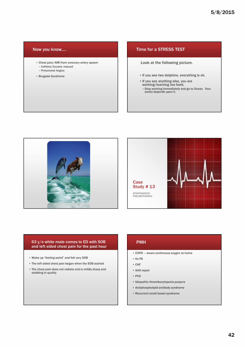
Look at the following picture.
▪ If you see two dolphins, everything is ok.
▪ If you see anything else, you are working/learning too hard.– Stop working immediately and go to Ocean. Your
sanity depends upon it.
Case Study # 13
SPONTANEOUS PNEUMOTHORAX
63 y/o white male comes to ED with SOB and left sided chest pain for the past hour
▪ Woke up “feeling weird” and felt very SOB
▪ The left sided chest pain began when the SOB started
▪ The chest pain does not radiate and is mildly sharp and stabbing in quality
PMH
▪ COPD – wears continuous oxygen at home
▪ Hx PE
▪ CHF
▪ AAA repair
▪ PVD
▪ Idiopathic thrombocytopenia purpura
▪ Antiphospholipid antibody syndrome
▪ Recurrent small bowel syndrome
5/8/2015
43
What is Antiphospholipid syndrome ?
▪ An autoimmune disease
▪ “Antiphospholipid antibodies" react against proteins that bind to anionic phospholipids on plasma membranes.
▪ The exact cause is not known, but activation of the system of coagulation is evident.
▪ Clinically important: antiphospholipid antibodies are associated with thrombosis and vascular disease.
Vital Signs in ED
▪ BP 136/77
▪ HR 134, regular
▪ RR 32
▪ Temp 97 oral
▪ SpO2 91% on 15 liters nonrebreather
▪ Pain 7/10
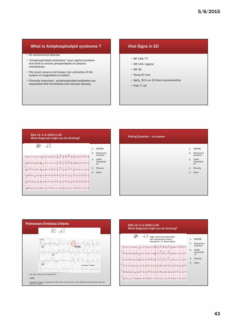
EKG 12- 2 at 2200 in EDWhat diagnosis might you be thinking?
1. NSTEMI
2. Pulmonary Embolus
3. COPD exacerbation
4. Pleurisy
5. Other
Polling Question – no answer
1. NSTEMI
2. Pulmonary Embolus
3. COPD exacerbation
4. Pleurisy
5. Other
Pulmonary Embolus Criteria
▪ S1, Q3 or S1,Q3, T3 (inverted T)
▪ RBBB
▪ Inverted T waves secondary to RV strain may be seen in the right precordial leads and can last for months
S1
Q3
T3
RBBB
Inverted T waves
EKG 12- 2 at 2200 in EDWhat diagnosis might you be thinking?
1. NSTEMI
2. Pulmonary Embolus
3. COPD exacerbation
4. Pleurisy
5. Other
Right ventricular hypertrophy with repolarization patternNonspecific ST abnormalities
5/8/2015
44
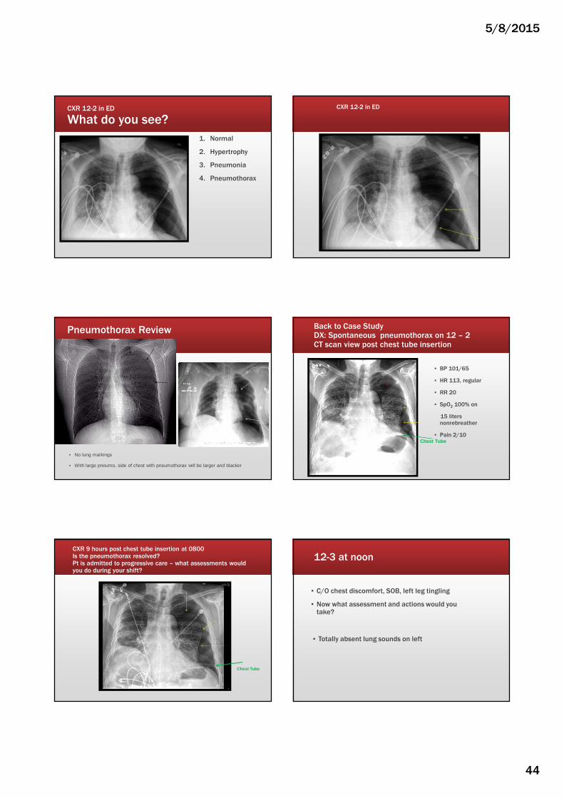
CXR 12-2 in ED
What do you see?
1. Normal
2. Hypertrophy
3. Pneumonia
4. Pneumothorax
CXR 12-2 in ED
Pneumothorax Review
▪ No lung markings
▪ With large pneumo, side of chest with pneumothorax will be larger and blacker
Back to Case StudyDX: Spontaneous pneumothorax on 12 – 2CT scan view post chest tube insertion
▪ BP 101/65
▪ HR 113, regular
▪ RR 20
▪ SpO2 100% on
15 liters nonrebreather
▪ Pain 2/10Chest Tube
CXR 9 hours post chest tube insertion at 0800Is the pneumothorax resolved?Pt is admitted to progressive care – what assessments would you do during your shift?
Chest Tube
12-3 at noon
▪ C/O chest discomfort, SOB, left leg tingling
▪ Now what assessment and actions would you take?
▪ Totally absent lung sounds on left
5/8/2015
45
CXR on 12 – 3 at 1215 after 2nd chest tube inserted
2nd Chest Tube
▪ Patient did not go to surgery for decoritication due to pulmonary hypertension – poor surgical candidate
▪ Sent home with Heimlich valve
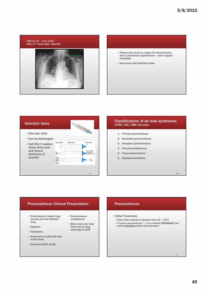
Heimlich Valve
▪ One way valve
▪ Can be discharged
▪ Call 911 if sudden sharp chest pain and severe shortness of breathe
267
Classifications of air leak syndromesCCRN, CSC, CMC test plan
1. Primary pneumothorax
2. Secondary pneumothorax
3. Iatrogenic pneumothorax
4. Pneumomediastinum
5. Pneumopericardium
6. Hydropneumothorax
268
Pneumothorax Clinical Presentation
▪ Diminished or absent lung sounds over the affected lung
▪ Dyspnea
▪ Tachypnea
▪ Acute pain on affected side of the chest
▪ Decreased Sp02 & p02
▪ Subcutaneous emphysema
▪ Black area over lung field with no lung markings on CXR
270
Pneumothorax
▪ Initial Treatment:
– Chest tube insertion if greater than 10 – 15 %
– If tension pneumothorax ---- it is a medical EMERGENCY and needs immediate needle decompression

5/8/2015
46
Primary Spontaneous Pneumothorax (PSP)
▪ Occurs without a precipitating event in a person who does not have lung disease
▪ Most individuals with PSP have unrecognized lung disease
271
Secondary Spontaneous Pneumothorax (SSP)
▪ A pneumothorax that occurs as a complication of an underlying lung disease
▪ Can be a complication of any lung disease. Most often occurs with:
– COPD
– Pneumocystis jirovecii infection
– Cystic fibrosis
– Tuberculosis
272
SSP Clinical Presentation
▪ C/O of dyspnea and chest pain on the same side as the pneumothorax
▪ Symptoms more severe than with PSP as SSP patients have less pulmonary reserve due to the underlying lung disease.
▪ Persistent air leaks are more common and tend to persist longer than PSP
273
SSP Treatment
▪ Should be hospitalized: diminished pulmonary reserve increases their risk for adverse outcomes.
▪ Initial Treatment
– Chest tube insertion
– Chest tube should remain in place until a procedure if performed to prevent recurrent SSP
274
SSP: Prevention of recurrence
▪ Video-Assisted Thoracoscopy (VAT) with stapling of blebs and pleural abrasion.
▪ Chemical pleurodesis
▪ Pleural Blood Patch
▪ Heimlich valve
275
Nursing Care of Chest Tubes
▪ Bubbling in the water seal chamber indicates air leak
▪ If suction is ordered for PSP or SSP, keep suction going even when ambulating!
276
5/8/2015
47
PSP and SSP – high risk activities
▪ Patients with resolving pneumothorax should be cautioned not to fly until intrapleural air has completely resolved.
▪ Deep sea diving should be avoided unless thoracotomy or pleurodesis has been performed
277
Case Study # 14
PNEUMOTHORAX AFTER PACEMAKER INSERTION
68 year old male
▪ PMH
– COPD
– Cardiomyopathy for past 7 years with EF 40%
– Recent EF 30% and now has Left Bundle Branch Block
▪ Plan: Insertion of Biventricular Pacemaker
I’m not a Cardiac Nurse!
▪ Biventricular Pacemaker is used in Stage 4 Heart Failure with Left Bundle Branch Block
– Three Leads: Third lead paces the left ventricle to provide ventricular synchrony
▪ During procedure a central line is inserted via the right internal jugular vein and the pacemaker leads via the left subclavian
▪ Key point --- two insertion site!
Routine Procedures?!?!?
▪ What are potential complications from central line and/or pacemaker Insertion?
▪ What Diagnostics should occur post procedure?
Potential Post Procedure Complications
Immediate
▪ Bleeding
▪ Arterial puncture
▪ Arrhythmia
▪ Air Embolism
▪ Pneumothorax
▪ Hemothorax
Delayed
▪ Infection
▪ Venous thrombosis/Pulmonary emboli
▪ Catheter migration
▪ Catheter embolization
▪ Myocardial perforation
▪ Nerve Injury
Central Line & Pacemaker Insertion
5/8/2015
48
Case Progression
▪ Post procedure vital signs started
▪ Initial Assessment
– B/P 110/70, HR 80, RR 16, Sp02 99%
– Clear lung sounds
– Right jugular and left subclavian dressings dry and intact
– No SQ emphysema noted
– Monitor shows paced rhythm
▪ Chest Xray orderedWhat are your actions?
▪When xray tech enters room:
–Acute onset SOB and wheezing
–Significant respiratory distress
–Calls RN
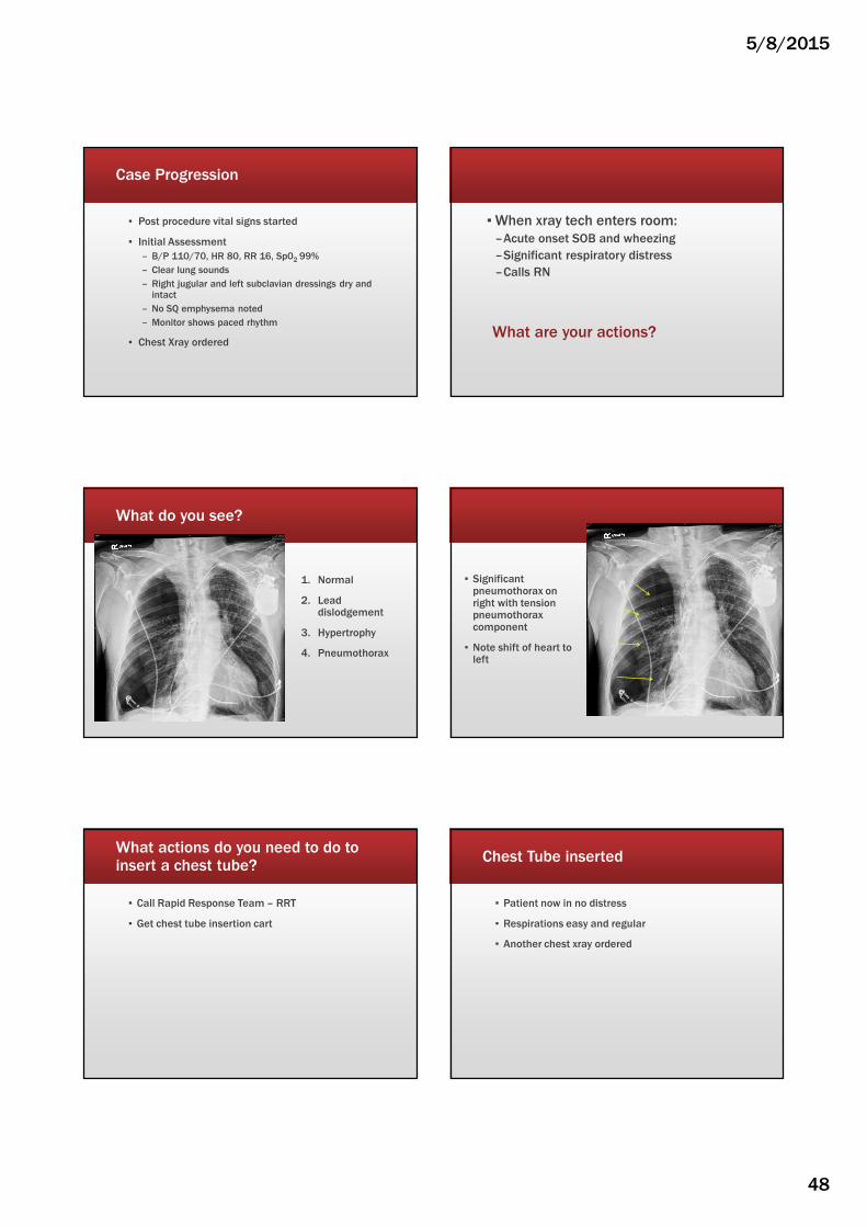
What do you see?
1. Normal
2. Lead dislodgement
3. Hypertrophy
4. Pneumothorax
▪ Significant pneumothorax on right with tension pneumothorax component
▪ Note shift of heart to left
What actions do you need to do to insert a chest tube?
▪ Call Rapid Response Team – RRT
▪ Get chest tube insertion cart
Chest Tube inserted
▪ Patient now in no distress
▪ Respirations easy and regular
▪ Another chest xray ordered
5/8/2015
49
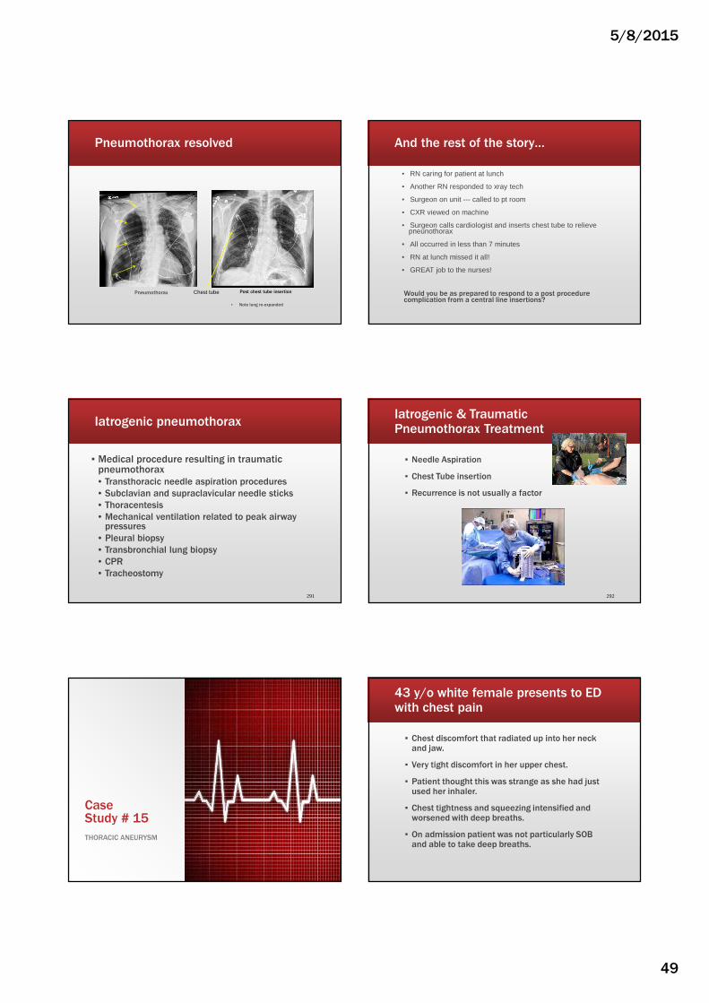
Pneumothorax resolved
Pneumothorax
▪ Note lung re-expanded
Post chest tube insertionChest tube
And the rest of the story…
▪ RN caring for patient at lunch
▪ Another RN responded to xray tech
▪ Surgeon on unit --- called to pt room
▪ CXR viewed on machine
▪ Surgeon calls cardiologist and inserts chest tube to relieve pneunothorax
▪ All occurred in less than 7 minutes
▪ RN at lunch missed it all!
▪ GREAT job to the nurses!
Would you be as prepared to respond to a post procedure complication from a central line insertions?
Iatrogenic pneumothorax
▪ Medical procedure resulting in traumatic pneumothorax• Transthoracic needle aspiration procedures
• Subclavian and supraclavicular needle sticks
• Thoracentesis
• Mechanical ventilation related to peak airway pressures
• Pleural biopsy
• Transbronchial lung biopsy
• CPR
• Tracheostomy
291
Iatrogenic & Traumatic Pneumothorax Treatment
▪ Needle Aspiration
▪ Chest Tube insertion
▪ Recurrence is not usually a factor
292
Case Study # 15
THORACIC ANEURYSM
▪ Chest discomfort that radiated up into her neck and jaw.
▪ Very tight discomfort in her upper chest.
▪ Patient thought this was strange as she had just used her inhaler.
▪ Chest tightness and squeezing intensified and worsened with deep breaths.
▪ On admission patient was not particularly SOB and able to take deep breaths.
43 y/o white female presents to ED with chest pain
5/8/2015
50
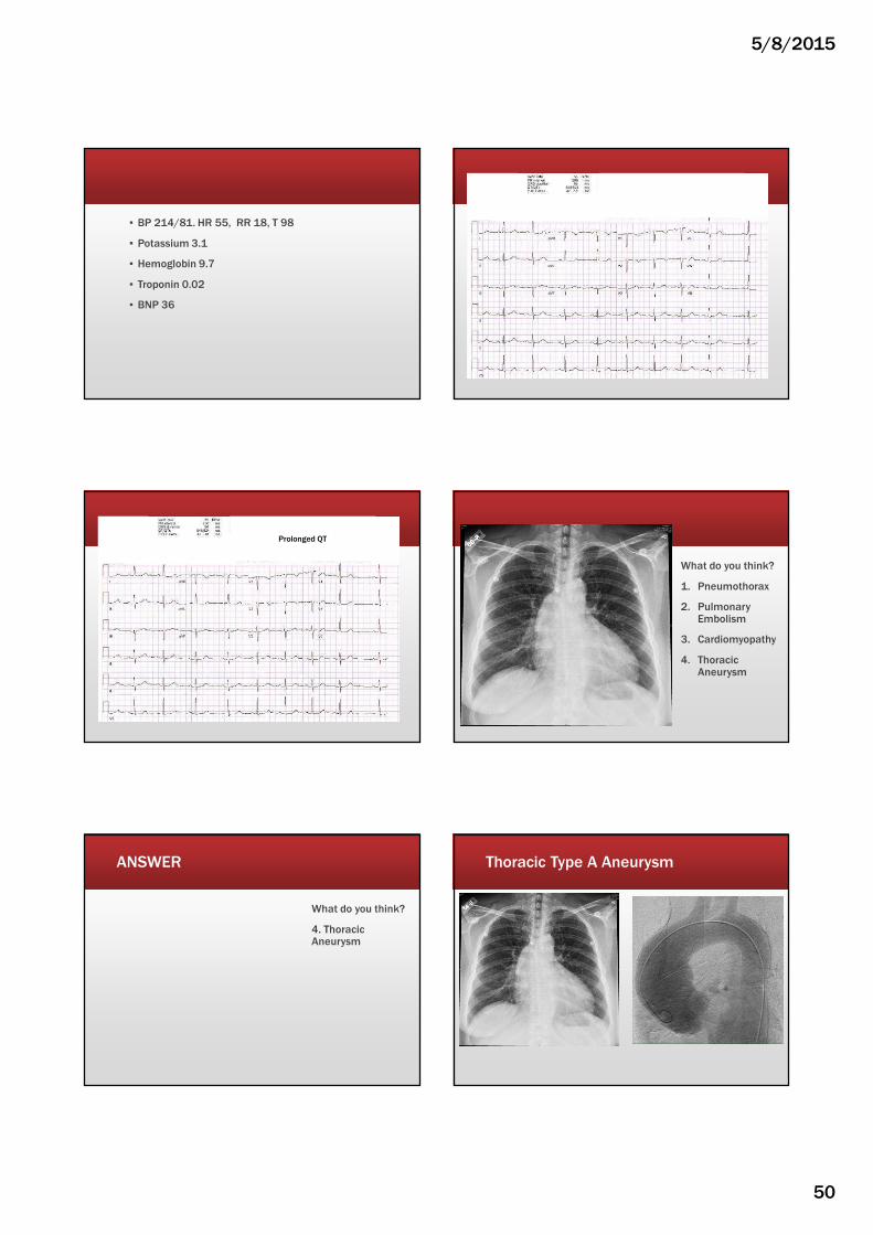
▪ BP 214/81. HR 55, RR 18, T 98
▪ Potassium 3.1
▪ Hemoglobin 9.7
▪ Troponin 0.02
▪ BNP 36
Prolonged QT
What do you think?
1. Pneumothorax
2. Pulmonary Embolism
3. Cardiomyopathy
4. Thoracic Aneurysm
ANSWER
What do you think?
4. Thoracic Aneurysm
Thoracic Type A Aneurysm
5/8/2015
51
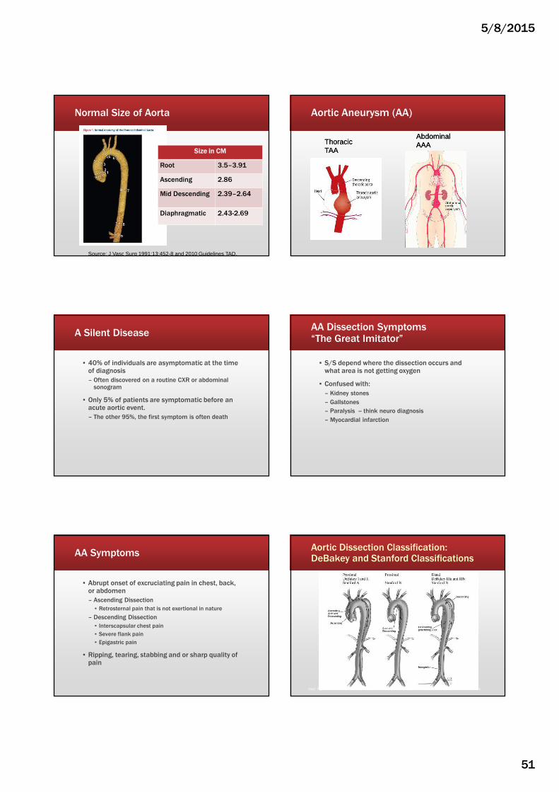
Normal Size of Aorta
Size in CM
Root 3.5–3.91
Ascending 2.86
Mid Descending 2.39–2.64
Diaphragmatic 2.43-2.69
Source: J Vasc Surg 1991:13:452-8 and 2010 Guidelines TAD.
Aortic Aneurysm (AA)
ThoracicTAAThoracicTAA
AbdominalAAAAbdominalAAA
A Silent Disease
▪ 40% of individuals are asymptomatic at the time of diagnosis
– Often discovered on a routine CXR or abdominal sonogram
▪ Only 5% of patients are symptomatic before an acute aortic event.
– The other 95%, the first symptom is often death
AA Dissection Symptoms“The Great Imitator”
▪ S/S depend where the dissection occurs and what area is not getting oxygen
▪ Confused with:
– Kidney stones
– Gallstones
– Paralysis -- think neuro diagnosis
– Myocardial infarction
AA Symptoms
▪ Abrupt onset of excruciating pain in chest, back, or abdomen
– Ascending Dissection
▪ Retrosternal pain that is not exertional in nature
– Descending Dissection
▪ Interscapsular chest pain
▪ Severe flank pain
▪ Epigastric pain
▪ Ripping, tearing, stabbing and or sharp quality of pain
Aortic Dissection Classification: DeBakey and Stanford Classifications
Note: Figure 20 in full-text version of TAD Guidelines. Reprinted with permission from The Cleveland Clinic Foundation.

5/8/2015
52
Dissections
▪ 62% are Type A
▪ Type B are typically older than Type A
▪ Type A
– Immediate operation room intervention
▪ Type B
– Medical management
2010 ACCF/AHA/AATS/ACR/ASA/
SCA/SCAI/SIR/STS/SVM Guidelines for
the Diagnosis and Management of Patients
with Thoracic Aortic Disease
Developed in partnership with the American College of Cardiology
Foundation/American Heart Association Task Force on Practice
Guidelines, American Association for Thoracic Surgery, American
College of Radiology, American Stroke Association, Society of
Cardiovascular Anesthesiologists, Society for Cardiovascular
Angiography and Interventions, Society of Interventional
Radiology, Society of Thoracic Surgeons, and Society for Vascular
Medicine.
Endorsed by the North American Society for Cardiovascular
Imaging.
Source: 2010 ACCF/AHA/AATS/ACR/ASA/SCA/SCAI/SIR/STA/SVM Guidelines for TAASource: 2010 ACCF/AHA/AATS/ACR/ASA/SCA/SCAI/SIR/STA/SVM Guidelines for TAA
Rate/Pressure Control
Intravenous beta blockadeor Labetalol
(If contraindication to beta blockadesubstitute diltiazem or verapamil)
Titrate to heart rate <60
1
Pain Control
Intravenous opiates
Titrate to pain control
Intravenous rate and pressure control
2
+
Hypotensionor shock state?
No
Yes
Systolic BP >120mm HG?
BP ControlIntravenous vasodilator
Titrate to BP <120mm HG (Goal is lowest possible BP that maintains adequate end organ perfusion)
Secondary pressure control
3
Anatomic based management
Acute AoD Management PathwaySTEP 2: Initial management of aortic wall stress
Acute AoD Management PathwaySTEP 2: Initial management of aortic wall stress
Anatomic based management
Urgent surgical consultation+
Arrange for expeditedoperative management
Intravenous fluid bolus•Titrate to MAP of 70mm HG
or Euvolemia(If still hypotensive begin
intravenous vasopressor agents)
Review imaging study for:• Pericardial tamponade• Contained rupture• Severe aortic insufficiency
1
2
3
Type A dissection
Intravenous fluid bolus•Titrate to MAP of 70mm HG
or Euvolemia(If still hypotensive begin
intravenous vasopressor agents)
Evaluate etiology of hypotension
• Review imaging study forevidence of contained rupture
• Consider TTE to evaluatecardiac function
Urgent surgical consultation
2
3
Type B dissection
1

5/8/2015
53
Case Study # 16
AZYGOS LOBE
You are admitting 66 y/o male after CABG x 1, AVRPMH: Diabetes, CAD, Hyperlipidemia
Admission
2122
pH 7.33
pCO2 50
pO2 79
TCO2 28
02 sat 95
BE 0.5
Hemoglobin 11.2
Hematocrit 36
Glucose 125
Potassium 4.8Based on ABGs and CXR,
what do you want to do?
0245 – about 5 hours post op
▪ SpO2 drops to 90 – 91%
▪ BP 95/62
▪ HR 106
▪ One hour prior:
– BP 118/81, HR 87, RR 12,
– Sp02 100 on 80% vent
Clear bilateral lung sounds except diminished right upper lobe
Admission
21220200 0248
Now
pH 7.33 7.47 7.49
pCO2 50 36 36
pO2 79 81 56
TCO2 28 27 29
02 sat 95 97 91
BE 0.5 2.5 4.1
Hemoglobin 11.2 11.5 10.9
Hematocrit 36 37 35
Glucose 125 147 133
Potassium 4.8 4.6 4.4
CXR 0315
CXR at 0530 after ET tube pulled back 2 cm suctioned with mucomyst for tan secretions.

5/8/2015
54
CXR at 0530 after ET tube pulled back 2 cm suctioned with mucomyst for tan secretions. Azygos Lobe
▪ Right upper lobe bronchus comes off trachea versus right main bronchus
▪ A rare congenital variation of the upper lobe of the right lung
▪ An anatomically separated part of the upper right lobe
▪ Not associated with any morbidity but can cause technical problems in thoracoscopic procedures
Tip of ET tube
Admission CXR
Post suction CXR
POD # 5 CXR
Case Study # 17LAST BUT NOT LEAST!
RIB FRACTURES
presents to ED post ground level fall
▪ Was up in the middle of the night, lost her balance while walking to the bathroom
▪ Fell backwards into a wooden table
▪ Struck the right lower side of her back and experienced severe pain
▪ Unable to stand and had to call for help
▪ Pain was excruciating
▪ Denies any preceding symptoms of dizziness, lightheadedness, chest pain, or vomiting

5/8/2015
55
Admission Vitals/Assessment
▪ BP 175/85, HR 84, irregular, RR 16, T 98
▪ SpO2 95 % on room air
▪ H & H 11/37
▪ Clear lung sounds, diminished in bases, No Wheezes
▪ MAE x 4 – with difficulty in right upper extremity
▪ Alert/ Oriented. Neuro assessment intact
▪ C/o SOB and severe pleuritic pain 7/10
▪ Can’t move around in bed without aggravating the pain in her right side of chest
▪ Given Fentanyl
PMH
▪ Atrial Fibrillation – currently on Xarelto
▪ Asthma
▪ Vertigo
▪ Hypertension
▪ Anxiety
▪ Spine surgery
▪ Hip fracture surgery
▪ Colectomy
Admission CXR
What do you see?
1. Pleural effusion
2. Cardiomyopathy
3. Normal
4. Other
ANSWER
What do you see?
4. Other
Admission CXR
▪ Acute rib fractures involving the right lateral and posterior 5th, 6th, 7th, 8th, 9th ribs with mild distraction at several of the rib fractures
▪ Small right pleural effusion and atelectasis
Diagnosis & Treatment
▪ Multiple right sided rib fractures
– Low dose fentanyl patch and lidocaine patch to right side
▪ Acute hypoxemia failure – secondary to rib fractures
▪ Incentive spirometer, oxygen 2 liters
▪ Atrial Fib – continue Xarelto
▪ DVT prophylaxis
– Lovenox
5/8/2015
56
Event progression
▪ 2000 (12 hours after admission)
▪ SpO2 92% on 2 liters
▪ 2100 – Acute SOB and increased pain on right side– Oxygen ↑ to 4 liters, SpO2 90%
▪ 2400
▪ Became very SOB and pain with position change SpO2 90%
▪ 0800
▪ SpO2 93% on 4 liters
– BP 108/55, HR 89, RR 18, T 98– H & H = 8.7/28.2 from 11/37– Diminished lung sounds on right – CXR ordered
24 hours later
▪ 1200
– Overnight has become more SOB
– Oxygen ↑ from 4 liters to 6 liters SpO2 91%
– Feels she “cannot take a deep breathe”
– Diminished lung sounds on right
– Cannot lie flat
▪ Complete opacification of the right chest secondary to a right pleural effusion
1500 ml drained immediately, then another 300 ml
CXR after CT inserted CXR 1 hour after CT inserted
Chest Tube insertion
▪ Bed side procedure
– Time Out
Post Procedure Complications Chest tube insertion/thoracentesis
▪ Pneumothorax
▪ Bleeding – hemothorax, hematoma, hemoperitoneum
▪ SQ emphysema
▪ Laceration of liver, spleen, or lung
▪ Hypovolemia
▪ Hypotension
▪ Dyspnea
▪ Re-expansion pulmonary edema
5/8/2015
57
In Summary…..
Challenging Complex
CardiacPulmonary
Case Studies
ALL chest pain is cardiac until proven otherwise
▪ Ask Questions to get a good history!
▪ No fibrinolytics for Broken Heart Syndrome or spasms
▪ Call for decreasing oxygen saturations and increasing oxygen needs
▪ Look for the obvious!
References
▪ Garcia, T.B., & Holtz, N.E. (2001). 12 lead ECG: The art of interpretation. Boston: Jones and Bartlett Publishers.
▪ Griffin, S. & Logue, B. Takotsubo Cardiomyopathy: A Nurse’s Guide. Critical Care Nurse.2009: 29(5):32-42.
▪ Lacey, G., Morley, S. , et Berman, L. (2008) The Chest X-ray: A Survival Guide. Philadelphia: Saunders/Elsevier.
▪ McCulloch, B. Transient Left Ventricular Apical Ballooning. Critical Care Nurse 2007:27(6):20-26.
▪ Moser, D. Reigal G (2008). Cardiac Nursing: A companion to Braunwald’s Heart Disease. St Louis MO: Sanders Elsevier.
▪ Sidebotham D, MeKee A, Gillham M et al. Cardiothoracic Critical Care 2007, Butterworth Heinemann Elsevier, Philadelaphia, PA
▪ Siela, D. Chest radiograph evaluation and interpretation. Advanced Critical Care. 2008: 19(4):444-475.
PPCM References
▪ Tsang, W, Bales A, et Lang R. Peripartum Cardiomyopathy: Treatment and Prognosis. Retrieved Feb 9, 2015 from Up To Date.
▪ Moser & Riegel. 2008. Cardiac Nursing
▪ Garg, J et al. 2015. Peripartum Cardiomyopathy. Cardiology in Review; 23(2).
▪ Sundin. C 2014. Peripartum Cardiomyopathy. MCN 39(4)