Embed Size (px)
Citation preview
Timothy M. Bateman M.D. Co-Director, Cardiovascular Radiologic Imaging
Mid America Heart Institute Professor of Medicine
University of Missouri-Kansas City Kansas City, Missouri
Challenges and Opportunities for SPECT & PET in 2013: Implementing Latest
Acquisition and Processing Protocols
Conflict of Interest Disclosure
Dr. Bateman declares that the following relationships constitute a potential conflict of interest with respect
to this presentation:
Research Grants: Astellas, GE, Lantheus, Philips, SpectrumDynamics Advisory Boards: Astellas, GE, Lantheus, SpectrumDynamics Royalties: ExSPECT IITM, ImagenPro/MD/QTM
Stock Ownership: Cardiovascular Imaging Technologies
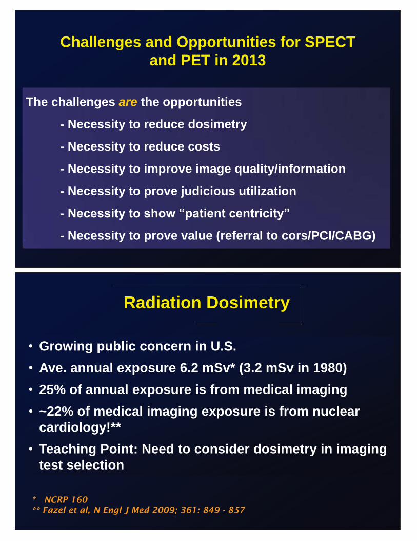
Challenges and Opportunities for SPECT and PET in 2013
The challenges are the opportunities
- Necessity to reduce dosimetry
- Necessity to reduce costs
- Necessity to improve image quality/information
- Necessity to prove judicious utilization
- Necessity to show “patient centricity”
- Necessity to prove value (referral to cors/PCI/CABG)
• Growing public concern in U.S. • Ave. annual exposure 6.2 mSv* (3.2 mSv in 1980) • 25% of annual exposure is from medical imaging • ~22% of medical imaging exposure is from nuclear
cardiology!** • Teaching Point: Need to consider dosimetry in imaging
test selection
Radiation Dosimetry
* NCRP 160 ** Fazel et al, N Engl J Med 2009; 361: 849 - 857
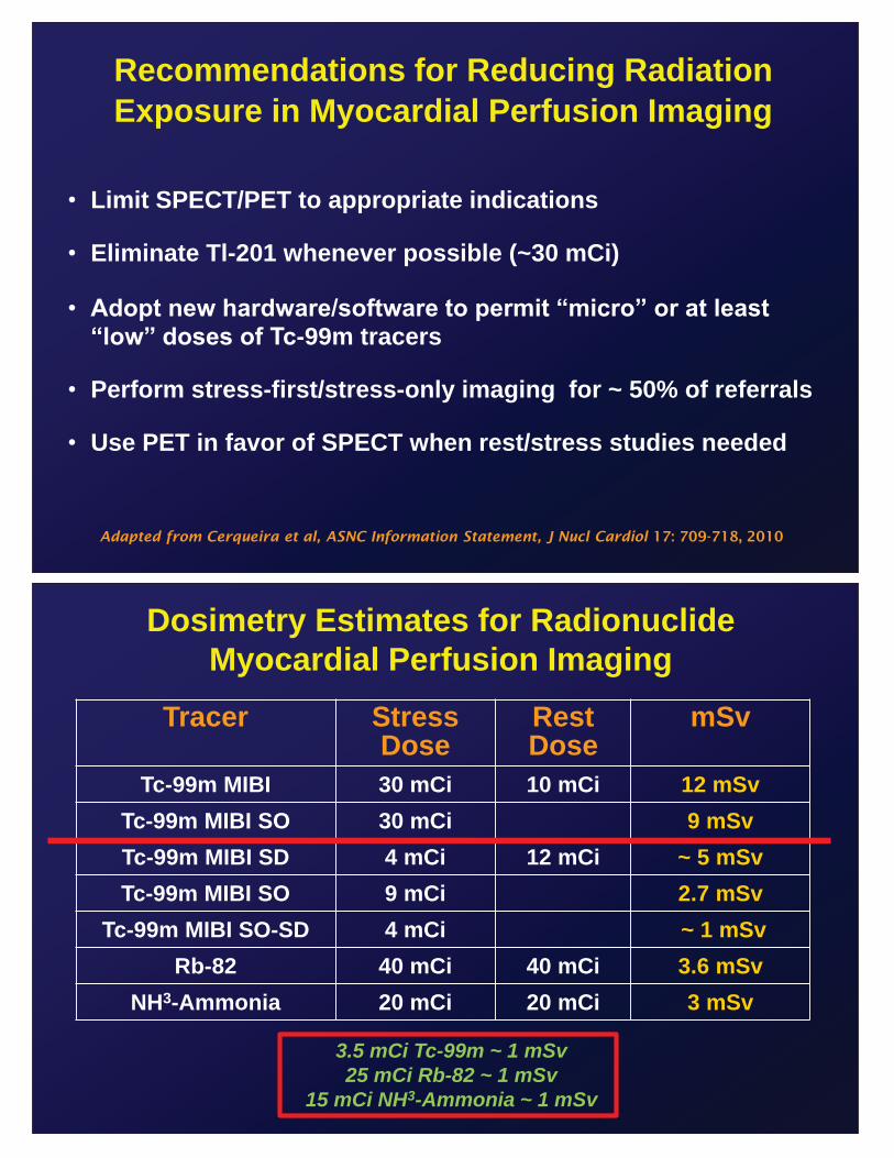
Recommendations for Reducing Radiation Exposure in Myocardial Perfusion Imaging
Adapted from Cerqueira et al, ASNC Information Statement, J Nucl Cardiol 17: 709-718, 2010
• Limit SPECT/PET to appropriate indications
• Eliminate Tl-201 whenever possible (~30 mCi)
• Adopt new hardware/software to permit “micro” or at least “low” doses of Tc-99m tracers
• Perform stress-first/stress-only imaging for ~ 50% of referrals
• Use PET in favor of SPECT when rest/stress studies needed
Dosimetry Estimates for Radionuclide Myocardial Perfusion Imaging
Tracer Stress Dose
Rest Dose
mSv
Tc-99m MIBI 30 mCi 10 mCi 12 mSv Tc-99m MIBI SO 30 mCi 9 mSv Tc-99m MIBI SD 4 mCi 12 mCi ~ 5 mSv Tc-99m MIBI SO 9 mCi 2.7 mSv
Tc-99m MIBI SO-SD 4 mCi ~ 1 mSv Rb-82 40 mCi 40 mCi 3.6 mSv
NH3-Ammonia 20 mCi 20 mCi 3 mSv
3.5 mCi Tc-99m ~ 1 mSv 25 mCi Rb-82 ~ 1 mSv
15 mCi NH3-Ammonia ~ 1 mSv
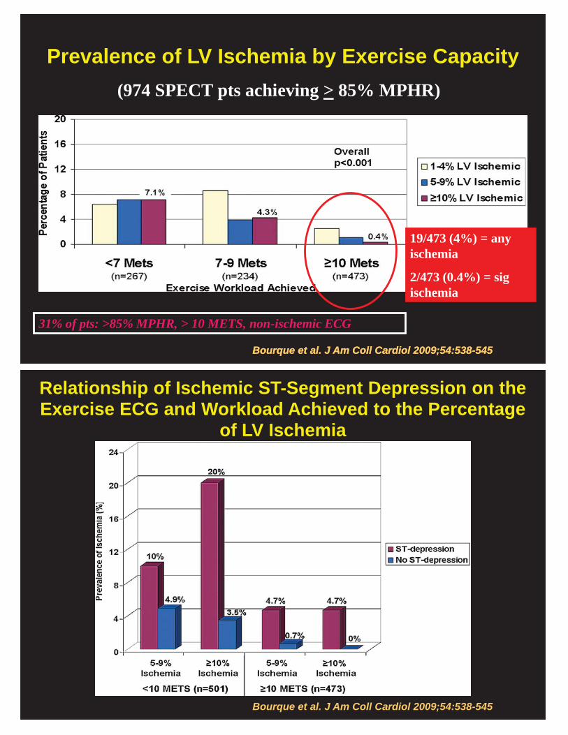
Bourque et al. J Am Coll Cardiol 2009;54:538-545
Prevalence of LV Ischemia by Exercise Capacity (974 SPECT pts achieving > 85% MPHR)
19/473 (4%) = any ischemia
2/473 (0.4%) = sig ischemia
Bourque et al. J Am Coll Cardiol 2009;54:538-545
31% of pts: >85% MPHR, > 10 METS, non-ischemic ECG
Relationship of Ischemic ST-Segment Depression on the Exercise ECG and Workload Achieved to the Percentage
of LV Ischemia
Bourque et al. J Am Coll Cardiol 2009;54:538-545
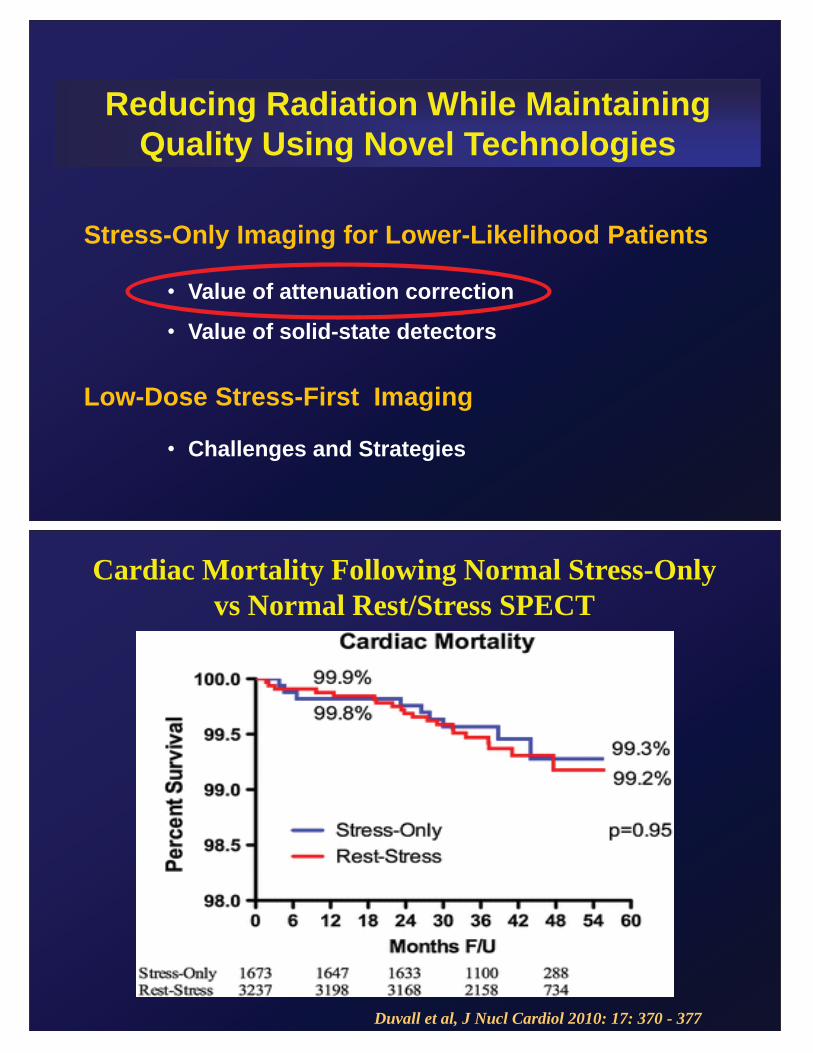
Reducing Radiation While Maintaining Quality Using Novel Technologies
Stress-Only Imaging for Lower-Likelihood Patients
• Value of attenuation correction
• Value of solid-state detectors
Low-Dose Stress-First Imaging
• Challenges and Strategies
Cardiac Mortality Following Normal Stress-Only vs Normal Rest/Stress SPECT
Duvall et al, J Nucl Cardiol 2010: 17: 370 - 377
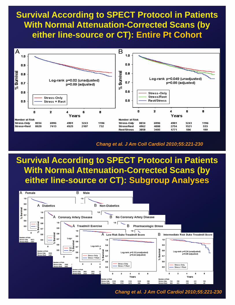
Chang et al. J Am Coll Cardiol 2010;55:221-230
Survival According to SPECT Protocol in Patients With Normal Attenuation-Corrected Scans (by
either line-source or CT): Entire Pt Cohort
Survival According to SPECT Protocol in Patients With Normal Attenuation-Corrected Scans (by either line-source or CT): Subgroup Analyses
Chang et al. J Am Coll Cardiol 2010;55:221-230
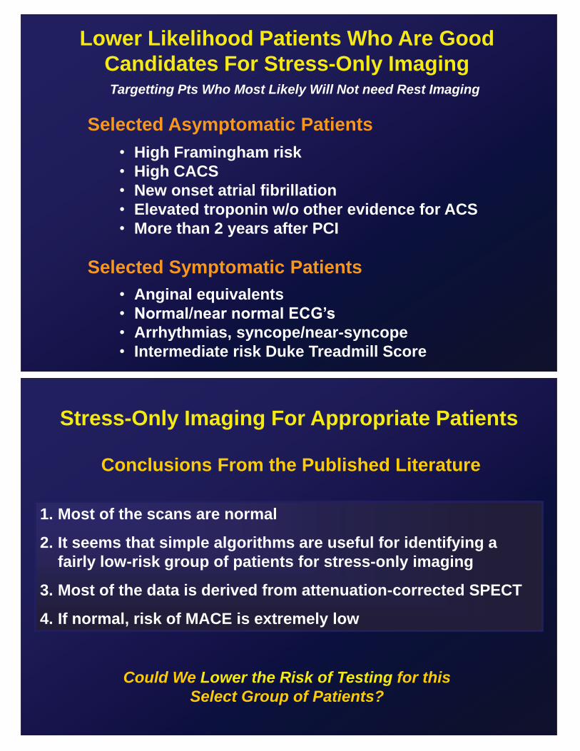
Lower Likelihood Patients Who Are Good Candidates For Stress-Only Imaging
Selected Asymptomatic Patients • High Framingham risk • High CACS • New onset atrial fibrillation • Elevated troponin w/o other evidence for ACS • More than 2 years after PCI
Selected Symptomatic Patients • Anginal equivalents • Normal/near normal ECG’s • Arrhythmias, syncope/near-syncope • Intermediate risk Duke Treadmill Score
Targetting Pts Who Most Likely Will Not need Rest Imaging
Stress-Only Imaging For Appropriate Patients
Conclusions From the Published Literature
1. Most of the scans are normal
2. It seems that simple algorithms are useful for identifying a fairly low-risk group of patients for stress-only imaging
3. Most of the data is derived from attenuation-corrected SPECT
4. If normal, risk of MACE is extremely low
Could We Lower the Risk of Testing for this Select Group of Patients?
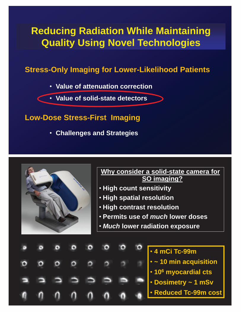
Reducing Radiation While Maintaining Quality Using Novel Technologies
Stress-Only Imaging for Lower-Likelihood Patients
• Value of attenuation correction
• Value of solid-state detectors
Low-Dose Stress-First Imaging
• Challenges and Strategies
Why consider a solid-state camera for SO imaging?
• High count sensitivity • High spatial resolution • High contrast resolution • Permits use of much lower doses •Much lower radiation exposure
• 4 mCi Tc-99m • ~ 10 min acquisition • 106 myocardial cts • Dosimetry ~ 1 mSv • Reduced Tc-99m cost
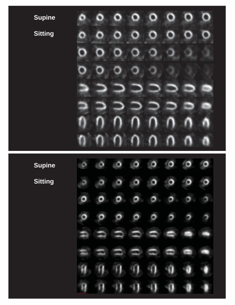
Supine
Sitting
Supine
Sitting
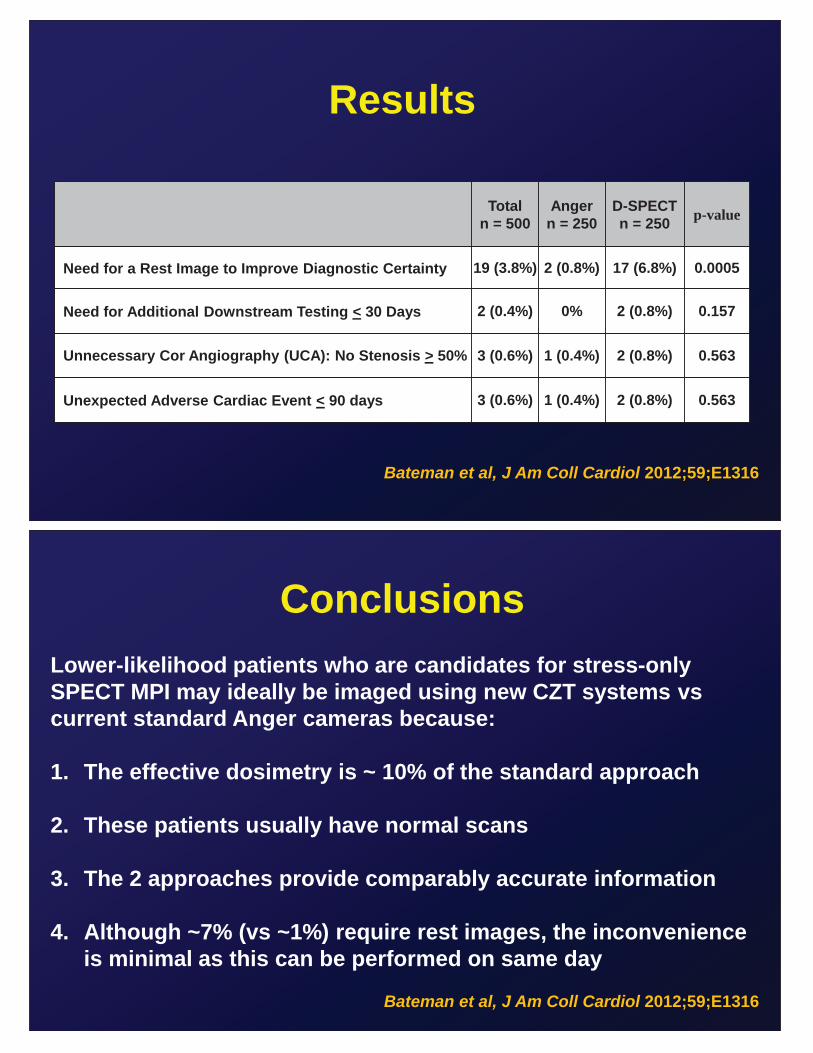
Results
Total
n = 500 Anger
n = 250 D-SPECT n = 250 p-value
Need for a Rest Image to Improve Diagnostic Certainty 19 (3.8%) 2 (0.8%) 17 (6.8%) 0.0005
Need for Additional Downstream Testing < 30 Days 2 (0.4%) 0% 2 (0.8%) 0.157
Unnecessary Cor Angiography (UCA): No Stenosis > 50% 3 (0.6%) 1 (0.4%) 2 (0.8%) 0.563
Unexpected Adverse Cardiac Event < 90 days 3 (0.6%) 1 (0.4%) 2 (0.8%) 0.563
Bateman et al, J Am Coll Cardiol 2012;59;E1316
Conclusions Lower-likelihood patients who are candidates for stress-only SPECT MPI may ideally be imaged using new CZT systems vs current standard Anger cameras because: 1. The effective dosimetry is ~ 10% of the standard approach
2. These patients usually have normal scans
3. The 2 approaches provide comparably accurate information
4. Although ~7% (vs ~1%) require rest images, the inconvenience
is minimal as this can be performed on same day
Bateman et al, J Am Coll Cardiol 2012;59;E1316
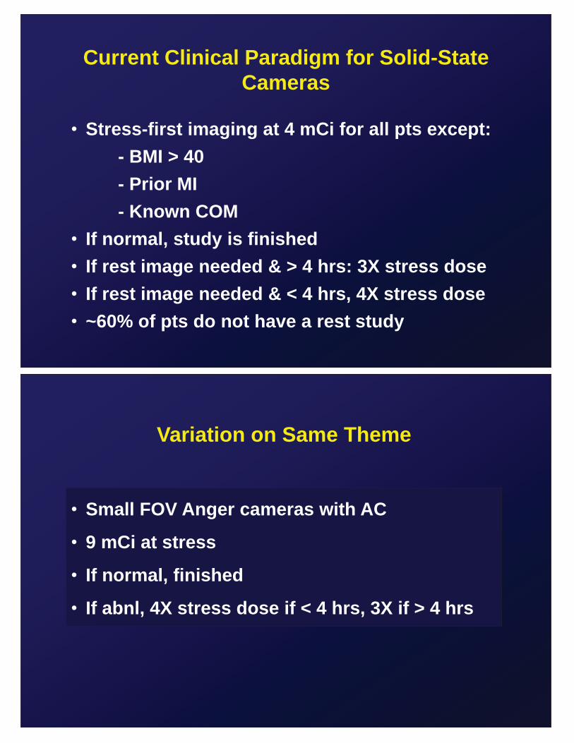
Current Clinical Paradigm for Solid-State Cameras
• Stress-first imaging at 4 mCi for all pts except: - BMI > 40 - Prior MI - Known COM • If normal, study is finished • If rest image needed & > 4 hrs: 3X stress dose • If rest image needed & < 4 hrs, 4X stress dose • ~60% of pts do not have a rest study
Variation on Same Theme
• Small FOV Anger cameras with AC
• 9 mCi at stress
• If normal, finished
• If abnl, 4X stress dose if < 4 hrs, 3X if > 4 hrs
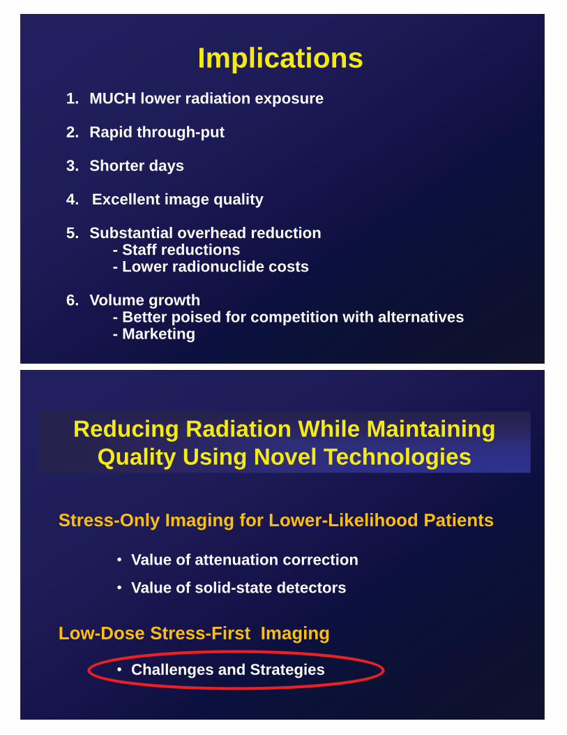
Implications 1. MUCH lower radiation exposure
2. Rapid through-put
3. Shorter days 4. Excellent image quality
5. Substantial overhead reduction - Staff reductions - Lower radionuclide costs 6. Volume growth - Better poised for competition with alternatives - Marketing
Reducing Radiation While Maintaining Quality Using Novel Technologies
Stress-Only Imaging for Lower-Likelihood Patients
• Value of attenuation correction
• Value of solid-state detectors
Low-Dose Stress-First Imaging
• Challenges and Strategies
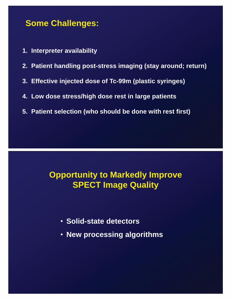
Some Challenges:
1. Interpreter availability
2. Patient handling post-stress imaging (stay around; return)
3. Effective injected dose of Tc-99m (plastic syringes)
4. Low dose stress/high dose rest in large patients
5. Patient selection (who should be done with rest first)
Opportunity to Markedly Improve SPECT Image Quality
• Solid-state detectors
• New processing algorithms
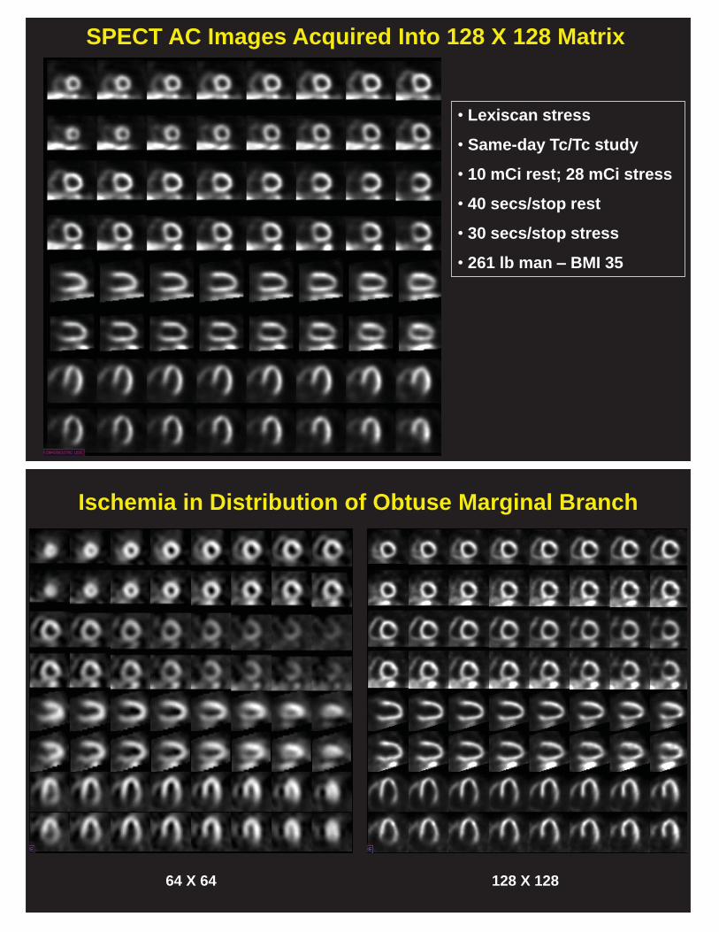
SPECT AC Images Acquired Into 128 X 128 Matrix
• Lexiscan stress
• Same-day Tc/Tc study
• 10 mCi rest; 28 mCi stress
• 40 secs/stop rest
• 30 secs/stop stress
• 261 lb man – BMI 35
Ischemia in Distribution of Obtuse Marginal Branch
64 X 64 128 X 128
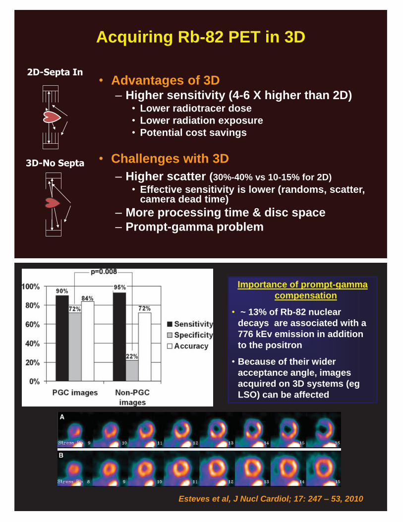
Acquiring Rb-82 PET in 3D
• Advantages of 3D – Higher sensitivity (4-6 X higher than 2D)
• Lower radiotracer dose • Lower radiation exposure • Potential cost savings
• Challenges with 3D – Higher scatter (30%-40% vs 10-15% for 2D)
• Effective sensitivity is lower (randoms, scatter, camera dead time)
– More processing time & disc space – Prompt-gamma problem
2D-Septa In
3D-No Septa
Esteves et al, J Nucl Cardiol; 17: 247 – 53, 2010
Importance of prompt-gamma compensation
• ~ 13% of Rb-82 nuclear decays are associated with a 776 kEv emission in addition to the positron
• Because of their wider acceptance angle, images acquired on 3D systems (eg LSO) can be affected
Stress-Only PET Imaging?
• Not so much benefit (dosimetry; efficiency)
• Longer wait-time vs SPECT
• No validation studies
• Lose stress/rest MBF information
Routine Flow Quantitation
• Overcomes issues with spatial-relativity
• Knowledge of stress adequacy
• Complete depiction of myocardial blood flow
• A physiologic measure resembling invasive FFR
• Numerous commercially-available packages