Embed Size (px)
DESCRIPTION
Ch 46 - Reproduction. Sexual reproduction results in genetic recombination, which provides potential advantages Asexual reproduction occurs through a variety of processes depending on the species. Mechanisms of Asexual Reproduction. - PowerPoint PPT Presentation
Citation preview
Copyright © 2008 Pearson Education, Inc., publishing as Pearson Benjamin Cummings
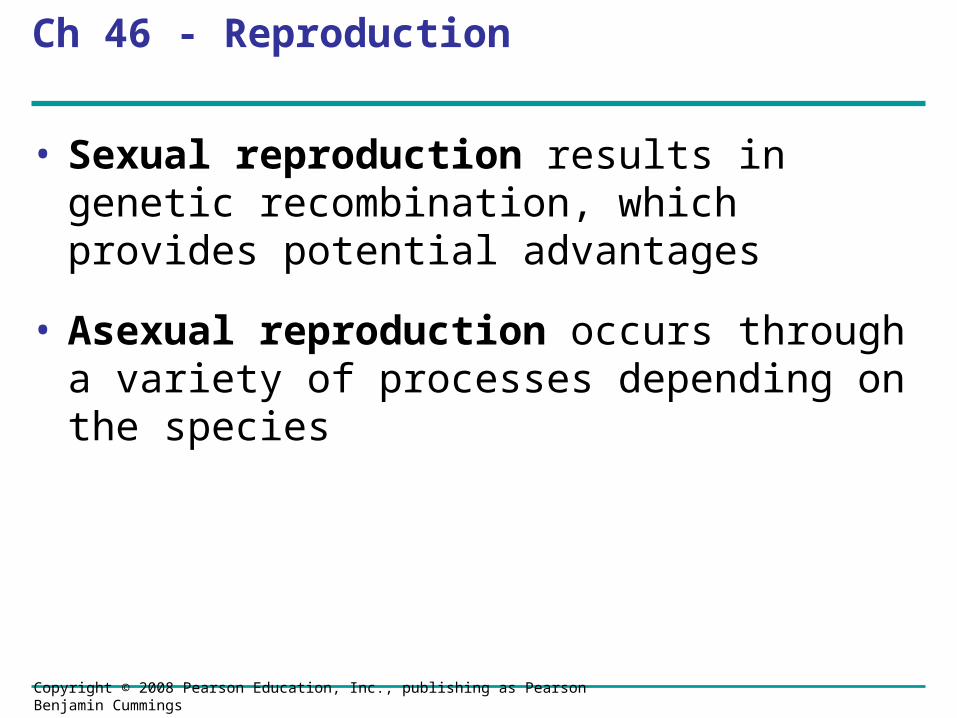
Ch 46 - Reproduction
• Sexual reproduction results in genetic recombination, which provides potential advantages
• Asexual reproduction occurs through a variety of processes depending on the species
Copyright © 2008 Pearson Education, Inc., publishing as Pearson Benjamin Cummings
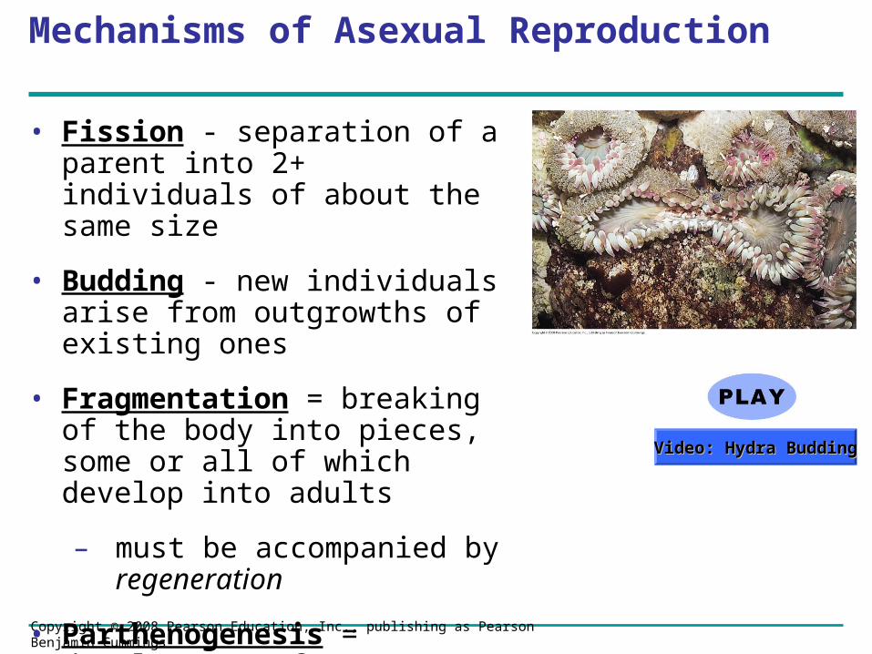
Mechanisms of Asexual Reproduction
• Fission - separation of a parent into 2+ individuals of about the same size
• Budding - new individuals arise from outgrowths of existing ones
• Fragmentation = breaking of the body into pieces, some or all of which develop into adults
– must be accompanied by regeneration
• Parthenogenesis = development of a new individual from an unfertilized egg
Video: Hydra BuddingVideo: Hydra Budding

Copyright © 2008 Pearson Education, Inc., publishing as Pearson Benjamin Cummings
• Sexual reproduction is a special problem for organisms that seldom encounter a mate
• One solution is hermaphroditism, in which each individual has male and female reproductive systems
– Some hermaphrodites can self-fertilize

Copyright © 2008 Pearson Education, Inc., publishing as Pearson Benjamin Cummings
• Individuals of some species undergo sex reversals
– Some species exhibit male to female reversal (ex: certain oysters)
– Others exhibit female to male reversal (ex: a coral reef fish)
Copyright © 2008 Pearson Education, Inc., publishing as Pearson Benjamin Cummings
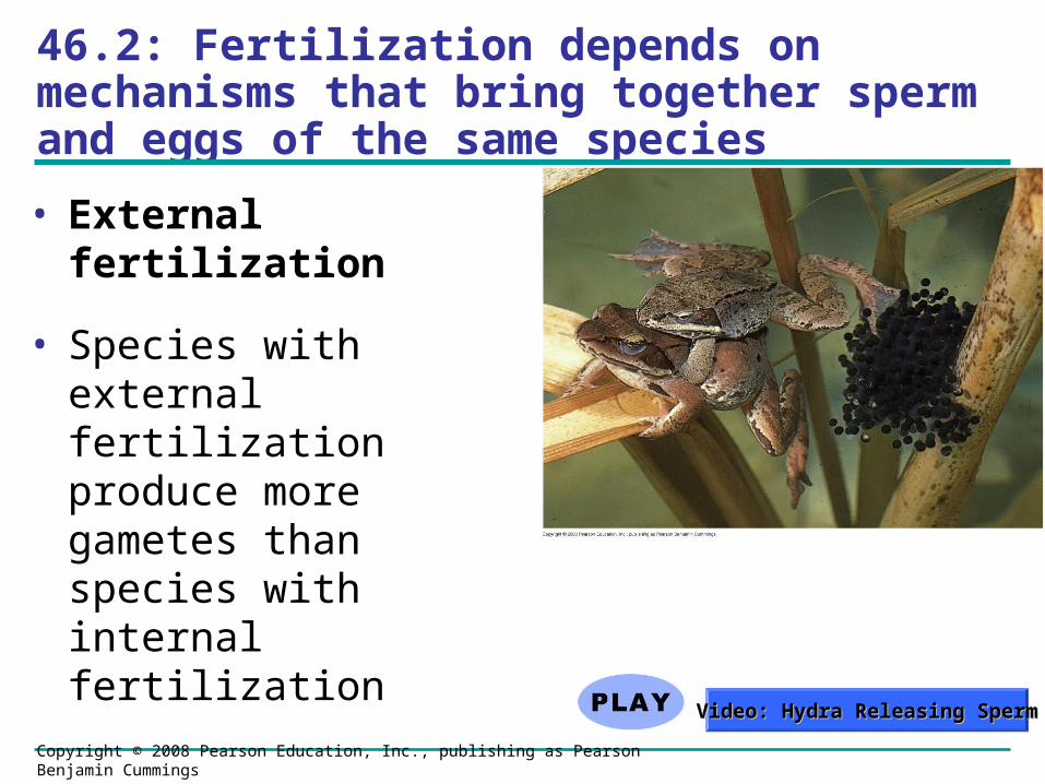
46.2: Fertilization depends on mechanisms that bring together sperm and eggs of the same species
• External fertilization
• Species with external fertilization produce more gametes than species with internal fertilization
Video: Hydra Releasing SpermVideo: Hydra Releasing Sperm
Copyright © 2008 Pearson Education, Inc., publishing as Pearson Benjamin Cummings
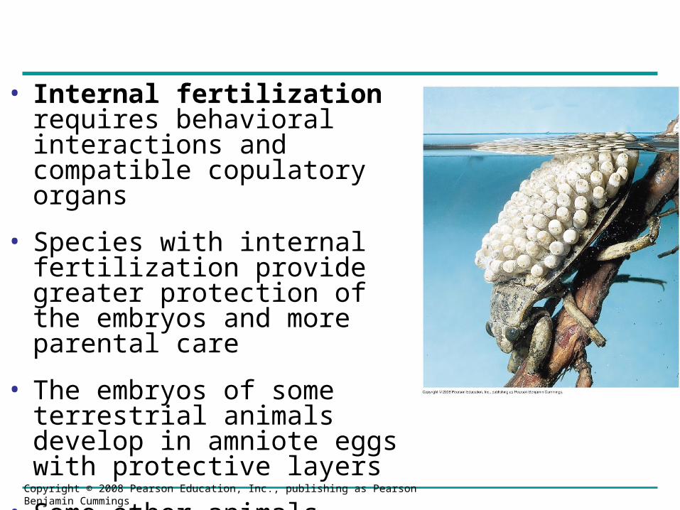
• Internal fertilization requires behavioral interactions and compatible copulatory organs
• Species with internal fertilization provide greater protection of the embryos and more parental care
• The embryos of some terrestrial animals develop in amniote eggs with protective layers
• Some other animals retain the embryo, which develops inside the female
Copyright © 2008 Pearson Education, Inc., publishing as Pearson Benjamin Cummings
Gamete Production and Delivery
• In most species, individuals have gonads that produce gametes
• Some simple systems do not have gonads, but gametes form from undifferentiated tissue
• The most complex systems contain many sets of accessory tubes and glands that carry, nourish, and protect gametes and developing embryos
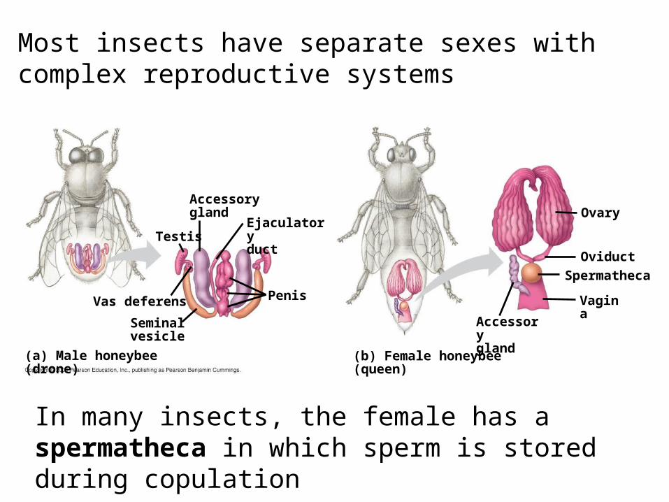
Most insects have separate sexes with complex reproductive systems
Accessorygland
EjaculatoryductTestis
Vas deferens
Seminalvesicle
Penis
Ovary
Oviduct
Spermatheca
Vagina
Accessorygland
(a) Male honeybee (drone) (b) Female honeybee (queen)
In many insects, the female has a spermatheca in which sperm is stored during copulation

Copyright © 2008 Pearson Education, Inc., publishing as Pearson Benjamin Cummings
• Even animals with simple body plans can have complex reproductive systems, for example parasitic flatworms
Copyright © 2008 Pearson Education, Inc., publishing as Pearson Benjamin Cummings
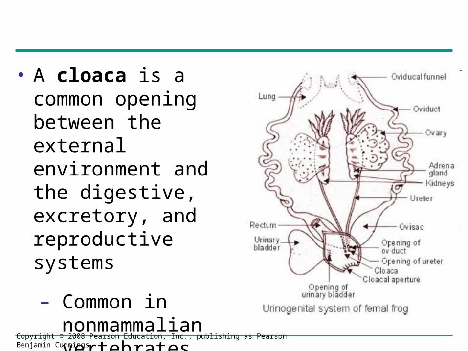
• A cloaca is a common opening between the external environment and the digestive, excretory, and reproductive systems
– Common in nonmammalian vertebrates
Copyright © 2008 Pearson Education, Inc., publishing as Pearson Benjamin Cummings
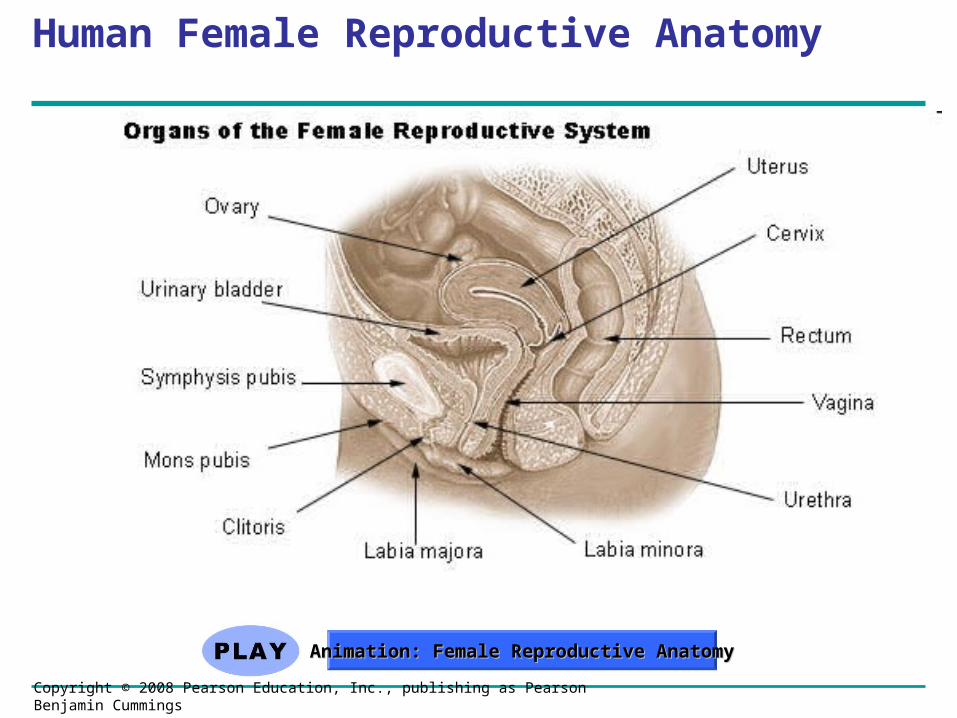
Human Female Reproductive Anatomy
Animation: Female Reproductive AnatomyAnimation: Female Reproductive Anatomy
Copyright © 2008 Pearson Education, Inc., publishing as Pearson Benjamin Cummings
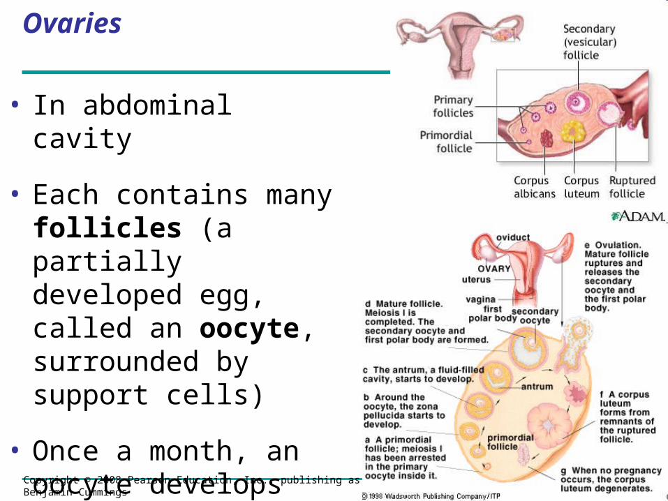
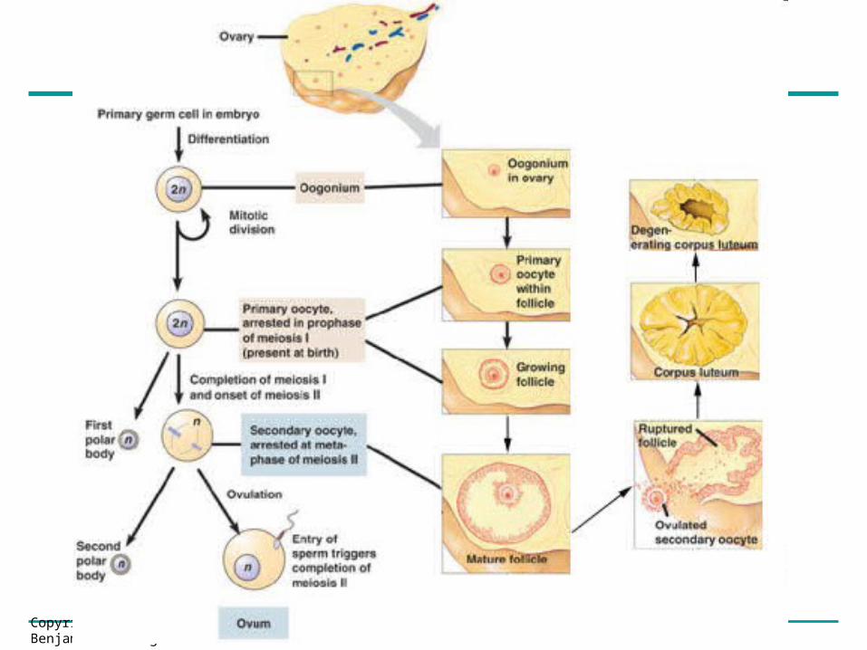
Ovaries
• In abdominal cavity
• Each contains many follicles (a partially developed egg, called an oocyte, surrounded by support cells)
• Once a month, an oocyte develops into an ovum (egg) by the process of oogenesis
Copyright © 2008 Pearson Education, Inc., publishing as Pearson Benjamin Cummings
Copyright © 2008 Pearson Education, Inc., publishing as Pearson Benjamin Cummings
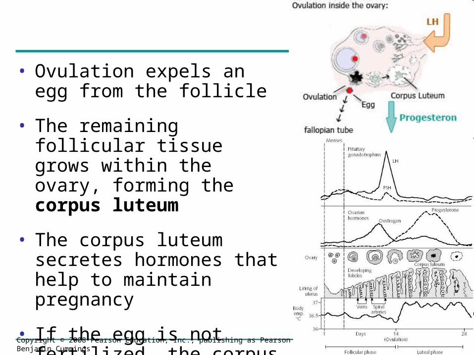
• Ovulation expels an egg from the follicle
• The remaining follicular tissue grows within the ovary, forming the corpus luteum
• The corpus luteum secretes hormones that help to maintain pregnancy
• If the egg is not fertilized, the corpus luteum degenerates
Copyright © 2008 Pearson Education, Inc., publishing as Pearson Benjamin Cummings
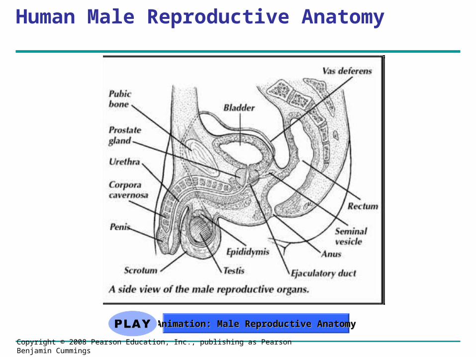
Human Male Reproductive Anatomy
Animation: Male Reproductive AnatomyAnimation: Male Reproductive Anatomy
Copyright © 2008 Pearson Education, Inc., publishing as Pearson Benjamin Cummings
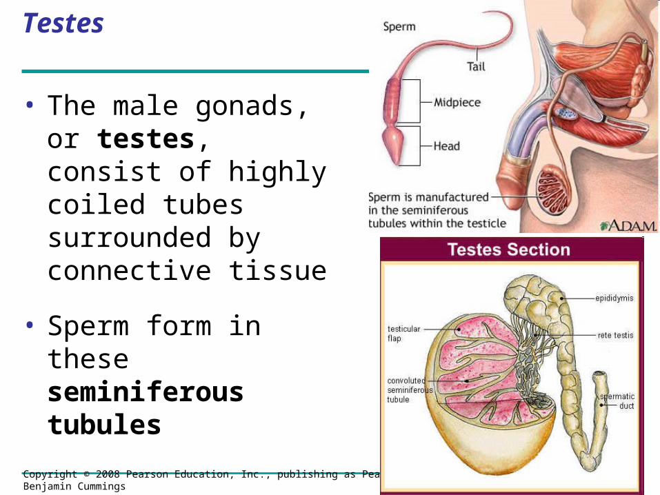
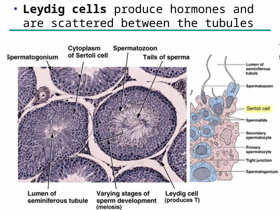
Testes
• The male gonads, or testes, consist of highly coiled tubes surrounded by connective tissue
• Sperm form in these seminiferous tubules
Copyright © 2008 Pearson Education, Inc., publishing as Pearson Benjamin Cummings
• Leydig cells produce hormones and are scattered between the tubules

Copyright © 2008 Pearson Education, Inc., publishing as Pearson Benjamin Cummings
Ducts
• From the seminiferous tubules, sperm pass into the epididymis
• During ejaculation, sperm are propelled through the muscular vas deferens and the ejaculatory duct, and then exit the penis through the urethra
Copyright © 2008 Pearson Education, Inc., publishing as Pearson Benjamin Cummings
Accessory Glands
• Semen = sperm + secretions from three sets of accessory glands
– seminal vesicles
– prostate gland
– bulbourethral glands
Copyright © 2008 Pearson Education, Inc., publishing as Pearson Benjamin Cummings
46.5: The interplay of tropic and sex hormones regulates mammalian reproduction
• Human reproduction is coordinated by hormones from the hypothalamus, anterior pituitary, and gonads
• Gonadotropin-releasing hormone (GnRH) is secreted by the hypothalamus and directs the release of FSH and LH from the anterior pituitary
• FSH and LH regulate processes in the gonads and the production of sex hormones

Copyright © 2008 Pearson Education, Inc., publishing as Pearson Benjamin Cummings
• The sex hormones are androgens, estrogens, and progesterone
• Sex hormones regulate:
– Development of primary sex characteristics (during embryogenesis)
– Development of secondary sex characteristics (at puberty)
– Sexual behavior and sex drive
Copyright © 2008 Pearson Education, Inc., publishing as Pearson Benjamin Cummings
Hormonal Control of the Male Reproductive System
• FSH promotes the activity of Sertoli cells, which nourish developing sperm and are located within the seminiferous tubules
• LH regulates Leydig cells, which secrete testosterone and other androgen hormones, which in turn promote spermatogenesis
Animation: Male HormonesAnimation: Male Hormones
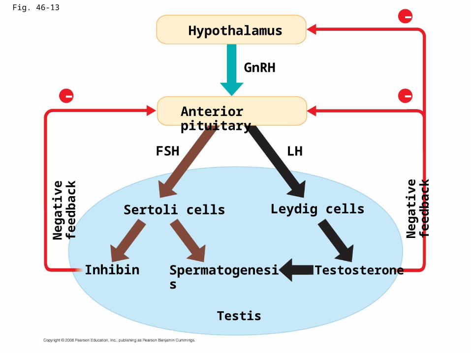
Fig. 46-13
Hypothalamus
GnRH
FSH
Anterior pituitary
Sertoli cells Leydig cells
Inhibin Spermatogenesis Testosterone
Testis
LH
Neg
ativ
e fe
edb
ack
Neg
ativ
e fe
edb
ack
– –
–
Copyright © 2008 Pearson Education, Inc., publishing as Pearson Benjamin Cummings
• Testosterone regulates the production of GnRH, FSH, and LH through negative feedback mechanisms
• Sertoli cells secrete the hormone inhibin, which reduces FSH secretion from the anterior pituitary
Copyright © 2008 Pearson Education, Inc., publishing as Pearson Benjamin Cummings
The Reproductive Cycles of Females
• In females, the secretion of hormones and the reproductive events they regulate are cyclic
• Prior to ovulation, the endometrium thickens with blood vessels in preparation for embryo implantation
• If an embryo does not implant in the endometrium, the endometrium is shed in a process called menstruation
Copyright © 2008 Pearson Education, Inc., publishing as Pearson Benjamin Cummings
• Hormones closely link the two cycles of female reproduction:
– Changes in the uterus define the menstrual cycle (also called the uterine cycle)
– Changes in the ovaries define the ovarian cycle
Control by hypothalamus Inhibited by combination of estradiol and progesterone
Stimulated by high levelsof estradiol
Inhibited by low levels of estradiol
Hypothalamus
GnRH
Anterior pituitary
FSH LH
Pituitary gonadotropinsin blood
LH
FSH
FSH and LH stimulatefollicle to grow
LH surge triggersovulation
Ovarian cycle
Growing follicle Maturingfollicle
Corpusluteum
Degeneratingcorpus luteum
Follicular phase Ovulation Luteal phase
(a)
(b)
(c)
Da
ys
0 5 10 14 15 20 25 28| | | | | | | |
–
–
+
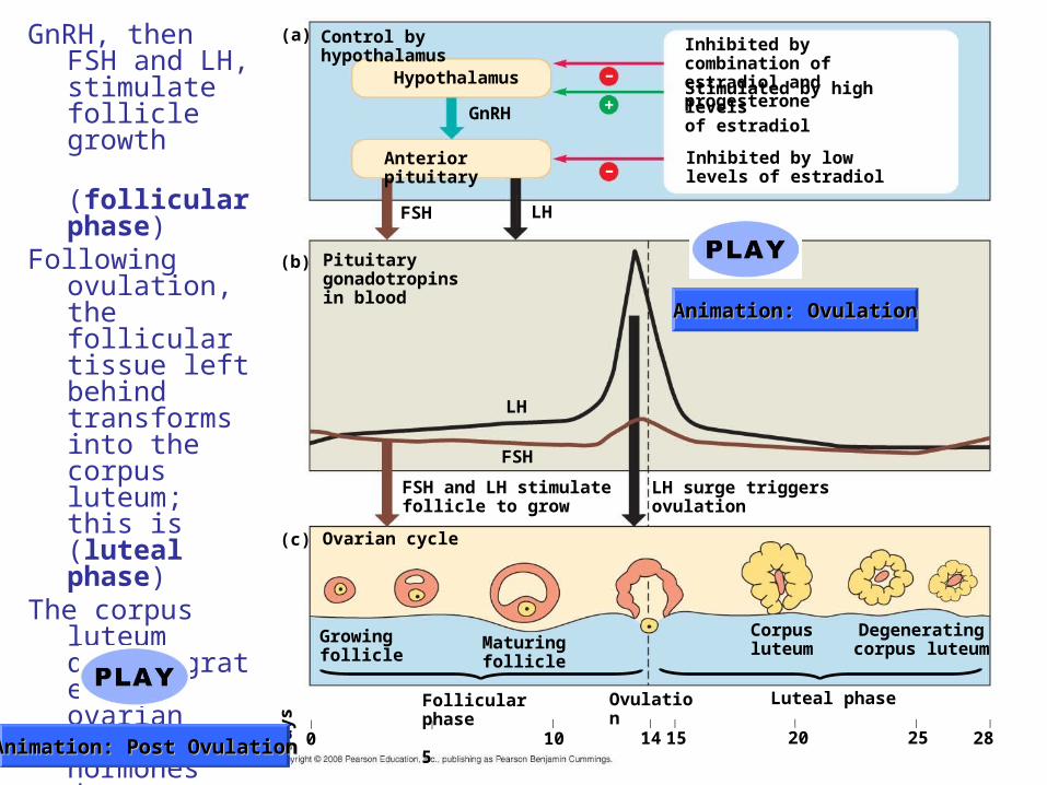
GnRH, then FSH and LH, stimulate follicle growth
(follicular phase)
Following ovulation, the follicular tissue left behind transforms into the corpus luteum; this is (luteal phase)
The corpus luteum disintegrates, and ovarian steroid hormones decrease
Animation: OvulationAnimation: Ovulation
Animation: Post OvulationAnimation: Post Ovulation
Ovarian hormones in blood
Peak causesLH surge
Estradiol level very low
Estradiol Progesterone
Ovulation Progesterone and estra-diol promote thickeningof endometrium
Uterine (menstrual) cycle
Endometrium
0 5 10 14 20 25 28| | | | | | | |
Da
ys
15
Menstrual flow phase Proliferative phase Secretory phase
(d)
(e)
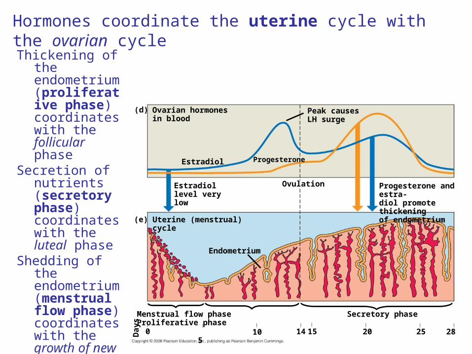
Thickening of the endometrium (proliferative phase) coordinates with the follicular phase
Secretion of nutrients (secretory phase) coordinates with the luteal phase
Shedding of the endometrium (menstrual flow phase) coordinates with the growth of new ovarian follicles
Hormones coordinate the uterine cycle with the ovarian cycle
Copyright © 2008 Pearson Education, Inc., publishing as Pearson Benjamin Cummings
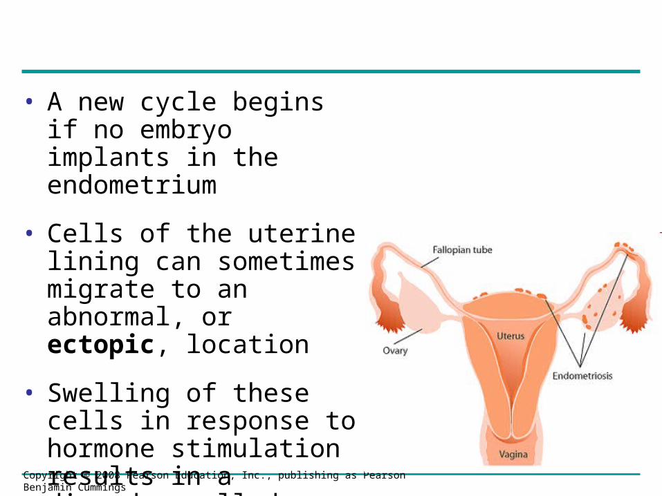
• A new cycle begins if no embryo implants in the endometrium
• Cells of the uterine lining can sometimes migrate to an abnormal, or ectopic, location
• Swelling of these cells in response to hormone stimulation results in a disorder called endometriosis
Copyright © 2008 Pearson Education, Inc., publishing as Pearson Benjamin Cummings
46.6: In placental mammals, an embryo develops fully within the mother’s uterus
• An egg develops into an embryo in a series of predictable events
• Conception, fertilization of an egg by a sperm, occurs in the oviduct
• The resulting zygote begins to divide by mitosis in a process called cleavage
• Division of cells gives rise to a blastocyst, a ball of cells with a cavity
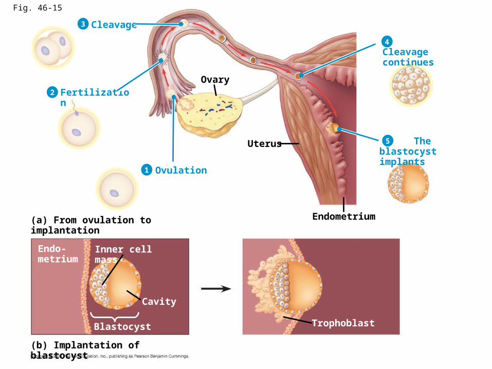
Fig. 46-15
Ovary
Uterus
Endometrium(a) From ovulation to implantation
(b) Implantation of blastocyst
Cleavage
Fertilization
Ovulation
Cleavage continues
The blastocystimplants
Trophoblast
Inner cell mass
Cavity
Blastocyst
Endo-metrium
1
2
3
4
5
Copyright © 2008 Pearson Education, Inc., publishing as Pearson Benjamin Cummings
• After blastocyst formation, the embryo implants into the endometrium
• The embryo releases human chorionic gonadotropin (hCG), which prevents menstruation
• Pregnancy, or gestation, is the condition of carrying one or more embryos in the uterus
• Duration of pregnancy in other species correlates with body size and maturity of the young at birth
Copyright © 2008 Pearson Education, Inc., publishing as Pearson Benjamin Cummings
First Trimester
• Human gestation can be divided into three trimesters of about three months each
• The first trimester is the time of most radical change for both the mother and the embryo
• During implantation, the endometrium grows over the blastocyst
Copyright © 2008 Pearson Education, Inc., publishing as Pearson Benjamin Cummings
• During its first 2 to 4 weeks, the embryo obtains nutrients directly from the endometrium
• Meanwhile, the outer layer of the blastocyst, called the trophoblast, mingles with the endometrium and eventually forms the placenta
• Blood from the embryo travels to the placenta through arteries of the umbilical cord and returns via the umbilical vein
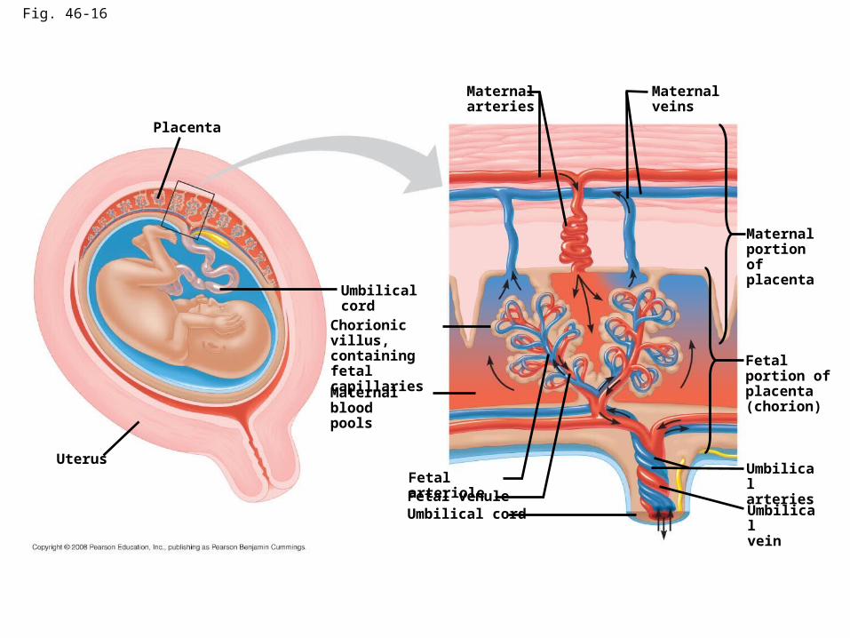
Fig. 46-16
Placenta
Uterus
Umbilical cord
Chorionic villus,containing fetalcapillaries
Maternal bloodpools
Maternalarteries
Maternalveins
Maternalportionof placenta
Fetal arterioleFetal venuleUmbilical cord
Fetalportion ofplacenta(chorion)
Umbilicalarteries
Umbilicalvein
Copyright © 2008 Pearson Education, Inc., publishing as Pearson Benjamin Cummings
• Splitting of the embryo during the first month of development results in genetically identical twins
• Release and fertilization of two eggs results in fraternal and genetically distinct twins

Copyright © 2008 Pearson Education, Inc., publishing as Pearson Benjamin Cummings
• The first trimester is the main period of organogenesis, development of the body organs
• All the major structures are present by 8 weeks, and the embryo is called a fetus
Copyright © 2008 Pearson Education, Inc., publishing as Pearson Benjamin Cummings
• Changes occur in the mother
– Growth of the placenta
– Cessation of ovulation and the menstrual cycle
– Breast enlargement
– Nausea is also very common
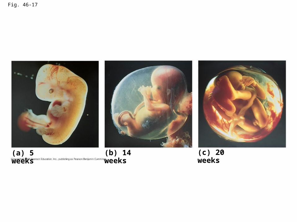
Fig. 46-17
(a) 5 weeks (b) 14 weeks (c) 20 weeks
Copyright © 2008 Pearson Education, Inc., publishing as Pearson Benjamin Cummings
Second Trimester
• During the second trimester
– The fetus grows and is very active
– The mother may feel fetal movements
– The uterus grows enough for the pregnancy to become obvious
Copyright © 2008 Pearson Education, Inc., publishing as Pearson Benjamin Cummings
Third Trimester
• During the third trimester, the fetus grows and fills the space within the embryonic membranes
• A complex interplay of local regulators and hormones induces and regulates labor, the process by which childbirth occurs

Fig. 46-18
Estradiol Oxytocin
fromovaries
Induces oxytocinreceptors on uterus
from fetusand mother’sposterior pituitary
Stimulates uterusto contract
Stimulates placenta to make
Prostaglandins
Stimulate morecontractions
of uterus
Po
siti
ve
fee
db
ac
k
+
+

3
2
1 Dilation of the cervix
Placenta
Umbilical cord
Uterus
Cervix
Expulsion: delivery of the infant
Uterus
Placenta(detaching)
Umbilicalcord
Delivery of the placenta
Birth, or parturition, is brought about by a series of strong, rhythmic uterine contractions
First the baby is delivered, and then the placenta
Copyright © 2008 Pearson Education, Inc., publishing as Pearson Benjamin Cummings
Detecting Disorders During Pregnancy
• Amniocentesis and chorionic villus sampling are invasive techniques in which amniotic fluid or fetal cells are obtained for genetic analysis
• Noninvasive procedures usually use ultrasound imaging to detect fetal condition
• Genetic testing of the fetus poses ethical questions and can present parents with difficult decisions
Video: Ultrasound of Human Fetus 1Video: Ultrasound of Human Fetus 1 Video: Ultrasound of Human Fetus 2Video: Ultrasound of Human Fetus 2
Copyright © 2008 Pearson Education, Inc., publishing as Pearson Benjamin Cummings
Treating Infertility
• Modern technology can provide infertile couples with assisted reproductive technologies
• In vitro fertilization (IVF) mixes eggs with sperm in culture dishes and returns the embryo to the uterus at the 8 cell stage
• Sperm are injected directly into an egg in a type of IVF called intracytoplasmic sperm injection (ICSI)