Embed Size (px)
DESCRIPTION
Presented here is the first Scientific Report of Basic Science Programs at the GRU Cancer Center of Georgia Regents University (GRU) in Augusta, Georgia. The following pages present the 2013 research achievements of Cancer Center members within the Cancer immunology, Inflammation, and Tolerance (CIT), Molecular Oncology and Biomarkers (MOB), and Tumor Signaling and Angiogenesis (TSA) basic science programs. The Cancer Prevention and Control Initiative is currently under construction and will be added to the Scientific Report as it achieves critical mass with inter-programmatic interactions. Similarly, the Translational Oncology Initiative is emerging with the creation and execution of infrastructure for a Phase I/II clinical trials unit to promote investigator-initiated trials that develop from within Cancer Center research laboratories.
Citation preview

2013 SCIENTIFIC REPORT OF BASIC SCIENCE PROGRAMS


// 01
CONTENTS
INTRODUCTION // 03-04
CANCER IMMUNOLOGY, INFLAMMATIONAND TOLERANCE PROGRAM // 05
OVERVIEW // 06CIT REPORTS // 07-20
MOLECULAR ONCOLOGY AND BIOMARKERS PROGRAM // 21
OVERVIEW // 22MOB REPORTS // 23-41
TUMOR SIGNALING AND ANGIOGENESIS PROGRAM // 43
OVERVIEW // 44TSA REPORTS // 45-57
CANCER CENTER SHARED RESOURCES // 59BIOINFORMATICS AND BIOSTATISTICS //60 BIOREPOSITORY AND CENTRAL SOURCE
FOR THE BIOREPOSITORY ALLIANCE OF GEORGIA-ONCOLOGY (BRAG-ONC) // 61
CORE IMAGING FACILITY FOR SMALL ANIMALS // 62FLOW CYTOMETRY RESOURCE // 63 INTEGRATED GENOMICS: MICROARRAY // 64 INTEGRATED GENOMICS: NEXT-GEN SEQUENCING // 65MICROSCOPY IMAGING CORE // 66 PROTEOMICS AND METABOLOMICS FACILITY // 67
2013 GRU CANCER CENTER PUBLICATIONS // 69-84
INDEX OF PROGRAM RESEARCHERS // 86-87

// 03
Presented here is the first Scientific Report of Basic Science Programs at the GRU Cancer Center of Georgia Regents University (GRU) in Augusta, Georgia. The following pages present the 2013 research achievements of Cancer Center members within the Cancer immunology, Inflammation, and Tolerance (CIT), Molecular Oncology and Biomarkers (MOB), and Tumor Signaling and Angiogenesis (TSA) basic science programs. The Cancer Prevention and Control Initiative is currently under construction and will be added to the Scientific Report as it achieves critical mass with inter-programmatic interactions. Similarly, the Translational Oncology Initiative is emerging with the creation and execution of infrastructure for a Phase I/II clinical trials unit to promote investigator-initiated trials that develop from within Cancer Center research laboratories.
Under the leadership of its new director, Dr. Samir Khleif, the Cancer Center has undergone unprecedented expansion in the basic and clinical sciences faculty over the past 18 months, with more than twenty new faculty, of which more than half conduct research in the basic sciences. The number of faculty is planned to increase by another twenty over the next three years as part of an effort to achieve NCI designation through NCI Cancer Center Support Grant (CCSG) funding before 2020. A critical component of NCI designation is the availability of Shared Resources to its members. Details of the facilities available and the services they offer are included in this report.
About the GRU Cancer CenterWith strong state support, GRU has invested substantially in developing the Cancer Center. In 2006, a $54 million, 151,000 square foot research building was erected to house Cancer Center scientific laboratories, shared facilities, and administrators. In 2010, a $31 million, 64,000 square foot adult cancer clinical facility opened, providing comprehensive outpatient oncology services and housing a dedicated clinical trials unit. In early 2012, the two units merged to become the GRU Cancer Center, with members derived from Colleges, Departments, and Institutes throughout the medical campus.
About GRUFounded in 1828, Georgia Regents University, previously Georgia Health Sciences University, is a comprehensive research university within the University System of Georgia. Nearly 10,000 students are enrolled in nine colleges that include the nationally ranked Hull College of Business, Georgia’s only College of Dental Medicine, and the sixth largest public medical school in the country – the Medical College of Georgia (MCG).
GRU CANCER CENTER 2013 SCIENTIFIC REPORT OF BASIC SCIENCE PROGRAMS

04 //
Nationally and internationally recognized leaders in clinical care and scientific research have been recruited to serve at the GRU Cancer Center in the following capacities:
Cancer Center Directorship
Samir N. Khleif, MDDirector, GRU Cancer CenterDirector, GRU Cancer Center Service Line
Olivier Rixe, MD, PhDMedical Director, GRU Cancer Center
David H. Munn, MDSenior Advisor to the Cancer Center Director
GRU CANCER CENTER SENIOR LEADERSHIP
Cancer Center Associate Directors
Michael Benedict, PharmDAssociate Cancer Center Director of Adminsitration
John K. Cowell, PhD, DSc, FRCPathAssociate Cancer Center Director of Basic Science
Sharad Ghamande, MDAssociate Cancer Center Director of Clinical Affairs
Nita J. Maihle, PhDAssociate Cancer Center Director of Education

// 05
Co-Leader // Dr. A. Mellor
CANCER IMMUNOLOGY, INFLAMMATION AND TOLERANCE (CIT) PROGRAM

06 //
The principal goals of the program are to elucidate molecular pathways and cellular processes active in tumor microenvironments in order to develop, characterize, and apply immunological approaches to prevention, diagnosis, treatment, and monitoring of premalignant and malignant diseases. Specific aims include:
• discovery research using models of chronic inflammatory diseases (i) to elucidate how immune responses are regulated to create tolerance (unresponsiveness) in tumor microenvironments that inhibit natural and vaccine-induced anti-tumor immunity and (ii) to identify novel targets for therapeutic intervention;
• development and characterization of molecular and immunological strategies for manipulating innate and adaptive immune responses to malignancy;
• translating new discoveries into clinical settings in collaboration with experimental oncologists in the GRU Cancer Center and corporate partners;
• evaluating the efficacy of immunotherapy and conventional therapies by developing a system to monitor immune response in conjunction with clinical outcomes;
• understanding how chronic inflammation creates tolerance to protect tumors and healthy tissues from immune-mediated injury; • manipulating tolerogenic mechanisms for clinical benefit; • developing better cancer vaccines;• elucidating novel targets to manipulate immune responses to treat cancer and other chronic inflammatory syndromes.
Pre-malignant lesions form tumors by evading natural immunity before clinical presentation, and established tumors are resistant to tumor vaccines because immune tolerance attenuates vaccine-induced immunity. Successful therapies must therefore disrupt local microenvironments that protect tumors. Hence, program research focuses on tolerogenic pathways in the innate and adaptive immune systems that protect healthy tissues and tumors from immune-mediated destruction, and on translational opportunities to treat cancer patients arising from this research.
Researchers in the CIT program use a range of techniques to study how the immune system influences tumorigenesis and cancer therapy. The immune system can inhibit or promote tumor progression in local tissues where pre-malignancies form. Major program themes are to elucidate, (i) how pre-malignancies create and sustain local immunologic tolerance necessary for tumor formation and, (ii) how to destroy local tolerance that protects tumors from natural and vaccine-induced anti-tumor immunity. Since loss of immune tolerance leads to autoimmune syndromes (e.g. type I diabetes, systemic lupus erythematosus, rheumatoid arthritis, colitis, multiple sclerosis), program investigators use mouse models of cancer and autoimmune progression to elucidate critical molecular and cellular pathways that either create or destroy immune tolerance. The scientific rationale for this dual approach is that pre-malignant cells create and sustain tolerance during tumor progression, while breaking tumor-associated tolerance is necessary for successful anti-tumor treatment. Hence, program goals are to elucidate molecular and cellular pathways at sites of inflammation that promote or break immune tolerance using pre-clinical mouse models of tumor progression and autoimmune syndromes, and developing novel immunotherapies to treat these syndromes more effectively by targeting tolerance pathways. To this end, program faculty also engage in promoting pre-clinical research and early-phase clinical trials of novel vaccine adjuvants to improve cancer immunotherapy, in some cases with corporate partners. To pursue these focused research themes and scientific goals, program faculty employ many state-of-the-art techniques, facilities, and unique resources, including flow cytometric sorting and analysis, a range of molecular imaging techniques, genomic analysis, and genetically modified mouse strains. Future program development will build on existing CIT program strengths by recruiting new investigators with expertise in inflammation, immunological, and metabolic research to complement current research focused on regulation of adaptive immunity.
cancer immunology, inflammation and tolerance program //OVERVIEW OF THE PROGRAM

// 07
DNA sensors that promote tolerance as regulators of immunity to tumor and tissue antigens: Inflammation associated with developing primary malignancies promotes immune tolerance that blocks innate anti-tumor immunity before clinical presentation and inhibits vaccine-induced immunity after clinical presentation. Paradoxically, inflammation also causes tolerance breakdown leading to autoimmune diseases. Pathways that promote or disrupt tolerance at sites of inflammation are poorly defined, even though they are pivotal drivers of cancer and autoimmune diseases. Dr. Mellor and his faculty colleague Dr. Lei Huang are studying the role of DNA in promoting tolerance based on our observation that DNA nanoparticles (DNPs) promote tolerogenic responses when administered systemically to mice. In a recent study (J Immunol 2013, 191:3509-13), they reported that nanoparticle cargo DNA was ingested and sensed to activate the molecular adaptor STimulator of INterferon Genes (STING). Once activated, STING incited interferon type I (IFN-I) release, which induced expression of the immune regulatory enzyme indoleamine 2,3 dioxygenase (IDO) by dendritic cells specialized to promote tolerogenic responses. In studies completed recently (Lemos et al., in revision), they showed that DNP treatments delayed onset of experimental autoimmune encephalitis (EAE), a mouse model of human multiple sclerosis, and reduced EAE severity substantially. Therapeutic responses to DNPs were abolished in mice lacking STING or IFN-I receptors but manifested normally in mice lacking IFN type II (IFNγ) receptors. Consistent with these findings, systemic treatments with the cyclic diguanylate monophosphate (cdiGMP), which binds and activates STING directly, also slowed EAE onset and reduced EAE severity. DNP treatments were also effective in preventing onset of autoimmune type I diabetes in susceptible NOD female mice (Lemos et al., in preparation). Thus, cargo DNA sensing to activate STING induced potent tolerogenic responses that overcame innate and experimentally induced autoimmunity that causes disease. In ongoing studies, they are evaluating if DNA sensing via STING also promotes tolerance to developing tumors using a mouse model of melanoma formation (Curr Opin Oncol 2013, 26:92-9).
The role of IDO and regulatory T cells in host control of acute and chronic viral infections: Viruses cause acute or chronic infections but pivotal pathways that allow some viruses to persist remain poorly defined. Drs. Mellor and Huang are studying the role of virus-induced IDO activity in host control of virus infections. Influenza virus infections induced IDO in lungs and lung-associated lymph nodes (LNs). Mice lacking IDO1 genes controlled influenza infections but exhibited less morbidity, recovered faster, and had altered T cell memory repertoires, relative to control mice (PLoS One. 2013, 8: e66546). Thus, virus-induced IDO activity modifies host responses and may enhance the risk of secondary microbial infections, which are lethal in some humans, following influenza infection. In other studies they are investigating the role of IDO in a model of chronic lentivirus infection using murine leukemia virus (MuLV). MuLV has some features of human HIV-1 infections, since MuLV-infected mice do not clear infections, succumb to progressive immune deficiency syndromes, and are at increased risk of developing malignancies. In ongoing studies (Huang et al., in preparation), they found that MuLV infection stimulated progressive increase in IDO activity in a small subset of dendritic cells, the same cells that expressed IDO in response to DNA sensing via STING. Mice lacking IDO1 genes exhibited worse control of MuLV infection, indicating that IDO has an anti-viral component. However, mice with functionally defective regulatory T cells (Tregs) were resistant to MuLV infection and developed effective anti-viral CD8 T cell responses, indicating that Tregs inhibit host control of MuLV infections. Hence, targeting Tregs, or metabolic pathways that control Treg functions, offer novel strategies to clear chronic infections that drive increased risk of cancer (Immunol Rev. 2013, 249:135-57).
Andrew Mellor, PhDCo-Leader, CIT ProgramProfessor
CN-4151
Lei Huang, PhDResearch ScientistMellor Laboratory
reports // cit program

08 //
Combinational anti-tumor immunotherapy: Tumors employ multiple mechanisms to escape immune surveillance and thus, successful cancer immunotherapy requires simultaneous targeting of both effector and suppressor arms of immune system. One of the major focuses of the Khleif laboratory is the development of immune corrective strategies to target multiple tumor-mediated immune inhibitory mechanisms that can enhance anti-tumor immunity and restructure the tumor microenvironment to allow effector cells to function potently. These strategies also include investigation of mechanisms of action for compounds within combinations.
The main targets that are currently being pursued for development of combinational immunotherapies include PD-1, PD-L1, OX40, IDO, CTLA-4, GITR, IL-10, TGFβ, and Akt/PI3K pathways. The compounds that used to target each of these molecules were and are being evaluated in combination with different vaccine formulations in different mouse tumor models for their immune and therapeutic efficacy. More specifically, it has been demonstrated (J ImmunoTherapy of Cancer. 2013, 1:15) that combinational treatment with Listeria-based vaccine and anti-PD-1 antibody synergistically leads to significant anti-tumor effect due to restructuring of tumor microenvironment (decrease of Treg cells and MDSCs and increase in antigen-specific CD8 T cells). In addition, promising results were generated when a peptide vaccine was combined with an anti-OX40 antibody and 1-MT (IDO inhibitor) and also when the vaccine was combined with different anti-PD-1 compounds and anti-IL-10 antibody). In both projects, in addition to showing synergistic enhancement of immune responses and therapeutic potency, new immune and molecular mechanisms involved in anti-tumor protection were identified.
Another major focus of the Khleif laboratory is the investigation of regulatory T cell biology, specifically the PI3K/Akt pathway for future development of strategies to selectively manipulate Treg cells. It has been demonstrated that PI3K and Akt are involved in regulation of different T cell subsets and thus, targeting of these isoforms can be used to selectively up- or down-regulate specific subpopulations.
Samir N. Khleif, MDCancer Center DirectorProfessor
CN-2101
Mikayel Mkrtichyan, PhDInstructorKhleif Laboratory
cit program // reports

// 09
The Munn laboratory, in conjunction with Dr. Madhav Sharma, focuses on tumor immunology and molecular mechanisms of immune suppression and tolerance. The overall focus is the regulation of T cell activation by tolerogenic dendritic cells and regulatory T cells (Tregs) in the setting of cancer. A major focus of the laboratory is the immunoregulatory role of tryptophan metabolism via the enzyme indoleamine 2,3-dioxygenase (IDO). Active projects include:
Treg reprogramming: Drs. Munn and Sharma have shown that at sites of inflammation, certain Tregs can undergo rapid reprogramming into helper-like cells without loss of the transcription factor Foxp3. They show that reprogramming is controlled by downregulation of the transcription factor Eos (Ikzf4), an obligate corepressor for Foxp3. Reprogramming was restricted to a specific subset of “Eos-labile” Treg cells that was present in the thymus and identifiable by characteristic surface markers and DNA methylation. Mice made deficient in this subset became impaired in their ability to provide help for presentation of new antigens to naive T cells. Downregulation of Eos required the proinflammatory cytokine interleukin-6 (IL-6), and mice lacking IL-6 had impaired development and function of the Eos-labile subset. Conversely, the immunoregulatory enzyme IDO blocked loss of Eos and prevented the Eos-labile Tregs from reprogramming. Thus, the Foxp3+ lineage contains a committed subset of Tregs capable of rapid conversion into biologically important helper cells (Immunity. 2013, 38:998-1012).
Other active preclinical projects include: Preclinical and basic-science studies of the role of IDO-expressing plasmacytoid dendritic cells (pDCs) in tumor immunology, with an emphasis on how these tolerogenic antigen-presenting cells suppress anti-tumor immune responses via IDO and downstream pathways (AhR and GCN2); Mechanistic studies of strategies to enhance anti-tumor immune response using vaccines in combination with inhibitors of immunosuppressive pathways (IDO, PD-1, and CTLA-4); and Control of Treg suppressor activity by IDO, and functional activity of CD4+ helper T cells and reprogrammed Tregs.
Translational studies: The Munn laboratory is involved in clinical/translational strategies (Phase I and II investigator-initiated trials) designed to enhance anti-tumor immune responses using IDO-inhibitor drugs. These are performed in collaboration with a variety of clinical investigators who are conducting trials of IDO-inhibitor drugs in the Phase I or Phase II setting.
David H. Munn, MDSenior Advisor to the GRU Cancer Center Director
Professor
CN-4141
Madhav Sharma, PhDSenior Research ScientistMunn Laboratory
reports // cit program

10 //
Dr. Johnson’s laboratory studies the indoleamine 2,3-dioxygenase (IDO) pathway of immune tolerance, which is co-opted by tumors to escape immune attack. They have developed a syngeneic orthotopic animal brain tumor model, which they have used to study the effects of IDO-blocking therapy on brain tumors. Using this approach, they have made the important discovery that conventional chemo-radiation therapy can drive intense intratumoral vasculitis and complement-mediated tumor rejection, but only if IDO is blocked.
Dr. Johnson has recently elucidated the molecular mechanisms by which IDO blockade synergizes with standard chemotherapy and radiation therapy. The central hypothesis is that IDO is a previously unrecognized vascular quiescence factor in tumor biology, and that blocking IDO allows standard chemo-radiation therapy to trigger vascular activation and innate inflammatory pathways leading to rapid microangiopathic tumor destruction. This is significant because understanding the mechanisms by which IDO shields tumors from the underlying immune-activating effects of standard chemo-radiation therapy will drive development of new strategies to combine these standard treatments with immunologic therapy. In fact, these novel findings from the Johnson laboratory have already opened up important new avenues of translational research, including an active ongoing collaboration with the adult Phase I program at the GRU Cancer Center to develop a first-in-human Phase I/II clinical trial using the IDO pathway inhibitor drug indoximod to treat adults with relapsed glioblastoma tumors. Dr. Johnson and his collaborators are also developing a first-in-children Phase I trial to use IDO-blockade with chemo-radiation therapy to treat pediatric patients with progressive brain tumors.
In addition, Dr. Johnson has considerable experience with rare pediatric hyper-immune disorders, such as hemophagocytic lymphohistiocytosis syndrome. In collaborations with groups at the Cincinnati Children’s Hospital (Cincinnati, Ohio) he determined the biological mechanism by which the cytolytic drug etoposide exerts therapeutic effect in an animal model of this disease (J Immunol. 2014, 192:84-91). Familial forms of hemophagocytic lymphohistiocytosis (HLH) are caused by frequently fatal defects of perforin-dependent cytotoxic T cell function. HLH patients have pathological immune responses that are characterized by unusually intense activation of T cells and macrophages. Although its therapeutic mechanism of action in treating HLH has been heretofore unknown, etoposide is a widely used chemotherapeutic drug that can inhibit topoisomerase II. Using an animal model of HLH, Dr. Johnson found that etoposide significantly alleviated animal HLH symptoms and prolonged survival. He then tested a variety of chemotherapeutic drugs, and determined that this therapeutic effect was relatively unique. In this model, etoposide suppressed effector cytokine production by deleting activated T cells, but sparing quiescent naive T cells and memory T cells. Thus, etoposide exerts therapeutic effect in HLH treatment by selectively ablating pathologic T cells, a novel immune modulatory property that may be useful in treating a broad spectrum of immunopathologic disorders in the future.
Theodore S. Johnson, MD, PhDAssistant Professor
CN-4155
cit program // reports

// 11
Dr. Yukai He and his group have been investigating the basic mechanisms and application of viral vector-mediated genetic immunization, focusing on both the priming and effector phases of antitumor immunity after genetic immunization. With innovative vaccine design, Dr. He and his group are able to activate potent tumor antigen-specific immune responses. Unfortunately, these effective vaccines, to date, have shown limited effect to control the growth of established tumors, due largely to effector failure of antitumor immune responses. The tumor lesion is characterized as chronic indolent inflammation in which the effector function of tumor-infiltrating lymphocytes (TILs) is severely impaired.
Rescue of the effector function of CD8 TILs by TLR3/9 ligands: In one study (J Immunol. 2013, 190, 5866-73), Dr. He showed a similarity in chronic inflammation and the tumor microenvironment, which exhibit the same chronic indolent inflammation that markedly inhibit the effector function of tissue-infiltrating CD8 T cells. He demonstrated that injection of TLR3/9 ligands (polyI:C/CpG) into a tumor during the effector phase of lentivector (lv) immunization effectively rescued the function of lv-activated CD8 TILs and decreased the percentage of T regulatory cells within the tumor, resulting in a marked improvement in the antitumor efficacy of lv immunization. Mechanistically, rescue of the effector function of CD8 TILs by TLR3/9 ligands is most likely dependent on production, within a tumor, of type-1 IFN that can mature and activate tumor-infiltrating dendritic cells. The effector function of CD8 TILs could not be rescued in mice lacking intact type I IFN signaling. These findings have important implications for tumor immunotherapy, suggesting that type I IFN-mediated activation of tumor-infiltrating dendritic cells within a tumor will most likely restore/enhance the effector function of CD8 TILs and thus improve the antitumor efficacy of current cancer vaccines.
In another study, along similar lines, Dr. He collaborated with investigators from the University of Pittsburgh Cancer Institute to explore the potential of using an oncolytic virus to enhance the antitumor effect of vaccines (Mol Cancer. 2013, 12:103).
Yukai He, MD, PhDAssociate Professor
CN-4150
reports // cit program

12 //
Cancer therapy-induced inflammatory monocytes and tumor relapse: In recent years, cancer immunotherapy has become an increasingly attractive treatment option for many types of cancer. However, in many cases, the long term efficacy of cancer immunotherapy is compromised by late relapse. The cellular and molecular mechanisms by which residual tumor cells escape immune surveillance are poorly understood. The Zhou laboratory has established a clinically relevant mouse model to study the immunologic events associated with relapse after cancer immunotherapy. In this model, mice with advanced B-cell lymphoma were treated with standard chemotherapy (Cytoxan) followed by adoptive transfer of tumor-specific CD4+ T cells. This treatment regimen led to initial tumor regression, but relapse was prevalent in most of the mice. The researchers found that tumor relapse was associated with acquisition of a tolerized phenotype in tumor-specific CD4+ effector cells, characterized by upregulation of PD1 and loss of function. They identified PD1 as the key molecule that mediates CD4+ effector cell tolerization. However, the identity of the cells that provided the relevant ligand for PD1 has not been defined. Recent studies have centered around this question, and data have shed light on a previously unappreciated mechanism that may contribute to relapse after initially promising effective cancer treatments. In particular, the Zhou laboratory discovered that certain widely-used chemotherapeutic agents can induce the expansion of inflammatory monocytes that exhibit potent suppressive activities on T cells. More alarmingly, seemingly effective immunotherapy, such as antitumor CD4+ effector cells, can further amplify these immunosuppressive monocytes. These suppressor cells act to attenuate antitumor immune responses by rendering CD4+ effector cells dysfunctional via the PD1-PDL1 axis. Based on these mechanistic findings, they have developed multiple clinically applicable approaches, including low-dose chemotherapy, therapeutic antibody and targeted inhibitors, to overcome the immunosuppressive effects associated with therapy-induced inflammation. These approaches, in combination with chemoimmunotherapy, are curative and generate immune memory.
Identifying chemotherapeutic agents that can potentiate antitumor CD4+ T cell responses: It has been increasingly recognized that the antitumor efficacy of some chemotherapeutic agents is at least in part contributable to their ability to elicit antitumor immune responses. The immunostimulatory effects of certain anticancer drugs have been characterized. We showed that cyclophosphamide (CTX) is unique in that its immunostimulatory effects can potentiate antitumor CD4+ T cell responses. However, CTX is suitable for only certain types of cancer. Therefore, it is desirable to identify additional drugs that possess CD4-potentiating effects. The Zhou laboratory has screened a panel of commonly used anticancer drugs of different classes. Among them, melphalan turned out to be equally potent as CTX in promoting CD4+ T cell effector differentiation. Exploring the molecular mechanisms implicated a combination of effects which included the melphalan-induced inflammatory immune milieu, lymphopenia and abrogation of immune suppression. Furthermore, the combination of melphalan and adoptive transfer of tumor-specific CD4+ T cells significantly delayed the growth of established B-cell lymphoma. The findings are readily translatable, and provide a mechanistic foundation for melphalan-based chemoimmunotherapy.
Overcoming immune regulatory mechanisms to induce and sustain polyfunctional antitumor CD4+ effector cells: CD4+ T cells play a central role in orchestrating antitumor immune responses. Therapeutic vaccines can successfully induce and expand tumor-reactive CD4+ T cells. However, these CD4+ effector cells are susceptible to tolerization by pre-established regulatory mechanisms in the tumor microenvironment, including Tregs, MDSCs, and tumors or tolerogenic APCs expressing IDO and PDL1. Dr. Zhou hypothesizes that overcoming these immune suppression mechanisms can lead to generation and maintenance of functional CD4+ effector cells and durable antitumor immunity. His team collaborated with Dr. Ron Levy’s group at Stanford University in a study reporting that depleting tumor-specific Tregs at a single site eradicates disseminated tumors (J Clin Invest. 2013;123, 2447-63). To study how an individual regulatory mechanism affects the phenotype and function of tumor-specific CD4+ T cells, they established a model system in which tumor-specific CD4+ T cells were adoptively transferred to mice with established lymphomas. The transferred CD4+ T cells expressed diphtheria toxin receptor (DTR) under the control of Foxp3 promoter and/or were deficient in PD1. These systems allow researchers to either singly or jointly remove tumor-induced Tregs and PDL1-dependent suppression. They found that blocking either regulatory pathway can partially restore CD4+ effector phenotypes, but CD4+ T cells exhibited better activities when both pathways were removed. To translate these findings to a clinically applicable setting, the Zhou laboratory is now testing in DEREG mice whether transient Treg removal can enhance GVAX vaccination to induce antitumor CD4+ effector cells in the endogenous repertoire, and whether PD1 Ab blockade can sustain these CD4+ effector cells to ensure prolonged therapeutic effects.
Gang Zhou, PhDAssistant Professor
CN-4140
cit program // reports

// 13
Dr. Kraj investigates how thymic development of Foxp+ regulatory T (TR) cells is regulated, what factors control maintenance and generation of TR cells in the periphery, and the mechanism of TR suppressor function. In particular, they study how peripheral tumor-derived antigens regulate the repertoire of TR cells. In collaboration with Dr. Ignatowicz, they investigated how the T cell receptor repertoire of conventional and regulatory CD4+ T cells in the gut is shaped by microbial antigens (Nature. 2013, 497: 258-62).
Comparing global gene expression profiles of conventional CD4+ T cells and TR cells identified genes that are TR cell-specific and that could be targeted to modulate TR cell function. One of the genes identified is connexin 43, which controls TR cell suppressor function. By deleting connexin 43 in T cells, Dr. Kraj’s laboratory found that it controls Foxp3 expression and thymic generation of TR cells. The laboratory has generated a connexin 43 reporter mouse and is investigating ways to block the function of connexin 43 in TR cells. In a collaborative effort with the laboratory of Dr. She, the Kraj laboratory participated in high throughput screening to identify drugs that alter the function of TR cells. The result of this screen was published recently (Biochem Pharmacol. 2013, 85: 1513-24).
The laboratory is also investigating how Bone Morphogenic Protein Receptor 1α (BMPR1α) signaling controls functions of conventional and TR cells. BMPR1α (Alk-3), expressed by activated effector and Foxp3+ regulatory CD4+ T cells, modulates the functions of both cell types. Bone Morphogenic Proteins (BMPs) belong to the TGF-β family of cytokines, which also includes TGF-β and activins. BMPs play crucial roles in embryonic development, tissue differentiation and homeostasis, and development of cancer. It was demonstrated that BMPs and activins synergize with TGF-β to regulate thymic T cell development and maintain TR cells and peripheral tolerance, but the precise mechanism of their function is not known. BMPR1α ligands, BMP2/4/7, are produced by multiple tumors, and the Kraj laboratory hypothesized that BMPR1α augments the suppressor function of existing TR cells, promotes conversion of activated conventional CD4+ T cells into TR cells, and modulates the balance between subsets of effector CD4+ T cells. Mice where BMPR1α is deleted in T cells (BMPR1αT- mice) had a decreased proportion of TR cells, and activated T cells produced higher levels of IFN-γ and lower levels of IL-4 than BMPR1α-sufficient cells. Moreover, B16 melanoma tumors grew smaller in BMPR1αT- mice, and tumors had very few infiltrating TR cells and a higher proportion of CD8+ T cells (J Immunotoxicol. 2013, Dec 19: Epub ahead of print).
To determine the molecular basis of the anti-tumor effect, the Kraj laboratory conducted exploratory studies of signal transduction pathways in BMPR1α-sufficient and -deficient CD4+ T cells. They found that phosphorylation of JNK, Erk, S6, and STAT4 was decreased and phosphorylation of STAT1 was increased in BMPR1α-deficient cells. Despite low activation of S6, which suggests low mTOR-C1 activity, Akt activation (phosphorylation) was the same in BMPR1α-sufficient and -deficient mice. In recent years, mTOR has emerged as a molecule that integrates environmental clues to direct T cell differentiation and function. The Kraj laboratory established that BMPR1α-deficient T cells (CD4+, CD8+) express lower levels of ICOS, 4-1BB, CCR4, and FR4 and higher levels of BTLA-4 than wild-type T cells. Expression of GARP, VISTA, CD73, CD39, CCR7, Il-6R, and Il-10R was the same. Molecules selected for phenotype studies are known to modulate functions of effector or regulatory T cells in tumors. Phenotype analysis was conducted on T cells isolated from prostate tumors growing in BMPR1α-deficient and -sufficient mice.
Piotr J. Kraj, PhD, DVMAssociate Professor
CN-4124
reports // cit program

14 //
A main theme in the Liu laboratory is studying the molecular mechanisms underlying myeloid-derived suppressor cell (MDSC) persistence in tumor-bearing mice and human colorectal cancer and breast cancer patients. MDSCs are a heterogeneous population of immature myeloid cells of various differentiation stages; MDSCs are induced under various pathological conditions, including cancer. In human cancer patients and mouse tumor models, massive accumulation of MDSCs in bone marrow, peripheral blood, lymphoid tissue, and tumor tissue is a hallmark of tumor progression. One key function of MDSCs is immune suppression. In addition, MDSCs make the tumor microenvironment favorable for angiogenesis, tumor growth, and progression through non-immunologic pathways. Therefore, by virtue of their functions as suppressors of anti-tumor immunity and producers of growth enhancers, MDSCs are widely recognized as potent tumor promoters that are key targets in cancer immunotherapy.
It is well-established that MDSCs arise from myeloid progenitor cells in response to pro-inflammatory mediators in the tumor microenvironment. However, the mechanism underlying MDSC turnover and persistence is largely unknown. The Liu laboratory discovered that tumor-induced MDSCs exhibit significantly decreased spontaneous apoptosis compared to myeloid cells with the same phenotype from tumor-free mice. Consistent with decreased apoptosis, cell surface Fas receptors decreased significantly in tumor-induced MDSCs. Screening for changes of key apoptosis mediators downstream of the Fas receptor revealed that expression levels of IRF8 and Bax are diminished, whereas expression of Bcl-xL is increased in tumor-induced MDSCs. ChIP assays determined that IRF8 binds directly to Bax and Bcl-x promoter in primary myeloid cells in vivo, and IRF8-deficient MDSCs-like cells also exhibit increased Bcl-xL and decreased Bax expression. IRF8 has been a study target of the Liu laboratory for the last eight years. They have now determined that IRF8 is essential for MDSC apoptosis. Their studies showed that IRF8 down-regulation is linked to MDSC accumulation in human breast cancer patients, and myeloid cell-specific overexpression of IRF8 in mice suppresses tumor-induced MDSC persistence in vivo. Analysis of CD69 and CD25 levels revealed that cytotoxic T lymphocytes (CTLs) are partially activated in the tumor-bearing host. Strikingly, FasL but not perforin and granzymes were selectively activated in CTLs in the tumor-bearing host. Treating MDSCs with the Bcl-xL inhibitor ABT-737 significantly increased sensitivity of MDSCs to Fas-mediated apoptosis in vitro. More importantly, ABT-737 therapy increased MDSC spontaneous apoptosis and decreased MDSC accumulation in vivo (J Bio Chem. 2013, 288:19103-19115; J Clin Invest. 2013, 123:4464-4478). Therefore, targeting Bcl-xL is potentially an effective approach to sensitize MDSCs to Fas-mediated apoptosis exerted by the CTLs of the host immune system in cancer therapy.
In another project, the Liu laboratory has been investigating the functions of the lymphotoxin β receptor (LTβR) pathways in apoptosis in the context of NF-κB activation (Carcinogenesis. 2013, 34:1105-1114). LTβR is a two-edged sword. Ligation of the LTβR has been shown to induce both tumor growth inhibition and promotion. The mechanism underlying LTβR functions in these two contrasting cellular processes is elusive. The Liu laboratory observed that mice deficient in LTβR ligands LTα, LIGHT, or both LTβ and LIGHT, exhibit greater susceptibility to methylcholanthrene-induced tumor development. LTα, LTβ, and LIGHT were expressed in tumor-infiltrating immune cells, and LTβR was expressed on human colon carcinoma and soft tissue sarcoma (STS) cells. Human LTβR agonist monoclonal antibody (mAb) BS-1 induced both growth inhibition and NF-κB activation in human colon carcinoma, mammary carcinoma, and STS cells. Interestingly, BS-1 also significantly inhibited growth of doxorubicin-resistant and radiation-resistant human STS cells in vitro. At the molecular level, the Liu laboratory demonstrated that BS-1 induces activation of caspases 8 and 3 and cytochrome c release in tumor cells, suggesting that the LTβR mediates apoptosis at least partially through a caspase-dependent mechanism. Furthermore, mouse LTβR mAb ACH6 suppressed colon carcinoma cell metastatic potential in an experimental metastasis mouse model. Although blocking NF-κB activation did not alter tumor cell growth rate and tumor cell response to LTβR mAb-induced growth inhibition in vitro, surprisingly, blocking NF-κB activation significantly enhanced colon carcinoma cell metastatic potential in vivo, suggesting that the LTβR-mediated apoptosis pathway and NF-κB signaling pathway might cooperate to suppress tumor growth in vivo. Liu laboratory findings thus determine that LTβR mediates tumor cell apoptosis in colon carcinoma, mammary carcinoma, and sarcoma and that LTβR-activated NF-κB potentially functions as a tumor suppressor.
Kebin Liu, PhDAssociate Professor
CN-1173
cit program // reports

// 15
Butyrate is a byproduct of dietary fiber fermentation of by gut microbiota. Epidemiological studies have shown a correlation between dietary fiber intake and decreased incidence of intestinal inflammation and colon cancers. Similarly, colons of human patients with ulcerative colitis and colon cancers possess fewer butyrate-producing gut microbiota than those of healthy individuals. However, molecular mechanisms underlying the protective effect of butyrate-producing bacteria or dietary fiber in suppressing colonic inflammation and colon cancer are poorly understood.
Niacin receptor 1 (Niacr1; Gpr109a), a G protein-coupled cell surface protein, functions as a receptor for both niacin and butyrate. The Singh laboratory found that Gpr109a plays an essential role in the butyrate-/niacin-mediated induction of anti-inflammatory molecules interleukin-10 (IL-10) and aldehyde dehydrogenase 1a (Aldh1a) in colonic dendritic cells and macrophages leading to differentiation of regulatory T cells (Tregs), in the colon (Immunity. 2014, 40:128-39). Moreover, Gpr109a also promoted expression of the wound-healing cytokine IL-18 in colon. Gpr109a agonist niacin, a water-soluble vitamin replaces the role of gut microbiota and dietary fiber in suppressing colonic inflammation and colon cancers. Therefore, Gpr109a connects dietary fiber and gut microbiota to the pathways that promote wound healing and the anti-inflammatory environment in colon, leading to maintanace of colonic health. The Singh laboratory’s findings suggest that niacin may be helpful in preventing and/or treating ulcerative colitis and colon cancers.
Nagendra Singh, PhDAssistant Professor
CN-1162
reports // cit program

16 //
The main research focus of the Manicassamy laboratory is to understand the critical mechanisms that regulate innate and adoptive immune responses at the mucosal surfaces of the gastro-intestinal track. My interest in this area stems from a central problem in immunology, how the immune system launches robust immunity against pathogens, while maintaining tolerance to self-antigens. This problem assumes a particular importance in the intestine because of the billions of commensal microorganisms, pathogenic microbes and dietary antigens that confront the intestinal immune system every day. At the mucosal sites, antigen presenting cells such as DCs and macrophages exist as distinct subsets based on their phenotype and microenvironmental localizations. However, the role of these specific APC subsets in modulating innate and adaptive immune responses under so-called “steady-state conditions” (i.e. in the absence of any detectable infection or overt inflammation) and “inflammatory-conditions” (i.e. in the presence of infection or tumor or overt inflammation) is largely unexplored and undefined. This raises several fundament questions: (i) what role does the distinct APCs subsets play in regulating immunity versus tolerance? (ii) are the functions and phenotypes of these cells fixed or do they display plasticity in their function and phenotype? (iii) how these APC subsets are programmed to promote tolerance or immunity? (iv) what are the signaling pathways in the APCs that are critical in promoting tolerance versus immunity? (v) what roles do the tumor environment and commensals play in shaping APCs function and anti-tumor immunity? New insights on these basic questions will shed light on interactions between commensal microorganisms and how these interactions can become dys-functional to cause increased risk of inflammatory bowel disease (Crohn’s disease and ulcerative colitis) and colon cancers. We are using systems biological tool, together with more traditional approaches, to identify the transcription factors in dendritic cells and macrophages in the intestine that critical for regulating immunity and inflammation. The work involves testing of several hypothesis using murine disease models of intestinal inflammation and colon cancer. This will provide the pre-clinical basis for future translation studies aimed at the development of an entierly new class of agents that may have significant therapeutic impact in treating IBD and cancer.
Santhakumar Manicassamy, PhDAssistant Professor
CN-4153
cit program // reports

// 17
The McGaha laboratory investigates mechanism of immune regulation and tolerance induced by apoptotic cell death in secondary lymphoid organs. Specifically, the laboratory is interested in delineating the role stromal (i.e. resident non-inflammatory) macrophages and dendritic cells play in initiating and propagating regulatory immunity. The recognition and processing of apoptotic cells by specialized macrophages in the marginal zone of the spleen and sub-capsular region of lymph nodes is thought to be critical for maintaining immune homeostasis and tolerance to particulate self-antigens. However, the underlying process driving the response on the cellular or molecular level remains poorly defined. Thus, the McGaha laboratory is exploring the early innate response to apoptotic cell phagocytosis in the spleen, examining the sequence of molecular and micro-environmental changes that are responsible for initial apoptotic cell-driven immune suppression and long-term immunologic tolerance to apoptotic cell-associated antigens.
Tumor-driven suppression of antigen-specific CD8+ T cells occurs in the marginal zone of the spleen as a result of myeloid derived suppressor cell (MDSC) expansion and exosome-derived antigen cross presentation. Since marginal zone macrophages are key mechanistic components of immune tolerance in the spleen, McGaha’s group surmised they may play a role in MDSC-dependent suppression. Their data suggests MDSCs are dependent on splenic macrophages for the recruitment and acquisition of suppressor function. Relatedly, they found that MDSC-driven suppression is dependent on genes associated with the integrated stress response. MDSCs genetically deficient in stress response proteins show an inability to inhibit antigen-specific CTL-mediated lysis of tumor cells in vitro or in vivo. Their interpretation of these observations is that cell stress is an integral component of MDSC expansion, differentiation, and function. Moreover, they believe there is a direct relationship between the action of macrophages in the spleen, initial stress responses in MDSC precursors, and ultimate expansion of immune-suppressive monocytic lineage cells. Thus, the second research interest outlined here is to understand the relationship between activation-induced stress and monocyte development and acquisition of suppressive function associated with tumor establishment and growth. Moreover, the McGaha laboratory is examining the contribution of stromal macrophages to the niche responsible for MDSC precursor recruitment and activation in secondary lymphoid organs, at the site of primary tumor growth, and at adjacent and distal metastatic sites.
Finally, the McGaha laboratory has a long-standing interest in systemic autoimmune disease pathophysiology. In particular, they focus on mechanisms of self-tolerance breakdown and development of target organ damage in systemic lupus erythematosus (SLE). They are investigating how defects in apoptotic cell recognition can drive lymphocyte dysfunction; the role of metabolic stress signals in inflammatory lymphocyte differentiation; and target pathology associated with lupus nephritis.
Tracy McGaha, PhDAssistant Professor
CN-4143
reports // cit program

18 //
The Celis laboratory is interested in the recognition and destruction of tumor cells by T-lymphocytes, with a research focus on the development of immune-based therapies and vaccines for cancer. Four areas of research are being investigated in his laboratory: 1) Identification of T-cell epitopes at the peptide level from known tumor-associated antigens (TAA); 2) Overcoming immunological tolerance to self, non-mutated TAA, as a way of eliciting strong and effective anti-cancer immunity; 3) Regulation of T-cell responses to tumor cells by lymphokines and costimulatory signals; and 4) Role of helper T cells in the regulation of cytotoxic T cell responses to tumor antigens.
The goal is to define the capacity of synthetic peptides to induce cytotoxic T lymphocyte (CTL) responses to TAA as a means of developing specific immunotherapy for various types of malignancies, including breast, colon, lung, prostate, and skin cancer. CTLs recognize antigenic peptides (epitopes) derived from “processed” proteins and bound to major histocompatibility complex (MHC) class I molecules. The Celis laboratory aims to identify CTL epitopes in various types of TAA that are expressed preferentially in tumor cells. Potential CTL epitopes have been selected from peptide sequences of tissue-specific proteins, oncogene products, and developmental antigens by screening for specific anchor binding motifs for MHC molecules and performing quantitative binding assays. The synthetic peptides from TAA that bind with sufficient affinity to purified MHC molecules are tested in vitro for their ability to induce tumor-specific CTL responses using human blood lymphocytes.
Because most of the known TAA are expressed in normal cells in lower quantities, the Celis laboratory is devoting a significant effort to the study of potential immune tolerance to these TAA. They wish to formulate possible approaches to overcome/minimize CTL tolerance in order to develop effective immunotherapy for cancer. To address immune tolerance to TAA, they utilize transgenic mouse models that will enable them to quantify and clinically evaluate immune responses induced by various modes of vaccination to CTL epitopes expressed in tumor cells and in some normal tissues. Identification of epitopes recognized by tumor-reactive CTL will allow the development of therapeutic vaccines to treat early disease, thereby preventing the establishment of metastatic disease and tumor recurrences. A recent strategy of the Celis laboratory to overcome immune tolerance to TAA is the use of immune inhibitory blockade using antibodies or small molecule inhibitors. Specifically, targeting the PD1, CTLA4, and TGFβ inhibitory pathways should help overcome immunological T cell tolerance to TAA. Furthermore, these studies will also lead to the development of adoptive cell-based therapies for the advanced metastatic state.
Esteban Celis, MD, PhDNew Recruit to the CIT Program Professor
CN-4121
cit program // reports

// 19
Transformation related protein 53 (Trp53, Tp53, or p53) is the most frequently inactivated tumor suppressor observed in tumor specimens. In fact, 50% of human tumors harbor p53 mutations leading to loss of p53 function. For the past 30 years, it is well illustrated that p53 suppresses tumor development via inducing cell cycle arrest and apoptosis. However, it is largely unexplored whether p53 dysfunction induces an imbalance of other biological systems, which also play a crucial role in its tumorigenic effects. Recent studies in the Cui laboratory and others have shown that p53 inactivation in immune cells, as well as tumor-associate fibroblasts (Cancer Res. 2013, 73:1668-75), skewed host innate and adaptive immune responses towards pro-inflammation. As chronic inflammation, one of the hallmarks of cancer, plays a vital role in tumor initiation, progression, and metastases, the Cui laboratory hypothesizes that the effects of p53 inactivation in modulating the host immunological environment is also a crucial factor in tumor initiation and progression. Currently, this laboratory’s major research projects focus on understanding the cellular and molecular mechanisms by which p53 dysfunction alters the immunological properties of the tumor microenvironment, thereby promoting tumorigenesis and tumor progression.
Fibroblastic – myeloid stromal cell interaction in inflammation and tumorigenesis: In addition to the frequently observed mutations in tumors, p53 mutations are also observed in “normal” fibroblasts surrounding cancer cells, the so-called stromal cells or cancer-associated fibroblasts (CAFs). Clinically, it has been reported that p53 mutations in CAFs are associated with increased cancer metastases and poor prognosis. So far, it is not entirely clear how these adjacent, non-tumorous cells that lack functional p53 affect the growth and replication of cancer cells. Recent studies in the Cui laboratory shed light onto one of the mechanisms by which loss of p53 function in CAFs promotes tumor progression through enhancing inflammation: Specifically, it was shown that p53 inactivation in CAFs enhanced their production of inflammatory cytokines and chemokines, which promoted the differentiation of myeloid derived suppressor cells (MDSCs), a type of immune cells that are strongly pro-inflammatory and pro-turmorgenic (Cancer Res. 2013, 73:1668-75).
Differential requirement of a functional p53 in tissue homeostasis and tumor suppression: Despite the overall high incidence of p53 mutations observed in human cancers, the frequency of p53 mutations is highly variable depending on the type of cancer. Whereas up to 80% of ovarian cancers and small cell lung carcinomas harbor p53 mutations, only a low percentage of hematopoietic malignancies incur p53 mutations. In addition to the involvement of other oncogenes and/or inactivation of other tumor suppressors, there is a clear tissue-specific difference in p53-mediated tissue homeostasis and tumor suppression. To understand this tissue specific requirement of p53 in tumor suppression, especially in the context of balanced host immune function, the Cui laboratory has generated several strains of conditional knockout mice with targeted inactivation of p53 in various immune cells, as well as non-hematopoietic cells. Their preliminary results demonstrated differential kinetics in tumor development, as well as tumor spectrum. Ongoing studies attempt to understand the contributions of various hematopoietic tissues versus non-hematopoietic tissues to these alternated tumorigenic processes and the particular immunological mechanisms involved. These studies will broaden the understanding and underscore the importance of this critical tumor suppressor p53 as a guardian and gatekeeper for the proper balance and function of patients’ immune systems.
Yan Cui, PhDNew Recruit to the CIT Program Professor
CN-4120
reports // cit program

20 //
The Kurago laboratory research areas include translational and laboratory-based studies on the mucosal immune system functions of the head and neck area in health and disease, particularly the human papillomavirus-negative squamous cell carcinoma (SCC), most of which are related to the use of tobacco products and alcohol. The poor success of patient treatment for HPV-negative SCC of the head and neck (<60% 5-yr survival) is in part due to tumor resistance to radiation and chemotherapy, the mechanisms of which are not understood. A very important aspect of oral SCC development includes chronic inflammation, possibly driven by recently identified extensive colonization with Gram-positive and Gram-negative bacteria, and the Kurago laboratory has focused on the outcomes of interactions among tumor cells, monocytes, dendritic cells and TLR-specific bacterial products that enhance tumor cell survival and promote cancer growth.
Immunoregulatory dendritic cells, periodontitis and oral cancer: Another project is focused on IDO and adenosine/adenosine receptors, which suppress specific inflammatory mediators capable of controlling infections and cancer growth. The Kurago team had previously identified oral squamous carcinoma cell-mediated interference with monocyte antimicrobial responses, as demonstrated by the profound suppression of TNF-alpha production by monocytes in response to toll-like receptor activation. Further studies in the laboratory implicated receptors involved in adenosine production and function, which is now the focus of this project. Our team is strengthened further by the newly developed collaboration with Dr. Andrew Mellor and Dr. Babak Baban here at GRU, who investigate IDO in the murine system.
Innate immune system and TLR functions during carcinogenesis in the upper aerodigestive tract: In collaboration with Dr. S. Manicassamy, the Kurago laboratory is developing a murine model of 4-NQO chemical carcinogenesis. This model has been shown to be representative of tobacco-induced carcinogenesis, and the Kurago laboratory will use it for mechanistic studies investigating the role that innate immune system cells and toll-like receptors play in carcinogenesis in the upper aerodigestive tract.
Impact of P53 on NF-kB activation in head and neck cancer cells: p53 mutations are found in well over 50% of squamous carcinomas of the head and neck. “Constitutively” activated NF-kB is the rule in these cancers, but mutation patterns in the malignant cells do not explain this activation. Previously, the Kurago laboratory found that NF-kB activity in cancer cells can be induced via several receptors, and ligands for these receptors are abundant in the tumor microenvironment. Because normal p53 suppresses NF-kB activity, the Kurago laboratory is exploring the possibility that certain p53 mutations may facilitate receptor-mediated NF-kB activation in head and neck cancer cells.
Zoya B. Kurago, DDS, PhDNew Recruit to the CIT Program Associate Professor
GC-2250
cit program // reports

// 21
Co-Leader // Dr. J.K. CowellCo-Leader // Dr. N. Mivechi
MOLECULAR ONCOLOGY AND BIOMARKERS (MOB) PROGRAM

22 //
The overall goals of this program are to understand the fundamental cellular and molecular processes that contribute to cancer development and progression. The research interests of the program can be divided into three broad themes: Cancer Genetics, Epigenetics, and Chaperone Biology. Collectively these themes address important topics of tumor cell and molecular biology:
• The genetic basis of cancer development and progression through the roles of specific genes and pathways;• The genetic basis of metastasis underlying the roles of metastasis suppressor genes, metastasis promoting genes, and microRNAs
involved in metastasis;• The role of transcription factors in promoting cancer progression;• Cancer genomics in primary human tumors and mouse models of cancer using gene expression and Next Gen sequencing;• Application of bioinformatics tools to study complex data sets;• The role of oncogenes and glycoconjugates in cancer cell progression;• Genome-wide analysis of epigenetic changes in cancer development as a tool to ideintfiy biomarkers for prediction of progression
and prognosis;• Analysis of heat shock chaperones and other stress proteins in cancer development and as targets for cancer therapies;• The role of obesity and metabolic changes in the development of cancer.
Research in this program uses a wide variety of state-of-the-art cell and molecular biology approaches to understand the fundamental events underlying tumorigenesis and to explore how this knowledge impacts the prediction of tumor progression and whether specific genetic changes affecting cancer development can guide targeted therapies, leading to investigator-initiated clinical trials.
molecular oncology and biomarkers program //OVERVIEW OF THE PROGRAM

// 23
FGFR1 gene and stem cell leukemia lymphoma syndrome: The Cowell laboratory, in collaboration with Dr. M. Ren, has developed a representative model for Stem Cell Leukemia Lymphoma (SCLL) syndrome, which is characterized by chromosome translocations involved in the FGFR1 gene in 8p11. These rearrangements invariably lead to ligand-independent, constitutively activated FGFR1 kinase, which is thought to drive the renewal of the stem cell compartment. Patients with this abnormality develop myelopoliferative disease, which progresses in most cases to AML with biphenotypic characteristics of myeloid and T cells. In some patients T lymphomas also develop, and in rare cases B lymphomas are seen. This trilineage, biphenotypic disease syndrome is relatively rare, and to overcome this, the Cowell laboratory has developed syngeneic mouse models of the disease in the past. In a recent report (Blood. 2013, 122: 1007-16), the Cowell laboratory describes a human cell model of this syndrome in immunocompromized mice. CD34+ stem cells, isolated from cord blood samples (supplied by the GRU cord blood bank) were transformed with chimeric CNTRL-FGFR1 kinase and transplanted into NOD/SCID/IL2g immunodeficient mice. After about seven months, these mice developed a myeloproliferative disease characterized by CD34+CD38-CD19+ immunophenotypes that progressed to AML within one year in 100% of mice. These tumors were transplantable through successive lineages. Gene expression studies using mRNASeq approaches identified sustained activation of genes that promote stemness and silencing of differentiation-promoting genes. The same genetic abnormalities were seen in syngeneic mouse models as in primary samples from human SCLL patients. These studies identified potential targets for therapeutic intervention, and the mouse models also provide an in vivo means to test drug regimens against FGFR1 disease (Leukemia. 2013, 27: 32-40). In these studies, significant suppression of leukemogenesis was observed in mouse models of SCLL.
WASF3 gene promotes metastasis: In another project, in collaboration with Dr. Y Teng, the Cowell laboratory has been studying the molecular basis by which the WASF3 gene promotes metastasis in a variety of human cancer cell types. WASF3 is involved in the regulation of the actin cytoskeleton in response to extracellular signals that promote invasion and migration in vitro and metastasis in vivo. Knockdown of WASF3 results in loss of invasion in metastatic cells, and overexpression of WASF3 in non-metastatic cells leads to an acquired ability to metastasize. Using gene expression analysis approaches, Dr. Cowell’s group demonstrated that in addition to reorganizing the expression profile, knockdown of WASF3 led to upregluation of the KISS1 metastasis suppressor gene (Oncogene. 2014, 33:203-11). KISS1 normally suppresses NFkB activity which, in its active form, is known to promote metastasis. Overexpression of WASF3 leads to down regulation of KISS1 with concomitant activation of NFkB and its relocation to the nucleus where it activates pro-invasion genes such as MMPs and members of the IL family of cytokines. WASF3 expression also promotes upregulation of ZEB1, which suppresses E-cadherin production, allowing the epithelial-to-mesenchyme transition. ZEB1 also downregulates the miR200 family of microRNAs, which controls WASF3 mRNA stability (Oncogene. 2014, 33:203-11). Thus, expression of WASF3 regulates a complex series of pathways that lead to increased invasion. WASF3 interacts with a number of proteins such as PI3Kinase and ABL kinase, and mass stereoscopy studies of WASF3 immunocomplexes identified JAK2 as an interacting protein (Carcinogenesis. 2013, 34:1994-9). IL6 treatment of cancer cells leads to activation of the JAK/STAT signaling pathway that is known to promote metastasis. STAT3 expression correlated with WASF3 expression, and analysis of the WSF3 promoter identified STAT3 binding sites, which were shown in reporter assays to increase WASF3 expression in response to IL6 treatment. Upregulation of STAT3 led to upregulation of STAT3. Independently, JAK2 was shown to activate WASF3 and promote invasion. Thus, in a WASF3-dependent NFkB-activated cell system, endogenous expression of IL6 leads to increased WASF3 expression levels as a result of STAT3 activation and increased activation of WASF3 through activation of JAK2. These experiments define a mechanism by which IL6 and activation of the JAK/STAT pathway leads to increased metastasis, and targeting this pathway with antiJAK2 (AG490) or antiSTAT3 (SI-301) reduces invasion potential, suggesting potential approaches to suppress metastasis.
continued on next page
John K. Cowell, PhD, DSc, FRCPathGRU Cancer Center Associate Director for Basic Science
Georgia Cancer Coalition Distinguished Cancer Scholar Professor
CN-2133
reports // mob program

24 //
Mingqiang Ren, PhDAssistant ProfessorCowell Laboratory
A zebrafish model of human cancer cell metastasis: Traditionally, mouse xenograft models have been used to evaluate the consequences of genetic manipulation of cancer cells on metastasis. These models are expensive, time-consuming and labor-intensive. To establish a more tractable model, Dr. Teng in collaboration with Dr. Cowell has investigated using zebrafish as an in vivo model system for metastasis (BMC Cancer. 2013, 13:453). Zebrafish do not develop an adaptive immune system until 10-14 days post-fertilization, during which time introduced human cancer cells will survive. In a series of experiments, the Cowell laboratory was able to demonstrate that when injected into the preivitelline space of 2-day-old embryos, metastatic cells could disseminate throughout the body of the fish within 24-48 hours. The transparency of the fish allows real-time monitoring of metastasis of single cells. Non-metastatic cells remain at the site of injection. Genetic manipulations that suppress metastasis, such as inactivation of the WASF3 gene, result in suppression of metastasis in the zebrafish model. Thus, this model provides a tractable and cost-effective alternative to complex mouse models to evaluate the metastatic potential of human cancer cells. The group also showed that primary human cancer cells could also metastasize in zebrafish, facilitating a screen for the metastatic potential of surgically resected tumors to predict the potential of recurrence as metastatic lesions. This zebrafish model was further used in a collaboration between the Cowell, Teng and Huang laboratories to demonstrate the role of the COP1 in metastasis through its regulation of JUN kinase (Neoplasia. 2013, 15:1075-85).
Yong Teng, PhDAssistant ProfessorCowell Laboratory
mob program // reports

// 25
Ongoing research interest in the Huang laboratory is elucidating molecular mechanisms important for cancer invasion and metastasis. Metastatic breast cancer cells often display sustained high levels of urokinase plasminogen activator (uPA). They found that sustained uPA expression is regulated by a mechanism distinct from the one that transiently upregulates uPA expression. Sustained uPA expression, but not transiently upregulated uPA expression, requires the presence of interleukin-linked factor 3 (ILF3). ILF3 regulates sustained uPA expression by serving as both a uPA-specific transcription factor and a blocker of uPA mRNA-targeting miRNA processing (Oncogene. 2013, 32:3933-43).
In another project, Dr. Huang’s group has demonstrated the overall levels of mature miRNAs are reduced in cancer cells versus normal cells. To define the cause of this phenomenon, they discovered that the p38 MAPK signaling pathway is critical for efficient pri-miRNA processing. Through MK2, a p38 downstream effector, this pathway regulates the nuclear localization of p68, a component of the miRNA processor that can facilitate miRNA processing only when it is in nucleus. By aiding the nuclear localization of p68, the p38 MAPK signaling pathway regulates miRNA processing and the abundance of mature miRNAs (Sci Signal. 2013, 6:ra16).
In a third project, the Huang group demonstrated that c-Jun is upregulated in metastatic breast cancer cells. They found that upregulation of c-Jun is due to lack of polyubiquitination of c-Jun in metastatic cancer cells and showed that COP1 is a c-Jun-specific E3 ubiquitin ligase in breast cancer cells (Neoplasia. 2013, 15:1075-85). By simultaneously elevating the level of c-Jun and the activity of GSK3β, they were able to downregulate c-Jun and successfully supressed breast tumorigenesis (growth, invasion and metastasis).
Shuang Huang, PhDProfessor
CN-2177
reports // mob program

26 //
Suppressing non-small cell lung cancer growth by targeting FGFR1: FGFR1 amplification and overexpression has been identified and confirmed as a recurrent theme in many lung cancer sequencing projects. Due to the role of FGFR1 in epithelial mesenchymal transformation and in cell growth, differentiation, and survival, FGFR1 is thought to drive disease, particularly in squamous cell lung cancer patients, 8% of whom present with FGFR1 amplification. The Hao laboratory found that FGFR1 is overexpressed in many lung cancer cell lines and in human lung cancer samples. The pan-FGFR1 inhibitor ponatinib effectively inhibited growth and clonogenicity of cell lines in which FGFR1 is overexpressed (Oncol Rep. 2013, 29:2181-90). The small molecule inhibitor ponatinib effectively blocked signal transduction of the FGFR1 pathway, and knockdown of FGFR1 gene expression using FGFR1 shRNA achieved the same results. Furthermore, ponatinib was able to suppress growth and shut down signal transduction through the FGFR1 cascade using primary lung cancer cells.
YM155 is a DNA damaging agent that synergizes with volasertib: In another project, the Hao laboratory has been studying YM155, initially developed as a survivin suppressant, in combination with other anti-neoplastic drugs in the treatment of lung cancer and other malignancies. Despite multiple early-phase clinical trials showing tolerance, YM155 did not pass the efficacy test. Although recent studies have shown synergy between YM155 and other antineoplastic drugs, its mechanism of action has remained controversial, with evidence suggesting YM155 is not a survivin suppressant. In a screen for antineoplastic drugs that synergize with volasertib, an inhibitor of the kinase Plk1, which plays a key role in cell cycle events, Dr. Hao’s group identified YM155. They found that YM155 potently synergized with volasertib in multiple lung cancer cell lines. Further studies have defined YM155 as a DNA damaging agent. YM155 triggers cell cycle arrest and checkpoint signaling and induces apoptosis in lung cancer cells. As a result, it delays cell cycle transition from G1 to S and G2 to M and back to G1 again. During treatment, DNA damage ensues with double strand breaks. YM155 does cause survivin downregulation at higher concentrations, but this is considered secondary to its DNA damage response. Survivin knockdown did not affect the extent of DNA damage. Currently, the mechanism by which YM155 causes DNA damage is being investigated in detail in collaboration with the David Wilson laboratory from Georgia State University.
Zhonglin Hao, MD, PhDCo-Leader, Thoracic Oncology ProgramAssistant Professor
CN-2132
mob program // reports

// 27
miRNAs and the regulation of cancer metastasis: The Shi laboratory studies the role of miRNAs and their target genes in regulating cancer metastasis. In a systematic evaluation of functional miRNA-mRNA interactions associated with the invasiveness of breast cancer cells using a combination of integrated miRNA and mRNA expression profiling, bioinformatics prediction, and functional assays (J Transl Med. 2013, 11:57), they identified 11 miRNAs that were differentially expressed, including 7 down-regulated (miR-200c, miR-205, miR-203, miR-141, miR-34a, miR-183, and miR-375) and 4 up-regulated miRNAs (miR-146a, miR-138, miR-125b1 and miR-100), in invasive cell lines compared to normal and less invasive cell lines. Re-introduction of three most down-regulated miRNAs (miR-200c, miR-205, and miR-375) into MDA-MB-231 breast cancer cells led to the inhibition of in vitro cell migration and invasion. Through integrated analysis of miRNA and mRNA expression, they identified 35 known and novel target genes of miR-200c, miR-205, and mir-375, including CFL2, LAMC1, TIMP2, ZEB1, CDH11, PRKCA, PTPRJ, PTPRM, LDHB, and SEC23A. Surprisingly, the majority of these genes (27 genes) were target genes of miR-200c, suggesting that miR-200c plays a pivotal role in regulating the invasiveness of breast cancer cells. The Shi laboratory characterized CFL2, one of the miR-200c target genes, and demonstrated that CFL2 is overexpressed in aggressive breast cancer cell lines and can be significantly down-regulated by exogenous miR-200c. Tissue microarray analysis further revealed that CFL2 expression in primary breast cancer tissue correlated with tumor grade. The results obtained from this study may improve the understanding of the role of these candidate miRNAs and their target genes in relation to breast cancer invasiveness and ultimately lead to the identification of novel biomarkers associated with prognosis.
Dissecting complex epigenetic regulation: The Shi laboratory has also spent a significant amount of effort on developing high-throughput technologies for dissecting complex epigenetic regulation in normal and cancer cells. One of their most recent innovations was the development of a targeted bisulfite sequencing method based on solution-based sequence capture technology (Cancer Lett. 2013, 340:171-8). Currently, they are utilizing these high-throughput bisulfite-sequencing methods for profiling DNA methylation patterns in leukemias and lymphomas. They demonstrated that aberrant epigenetic gene regulation in lymphoid malignancies involves DNA methylation, histone modifications, and spatial conformation of the genome (Semin Hematol. 2013, 50:38-47). In collaboration with Dr. Munn’s laboratory (CIT Program), they have also used genome-wide DNA methylation profiles to define a novel subset of Foxp3+ regulatory T-cells (Tregs) discovered by Dr. Munn’s group (Immunity. 2013, 38:998-1012). This novel subset of Treg cells shows labile expression of the Foxp3 corepressor Eos, and they can reprogram into helper cells without losing Foxp3. The Eos-labile Treg cells are highly important because they help support priming of effector T cells to new antigens. Genome-wide bisulfite sequencing analysis revealed highly similar methylation profiles between Eos-labile Treg and Eos-stable Treg cells, but also identified subtle differences between the two subpopulations of Treg cells (Immunity. 2013, 38:998-1012). The methylation data provides independent evidence suggesting that Eos-labile Treg cells formed a distinct developmental subset related to, but not identical with, the Eos-stable subset. The Shi laboratory is also interested in applying the genome-wide bisulfite sequencing approach to epigenome-wide association studies (EWAS). Currently, they are collaborating with a group of epidemiologists and biostatisticians at GRU to develop analytical and statistical approaches to analyzing the EWAS data (Genet Epidemiol. 2013, 3: 377-82; PLoS One. 2013, 8:e53938; J Pediatr. 2013, 162:1004-9; Epigenetics. 2013, 8:522-33).
Huidong Shi, PhDGeorgia Cancer Coalition Distinguished Cancer ScholarAssociate Professor
CN-2138
reports // mob program

28 //
The Choi laboratory has developed modules and pipelines to analyze GeneChip, RNA-seq, ChIP-seq, and BS-seq, which have been applied to various projects. In collaboration with Dr. D. Munn, the pipeline has performed epigenetic analysis using RRBS to show that Eos-labile Tregs (regulatory T cells) are distinct from Eos-stable Tregs (Immunity. 2013, 38:998-1012). Hierarchal clustering of regions with statistically significant differences in methylation showed that all four Treg populations clustered together and that the Eos-stable Tregs from thymus and spleen were similar to each other, while the Eos-labile Tregs were distinct from the Eos-stable Tregs. They show that reprogramming is controlled by downregulation of the transcription factor Eos (Ikzf4), an obligate corepressor of Foxp3. Reprogramming was restricted to a specific subset of “Eos-labile” Treg cells that was present in the thymus and identifiable by characteristic surface markers and DNA methylation. The Foxp3+ lineage contains a committed subset of Treg cells capable of rapid conversion into biologically important helper cells.
In collaboration with Dr H. Ding, the pipeline has been used to analyze ChIP-seq and RNA-seq to reveal an essential role of G9A in sustaining cancer cell survival and proliferation by transcriptional activation of the serine-glycine biosynthetic pathway (Cell Metab. 2013, 18: 896-907). G9A, also known as EHMT2, is a H3K9 methyltransferase that has a primary role in catalyzing monomethylation and dimethylation of H3K9 (H3K9me1 and H3K9me2) in euchromatin with H3K9me1 being associated with transcriptional activation and H3K9me2 with transcriptional repression. Based on experiments in human cancer cell lines of different tissue origins including bladder (J82), bone (U2OS), brain (U251), breast (MCF10A and MCF7), cervix (HeLa), colon (HCT116 and RKO), liver (Hep2G),lung (H1299), and sympathetic nervous system (BE(2)-C, SMSKCNR, and SHEP1), they concluded that G9A sustains cancer cell survival and proliferation, epigenetically activates the serine-glycine biosynthetic pathway, and has an oncogenic function in tumorigenesis.
Justin Choi, PhDAssistant Professor
CN-2152
mob program // reports

// 29
Studies in the Hawthorn laboratory focus on the use of genomics tools to study cancer development and progression and identify biomarkers to predict progression and prognosis in cancer patients. In a recent study, they investigated whether field effect cancerization is detectable in colorectal cancer. The terms “field cancerization” and “field defect” have been used to describe pre-malignant tissue in which new cancers are more likely to arise. The logic for studying this phenomenon in colon cancer is motivated by the exceedingly high incidence of second primary colorectal cancers occurring in approximately 300 to 400/100,000 patients between age 30-39 and 70 or over. To study field effect cancerization, Dr. Hawthorn’s group compared gene expression, copy number, and loss of heterozygosity (LOH) in a series of colorectal tumors and sites distal to the tumor to determine if there is evidence of field effect cancerization. In the primary tumor cells, they found chromosomal abnormalities that had been previously reported in colorectal cancer. Epithelial cells were isolated from regions surrounding the tumor ranging from 1-10 cm for each of 12 patients. The number and size of the chromosomal abnormalities were greatly reduced in these cells; however, many copy number and LOH events were discernable. Interestingly, these abnormalities were not consistent across the field in the same patient samples, suggesting a field of chromosomal instability surrounding the tumor. A mutator phenotype has been proposed to account for this instability. This theory states that the genotypes of most cells within a tumor would not be identical, but would share at least a single mutation in any number of genes. Alternatively, this could be a collection of genes affecting a specific pathway which provide a proliferative advantage. In this scenario, the tumor would develop as a heterogeneous collection of cells all sharing a common feature of chromosomal instability. Another theory suggests that the mutator phenotype results in genetically altered cells that then clonally expand to produce tumorigenesis, but the resultant tumor carries many different clones of these original cells. Findings from the Hawthorn laboratory show that copy number events strongly reflected widespread chromosome instability that were not consistent across sites distal to the tumor ranging from 1-10 cm, supporting one of the mutator phenotype models for field cancerization and tumorigenesis in colorectal cancer. (Genomics. 2013, Dec 3: Epub ahead of print).
Lesleyann Hawthorn, PhDDirector of Cancer Center Shared ResourcesAssociate Professor
CN-2135
reports // mob program

30 //
Dnmt3b is direct target of MYC and required for tumor maintenance in T cell acute lymphoblastic leukemia: The van Riggelen laboratory has been using the Myc/Miz1 network and Smad/TGF-beta signaling pathway in hematopoietic malignancies as a model system to study how dynamics in the epigenetic landscape affect the properties of key transcriptional regulators to drive proliferation versus cellular senescence.
Using ChIP, they performed a genome-wide search for direct MYC target genes in their conditional mouse model (Eµ-tTA/tetO-MYC) for T cell lymphoblastic leukemia (T-ALL). They identified the de novo DNA methyltransferase 3b (Dnmt3b) as a novel, direct target of the MYC oncogene in T-ALL and found that MYC binds to a non-E-box site within the Dnmt3b promoter. Indeed, MYC activates transcription of Dnmt3b, resulting in high expression levels not only in murine T-ALL but also in human T-ALL and Burkitt’s lymphoma. However, Dnmt3b levels decreased significantly upon MYC inactivation when tumors started to regress. Similarly, shRNA-mediated knockdown of MYC in human T-ALL and Burkitt’s lymphoma cell lines resulted in significantly diminished Dnmt3b expression. Furthermore, shRNA-mediated knockdown of Dnmt3b in murine T-ALL induced cellular senescence and tumor regression. The van Riggelen laboratory’s results demonstrate that MYC directly regulates Dnnmt3b expression and that Dnmt3b is required by MYC to maintain a neoplastic phenotype in T-ALL.
Jan van Riggelen, PhDAssistant Professor
CN-2175
mob program // reports

// 31
The Mivechi laboratory focuses on understanding the role of molecular chaperones in metabolism and cancer. They generated a number of conventional and tissue-specific mutant mouse models of molecular chaperones to investigate the role of these molecules in lymphomagenesis and liver, breast, and lung cancers.
A major focus is on hepatocellular carcinoma (HCC) occurrence and progression, which are tightly linked to progressive hepatic metabolic syndrome associated with insulin resistance, hepatic steatosis, and chronic inflammation. Hsf1, a major transactivator of stress proteins, has been implicated in the pathogenesis of cancer, but specific mechanisms by which Hsf1 may support cancer development remains elusive. In this project, Dr. Mivechi’s group is examining the inhibitory role of Hsf1 deletion in metabolism (gluconeogenesis and lipogenesis) and liver cancer. They are using tissue-specific deletion of Hsf1 from hepatocytes, adipocytes, and muscle cells to investigate the cell-type-specific metabolic changes in protecting Hsf1-deficient mice resistance to liver cancer and obesity.
Another project focuses on acute lymphoblastic leukemia (T-ALL) originating from the T cell lineage. The disease represents 15% of pediatrics and 25% of adult ALL cases annually, making it the most common cancer in the very young and elderly populations. Disease relapse occurs frequently, and more than 80% of relapse T-ALL cases harbor the TP53 mutation. These patients develop resistance to chemotherapy that is associated with very poor prognosis. Furthermore, those patients who do go into remission are faced with severe complications due to their prior aggressive chemotherapy. In this project, the Mivechi group is investigating the role three heat shock factors (Hsf1, Hsf2, and Hsf4) in p53-mediated lymphomagenesis. T-ALLs are also induced in Hsf1, Hsf2, and Hsf4 mutant mice through inactivation of PTEN, or exposure of mice to ionizing radiation (IR). The role of Hsfs in development of hematopoietic stem cell development is also being investigated.
Nahid F. Mivechi, PhDProfessor
CN-3153
reports // mob program

32 //
The focus in the Chadli laboratory is on the identification of heat shock protein 90 (Hsp90) inhibitors as anti-cancer reagents. The Hsp90 chaperoning machine maintains the conformation and stability of many oncogenic proteins, transcription factors, steroid receptors, metalloproteases, and nitric oxide synthases that are essential for cancer cell survival and proliferation. The Hsp90 machine is therefore an exciting therapeutic target, the inactivation of which would deliver a combinatorial attack on multiple signaling pathways, leading to a more efficient killing of cancer cells and reducing resistance to chemotherapy. Most of the inhibitors that are currently in clinical trials inactivate the ATPase activity of Hsp90, causing proteasomal degradation of its “client” proteins. Unfortunately, all these N-terminus inhibitors, such as 17-AAG, also induce overexpression of apoptosis inhibitor proteins Hsp70 and Hsp27, which are thought to contribute to the modest outcomes observed in the clinic. Thus, novel inhibitors of the Hsp90 chaperoning machine with different mechanisms of action are urgently needed. These could target drugable pockets on Hsp90 other than the N-terminal ATP binding site, or the complex Hsp90/client/co-chaperones, or alternatively inactivate Hsp90 co-chaperones. To identify novel small molecule inhibitors of molecular chaperones, the Chadli laboratory is developing a high-throughput functional screen based on isoform A of progesterone receptor (PRA), a well-established physiological client of Hsp90, and rabbit reticulocyte lysate (RRL) as a source of molecular chaperones. They have also established the assay using the five chaperones Hsp90, Hsp70, Hsp40, Hop, and p23. This comprehensive functional assay measures the recovery of hormone binding activity of PR after mild heat treatment. This assay was used to screen the NIH clinical library of small molecules. They also used it to screen 175 natural products of different chemical scaffolds from Moroccan medicinal plants and their endophytes (J Biol Chem. 2013, 288:7313-25; Tetrahedron Lett. 2013, 54:4210-14). Six compounds have been identified as novel potent inhibitors of the Hsp90 chaperoning machine in vitro and in cells.
Ahmed Chadli, PhDAssociate Professor
CN-3151
mob program // reports

// 33
Targeting TREM1 gene in hepatocellular carcinoma: The Horuzsko laboratory has developed a model to study the role of the Trem1 gene, an amplifier of inflammatory responses, during initiation and progression of cancer development. In a series of experiments, they were able to demonstrate the critical role of TREM1 in the crosstalk between Kupffer cells and hepatocytes in a classical inflammation-associated tumor model of liver tumorigenesis. These experiments define the novel induction mechanisms of chronic inflammation leading to carcinogenesis.
Role of TREM-1 in inflammation-associated hepatocarcinogenesis: Injury induces hepatocyte damage. Products of dead hepatocytes activate Kupffer cells, which generate inflammatory signals with up-regulation of TREM-1. TREM-1/HMGB1 interaction, in turn, controls inflammatory signals, which amplify liver damage as well as increase compensatory proliferation and hepatocarcinogenesis.
They also discovered High Mobility Group Box 1 (HMGB1) protein is a TREM-1 ligand released by necrotic hepatocytes involved in their ability to activate Kupffer cells, and TREM-1/HMGB1 interaction plays an important role in promotion of inflammatory response, liver damage, and hepatocarcinogenesis, deepening mechanistic insights into how chronic inflammation underpins the development and progression of liver cancer. These studies provide new insights into the molecular link between inflammation and tumorigenesis. The restricted expression of TREM-1 in certain cells makes TREM-1 a rational target for clinical situations that involve inflammation, resistance to infections, tumors, transplantation, allergy, autoimmune disease, immunodeficiency, and vaccines.
HLA-G dimers promote kidney allograft survival: In another project, the Horuzsko laboratory has investigated the role of soluble HLA-G in prolongation of kidney allograft survival. Current models of the interactions of HLA-G and its specific receptors explain its functioning as a monomer. However, in recent years, new data, including their own (FASEB J. 2013, 9:3643-51), has revealed the ability of HLA-G to form disulfide-linked dimeric complexes with high preferential binding and functional activities. In a series of experiments, they were able to demonstrate that HLA-G dimers are present in kidney transplant patients. The high level of HLA-G dimers in plasma and the expression of a membrane-bound form of HLA-G on monocytes associated with prolongation of kidney allograft survival. The presence of HLA-G dimers was correlated also with lower levels of pro-inflammatory cytokines, suggesting the potential role of HLA-G dimers in controlling an accompanying inflammatory state.
Anatolij Horuzsko, MD, PhDAssociate Professor
CN-3154
reports // mob program

34 //
The Yu laboratory studies the biological functions of glycoconjugates in neural stem cell fate determination and in brain development. These cell surface glycoconjugates also serve as specific biomarkers of metastatic tumor cells and have been targeted for tumor immunotherapy. In addition to the above, another major area of research concerns the immunological properties of glycoconjugates and the immunopathogenic mechanisms of autoimmune diseases, particularly as related to peripheral neuropathies and paraneoplastic disorders. Most significantly, this lab has provided evidence linking those diseases to infectious agents through the mechanism of molecular mimicry. Based on the work, they have developed novel treatment strategies for Guillain-Barré Syndrome (GBS), a major form of immune-mediated peripheral neuropathy, employing anti-idiotypic antibodies or peptide mimics to target pathogenic antibodies for elimination. Those novel treatment strategies have led to development of three patents that are in different stages of clinical trials.
Regulation of the EGF-induced proliferation of neural stem cells: Interaction of ganglioside GD3 and an EGF receptor Mounting evidence supports the notion that gangliosides, sialic acid containing glycosphingolipids, serve regulatory roles in cellular functions, including cell fate determination. The Yu laboratory previously demonstrated that ganglioside GD3 is a tumor-associated ganglioside in human melanoma cells that are rapidly dividing, but the slow-growing normal melanocytes express primarily ganglioside GM3. They further showed that GD3 is a dominant ganglioside in mouse neural stem cells (NSCs) that proliferate by treatment with the mitogen EGF. Thus, GD3 represents a unique cell surface marker of rapidly proliferating cells, including NSCs. Experiments using GD3-sythase knockout (GD3S-KO) mice indicate that GD3 interacts with EGFR in the lipid raft of cell surface membranes of NSCs, and this interaction is responsible for sustaining EGFR function and the downstream signaling in maintaining the self-renewal ability of NSCs. This finding is of fundamental importance in understanding normal brain development and neuro-oncogenesis. It also suggests the possibility of developing treatment strategies for neural tumors by targeting EGFR function by a known inhibitor, such as Tamiflu, or alternatively by converting GD3 by a specific neuraminidase to ganglioside GM3 that is known to inhibit the EGFR tyrosine kinase activity (Proc Natl Acad Sci U S A. 2013, 110:19137-42).
O-Acetylated N-acetylneuraminic acid as a novel target for therapy in human pre-B-acute lymphoblastic leukemia: The development of resistance to chemotherapy is a major cause of relapse in acute lymphoblastic leukemia (ALL). Though several mechanisms associated with drug resistance have been studied in detail, the role of carbohydrate modification remains unexplored. The Yu laboratory demonstrated that strong induction of 9-O-acetylated Neu5Ac including 9-O-acetyl GD3 was detected in ALL cells that developed resistance against vincristine or nilotinib, drugs with distinct cytotoxic mechanisms. Removal of 9-O-acetyl residues from Neu5Ac on the cell surface by an O-acetylesterase made ALL cells more vulnerable to such drugs. Moreover, removal of intracellular and cell surface-resident 9-O-acetyl Neu5Ac by lentiviral transduction of the esterase was lethal to ALL cells in vitro, even in the presence of stromal protection. Interestingly, expression of the esterase in normal fibroblasts or endothelial cells had no effect on their survival. Transplanted mice induced for expression of the O-acetylesterase in the ALL cells exhibited a reduction of leukemia to minimal cell numbers and significantly increased survival. This demonstrates that Neu5Ac 9-O-acetylation is essential for survival of these cells and suggests that Neu5Ac de-O-acetylation could be used as therapy to eradicate drug-resistant ALL cells (J Exp Med. 2013, 210:805-19).
Robert Yu, MD, PhDGRA Eminent Scholar in Molecular and Cellular NeurobiologyProfessor
CA-1006
mob program // reports

// 35
Interaction between astral microtubules and the cell cortex: mechanisms of spindle positioning and chromosome separation: During mitosis, the positioning of the mitotic spindle and anaphase chromosome separation are believed to be regulated by the interaction between astral microtubules and the cell cortex and to involve cortically anchored motor protein dynein. How dynein is recruited to and regulated at the cell cortex to generate forces on astral microtu¬bules was not clear. In a recent report (Mol Biol Cell. 2013, 24:901-13), the Du laboratory revealed that cytoplasmic dynein is recruited to the cell cortex by the Gαi/LGN/NuMA complex. Interestingly, while LGN is required for the mitotic cortical localization of dynein, dynein also modulates the cortical accumulation of LGN. Using fluorescence recovery after photobleach¬ing (FRAP) analysis, the Du laboratory showed that cortical LGN is dynamic and that the turnover of LGN relies on astral microtubules and dynein. The Du laboratory further provided strong evidence for dynein- and astral microtubule–mediated transport of the Gαi/LGN/NuMA complex from the cell cortex to spindle poles and showed that actin filaments counteract such transport by maintaining Gαi/LGN/NuMA and dynein at the cell cortex. These results provide mechanistic insights in the relationship between astral microtubules and cortical actin filaments and suggest that regulated cortical release and transport of the LGN complex along astral microtubules may contribute to spindle positioning in mammalian cells. In another recent report (Mol Biol Cell. 2013, Dec 26:Epub ahead of print), the Du lab uncovered a Gα/LGN-independent lipid- and membrane-binding domain at the C terminus of NuMA. Importantly, the membrane binding of NuMA is cell cycle-regulated—it is inhibited during prophase and metaphase by CDK1-mediated phosphorylation of threonine 2041, and occurs only after anaphase onset when CDK1 activity is downregulated. Further studies indicate that cell cycle-regulated membrane association of NuMA underlies anaphase-specific enhancement of cortical NuMA and dynein. By replacing endogenous NuMA with a membrane-binding-deficient NuMA, the Du lab can specifically reduce the cortical accumulation of NuMA and dynein during anaphase and demonstrate a definitive contribution of cortical NuMA and dynein to efficient chromosome separation in mammalian cells.
Quansheng Du, PhDAssociate Professor
CA-2010
reports // mob program

36 //
Identifying early-phase hepatocellular carcinoma: The Kolhe laboratory has developed a mir-21 based rapid chromogenic in situ hybridization (CISH) test to identify early-phase hepatocellular carcinoma (HCC). This test will help pathologists differentiate dysplasia arising in a background of post infective cirrhosis versus liver cancer. HCC, also known as malignant hepatoma, is the third leading cause of cancer deaths worldwide, with more than 500,000 people affected. The majority of HCCs arise from the cirrhotic liver, especially post chronic Hepatitis B or C viral infections in developing countries. Identifying well-differentiated (WD) HCC arising in the background of cirrhosis is somewhat challenging for the pathologist – especially in tiny core biopsies. In addition, the presence of dysplasia (micro/macro) in cirrhotic nodules makes the task more difficult. Early diagnosis and prompt treatment could change the clinical course of this deadly cancer. The goal of this biomarker development is to evaluate the usefulness of a relatively new molecular marker called microRNA in HCC, utilizing a new diagnostic tool known as chromogenic in situ hybridization (CISH). CISH is a cutting-edge, practical, cost-effective, and highly valid alternative to fluorescent in situ hybridization in testing for DNA/RNA alteration, especially in centers primarily working with immunohistochemistry (IHC). It does not require a cytogenetics laboratory to perform the test. CISH is a promising alternative to fluorescence in situ hybridization (FISH) because CISH is evaluated by bright-field microscopy, and the generated chromogenic signals are stable. Unlike FISH, CISH can be used to evaluate cell morphology and locate signals. Methods of sub-cellular and tissue localization of mi-RNAs are essential to understanding their biological roles and their contribution to disease along with diagnosis.
In a current study, the Kolhe laboratory examined 188 specimens from patients with HCC, which were compared to non-neoplastic liver. In order to develop this novel diagnostic test, they collaborated with a company called Biogenex (Fremont, CA) to design and produce the mir-21 probes. In all cases of HCC, cancer cells were highlighted by the mir-21 marker probe, helping to make the diagnosis. This marker was absent in normal liver. Other than morphology, there are currently no definite helpful markers aiding in differentiating dysplasia (micro/macro) versus WD HCC on a core biopsy in a post infectious cirrhotic liver.
Ravindra Kolhe, MBBS, PhDClinical Assistant Professor
BAE-2575
mob program // reports
// 37
Liver regeneration and liver tumorigenesis: Several lines of evidence suggest that the same molecular pathways that operate accurately under very tight control during liver development and liver regeneration are disrupted during liver tumorigenesis. Therefore, studying the molecular mechanisms that precisely control liver regeneration greatly facilitates understanding liver cancer initiation. Dr. Ande’s laboratory uses the 70% partial hepatectomy (PH) model in knockout and transgenic mouse models to discover the functions of specific genes in the liver regeneration process. Using this model, they previously dissected the specific functions of telomerase and cyclin dependent kinases (CDKs) in liver regeneration, which turned out to be essential for liver cancer progression. Recently, they identified that Id1 (inhibitor of DNA binding 1) is strongly overexpressed in DEN-induced liver tumors and in hepatoma cancer cell lines, whereas its expression is completely absent in non-tumorous liver tissues. Currently, the Ande laboratory is investigating the molecular regulation of Id1 in hepatocellular carcinoma and its specific role in liver regeneration, liver tumor initiation, and progression. For this purpose, they are utilizing Id1fl/fl-AlbCre conditional knockout mice and Id1 transgenic mice (Alb-Id1Tg) and conducting liver regeneration and liver cancer studies in these mouse models. In a separate study, Dr. Ande’s laboratory identified Scrib tumor suppressor regulating cell cycle progression by controlling the expression and translocation of the cell cycle regulator cyclin D1. Therefore, they are investigating the regulation and function of the Scrib tumor suppressor gene in cell cycle regulation, liver regeneration, and liver tumorigenesis. In particular, they are trying to understand the molecular link between Scrib and the oncogene cyclin D1 and the potential impact of dysregulation of this singling pathway in liver tumor initiation and progression. To this end, they are utilizing Scribfl/fl–AlbCre conditional knockout mice, 70% PH, and DEN-induced liver tumorigenesis models.
Liver tumor angiogenesis: Liver tumors are some of the most vascular solid tumors, and new blood vessel regeneration (angiogenesis) is absolutely essential for their growth. The Ande laboratory is interested in discovering genes that play an important role in blood vessel regeneration, since anti-angiogenic strategies offer promise to prevent liver tumor progression and metastasis. Recently, Dr. Ande discovered that RapGEF2 has a significant role in embryonic and extra-embryonic vascular development. RapGEF2 is a guanine nucleotide exchange factor (GEF) that specifically activates Rap1. The vascular defects observed in the RapGEF2-/- mice are surprisingly very similar to that of Scl and Gata transcription factor-deficient mice. These transcription factors are essential for normal as well as tumor angiogenesis. Therefore, the Ande laboratory is investigating the molecular links between RapGEF2/Rap1 signaling and SCL/Gata regulation and the potential role of this signaling pathway in liver tumor angiogenesis by utilizing RapGEF2fl/fl–Mx1Cre conditional knockout mice.
Molecular links between obesity and liver cancer: Epidemiological studies suggest that obesity results in a substantial increase in cancer risk, and the largest increase (>4 fold) was seen for hepatocellular carcinoma. The Ande laboratory is investigating whether obesity-associated chronic inflammation is mainly responsible for liver cancer. They are identifying molecular targets in obesity-associated cancers with the view to i) suppress cancer cell proliferation, and ii) reduce body adiposity and maintain body energy levels and circulating inflammatory cytokines at a level that do not promote the growth of cancer cells. Thus, they are exploring upstream targets of the PGC1α/UCP1 thermogenesis pathway, which regulates energy metabolism and thus directly influences cancer progression. Recently, Dr. Ande discovered that Id1 is strongly expressed in brown adipose tissue (BAT) compared to other metabolic organs. Id1 appears to be a potential regulator of the PGC1α/UCP1 pathway, and his laboratory is investigating the role of Id1 and its downstream targets in BAT differentiation and BAT-induced thermogenesis. In addition, they are interested in understanding how changes in energy balance influence liver tumor progression in obesity-associated liver cancers.
Satyanarayana Ande, PhDNew Recruit to the MOB Program Assistant Professor
CN-3150
reports // mob program

38 //
Breast cancer stem cell-specific pathways and molecular targets for therapeutics: Dr. Korkaya and colleagues have demonstrated that trastuzumab resistance in HER2+ breast cancers is mediated by breast cancer stem cells (CSCs). Furthermore, his research identified a number of molecular targets that are important regulators of breast CSCs.
Patient derived xenograft models of metastatic breast cancer: The Korkaya laboratory has previously developed in vitro and mouse xenograft models of trastuzumab resistance and investigated the molecular mechanism of aggressive/metastatic phenotype. His team was the first to identify the role of the inflammatory cytokine IL6 in trastuzumab resistance and demonstrated that blocking IL6 signaling pathway in these mouse xenograft models overcomes resistance and inhibits tumor growth and metastasis. His lab currently investigates the molecular characterization of metastatic triple negative breast cancer in mouse models that were developed by his team.
Inflammatory cytokines as key therapeutic targets in metastatic breast cancer: The Korkaya laboratory also investigates how inflammatory cytokines play a crucial role in breast cancer progression as well as in therapeutic resistance. Although the importance of inflammatory cytokines has been known in these processes, the mechanism of the activation of the inflammatory cytokine loop in breast cancer has been elusive. Dr. Korkaya’s preliminary findings suggest that Suppressor of Cytokine Signaling 3 (SOCS3, a negative regulator of inflammatory cytokines) is silenced in aggressive/metastatic breast cancers. Reduced or silenced SOCS3 leads to activation of the inflammatory feedback loop in these breast cancers and is strongly associated with aggressive/metastatic behavior.
Hasan Korkaya, DVM, PhDNew Recruit to the MOB Program Assistant Professor
CN-2136
mob program // reports

// 39
Dr. Yan studies transcriptional regulation networks in cancer using a variety of molecular biology and genomics approaches and genetically-engineered mouse models. His current research interests include the regulation of the p53 tumor suppressor pathway and roles of protein-protein interactions and protein posttranslational modifications (e.g., ubiquitination and acetylation) in cellular responses to DNA damage and metabolic stresses. Dr. Yan has identified a common stress-responsive transcription factor ATF3 as a major p53 regulator and thus hypothesizes that ATF3 protects a normal cell from transformation into the malignant state. He has also demonstrated that ATF3 represses androgen receptor signaling and unveiled a role that ATF3 plays in the development of prostate cancer. Dr. Yan is currently investigating the ATF3 interaction network with an ultimate goal of characterizing ATF3 as a tumor biomarker and/or an anti-cancer drug target.
Dr. Yan is also interested in developing novel, high-throughput screening assays for anti-cancer drug discovery. Given that aberrant gene expression is a hallmark of cancer, the Yan laboratory is in the process of identifying small molecules that can rectify malignancy-associated aberration in gene expression through reporter-based drug screening, and subsequently develop transcription-targeted drugs for cancer treatment.
Chunhong Yan, PhDNew Recruit to the MOB Program Associate Professor
CN-2134
reports // mob program

40 //
The main focus of the Salman laboratory is to study the embryonic stem cell (ES) transcription factor SALL1 in acute myelogenous leukemia (AML). SALL1 is a stem cell factor that gets downregulated upon ES differentiation together with other stem cell factors such as Nanog. The Salman group has found that mice with a gain-of-function, heterozygous truncation of SALL1 develop myeloproliferative disorder (MPD). An N-terminal truncated form of the SALL1 protein has also been shown to be expressed in human AML and in human AML cell lines but not in normal human bone marrow. SALL1 knockdown in at least three AML cell lines using stable transfection with SALL1-specific sh-RNA yielded cells with less proliferative capacity and with more terminal differentiation compared to SALL1-intact cell lines. This includes HEK (293T) cells that normally express full-length SALL1 protein. One very intriguing finding that could be functionally important to the role of SALL1 in leukemogenesis is that it downregulates PTEN. In stable SALL1 knockdown AML cell lines, a significant increase in PTEN expression was observed, which was associated with downregulation of both of AKT and mTOR. SALL1 expression in primary AML and in cell lines is largely confined to, or preferentially expressed on, the CD34+/CD38- cell fraction, which is postulated to contain leukemia-initiating (stem) cells.
Huda S. Salman, MDNew Recruit to the MOB Program Associate Professor
CN-2174
mob program // reports

// 41
The Wu laboratory focuses on the mechanism of prostate cancer metastasis. They have identified the epithelial protein lost in neoplasm (EPLIN) as a metastasis suppressor, whose downregulation is associated with clinical metastasis. Currently, this group is studying the signaling mechanism by which EPLIN negatively regulates epithelial-to-mesenchymal transition (EMT) and tumor cell stemness. As part of this project, they have demonstrated the ability of genistein to inhibition tumor metastasis through upregulation of EPLIN and E-cadherin. Also, EPLIN is significantly increased in a set of human prostate cancer tissues pre-treated with genistein, which may result from activation of EPLIN transcription. With an EPLIN-based cell culture system in place, high throughput screening has identified LG1980 lead compound that is under development as an anti-metastasis agent.
A second project in the Wu laboratory involves identification of small-molecule inhibitors of bone metastatic prostate cancer. They showed that the combination of BKM1644 and taxotere is effective in inhibiting tumor growth in mouse bone and also completed an NCI60 screening that indicated this lead compound is effective against p53-mutated lung cancer cells.
A third project involves dietary intervention in prostate cancer using a formulation (ProFine), which co-targets androgen receptor and Akt signaling and induces apoptosis in prostate cancer cells. They also showed ProFine inhibits tumor growth in an animal model.
Daqing Wu, PhDNew Recruit to the MOB Program Associate Professor
CN-2153
reports // mob program


// 43
Co-Leader // Dr. V. Ganapathy
TUMOR SIGNALING AND ANGIOGENESIS (TSA) PROGRAM

44 //
The unifying theme of this program is to build translational clinical trials based on innovative and novel research projects that focus on signaling cascades leading to uncontrolled cell growth and resistance to apoptosis. The program goals are to identify dysregulated molecular signaling pathways that can be used as cancer-specific targets. Collectively the members of this program work cooperatively to study:
• A variety of kinase targets involved in cancer cell proliferation and progression;• The basic undelying mechanisms whereby a variety of membrane transporters promote cancer cell survival and how these can be
targeted in therapeutic strategies;• Growth factors and lipid signaling molecules that stimulate growth and invasion;• The role of G protein-coupled receptors that are involved in a variety of cancer cell functions and metabolism; • Epigenetic regulation of biosynthetic pathways in cancer cell metabolism and proliferation;• The role of the MYCN oncogene in the development of pediatric tumors;• Novel regualtors of apoptosis.
Targets identified in this program can be exploited to develop innovative approaches to cancer prevention and therapy that can be translated into clinical trials. The research into cancer cell signaling incorporates animal models in breast and colon cancer, as well as the pediatric cancer neuroblastoma, to study how specific signaling pathways are involved in the progression of cancer.
The GRU medical school has a strong history in the basic underlying mechanisms of vascular biology, which is an important aspect of neovascularization in developing tumors. To complement this expertise, the GRU Cancer Center is in the process of developing a tumor microenvironment theme within the signaling program with one emphasis on angiogenesis. This will build on the expertise in the basic biology of the vascular biology group to devise ways of understanding the mechanisms of tumor vascularization and how this is affected by the microenvironment and how novel targets for intervention can be designed and used as anti-cancer therapies. In this area, the biology of tumor angiogenesis in gliomas is a special focus, particularly as it relates to hypoxia, with a view to targeting critical molecules essential for endothelial cell function in animal models. Tumor neovascularization in these models is visualized in vivo using imaging approaches such as MRI and SPECT.
tumor signaling and angiogenesis program //OVERVIEW OF THE PROGRAM

// 45
Butyrate functions as an anti-inflammatory and anticancer agent in the colon: The Ganapathy laboratory in collaboration with Dr. N. Singh, has demonstrated that butyrate, produced by colonic bacteria, functions as an anti-inflammatory and anticancer agent in the colon by serving as an agonist for the G-protein-coupled receptor GPR109A (Immunity. 2014, 40:128-39). This is an important finding because it provides a molecular mechanism for the biological effects of butyrate, linking colonic bacteria to colonic health. Deletion of Gpr109a in mice increases the risk for colitis and colon cancer in experimental models. Oral administration of butyrate in the form of tributyrin effectively reduces antibiotic-induced intestinal injury. While butyrate is the physiologic agonist for GPR109A, the B-complex vitamin niacin is a pharmacologic agonist. Oral administration of niacin to mice suppresses colonic inflammation and colon cancer in experimental models. This vitamin also reduces antibiotic-induced diarrhea (J Parenter Enteral Nutr. 2013, 37:763-74).
Butyrate also functions as an inhibitor of histone deacetylases 1 and 3: The bacterial metabolite butyrate also functions as an inhibitor of histone deacetylases 1 and 3. This represents an intracellular action of the compound. The plasma membrane transporter SLC5A8 is critical to mediating the entry of this metabolite from the colonic lumen into colonic epithelial cells. Deletion of Slc5a8 in mice increases the risk for colon cancer in experimental models. The effect is even higher when mice are fed a low-fiber diet. This is because Slc5a8 is a high-affinity transporter for butyrate. With a normal fiber diet, butyrate levels in colonic lumen are very high, and the metabolite can enter colonic epithelial cells even without Slc5a8 via diffusion. In contrast, with low-fiber diets, butyrate levels in the colonic lumen are markedly reduced. Under these conditions, the high-affinity transporter Slc5a8 becomes obligatory for entry of butyrate into colonic epithelial cells. Butyrate also has marked effects on the mucosal immune system. This bacterial metabolite effectively alters the biological phenotype of the dentritic cells in that after exposure to butyrate, dendritic cells acquire the ability to convert naïve T cells into immunosuppressive Tregs. This does not occur in dendritic cells isolated from Slc5a8-null mice, indicating that inhibition of histone deacetylases might be critical for this phenomenon.
Dr. Ganapathy’s laboratory also observed that SLC5A8 elicits biological effects independent of its transport function. This is a surprising finding because there is no precedence for this phenomenon. SLC5A8 interacts with survivin and sequesters this anti-apoptotic protein in the plasma membrane. In breast cancer cells, SLC5a8 is silenced. When the transporter is expressed ectopically in breast cancer cells, survivin moves to the plasma membrane, thus reducing the intracellular and nuclear levels of this protein. This suppresses the growth of breast cancer cells (Biochem J. 2013, 450:169-78; Curr Opin Pharmacol. 2013, 13:869-74).
Plasma membrane transporter xCT and cancer: The plasma membrane transporter xCT is gaining increasing attention in the field of cancer biology. This transporter mediates the cellular entry of the amino acid cystine and thereby increases cellular levels of the antioxidant molecule glutathione. This transporter is upregulated in a wide variety of cancers. The prevailing notion is that this transporter is critical for tumor cell proliferation and protection against oxidative stress. It is believed that that is why tumor cells upregulate this transporter. xCT is an exchanger that mediates the entry of cystine into cells coupled with efflux of glutamate out of the cells. Therefore, intracellular levels of glutamate control the activity of xCT. Dr. Ganapathy’s team has shown that intracellular levels of glutamate are controlled by glutamine transporters in the plasma membrane. They are focusing on two selective glutamine transporters, namely SLC6A14 and SLC38A5. They have shown that SLC6A14 is upregulated in estrogen receptor-positive breast cancer, colon cancer, cervical cancer, and pancreatic cancer, whereas SLC38A5 is upregulated in Triple-negative breast cancer. Therefore, the function of xCT is coupled to that of SLC6A14 in certain cancers but to that of SLC38A5 in certain other cancers. The laboratory is currently testing a hypothesis that simultaneous blockade of xCT and the glutamine transporters would be effective in preventing tumor growth (Antioxid Redox Signal. 2013, 18:522-55).
Vadivel Ganapathy, PhDChair, Department of Biochemistry and Molecular BiologyRegents’ Professor of Biochemistry and Molecular BiologyProfessorCN-1177A
reports // tsa program

46 //
The main focus of the Thangaraju laboratory is to demonstrate the tumor suppressor function of SLC5A8, a transporter for short-chain fatty acids (SCFA) and monocarboxylates, in breast cancer. SLC5A8 is a putative tumor suppressor that is inactivated in more than 10 different types of cancer, but neither the oncogenic signaling responsible for SLC5A8 inactivation nor the functional relevance of SLC5A8 loss to tumor growth has been elucidated. Recently, the Thangaraju laboratory identified oncogenic HRAS (HRASG12V) as a potent mediator of SLC5A8 silencing in human non-transformed normal mammary epithelial cell lines and in mouse mammary tumor through DNMT1 (Mol Cell Biol. 2013, 33: 3920-35). Further, they demonstrated that loss of Slc5a8 increases cancer-initiating stem cell formation and promotes mammary tumorigenesis and lung metastasis in MMTV-HRAS-driven murine model of mammary tumors. Mammary gland-specific overexpression of SLC5A8 (MMTV-Slc5a8 transgenic mouse), as well as induction of endogenous Slc5a8 in mice with inhibitors of DNA methylation, protect against HRAS-driven mammary tumors. Data show the tumor-suppressive role of SLC5A8 and identify oncogenic HRAS as a mediator of tumor-associated silencing of this tumor suppressor in mammary gland. These findings suggest that pharmacological approaches to reactivate SLC5A8 expression in tumor cells have potential as a novel therapeutic strategy for breast cancer treatment.
Another project in the Thangaraju laboratory explores the tumor suppressor function of GPR109A, a G protein-coupled receptor for niacin and butyrate, in breast cancer. Upon activation in colonocytes, GPR109A potentiates anti-inflammatory pathways, induces apoptosis, and protects against inflammation-induced colon cancer. In contrast, GPR109A activation in keratinocytes induces flushing by activating Cox-2-dependent inflammatory signaling, and the receptor expression is upregulated in human epidermoid carcinoma. Thus, depending on the cellular context and tissue, GPR109A functions either as a tumor suppressor or a tumor promoter. Dr. Thangaraju’s laboratory has recently shown that GPR109A is expressed in normal mammary tissue and that, irrespective of the hormone receptor status, its expression is silenced in human primary breast tumor tissues, breast cancer cell lines, and in tumor tissues of three different murine mammary tumor models. Functional expression of this receptor in human breast cancer cell lines decreases cAMP production, induces apoptosis, and blocks colony formation and mammary tumor growth. Transcriptome analysis revealed that GPR109A activation inhibits genes that are involved in cell survival and anti-apoptotic signaling in human breast cancer cells. In addition, deletion of Gpr109a in mice increased tumor incidence and triggered early onset of mammary tumorigenesis with increased lung metastasis in MMTV-Neu mouse model of spontaneous breast cancer. These findings suggest that GPR109A is a tumor suppressor in the mammary gland and that pharmacological induction of this gene in tumor tissues followed by its activation with agonists could be an effective therapeutic strategy to treat breast cancer (Cancer Res. 2014, 74:1166-78. Epub 2013 Dec 26).
Muthusamy Thangaraju, PhDAssociate Professor
CN-1161
tsa program // reports

// 47
Research in the Browning laboratory focuses on signaling through cGMP-dependent protein kinases (PKG) in the gastrointestinal tract. Their work has highlighted cGMP signaling through PKG as a novel therapeutic target for colon cancer prevention and treatment. A major focus has been on characterizing the function of type 2 PKG (PKG2) in the colon epithelium. Using PKG2 knockout mice, an important role for PKG2 in regulating epithelial homeostasis has been identified.
The Browning laboratory has also recently discovered that PKG2 signaling can be activated in the colon mucosa using FDA-approved phosphodiesterase inhibitors (e.g. Vardenafil). They further showed that this suppresses proliferation and promotes differentiation in the colon epithelium in mice by inhibiting JNK activity (Cell Death Differ. 2013, 21:427-37; Epub 2013 Nov 22).
In another project the Browning group studies inhibition of JNK-dependent apoptosis in the colon mucosa using PDE5 inhibitors in attempts to suppress both colitis and colon cancer (Patent # US 13/654,118; publication US 20130123264 A1, May 16, 2013).
Darren D. Browning, PhDAssociate Professor
CN-1164
reports // tsa program

48 //
The research focus of the Bieberich laboratory is on the role of lipids, particularly the sphingolipid ceramide and cholesterol derivatives, in cell signaling of cancer cells. More specifically, the Bieberich laboratory is interested in the design of lipid-based drug regimens that elevate endogenous ceramide for induction of apoptosis in breast cancer cells. This research is based on Dr. Bieberich’s original and surprising discovery that secondary bile acids, cholesterol derivatives synthesized by intestinal bacteria and taken up into the blood stream, induce migration and promote survival of breast cancer stem-like cells. The Bieberich laboratory has found that this pro-survival effect is caused by reducing ceramide levels in cancer stem-like cells. They have also found that secondary bile acids are enriched in bone tissue, suggesting that there is a novel “gut-to-bone” connection that may be involved in the induction of breast cancer cell migration and metastasis to the bone. The Bieberich laboratory is now investigating the mechanism by which bile acids reduce ceramide and how this can be reversed to elevate ceramide and kill cancer (stem) cells. Their data suggests that antagonists of the nuclear bile acid receptor such as the lipid guggulsterone are important drug leads to achieve this goal. Currently, the Bieberich laboratory is testing various combinations of ceramide-elevating lipids for treatment of breast cancer. In addition, they are involved in several collaborative projects to utilize ceramide-induced apoptosis for treatment of other cancers such as colon cancer.
Erhard Bieberich, PhDProfessor
CA-4012
tsa program // reports

// 49
Non-melanoma skin cancer (NMSC), including basal and squamous cell carcinoma, is the most common cancer in the world, with more than three million new cases diagnosed each year in the United States alone. The Bollag laboratory previously demonstrated that the serine/threonine protein kinase protein kinase D (PKD) is upregulated in epidermal keratinocytes in human basal cell carcinoma lesions. They also showed that PKD1 is activated in keratinocytes exposed to ultraviolet (UV) irradiation, the primary risk factor for the development of NMSC, and that PKD1 has a critical role in protecting keratinocytes from UV-induced apoptosis. Therefore, they investigated the role of PKD1 in regulating the balance between proliferation and differentiation essential for normal skin function, with the idea that dysregulation of these processes may contribute to tumorigenesis. Based on their previous results, they hypothesized that PKD1 exerts pro-proliferative, anti-differentiative effects on epidermal keratinocytes such that its loss will result in decreased proliferation and increased differentiation. Using a floxed PKD1 mouse model targeting Cre-recombinase to epidermal keratinocytes, they showed that PKD1 deficiency resulted in a significant increase in the mRNA and protein expression of various differentiation markers such as loricrin, involucrin, and keratin 10 under basal and/or elevated extracellular calcium concentration-induced differentiation. PKD1-deficient keratinocytes also showed an increase in transglutaminase 1 expression and activity, confirming an anti-differentiative role of PKD1. Finally, the PKD1-deficient keratinocytes exhibited decreased proliferation. Their results confirm that PKD1 is an anti-differentiative, pro-proliferative signaling molecule in mouse keratinocytes, and suggest that this enzyme may play a role in cancer development in the epidermis as well.
In a second project (PLoS One. 2013, 8:e80946), the Bollag laboratory demonstrated the ability of the caveolin-1 scaffolding domain peptide to disrupt the functional interaction between AQP3 and PLD2 and inhibit keratinocyte differentiation. PLD2 is localized in low-density, caveolin-rich membrane microdomains, and their previous study suggested that PLD2 and aquaporin 3 (AQP3) interact in these domains to inhibit keratinocyte proliferation and promote differentiation by cooperating to produce phosphatidylglycerol. To examine the effect of membrane microdomain localization on the PLD2/AQP3 signaling module and keratinocyte proliferation and differentiation, they treated mouse keratinocytes with cell-permeable caveolin-1 scaffolding domain peptide or a negative control peptide and stimulated cell differentiation using a moderately elevated extracellular calcium concentration. Cell proliferation, differentiation, total PLD activity, phosphatidylglycerol levels, and AQP3 activity were monitored. Localization of AQP3, PLD2, and caveolin-1 in membrane fractions was determined using sucrose gradient ultracentrifugation, and lipid rafts/membrane microdomains were visualized by fluorescence microscopy. They found that, on its own, caveolin-1 scaffolding domain peptide disrupted the lipid rafts in the cell membrane but when combined with calcium lipid rafts were restored the to their basal appearance. The caveolin-1 scaffolding domain peptide itself had no effect on phosphatidylglycerol levels or keratinocyte proliferation or differentiation but prevented the changes induced by a moderately elevated calcium concentration, whereas a negative control did not. The caveolin-1 scaffolding domain peptide altered PLD2 distribution within membrane microdomains, but had little effect on AQP3 distribution or on total PLD activity or glycerol uptake (AQP3 activity). They conclude that the caveolin-1 scaffolding domain peptide disrupts membrane microdomains to regulate both calcium-inhibited proliferation and –stimulated differentiation, possibly through effects on the PLD2/AQP3 signaling module and phosphatidylglycerol levels. This result suggests the likely importance of lipid rafts and the PLD2/AQP3 signaling unit in regulating normal proliferation, suggesting that abnormalities in these complexes could be involved in cancer development.
Wendy B. Bollag, PhD, FAHAProfessor
CA-1008
reports // tsa program

50 //
Epigenetic control of cancer metabolism: Increased activation of the serine-glycine biosynthetic pathway is an integral part of cancer metabolism that drives macromolecule synthesis needed for cell proliferation. Whether this pathway is under epigenetic control is unknown. In a study recently published in Cell Metabolism (2013, 18:896-907), the Ding laboratory reports that the histone H3 lysine 9 (H3K9) methyltransferase G9A is required for maintaining the serine-glycine pathway enzyme genes in an active state marked by H3K9 monomethylation and for the transcriptional activation of this pathway in response to serine deprivation. G9A inactivation depletes serine and its downstream metabolites, triggering cell death with autophagy in cancer cell lines of different tissue origins. Higher G9A expression, which is observed in various cancers and is associated with greater mortality in cancer patients, increases serine production and enhances the proliferation and tumorigenicity of cancer cells. These findings identify a G9A-dependent epigenetic program in the control of cancer metabolism, providing the molecular basis for G9A inhibition as a therapeutic strategy for cancer.
Molecular basis of pediatric cancer development: Embryonal cancer can arise from postnatally persistent embryonal remnant or rest cells, which are uniquely characterized by the absence of p53 mutations. Perinatal overexpression of the MycN oncoprotein in embryonal cancer precursor cells causes postnatal rests, and later tumor formation through unknown mechanisms. However, overexpression of Myc in adult tissues normally activates apoptosis and/or senescence signals as an organismal defense mechanism against cancer. In a recently published study (Oncogene. 2013, 32:3616-26), the Ding laboratory and a research team at the Children’s Cancer Institute Australia for Medical Research report that perinatal neuroblastoma precursor cells exhibited a transiently diminished p53 response to MycN oncoprotein stress and resistance to trophic factor withdrawal, compared with their adult counterpart cells from the TH-MYCN transgenic mouse model of neuroblastoma. The adult stem cell maintenance factor and Polycomb group protein Bmi1 (B-cell-specific Moloney murine leukemia virus integration site) had a critical role in neuroblastoma initiation in the model, by repressing p53 responses in precursor cells. The Ding laboratory further shows in neuroblastoma tumor cells that Bmi1 could directly bind p53 in a complex with other Polycomb complex proteins, Ring1A or Ring1B, leading to increased p53 ubiquitination and degradation. Repressed p53 signal responses were also seen in precursor cells for other embryonal cancer types, medulloblastoma and acute lymphoblastic leukemia. Collectively, these date indicate a general mechanism for p53 inactivation in some embryonal cell types and consequent susceptibility to MycN oncogenesis at the point of embryonal tumor initiation.
Targeting cancer metabolism as a therapeutic strategy: Leflunomide is a small molecule inhibitor of DHODH (dihydroorotate dehydrogenase), an essential enzyme for de novo pyrimidine biosynthesis. DHODH is highly expressed in cancers of different tissue origins, including the pediatric cancer neuroblastoma. In a recently published study (PLoS ONE. 2013, 8: e71555), the Ding laboratory and a research team at the Southwest University in China report that leflunomide inhibits the growth and proliferation of human neuroblastoma cells by inducing apoptosis. Importantly, leflunomide markedly inhibits tumor growth in an in vivo xenograft model of neuroblastoma. Given that leflunomide has been used in clinic for treatment of rheumatoid arthritis, these findings are expected to facilitate the clinical trial of leflunomide for neuroblastoma.
Han-Fei Ding, PhDGeorgia Cancer Coalition Distinguished Cancer ScholarProfessor
CN-2134
tsa program // reports

// 51
Aberrant Wnt signaling has been implicated in many cancers, and increasing attention has been paid to stability of secreted Wnt proteins and to negative regulatory factors present within the extracellular environment as potential therapeutic targets. With developmental biology as the LeMosy laboratory’s principal disciplinary approach, they have been defining developmental requirements of a putative Wnt positive cofactor in drosophila and zebrafish models. In zebrafish, knockdown of this cofactor results in defects in multiple organ systems, while in fly, a knockout allele appears to principally disrupt nervous system function. Based on the defects observed, Dr. LeMosy’s researchers are pursuing mechanistic studies to determine the role of this protein in specific Wnt-regulated cellular processes.
Transforming growth factor-alpha (TGF-a) is a ligand for the epidermal growth factor receptor (EGF-R) whose signaling is important for development and in many cancers. The LeMosy laboratory has found that blocking synthesis of glycosaminoglycans (GAGs) increases TGF-a/EGF-R signaling responsible for determining epithelial cell fates in the dorsoventral axis of the fly ovary. This increased signaling is associated with apparently increased TGF-a levels in the extracellular space. GAGs have not previously been linked to EGF-R/HER family receptor activation apart from a few ligands that have discrete GAG-binding domains absent in TGF-a.
Ellen K. LeMosy, MD, PhDAssociate Professor
CB-2916
reports // tsa program

52 //
The Li laboratory studies the role of the C53 gene in regulation of apoptosis. Apoptosis is a highly conserved cell death program occurring during normal development and pathological conditions. The apoptotic nucleus undergoes distinct morphological and biochemical changes including nuclear shrinkage, chromatin condensation, and DNA fragmentation, and these changes are attributed to caspase-mediated cleavage of several nuclear substrates such as lamins. In a recent study, the Li laboratory found that caspase-mediated cleavage of C53, a protein that has been implicated in multiple signaling pathways, plays a key role in disrupting the nuclear-cytoplasmic barrier during apoptosis. This finding may shed a light on a novel approach to facilitate chemotherapy-induced apoptosis of cancer cells (Cell Res. 2013, 23:691-704).
Honglin Li, PhDAssociate Professor
CB-2503
tsa program // reports

// 53
The Sakamuro laboratory is interested in the role of the bridging integrator 1 (BIN1) protein in the DNA damage response. BIN1 is a c-MYC oncoprotein-interacting adaptor protein with features of a proapoptotic tumor suppressor as well as a poly(ADP-ribose) polymerase 1 (PARP1)-interacting cancer chemosensitizer. Deregulated c-MYC induces genomic instability and causes cancer initiation and promotion. However, in recurrent tumors, oncogenic c-MYC protects cancer cells from DNA-damaging therapeutic agents. To better understand the opposing roles of c-MYC in late-stage tumor cells, the Sakamuro laboratory investigated the functions and expression of BIN1 and showed that BIN1 acts not only as an upstream regulator of the c-MYC-PARP1 axis, but also as a downstream effector of c-MYC-PARP1, implying a BIN1-associated positive feedback loop mechanism for cancer suppression in response to c-MYC and PARP inhibition (Med Sci. 2013, 29:133-35).
Daitoku Sakamuro, PhDAssociate Professor
CN-1176
reports // tsa program

54 //
The Schoenlein laboratory is focused on novel approaches toward blocking pro-survival autophagy in breast cancer cells undergoing conventional therapies. They are utilizing HDAC inhibitors such as SAHA to achieve high-level BimEL expression in antiestrogen or chemotherapy-treated breast cancer cells to test the hypothesis that combining an HDAC inhibitor with conventional therapies will robustly activate apoptosis, block autophagy, and prevent the emergence of resistant breast cancer cells. In vitro studies have determined that breast cancer cell lines show variable expression of BimEL and that this differential regulation of BimEL expression, in part, occurs at the transcriptional level. Breast cancer cells with low level BimEL expression are resistant to antiestrogen and paclitaxel-induced death. To overcome this resistance, they have utlilized the HDAC inhibitor SAHA to “activate” transcription of BimEL in breast cancer cells showing low-level BimEL expression. SAHA treatment robustly leads to the upregulation of BimEL expression and apoptotic cell death, and siRNA targeting of BimEL prior to SAHA treatment significantly attenuates the efficacy of SAHA treatment. Thus, their studies show that SAHA-mediated BimEL expression is a critical effector of HDAC inhibitor efficacy in ER+ breast cancer cells.
In a second study, the Schoenlein group has targeted sphingosine kinases (SK 1 & 2) to block pro-survival autophagy in breast cancer cells undergoing conventional hormone treatments or chemotherapies. They have determined that small molecule inhibitors of SK1 & 2 effectively block pro-survival autophagy and robustly induce apoptotic cell death in antiestrogen-treated breast cancer cells and are currently elucidating the molecular mechanism of SK inhibition, which appears to involve lysosomal mediated permeability-activation of cathepsin B.
Patricia V. Schoenlein, PhDAssociate Professor
CN-1176
tsa program // reports

// 55
G protein-coupled receptors (GPCRs) constitute the largest superfamily of cell surface receptors. They regulate cellular responses to a broad spectrum of extracellular signals, such as hormones, neurotransmitters, chemokines, proteinases, odorants, light, and calcium ions. All GPCRs share a common molecular topology with a hydrophobic core of seven membrane-spanning α-helices. The proper function of GPCRs is largely determined by the highly regulated intracellular trafficking of the receptors. GPCRs are synthesized in the ER, and after proper folding and correct assembly, they transport to the cell surface en route through the Golgi apparatus and trans-Golgi network. As the first step in post-translational biogenesis, the efficiency of ER export of nascent GPCRs plays a crucial role in the regulation of maturation, cell-surface expression, and physiological functions of the receptors. However, the underlying molecular mechanisms remain poorly understood. The Wu laboratory demonstrated that the C-terminus (CT) of the angiotensin II type 1 receptor (AT1R) directly and strongly binds to tubulin (PLoS ONE. 2013, 8:e57805). They mapped the binding domains to two consecutive Lys residues at positions 310 and 311 in the CT membrane-proximal region of AT1R and the acidic CT of tubulin, suggesting essentially ionic interactions between AT1R and tubulin. Furthermore, mutation to disrupt tubulin binding dramatically inhibits the cell surface expression of AT1R, arrests AT1R in the ER, and attenuates AT1R-mediated signaling. These data demonstrate for the first time that specific Lys residues in the CT juxtamembrane region regulate the processing of AT1R through interacting with tubulin. These data also suggest an important role of the microtubule network in the cell surface transport of AT1R.
In an effort to improve the understanding of specific motifs regulating protein exit from the endoplasmic reticulum (ER), the Wu laboratory used experimental approaches to identify new motifs, using their recent discovery of the novel α(2B)-adrenergic receptor ER export motif as a model (Methods Enzymol. 2013, 521:189-202). The laboratory also performed coimmunoprecipitation and GST fusion protein pull-down approaches that provide critical insight on molecular mechanisms underlying important GPCR-small GTPase interactions (Methods Enzymol. 2013, 522:97-108).
Guangyu Wu, PhDProfessor
CB-3528
reports // tsa program

56 //
The primary focus of the Arbab laboratory is tumor neovascularization. Because of hypervascular nature of malignant tumors and associated active angiogenesis, antiangiogenic treatment has been added as an adjuvant to normalize blood vessels and to control abnormal angiogenesis and tumor growth. Regrettably, benefits of antiangiogenic therapy are at best transitory, and this period of clinical benefit (measured in weeks or months) is followed by restoration of tumor growth and progression. Antiangiogenic therapy disturbs tumor vasculature, leading to marked hypoxia. In glioblastoma multiforme (GBM), hypoxia leads to up-regulation of hypoxia inducible factor 1-alpha (HIF-1α). HIF-1α up-regulates stromal-cell derived factor-1α (SDF-1α), which in turn may recruit various pro-angiogenic bone marrow-derived cells. SDF-1α is one of the potent chemo-attractants for bone marrow-derived endothelial progenitor cells (EPCs) due to the presence of CXCR4 receptors in these cells. Activation of this pathway provides a mechanistic rationale for how hypoxia can promote tumor resistance to anti VEGF therapy. Any therapy that invites more EPCs might promote neovascularization and pro-growth, a paradoxical effect of anti-angiogenic therapy. Hence, deciphering the role of EPC-mediated glioma vasculogenesis/ angiogenesis is critically important for developing complex future anti-tumor therapies that would successfully inhibit glioma neovascularization and growth by targeting not only angiogenesis, but vasculogenesis as well. Recently, the Arbab laboratory also indicated the involvement of vasculogenic mimicry in glioma following anti-angiogenic treatments.
Dr. Arbab’s group is involved in making different orthotropic animal models for human glioma. They extensively use in vivo MRI, SPECT, and optical imaging modalities to determine the tumor growth, tumor vascular parameters, migration and accumulation of endogenous or exogenously administered stem/progenitor cells in the tumor neovascularization, and accumulation of laminin avid nanoparticle based contrast agents. All these findings are correlated with imaging and molecular biomarkers as well as with immunohistochemistry.
Ali Arbab, MD, PhDNew Recruit to the TSA Program Professor
CN-3141
tsa program // reports

// 57
Studies in Dr. Maihle’s laboratory are focused on the discovery of disruptive innovations in the rapidly emerging field of precision medicine: innovations that will allow the more efficient coupling of biologically-targeted therapeutics to the right cancer patients, through the development and application of biochemical and molecular diagnostics. Toward this end, Dr. Maihle’s laboratory has developed exquisitely sensitive biochemical assays that may one day be useful for the detection of cancer cells anywhere in the body, using a simple blood test to detect the tumor long before it is clinically detectable. Such early detection would allow even currently available treatments to be more effective—potentially alleviating much human suffering. These early detection studies in Dr. Maihle’s laboratory are particularly advanced in breast, ovarian and endometrial cancer patients, and also show promise for lung, pancreatic, and colorectal cancer, as well as for certain types of brain cancer. Current projects in her laboratory include: (1) Discovery and validation of novel serum biomarkers for early cancer detection, (2) Improved diagnostic and theragnostic markers for predicting response to EGF/HER receptor - targeted therapeutics, (3) Molecular basis for primary and acquired resistance to targeted therapies, and (4) The role of ligand-independent EGFR-dependent pro-survival signaling and the tumor microenvironment in responsiveness to biologically targeted and radiation therapies.
Nita Maihle, PhDNew Recruit to the TSA Program GRU Cancer Center Associate Director for EducationProfessorCN-3114
reports // tsa program


// 59
Bioinformatics and Biostatistics
Biorepository and Central Source for the Biorepository Alliance of Georgia-Oncology (BRAG-ONC)
Core Imaging Facility for Small Animals
Flow Cytometry Resource
Integrated Genomics Microarray Next-Gen Sequencing
Microscopy Imaging Core
Proteomics and Metabolomics Facility
GRU CANCER CENTER SHARED RESOURCES

60 //
gru cancer center shared resources //BIOINFORMATICS AND BIOSTATISTICS
Manager // Justin Choi, PhD Website // www.gru.edu/cancer/research/shared/bioinformaticsManager // Ramses F. Sadek, PhD Website // www.gru.edu/cancer/research/shared/biostatistics
ServicesBioinformatics is an interdisciplinary scientific field that develops methods for storing, retrieving, organizing and analyzing biological data. A major activity in bioinformatics is to develop software tools to generate useful biological knowledge. Our mission is to provide expertise in integrative computational-based analysis solutions to basic, clinical, and translational research applications. Bioinformatics support ranges in scope from simple consultations to more in-depth collaborations. We require the participation of the investigator during the course of our data analysis because we believe that input into the biological parameters are tantamount to success of the analysis.
In addition to conducting independently sponsored research in statistical analysis and quantitative methods to develop novel methodologies, the Bioinformatics and Biostatistics Core provides:
• Expertise for the planning, conducting, analysis, and reporting of data related to clinical trials, as well as epidemiologic- and population-based studies in areas such as cancer biology and genetic susceptibility
• Database design and management of clinical research data for the Quality Assurance Office for Clinical Trials (QACT) and the Office for Protection of Research Subjects (OPRS)
• Microarray data analysis • NGS data analysis• Design and monitor clinical trials, experimental design, power, and sample size calculation• Collaborative research support throughout all phases of grant proposal preparations and funded research• Education in the areas of study design, data collection, computerization, and statistical methods for laboratory, clinical,
and population based studies• Methodological research in quantitative methods
Statistical Software• SAS• R• Partek• NCSS/PASS

// 61
Director // Roni Bollag, MD Website // www.gru.edu/cancer/research/shared/tumor
Our MissionThe repository (tumor tissue and serum) was established to provide a centralized service for biospecimen procurement and distribution to support basic and translational research. The repository collects and stores specimens under standardized conditions with accompanying clinical and demographic information. The collection is supported by a web-accessible database for inventory management and annotation, and a long-term storage facility with backups of cryopreserved specimens. In addition to serving the GRU research community, the repository serves as the central coordinating center for the statewide network. BRAG-Onc was established with funds from the Georgia Cancer Coalition to represent the diversity of the cancer patient population in Georgia and to enhance cancer research in the state.
A Collaborative EffortThe collection of specimens, coordinated by the tumor bank, requires the collaboration of many individuals, such as surgical oncologists, surgery staff, and pathologists. Most important in this process are the patients who donate specimens for future research. It is an opportunity for patients to contribute to science that may lead to better and earlier cancer detection and treatments. And donating makes use of tissue or other material that is not needed for diagnosis and that would otherwise be discarded.
Specimens and Services• Tumors from all sites, as well as any specimens that can be used as controls (including; tissues, blood, saliva, urine, etc.), are collected and
banked following appropriate patient consent. • Other types of specimens may be procured as needed by specific studies following approval of the tissue biorepository committee. • Most tumor specimens and adjacent normal tissues are flash frozen in liquid nitrogen.• Blood samples are routinely separated into plasma and buffy coat components prior to freezing.• Alternative methods of tissue collect are considered for specific studies.• Tissues are maintained at -150oC to -190oC liquid nitrogen vapor phase; blood derivatives and biofluids are maintained at -50oC to -90oC.• A specialized bone marrow repository has been established for hematopoietic malignancies and disorders. The collection consists of viable,
frozen mononuclear cells enriched using density centrifugation.• The database of samples is managed using TissueMetrix biorepository management system for tracking procurement and distribution of
samples with annotated clinical information.
gru cancer center shared resources //BIOREPOSITORY AND CENTRAL SOURCE FOR THE BRAG-ONC

62 //
gru cancer center shared resources //CORE IMAGING FACILITY FOR SMALL ANIMALS
Perkin Elmer IVIS 100The Perkin Elmer IVIS 100 optical imaging device is capable of in vivo bioluminescence and fluorescence imaging in mice and rats. The system includes animal handling features such as a heated sample shelf and a full gas anesthesia system. It is highly automated with all hardware motor movement, imaging parameters, and image analysis controlled via Living Image® software.
• Adjustable field of view of 10-25 cm• 25 mm (1.0 inch) square back-thinned, back-illuminated, cooled CCD camera• Signals detectable from 500-900 nm• Extremely light-tight, low background imaging chamber can be used in standard
lab lighting environments
EquipmentBruker Biospin 7T horizontal bore scanner
• Bore diameter of 30 cm• Gradient strength of 200 mT/m and Max
slew rate of 640 T/m/s• ParaVision® software package provides
a framework for multi-dimensional MRI/MRS data acquisition, reconstruction, analysis and visualization
• Two gradients are available to accom-modate both small and large rodents
• Numerous ready-to-use MRI methods and sequences available
• Small animal anesthesia and monitor system available
Infusion & Animal Prep StationUseful for control of animal anesthesia and minimal surgical preparation, if necessary.
In-House Data Analysis Packages• Imagei• Paravision
• Matlab• Amira
• MRIcro• Spin
• Bioimage Suite• Andor Solis
• Itk-Snap• Volview
Our MissionThe Core Imaging Facility for Small Animals (CIFSA), commissioned to provide MRI and optical imaging resources of small animals, provides a mechanism for studying animal models in vivo and ex vivo samples for the GRU Cancer Center research community. In particular with regard to MRI, the core facilities mirror clinical imaging capabilities on campus in order to advance the translation of clinical and biomedical sciences. Efforts of the CIFSA are focused on elucidating the pathophysiology of disease and on providing a better evaluation of the efficacy of pharmaceutical interventions in the battle of those diseases.
Services• Utilize established imaging protocols and develop new ones, to include
segmentation and quantitative analysis of structures of interest.• Offer image data analysis and processing for quantifying and qualifying in vivo
and in vitro research.• Offer biomedical project consultation that will help better the understanding of the role
non-invasive whole body magnetic resonance, bioluminescent, and fluorescent imaging can play in achieving research objectives.
Director // Nathan Yanasek, PhD Website // www.gru.edu/cancer/research/shared/smallanimalManager // Christopher Middleton, MBA

// 63
Manager // William King Website // www.gru.edu/cancer/research/shared/flow
EquipmentThe Cancer Center Flow Cytometry Core Facility is equipped with 5 flow cytometers that are categorized into 3 types:
• Four analyzer flow cytometers that are typically operated by investigators themselves
• A state-of-the-art imaging flow cytometer that is typically operated by investigators themselves.
• A cell sorter flow cytometer that is typically provided as a service to investigators. In order to select the best fluorophores for use in a specific application on a particular flow cytometer, it is necessary to know the laser configuration of the cytometer and its optical configuration and detectors. The facility’s lasers are summarized in the following table. Each cytometer’s configuration is detailed in the section specific to the cytometer on the website. The facility is able to accommodate the majority of flow cytometry protocols.
SoftwareFlowJo v10 and v9ModFit LT v4IDEAS v6 (for ImageStream× data file analysis)
Cyflogic v1.2.1Flowing Software v2.5
gru cancer center shared resources //FLOW CYTOMETRY RESOURCE
Flow Cytometers And Laser LinesANALYZER FLOW CYTOMETERS
(Becton Dickson)IMAGING FLOW
CYTOMETERCELL SORTER
FLOW CYTOMETERLaser Accuri C6 FACSCanto LSR II SORP LSR II SORP
w/ HTSAmnis ImageStream Becton Dickson
FACSAria II SORPUV (355 nm)
Violet (405 nm)
Cyan (457 nm)
Blue (488 nm)
Green (514-532 nm)
Yellow (561 nm)
Orange (592 nm)
Red (633/640/658 nm)
Infrared (780 nm)

64 //
gru cancer center shared resources //INTEGRATED GENOMICS: MICROARRAY
Director // Eiko Kitamura, PhD Website // www.gru.edu/cancer/research/shared/genomics
ScopeGene expression profiling, genotyping, cytogenetics, and epigenetic analysis using microarray technologies.
Equipment• Affymetrix GeneChip Scanner 3000 7G Plus• Affymetrix Hybridization Oven 640• Affymetrix Fulidics Station 450• Agilent Microarray Scanner• Agilent Hybridization Oven• Agilent 2100 Bioanalyzer• Agilent 2200 TapeStation• Applied Biosystems GeneAmp 9700 Thermocycler• Applied Biosystems StepOnePlus Real-Time PCR system• NanoDrop 1000• Pyrosequencer, Qiagen PyroMark MD / PyroMark Q96 workstation
ServicesGeneChip Human Gene 2.0 ST GeneChip miRNA 3.0 ArrayOther Arrays
• GeneChip Human Exon 1.0 ST Array• GeneChip Mouse Exon 1.0 ST Array• GeneChip Rat Gene 2.0 ST Array• GeneChip Rat Exon 2.0 ST Array• Mode and applied research organisms Gene 1.0 ST Array• Arabidopsis Gene 1.0 ST Array• Bovine Gene 1.0 ST Array• C. elegans Gene 1.0 ST Array• Canine Gene 1.0 ST Array• Chicken Gene 1.0 ST Array• Drosophila (melanogaster) Gene 1.0 ST Array• Guinea Pig Gene 1.0 Gene 1.0 ST Array• Genome-Wide Human SNP Array
Software• Affymetrix Expression Console• Partek Genomics Suite 6.6• Partek Pathway anaysis• Ingenuity Pathway Analysis (IPA)• Gene Set Enrichment Analysis (GSEA)• Pyromark
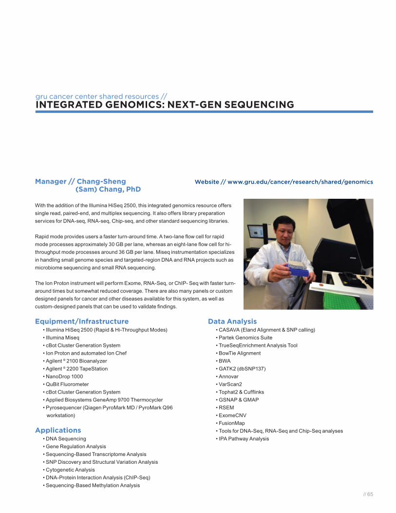
// 65
gru cancer center shared resources //INTEGRATED GENOMICS: NEXT-GEN SEQUENCING
Manager // Chang-Sheng Website // www.gru.edu/cancer/research/shared/genomics (Sam) Chang, PhD
With the addition of the Illumina HiSeq 2500, this integrated genomics resource offers single read, paired-end, and multiplex sequencing. It also offers library preparation services for DNA-seq, RNA-seq, Chip-seq, and other standard sequencing libraries.
Rapid mode provides users a faster turn-around time. A two-lane flow cell for rapid mode processes approximately 30 GB per lane, whereas an eight-lane flow cell for hi-throughput mode processes around 36 GB per lane. Miseq instrumentation specializes in handling small genome species and targeted-region DNA and RNA projects such as microbiome sequencing and small RNA sequencing.
The Ion Proton instrument will perform Exome, RNA-Seq, or ChIP- Seq with faster turn-around times but somewhat reduced coverage. There are also many panels or custom designed panels for cancer and other diseases available for this system, as well as custom-designed panels that can be used to validate findings.
Equipment/Infrastructure• Illumina HiSeq 2500 (Rapid & Hi-Throughput Modes)• Illumina Miseq• cBot Cluster Generation System• Ion Proton and automated Ion Chef• Agilent ® 2100 Bioanalyzer • Agilent ® 2200 TapeStation• NanoDrop 1000• QuBit Fluorometer• cBot Cluster Generation System• Applied Biosystems GeneAmp 9700 Thermocycler• Pyrosequencer (Qiagen PyroMark MD / PyroMark Q96
workstation)
Applications• DNA Sequencing• Gene Regulation Analysis• Sequencing-Based Transcriptome Analysis• SNP Discovery and Structural Variation Analysis• Cytogenetic Analysis• DNA-Protein Interaction Analysis (ChIP-Seq)• Sequencing-Based Methylation Analysis
Data Analysis• CASAVA (Eland Alignment & SNP calling)• Partek Genomics Suite • TrueSeqEnrichment Analysis Tool• BowTie Alignment• BWA• GATK2 (dbSNP137)• Annovar• VarScan2• Tophat2 & Cufflinks • GSNAP & GMAP• RSEM • ExomeCNV• FusionMap • Tools for DNA-Seq, RNA-Seq and Chip-Seq analyses• IPA Pathway Analysis

66 //
gru cancer center shared resources //MICROSCOPY IMAGING CORE
Director // Jeane Silva, PhD Website // www.gru.edu/cancer/research/shared/microscopy
Services• Confocal Microscopy• Fluorescence Microscopy• Digital Imaging scanner• Imaging processing
Equipment• Scanning confocal microscope: Zeiss LSM 510
meta-inverted microscope • Sub-resolution fluorescence microscope: Zeiss AxioObserver.D1• Digital slide scanning system: Ariol DM6000 B Digital Slide Scanner• Two image analysis workstations
Software• Zen 2009 Imaging acquisition and analysis• Digital Imaging Hub – Slide Path software

// 67
gru cancer center shared resources //PROTEOMICS AND METABOLOMICS FACILITY
SoftwareThermo Scientific LTQ Orbitrap Discovery™ Hybrid FT MS
• Agilent ChemStation for LC systems Rev. B.04.01 SP1 (647) (*.M; *.S)
• Thermo Fisher Xcalibur™ 2.0.7 (*.RAW)• Thermo Fisher Proteome Discoverer 1.4 v1.4.0.288 (*.MSF)• Thermo Fisher BioWorks™ Rev.3.3.1 SP1 (*.SRF)• Proteome Software Scaffold 3.6.5 (*.Sf3)
Agilent 6410 Triple Quad LC/MS System• Agilent MassHunter Workstation Software, Qualitative
Analysis, Version B.01.03; Build 1.3.157.0; Patch 2• Agilent MassHunter Workstation Software, Data
Acquisition for 6400 Series Triple Quadrupole, Version B.01.04
• Agilent MassHunter Workstation Software, Quantitative Analysis, Version B.01.04
Software for Agilent 6520 Accurate-Mass Quadrupole Time-of-Flight MS
• Agilent MassHunter Workstation Software, LC/MS Data Acquisition Version B.02.00, for 6200 Series TOF & 6500 Series Q-TOFBuild 1.3.157.0; Patch 2
• Agilent MassHunter Workstation Software, Qualitative Analysis, Version B.03.01; Build 3.1.346.6; Service Pack 2
• Agilent MassHunter Workstation Software, Quantitative Analysis; Version B.01.04
Independently Licensed Software• Proteome Discoverer 1.2 (Thermo Fisher scientific)• Bioworks 3.3.1 SR1 (Thermo Fisher scientific)• Scaffold 3.6.5 (Proteome Software, Inc.)• Scaffold PTM 1.0 (Proteome Software, Inc.)• G3835-64000 Mass Profiler Professional B.02.01.
Rev. B.02.01 G3835-6003 USK0183590 (Agilent Technologies)
• G6825AA Personal METLIN Metabolite Database (Agilent Technologies)
Manager // Lambert Ngoka, PhD Website // www.gru.edu/cancer/research/shared/proteomics
InstrumentationThree LC/MS systems
Proteomics• Thermo Scientific Orbitrap Velos Pro Hybrid Mass Spectrometer/ plus
a new Nanospray Flex Ion Source ES071• Thermo Scientific-Dionex UltiMate 3000 RSLCnano+(TBPLFC)+(MS-
THM)+(nAO2D)• Agilent 1200 Series Nanoflow/capillary LC System for MS
Discovery Metabolomics• Agilent 6520 Accurate-Mass Quadrupole Time-of-Flight MS• Agilent 1200 Series Binary LC System
Targeted Metabolomics• Agilent 6410 Triple Quad LC/MS System• Agilent 1200 Series Binary LC System
ServicesProteomics
• Protein extraction from cell lines, tissue and biofluids• Co-immunoprecipitation• Trypsin digestion• Nano-LC/MS/MS• Protein identification• Detection and characterization of posttranslational modifications of proteins
- structural characterization of modified proteins, lipids and DNA in disease (e.g. the identification and quantification of oxidative damage to proteins, lipids and DNA)
• Screening for genetic mutations in proteinsMetabolomics
• Sample extraction and purification for metabolomics• Metabolome profiling• LC/MS analysis of the different sample sets (i.e. disease vs. matched controls)• XCMS differential analysis of the LC/MS analyses.• Provide XCMS output (m/z and RT) highlighting the ions that differed the
most in intensity between data sets.• Provide tentative identification based on accurate mass and molecules
available in the METLIN database.• Provide more firm identification based on comparative MS/MS and high
accuracy analysis of ‘unknown’ with a standard provided by the client.


// 69
2013 GRU CANCER CENTER PUBLICATIONS

70 //
Alamanda VK, Delisca GO, Mathis SL, Archer KR, Ehrenfeld JM, Miller MW, Homlar KC, Halpern JL, Schwartz HS, Holt GE. The financial burden of reexcising incompletely excised soft tissue sarcomas: a cost analysis. Ann Surg Oncol. 2013 Sep;20(9):2808-14.
Aoyama K, Saha A, Tolar J, Riddle MJ, Veenstra RG, Taylor PA, Blomhoff R, Panoskaltsis-Mortari A, Klebanoff CA, Socié G, Munn DH, Murphy WJ, Serody JS, Fulton LM, Teshima T, Chandraratna RA, Dmitrovsky E, Guo Y, Noelle RJ, Blazar BR. Inhibiting retinoic acid signaling ameliorates graft-versus-host disease by modifying T-cell differentiation and intestinal migration. Blood. 2013 Sep 19;122(12):2125-34.
Ariga T, Itokazu Y, McDonald MP, Hirabayashi Y, Ando S, Yu RK. Brain gangliosides of a transgenic mouse model of Alzheimer’s disease with deficiency in GD3-synthase: expression of elevated levels of a cholinergic-specific ganglioside, GT1aα. ASN Neuro. 2013 May 30;5(2):141-8.
Ariga T, Kubota M, Nakane M, Oguro K, Yu RK, Ando S. Anti-Chol-1 antigen, GQ1bα, antibodies are associated with Alzheimer’s disease. PLoS One. 2013 May 23;8(5):e63326.
Arun SN, Xie D, Howard AC, Zhong Q, Zhong X, McNeil PL, Bollag WB. Cell wounding activates phospholipase D in primary mouse keratinocytes. J Lipid Res. 2013 Mar;54(3):581-91.
Bahassi el M, Li YQ, Wise-Draper TM, Deng L, Wang J, Darnell CN, Wilson KM, Wells SI, Stambrook PJ, Rixe O. A patient-derived somatic mutation in the epidermal growth factor receptor ligand-binding domain confers increased sensitivity to cetuximab in head and neck cancer. Eur J Cancer. 2013 Jul;49(10):2345-55.
Barabutis N, Handa V, Dimitropoulou C, Rafikov R, Snead C, Kumar S, Joshi A, Thangjam G, Fulton D, Black SM, Patel V, Catravas JD. LPS induces pp60c-src-mediated tyrosine phosphorylation of Hsp90 in lung vascular endothelial cells and mouse lung. Am J Physiol Lung Cell Mol Physiol. 2013 Jun 15;304(12):L883-93.
Bardhan K, Liu K. Epigenetics and colorectal cancer pathogenesis. Cancers (Basel). 2013 Jun 5;5(2):676-713.
Barnes VA, Maria BL, Caldwell AL, Hopkins I. Prevention of Traumatic Brain Injury in Youth and Adolescents. J Child Neurol. 2013 Nov;28(11):1412-1417.
Barnes VA, Rigg JL, Williams JJ. Clinical case series: treatment of PTSD with transcendental meditation in active duty military personnel. Mil Med. 2013 Jul;178(7):e836-40.
Bartlett DL, Liu Z, Sathaiah M, Ravindranathan R, Guo Z, He Y, Guo ZS. Oncolytic viruses as therapeutic cancer vaccines. Mol Cancer. 2013 Sep 11;12(1):103.
Berger PK, Principe JL, Laing EM, Henley EC, Pollock NK, Taylor RG, Blair RM, Baile CA, Hall DB, Lewis RD. Weight gain in college females is not prevented by isoflavone-rich soy protein: a randomized controlled trial. Nutr Res. 2014 Jan;34(1):66-73.
Berning A, Eason A, Gilley N, Takhar S, ElShafey S, Thangaraju M, Schoenlein P. Abstract 1725: HDAC inhibition induces Bim expression and apoptosis in breast cancer cells undergoing paclitaxel or antiestrogen treatment. Cancer Res. 2013 Apr 15;73(8):Supplement 1.
Bharucha AE, Rao SS. An update on anorectal disorders for gastroenterologists. Gastroenterology. 2014 Jan;146(1):37-45.e2.
Binder DC, Engels B, Arina A, Yu P, Slauch JM, Fu YX, Karrison T, Burnette B, Idel C, Zhao M, Hoffman RM, Munn DH, Rowley DA, Schreiber H. Antigen-specific bacterial vaccine combined with anti-PD-L1 rescues dysfunctional endogenous T cells to reject long-established cancer. Cancer Immunol Res. 2013 Aug;1:123-133.
Bollag WB. COUP-TF-interacting protein-2 (Ctip2): driving the coupé of sphingolipid biosynthesis in the epidermis. J Invest Dermatol. 2013 Mar;133(3):593-4.
Bollag WB, Hill WD. CXCR4 in epidermal keratinocytes: crosstalk within the skin. J Invest Dermatol. 2013 Nov;133(11):2505-8.
Bollag WB, Isales CM. GRowing an epidermal tumor. J Invest Dermatol. 2013 Dec;133(12):2659-62.
Britten CM, Singh-Jasuja H, Flamion B, Hoos A, Huber C, Kallen KJ, Khleif SN, Kreiter S, Nielsen M, Rammensee HG, Sahin U, Hinz T, Kalinke U. The regulatory landscape for actively personalized cancer immunotherapies. Nat Biotechnol. 2013 Oct;31(10):880-2.
Burnette JO, Klaassen Z, Hatley RM, Neunert CE, Williams H, Donohoe JM. Staging paratesticular rhabdomyosarcoma in the “as low as reasonably achievable” age: the case for PET-CT. Urology. 2013 Jul;82(1):220-3.
2013 gru cancer center publications

// 71
Byeon B, Wang W, Barski A, Ranallo RT, Bao K, Schones DE, Zhao K, Wu C, Wu WH. The ATP-dependent chromatin remodeling enzyme Fun30 represses transcription by sliding promoter-proximal nucleosomes. J Biol Chem. 2013 Aug 9;288(32):23182-93.
Calao M, Sekyere EO, Cui H J , Cheung BB, Thomas WD, Keating J, Chen JB, Raif A, Jankowski K, Davies NP, Bekkum MV, Chen B, Tan O, Ellis T, Norris MD, Haber M, Kim ES, Shohet JM, Trahair TN, Liu T, Wainwright BJ, Ding HF, Marshall GM. Direct effects of Bmi1 on p53 protein stability inactivates oncoprotein stress responses in embryonal cancer precursor cells at tumor initiation. Oncogene (2013) 32, 3616–3626.Cassuto J, Dou H, Czikora I, Szabo A, Patel VS, Kamath V, Belin de Chantemele E, Feher A, Romero MJ, Bagi Z. Peroxynitrite Disrupts Endothelial Caveolae Leading to eNOS Uncoupling and Diminished Flow-Mediated Dilation in Coronary Arterioles of Diabetic Patients. Diabetes. 2013 Dec 18. [Epub ahead of print]
Cassuto J, Feher A, Lan L, Patel VS, Kamath V, Anthony DC, Bagi Z. Obesity and statins are both independent predictors of enhanced coronary arteriolar dilation in patients undergoing heart surgery. J Cardiothorac Surg. 2013 Apr 30;8(1):117
Cebula A, Seweryn M, Rempala GA, Pabla SS, McIndoe RA, Denning TL, Bry L, Kraj P, Kisielow P, Ignatowicz L. Thymus-derived regulatory T cells contribute to tolerance to commensal microbiota. Nature. 2013 May 9;497(7448):258-62.
Chaudhary K, Kleven DT, McGaha TL, Madaio MP. A human monoclonal antibody against the collagen type IV α3NC1 domain is a non-invasive optical biomarker for glomerular diseases. Kidney Int. 2013 Aug;84(2):403-8.
Chen B, Yi B, Mao R, Liu H, Wang J, Sharma A, Peiper S, Leonard WJ, She JX. Enhanced T cell lymphoma in NOD.Stat5b transgenic mice is caused by hyperactivation of Stat5b in CD8+ thymocytes. PLoS One. 2013;8(2):e56600.
Chernecky CC, Waller JL, Jarvis WR. In vitro study assessing the antibacterial activity of three silver-impregnated/coated mechanical valve needleless connectors after blood exposure. Am J Infect Control. 2013 Mar;41(3):278-80.
Chinnaiyan P, Won M, Wen PY, Rojiani AM, Wendland M, Dipetrillo TA, Corn BW, Mehta MP. RTOG 0913: a phase 1 study of daily everolimus (RAD001) in combination with radiation therapy and temozolomide in patients with newly diagnosed glioblastoma. Int J Radiat Oncol Biol Phys. 2013 Aug 1;86(5):880-4.
Chothe PP, Chutkan N, Sangani R, Wenger KH, Prasad PD, Thangaraju M, Hamrick MW, Isales CM, Ganapathy V, Fulzele S. Sodium-coupled vitamin C transporter (SVCT2): expression, function, and regulation in intervertebral disc cells. Spine J. 2013 May;13(5):549-57.
Ciarrocca K, Jackson LL, De Rossi SS. Human papillomavirus: the fundamentals of HPV for oral health care providers. J Calif Dent Assoc. 2013 May;41(5):349-55.
Coothankandaswamy V, Elangovan S, Singh N, Prasad PD, Thangaraju M, Ganapathy V. The plasma membrane transporter SLC5A8 suppresses tumour progression through depletion of survivin without involving its transport function. Biochem J. 2013 Feb 15;450(1):169-78.
Cosentino-Boehm AL, *Lafky JM, Greenwood T, Kimbler K, Buenafe MC, Wang Y, Branscum AJ, Yang P, Maihle NJ, Baron AT. Soluble HER2 (sHER2) is a potential risk assessment, screening, and diagnostic biomarker of non-small cell lung cancer. Diagnostics 2013 3:13-32. Coss-Adame E, Erdogan A, Rao SS. Treatment of Esophageal (Noncardiac) Chest Pain: A Review. Clin Gastroenterol Hepatol. 2013 Aug 28. pii: S1542-3565(13)01255-X. doi: 10.1016/j.cgh.2013.08.036. [Epub ahead of print]
Cresci G, Nagy LE, Ganapathy V. Lactobacillus GG and tributyrin supplementation reduce antibiotic-induced intestinal injury. JPEN J Parenter Enteral Nutr. 2013 Nov;37(6):763-74.
Cronin P, Rawson JV. Health services research in radiology: meeting the needs of the professions and the patients. Acad Radiol. 2013 Sep;20(9):1061-2.
Dasgupta S, Kong J, Bieberich E. Phytoceramide in vertebrate tissues: one step chromatography separation for molecular characterization of ceramide species. PLoS One. 2013 Nov 29;8(11):e80841.
Davis CL, Waller JL, Pollock NK. Exercise for overweight children and diabetes risk--reply. JAMA. 2013 Jan 9;309(2):133-4.
Dennis BA, Ergul A, Gower BA, Allison JD, Davis CL. Oxidative stress and cardiovascular risk in overweight children in an exercise intervention program. Child Obes. 2013 Feb;9(1):15-21.
Despotovic JM, Neunert CE. Is anti-D immunoglobulin still a frontline treatment option for immune thrombocytopenia? Hematology Am Soc Hematol Educ Program. 2013;2013:283-5.
2013 gru cancer center publications

72 //
Ding J, Li T, Wang X, Zhao E, Choi JH, Yang L, Zha Y, Dong Z, Huang S, Asara JM, Cui H, Ding HF. The histone H3 methyltransferase G9A epigenetically activates the serine-glycine synthesis pathway to sustain cancer cell survival and proliferation. Cell Metab. 2013 Dec 3;18(6):896-907.
Doi K, Noiri E, Nangaku M, Yahagi N, Jayakumar C, Ramesh G. Repulsive guidance cue semaphorin 3A in urine predicts the progression of acute kidney injury in adult patients from a mixed intensive care unit. Nephrol Dial Transplant. 2014 Jan;29(1):73-80.
Dolisca SB, Mehta M, Pearce DA, Mink JW, Maria BL. Batten disease: clinical aspects, molecular mechanisms, translational science, and future directions. J Child Neurol. 2013 Sep;28(9):1074-100.
Dong C, Wu G. G-protein-coupled receptor interaction with small GTPases. Methods Enzymol. 2013;522:97-108.
Dowling AR, Nedorezov LB, Qiu X, Marino JS, Hill JW. Genetic factors modulate the impact of pubertal androgen excess on insulin sensitivity and fertility. PLoS One. 2013 Nov 20;8(11):e79849.
Duke WS, Terris DJ. Simplifying nerve monitoring. Otolaryngol Head Neck Surg. 2013 Sep;149(3):517.
Dun B, Sharma A, Xu H, Liu H, Bai S, Zeng L, She JX. Transcriptomic changes induced by mycophenolic acid in gastric cancer cells. Am J Transl Res. 2013 Dec 1;6(1):28-42.
Dun B, Xu H, Sharma A, Liu H, Yu H, Yi B, Liu X, He M, Zeng L, She JX. Delineation of biological and molecular mechanisms underlying the diverse anticancer activities of mycophenolic acid. Int J Clin Exp Pathol. 2013 Nov 15;6(12):2880-6.
Dun B, Sharma A, Teng Y, Liu H, Purohit S, Xu H, Zeng L, She JX. Mycophenolic acid inhibits migration and invasion of gastric cancer cells via multiple molecular pathways. PLoS One. 2013 Nov 15;8(11):e81702.
Duncan MB. Extracellular matrix transcriptome dynamics in hepatocellular carcinoma. Matrix Biol. 2013 Oct-Nov;32(7-8):393-8.
Elangovan S, Pathania R, Ramachandran S, Ananth S, Padia RN, Lan L, Singh N, Martin PM, Hawthorn L, Prasad PD, Ganapathy V, Thangaraju M. The Niacin/Butyrate Receptor GPR109A Suppresses Mammary Tumorigenesis by Inhibiting Cell Survival. Cancer Res. 2014 Feb 15;74(4):1166-78. Elangovan S, Pathania R, Ramachandran S, Ananth S, Padia RN, Srinivas SR, Babu E, Hawthorn L, Schoenlein PV, Boettger T, Smith SB, Prasad PD, Ganapathy V, Thangaraju M. Molecular mechanism of SLC5A8 inactivation in breast cancer. Mol Cell Biol. 2013 Oct;33(19):3920-35.
Elding Larsson H, Vehik K, Gesualdo P, Akolkar B, Hagopian W, Krischer J, Lernmark A, Rewers M, Simell O, She JX, Ziegler A, Haller MJ; the TEDDY Study Group. Children followed in the TEDDY study are diagnosed with type 1 diabetes at an early stage of disease. Pediatr Diabetes. 2013 Aug 27. doi: 10.1111/pedi.12066. [Epub ahead of print]
Eroglu B, Min JN, Zhang Y, Szurek E, Moskophidis D, Eroglu A, Mivechi NF. An essential role for heat shock transcription factor binding protein 1 (HSBP1) during early embryonic development. Dev Biol. 2014 Feb 15;386(2):448-60.
Ezeoke A, Mellor A, Buckley P, Miller B. A systematic, quantitative review of blood autoantibodies in schizophrenia. Schizophr Res. 2013 Oct;150(1):245-51.
Fahmy CE, Carrau R, Kirsch C, Meeks D, de Lara D, Solares CA, Otto BA, Prevedello DM. Volumetric analysis of endoscopic and traditional surgical approaches to the infratemporal fossa. Laryngoscope. 2013 Oct 1. doi: 10.1002/lary.24428. [Epub ahead of print]
Feher A, Cassuto J, Szabo A, Patel V, Vinayak Kamath M, Bagi Z. Increased tissue angiotensin-converting enzyme activity impairs bradykinin-induced dilation of coronary arterioles in obesity. Circ J. 2013;77(7):1867-76.
Felema GG, Bryskin RB, Heger IM, Saswata R. Venous air embolism from Tisseel use during endoscopic cranial vault remodeling for craniosynostosis repair: a case report. Paediatr Anaesth. 2013 Aug;23(8):754-6.
Fick DM, Steis MR, Waller JL, Inouye SK. Delirium superimposed on dementia is associated with prolonged length of stay and poor outcomes in hospitalized older adults. J Hosp Med. 2013 Sep;8(9):500-5.
Fizazi K, Delva R, Caty A, Chevreau C, Kerbrat P, Rolland F, Priou F, Geoffrois L, Rixe O, Beuzeboc P, Malhaire JP, Culine S, Aubelle MS, Laplanche A. A risk-adapted study of cisplatin and etoposide, with or without ifosfamide, in patients with metastatic seminoma: results of the GETUG S99 multicenter prospective study. Eur Urol. 2014 Feb;65(2):381-6.
2013 gru cancer center publications

// 73
Fox JM, Sage LK, Huang L, Barber J, Klonowski KD, Mellor AL, Tompkins SM, Tripp RA. Inhibition of indoleamine 2,3-dioxygenase enhances the T-cell response to influenza virus infection. J Gen Virol. 2013 Jul;94(Pt 7):1451-61.
Fulzele S, Chothe P, Sangani R, Chutkan N, Hamrick M, Bhattacharyya M, Prasad PD, Zakhary I, Bowser M, Isales C, Ganapathy V. Sodium-dependent vitamin C transporter SVCT2: expression and function in bone marrow stromal cells and in osteogenesis. Stem Cell Res. 2013 Jan;10(1):36-47.
Galarneau G, Coady S, Garrett ME, Jeffries N, Puggal M, Paltoo D, Soldano K, Guasch A, Ashley-Koch AE, Telen MJ, Kutlar A, Lettre G, Papanicolaou GJ. Gene-centric association study of acute chest syndrome and painful crisis in sickle cell disease patients. Blood. 2013 Jul 18;122(3):434-42.
Ganapathy V, Thangaraju M, Prasad PD, Martin PM, Singh N. Transporters and receptors for short-chain fatty acids as the molecular link between colonic bacteria and the host. Curr Opin Pharmacol. 2013 Dec;13(6):869-74.
Ganesh SK, Tragante V, Guo W, Guo Y, Lanktree MB, Smith EN, Johnson T, Castillo BA, Barnard J, Baumert J, Chang YP, Elbers CC, Farrall M, Fischer ME, Franceschini N, Gaunt TR, Gho JM, Gieger C, Gong Y, Isaacs A, Kleber ME, Mateo Leach I, McDonough CW, Meijs MF, Mellander O, Molony CM, Nolte IM, Padmanabhan S, Price TS, Rajagopalan R, Shaffer J, Shah S, Shen H, Soranzo N, van der Most PJ, Van Iperen EP, Van Setten J, Vonk JM, Zhang L, Beitelshees AL, Berenson GS, Bhatt DL, Boer JM, Boerwinkle E, Burkley B, Burt A, Chakravarti A, Chen W, Cooper-Dehoff RM, Curtis SP, Dreisbach A, Duggan D, Ehret GB, Fabsitz RR, Fornage M, Fox E, Furlong CE, Gansevoort RT, Hofker MH, Hovingh GK, Kirkland SA, Kottke-Marchant K, Kutlar A, Lacroix AZ, Langaee TY, Li YR, Lin H, Liu K, Maiwald S, Malik R; CARDIOGRAM, METASTROKE, Murugesan G, Newton-Cheh C, O’Connell JR, Onland-Moret NC, Ouwehand WH, Palmas W, Penninx BW, Pepine CJ, Pettinger M, Polak JF, Ramachandran VS, Ranchalis J, Redline S, Ridker PM, Rose LM, Scharnag H, Schork NJ, Shimbo D, Shuldiner AR, Srinivasan SR, Stolk RP, Taylor HA, Thorand B, Trip MD, van Duijn CM, Verschuren WM, Wijmenga C, Winkelmann BR, Wyatt S, Young JH, Boehm BO, Caulfield MJ, Chasman DI, Davidson KW, Doevendans PA, Fitzgerald GA, Gums JG, Hakonarson H, Hillege HL, Illig T, Jarvik GP, Johnson JA, Kastelein JJ, Koenig W; LifeLines Cohort Study, März W, Mitchell BD, Murray SS, Oldehinkel AJ, Rader DJ, Reilly MP, Reiner AP, Schadt EE, Silverstein RL, Snieder H, Stanton AV, Uitterlinden AG, van der Harst P, van der Schouw YT, Samani NJ, Johnson AD, Munroe PB, de Bakker PI, Zhu X, Levy D, Keating BJ, Asselbergs FW. Loci influencing blood pressure identified using a cardiovascular gene-centric array. Hum Mol Genet. 2013 Apr 15;22(8):1663-78.
Gnana-Prakasam JP, Veeranan-Karmegam R, Coothankandaswamy V, Reddy SK, Martin PM, Thangaraju M, Smith SB, Ganapathy V. Loss of Hfe leads to progression of tumor phenotype in primary retinal pigment epithelial cells. Invest Ophthalmol Vis Sci. 2013 Jan 7;54(1):63-71.
Goldstone SE, Jessen H, Palefsky JM, Giuliano AR, Moreira ED Jr, Vardas E, Aranda C, Hillman RJ, Ferris DG, Coutlee F, Marshall JB, Vuocolo S, Haupt RM, Guris D, Garner E. Quadrivalent HPV vaccine efficacy against disease related to vaccine and non-vaccine HPV types in males. Vaccine. 2013 Aug 20;31(37):3849-55.
Gower BA, Pollock NK, Casazza K, Clemens TL, Goree LL, Granger WM. Associations of total and undercarboxylated osteocalcin with peripheral and hepatic insulin sensitivity and β-cell function in overweight adults. J Clin Endocrinol Metab. 2013 Jul;98(7):E1173-80.
Guo DH, Parikh SJ, Chao J, Pollock NK, Wang X, Snieder H, Navis G, Wilson JG, Bhagatwala J, Zhu H, Dong Y. Urinary prostasin excretion is associated with adiposity in nonhypertensive African-American adolescents. Pediatr Res. 2013 Aug;74(2):206-10.
Guo G, Marrero L, Rodriguez P, Del Valle L, Ochoa A, Cui Y. Trp53 inactivation in the tumor microenvironment promotes tumor progression by expanding the immunosuppressive lymphoid-like stromal network. Cancer Res. 2013 Mar 15;73(6):1668-75.
Hao Z, Dillard T, Biddinger P, Patel V. Suppression of respiratory papillomatosis with malignant transformation by erlotinib in a kidney transplant recipient. BMJ Case Rep. 2013 Aug 28;2013. pii: bcr2013008757.
Harbarger CF, Weinberger PM, Borders JC, Hughes CA. Prenatal ultrasound exposure and association with postnatal hearing outcomes. J Otolaryngol Head Neck Surg. 2013 Jan 31;42:3.
Hawkins CM, Duszak R, Rawson JV. Social Media in Radiology: Early Trends in Twitter Microblogging at Radiology’s Largest International Meeting. J Am Coll Radiol. 2013 Oct 17. pii: S1546-1440(13)00438-9.
Hawthorn L, Lan L, Mojica W. Evidence for Field Effect Cancerization in Colorectal Cancer. Genomics. 2013 Dec 3. pii: S0888-7543(13)00206-1. [Epub ahead of print]
Heilbrun ME, Rawson JV, Shah M. Using health services research to meet ACGME resident research requirements. Acad Radiol. 2013 Sep;20(9):1077-82.
2013 gru cancer center publications

74 //
Hepworth MR, Monticelli LA, Fung TC, Ziegler CG, Grunberg S, Sinha R, Mantegazza AR, Ma HL, Crawford A, Angelosanto JM, Wherry EJ, Koni PA, Bushman FD, Elson CO, Eberl G, Artis D, Sonnenberg GF. Innate lymphoid cells regulate CD4+ T-cell responses to intestinal commensal bacteria. Nature. 2013 Jun 6;498(7452):113-7.
Holmgaard RB, Zamarin D, Munn DH, Wolchok JD, Allison JP. Indoleamine 2,3-dioxygenase is a critical resistance mechanism in antitumor T cell immunotherapy targeting CTLA-4. J Exp Med. 2013 Jul 1;210(7):1389-402.
Homlar KC, Sellers MH, Halpern JL, Seeley EH, Holt GE. Serum levels of methyl methacrylate following inhalational exposure to polymethylmethacrylate bone cement. J Arthroplasty. 2013 Mar;28(3):406-9.
Hong S, Noh H, Chen H, Padia R, Pan ZK, Su SB, Jing Q, Ding HF, Huang S. Signaling by p38 MAPK stimulates nuclear localization of the microprocessor component p68 for processing of selected primary microRNAs. Sci Signal. 2013 Mar 12;6(266):ra16.
Hong Y, Peng Y, Guo ZS, Guevara-Patino J, Pang J, Butterfield LH, Mivechi NF, Munn DH, Bartlett DL, He Y. Epitope-optimized alpha-fetoprotein genetic vaccines prevent carcinogen-induced murine autochthonous hepatocellular carcinoma. Hepatology. 2013 Oct 12. doi: 10.1002/hep.26893. [Epub ahead of print]
Hosseini SM, McLaughlin N, Carrau RL, Otto B, Prevedello DM, Solares CA, Zanation AM, Kassam AB. Endoscopic transpterygoid nasopharyngectomy: correlation of surgical anatomy with multiplanar CT. Head Neck. 2013 May;35(5):704-14.
Hu X, Zimmerman MA, Bardhan K, Yang D, Waller JL, Liles GB, Lee JR, Pollock R, Lev D, Ware CF, Garber E, Bailly V, Browning JL, Liu K. Lymphotoxin β receptor mediates caspase-dependent tumor cell apoptosis in vitro and tumor suppression in vivo despite induction of NF-κB activation. Carcinogenesis. 2013 May;34(5):1105-14.
Hu X, Bardhan K, Paschall AV, Yang D, Waller JL, Park MA, Nayak-Kapoor A, Samuel TA, Abrams SI, Liu K. Deregulation of apoptotic factors Bcl-xL and Bax confers apoptotic resistance to myeloid-derived suppressor cells and contributes to their persistence in cancer. J Biol Chem. 2013 Jun 28;288(26):19103-15.
Huang L, Mellor AL. Metabolic control of tumour progression and antitumour immunity. Curr Opin Oncol. 2014 Jan;26(1):92-9.
Huang L, Li L, Klonowski KD, Tompkins SM, Tripp RA, Mellor AL. Induction and role of indoleamine 2,3 dioxygenase in mouse models of influenza a virus infection. PLoS One. 2013 Jun 13;8(6):e66546.
Huang L, Li L, Lemos H, Chandler PR, Pacholczyk G, Baban B, Barber GN, Hayakawa Y, McGaha TL, Ravishankar B, Munn DH, Mellor AL. Cutting edge: DNA sensing via the STING adaptor in myeloid dendritic cells induces potent tolerogenic responses. J Immunol. 2013 Oct 1;191(7):3509-13.
Hutchings EJ, Waller JL, Terry AV Jr. Differential long-term effects of haloperidol and risperidone on the acquisition and performance of tasks of spatial working and short-term memory and sustained attention in rats. J Pharmacol Exp Ther. 2013 Dec;347(3):547-56.
Itokazu Y, Kato-Negishi M, Nakatani Y, Ariga T, Yu RK. Effects of amyloid β-peptides and gangliosides on mouse neural stem cells. Neurochem Res. 2013 Oct;38(10):2019-27.
Jana T, Khabbaz E, Bush CM, Prosser JD, Birchall MA, Nichols CA, Postma GN, Weinberger PM. The body as a living bioreactor: a feasibility study of pedicle flaps for tracheal transplantation. Eur Arch Otorhinolaryngol. 2013 Jan;270(1):181-6.
Jayakumar C, Ranganathan P, Devarajan P, Krawczeski CD, Looney S, Ramesh G. Semaphorin 3A is a new early diagnostic biomarker of experimental and pediatric acute kidney injury. PLoS One. 2013;8(3):e58446.
Jin B, Robertson KD. DNA methyltransferases, DNA damage repair, and cancer. Adv Exp Med Biol. 2013;754:3-29.
Jin Y, Sharma A, Carey C, Hopkins D, Wang X, Robertson DG, Bode B, Anderson SW, Reed JC, Steed RD, Steed L, She JX. The expression of inflammatory genes is upregulated in peripheral blood of patients with type 1 diabetes. Diabetes Care. 2013 Sep;36(9):2794-802.
Johnson TS, Terrell CE, Millen SH, Katz JD, Hildeman DA, Jordan MB. Etoposide selectively ablates activated T cells to control the immunoregulatory disorder hemophagocytic lymphohistiocytosis. J Immunol. 2014 Jan 1;192(1):84-91.
Jrad-Lamine A, Henry-Berger J, Damon-Soubeyrand C, Saez F, Kocer A, Janny L, Pons-Rejraji H, Munn DH, Mellor AL, Gharbi N, Cadet R, Guiton R, Aitken RJ, Drevet JR. Indoleamine 2,3-dioxygenase 1 (ido1) is involved in the control of mouse caput epididymis immune environment. PLoS One. 2013 Jun 20;8(6):e66494.
2013 gru cancer center publications

// 75
Kaji T, Furukawa K, Ishige A, Toyokura I, Nomura M, Okada M, Takahashi Y, Shimoda M, Takemori T. Both mutated and unmutated memory B cells accumulate mutations in the course of the secondary response and develop a new antibody repertoire optimally adapted to the secondary stimulus. Int Immunol. 2013 Dec;25(12):683-95.
Kalpatthi R, Thompson B, Lu M, Wang WC, Patel N, Kutlar A, Howard T, Luchtman-Jones L, Miller ST; BABY HUG Investigators. Comparison of hematologic measurements between local and central laboratories: data from the BABY HUG trial. Clin Biochem. 2013 Feb;46(3):278-81.
Kang M, Shen XJ, Kim S, Araujo-Perez F, Galanko JA, Martin CF, Sandler RS, Keku TO. Somatic gene mutations in African Americans may predict worse outcomes in colorectal cancer. Cancer Biomark. 2013;13(5):359-66.
Kasemsiri P, Solares CA, Carrau RL, Prosser JD, Prevedello DM, Otto BA, Old M, Kassam AB. Endoscopic endonasal transpterygoid approaches: anatomical landmarks for planning the surgical corridor. Laryngoscope. 2013 Apr;123(4):811-5.
Kassing P, Mulaik MW, Rawson J. Pricing radiology bundled CPT codes accurately. Radiol Manage. 2013 Mar-Apr;35(2):9-15.
Kazi AA, Flowers WJ, Barrett JM, O’Rourke AK, Postma GN, Weinberger PM. Ethical issues in laryngology: Tracheal stenting as palliative care. Laryngoscope. 2013 Nov 20. doi: 10.1002/lary.24531. [Epub ahead of print]
Kazi AA, Solowski NL, Postma GN, Weinberger PM. Esophageal perforation in a patient with diverticulum following anterior discectomy and fusion. Ear Nose Throat J. 2013 Oct-Nov;92(10-11):506-7.
Kim S, Taylor JA, Milne GL, Sandler DP. Association between urinary prostaglandin E2 metabolite and breast cancer risk: a prospective, case-cohort study of postmenopausal women. Cancer Prev Res (Phila). 2013 Jun;6(6):511-8.
King MD, Alleyne CH Jr, Dhandapani KM. TNF-alpha receptor antagonist, R-7050, improves neurological outcomes following intracerebral hemorrhage in mice. Neurosci Lett. 2013 May 10;542:92-6.
Kolhe R, Reid MD, Lee JR, Cohen C, Ramalingam P. Immunohistochemical expression of PAX5 and TdT by Merkel cell carcinoma and pulmonary small cell carcinoma: a potential diagnostic pitfall but useful discriminatory marker. Int J Clin Exp Pathol. 2013;6(2):142-7.
Koni PA, Bolduc A, Takezaki M, Ametani Y, Huang L, Lee JR, Nutt SL, Kamanaka M, Flavell RA, Mellor AL, Tsubata T, Shimoda M. Constitutively CD40-activated B cells regulate CD8 T cell inflammatory response by IL-10 induction. J Immunol. 2013 Apr 1;190(7):3189-96.
Krafft CE, Schwarz NF, Chi L, Weinberger AL, Schaeffer DJ, Pierce JE, Rodrigue AL, Yanasak NE, Miller PH, Tomporowski PD, Davis CL, McDowell JE. An 8-month randomized controlled exercise trial alters brain activation during cognitive tasks in overweight children. Obesity (Silver Spring). 2014 Jan;22(1):232-42.
Kuczma M, Kurczewska A, Kraj P. Modulation of bone morphogenic protein signaling in T-cells for cancer immunotherapy. J Immunotoxicol. 2013 Dec 19. [Epub ahead of print]
Kutlar A. GLEE-ful for sickle cell pain? Blood. 2013 Sep 12;122(11):1846-7.
Kutlar A, Reid ME, Inati A, Taher AT, Abboud MR, El-Beshlawy A, Buchanan GR, Smith H, Ataga KI, Perrine SP, Ghalie RG. A dose-escalation phase IIa study of 2,2-dimethylbutyrate (HQK-1001), an oral fetal globin inducer, in sickle cell disease. Am J Hematol. 2013 Nov;88(11):E255-60.
Laing EM, Tripp RA, Pollock NK, Baile CA, Della-Fera MA, Rayalam S, Tompkins SM, Keys DA, Lewis RD. Adenovirus 36, adiposity, and bone strength in late-adolescent females. J Bone Miner Res. 2013 Mar;28(3):489-96.
Laird MD, Shields JS, Sukumari-Ramesh S, Kimbler DE, Fessler RD, Shakir B, Youssef P, Yanasak N, Vender JR, Dhandapani KM. High mobility group box protein-1 promotes cerebral edema after traumatic brain injury via activation of toll-like receptor 4. Glia. 2014 Jan;62(1):26-38. Lee D, Xiong S, Xiong WC. General introduction to in situ hybridization protocol using nonradioactively labeled probes to detect mRNAs on tissue sections. Methods Mol Biol. 2013;1018:165-74.
Lee EJ, Luo J, Wilson JM, Shi H. Analyzing the cancer methylome through targeted bisulfite sequencing. Cancer Lett. 2013 Nov 1;340(2):171-8.
Lee HS, Burkhardt BR, McLeod W, Smith S, Eberhard C, Lynch K, Hadley D, Rewers M, Simell O, She JX, Hagopian B, Lernmark A, Akolkar B, Ziegler AG, Krischer JP; the TEDDY Study Group. Biomarker discovery study design for type 1 diabetes in The Environmental Determinants of Diabetes in the Young (TEDDY) study. Diabetes Metab Res Rev. 2013 Dec 15. doi: 10.1002/dmrr.2510. [Epub ahead of print]
2013 gru cancer center publications

76 //
Lee YY, Erdogan A, Rao SS. High resolution and high definition anorectal manometry and pressure topography: diagnostic advance or a new kid on the block? Curr Gastroenterol Rep. 2013 Dec;15(12):360.
LeMaoult J, Daouya M, Wu J, Loustau M, Horuzsko A, Carosella ED. Synthetic HLA-G proteins for therapeutic use in transplantation. FASEB J. 2013 Sep;27(9):3643-51.
Leslie KK, Sill MW, Darcy KM, Baron AT, *Wilken JA, Godwin AK, Cook L, Schilder RJ, Schilder JM, Maihle NJ. Prognostic value of serum sEGFR and EGFR mutations in a phase II trial of gefitinib (Iressa®, ZD1839) in women with endometrial cancer. Gynecologic Oncology. 2013 129:486-484.
Lewerenz J, Hewett SJ, Huang Y, Lambros M, Gout PW, Kalivas PW, Massie A, Smolders I, Methner A, Pergande M, Smith SB, Ganapathy V, Maher P. The cystine/glutamate antiporter system x(c)(-) in health and disease: from molecular mechanisms to novel therapeutic opportunities. Antioxid Redox Signal. 2013 Feb 10;18(5):522-55.
Lin L, Hutzen B, Lee HF, Peng Z, Wang W, Zhao C, Lin HJ, Sun D, Li PK, Li C, Korkaya H, Wicha MS, Lin J. Evaluation of STAT3 Signaling in ALDH+ and ALDH+/CD44+/CD24- Subpopulations of Breast Cancer Cells. PLoS One. 2013 Dec 23;8(12):e82821.
Liu H, Huang L, Bradley J, Liu K, Bardhan K, Ron D, Mellor AL, Munn DH, McGaha TL. GCN2-dependent metabolic stress is essential for endotoxemic cytokine induction and pathology. Mol Cell Biol. 2014 Feb;34(3):428-38.
Liu L, Karmakar S, Dhar R, Mahajan M, Choudhury A, Weissman S, Pace BS. Regulation of Gγ-globin gene by ATF2 and its associated proteins through the cAMP-response element. PLoS One. 2013 Nov 6;8(11):e78253.
Lucas R, Parikh SJ, Sridhar S, Guo DH, Bhagatwala J, Dong Y, Caldwell R, Mellor A, Caldwell W, Zhu H, Dong Y. Cytokine profiling of young overweight and obese female African American adults with prediabetes. Cytokine. 2013 Oct;64(1):310-5.
Luo D, Wilson JM, Harvel N, Liu J, Pei L, Huang S, Hawthorn L, Shi H. A systematic evaluation of miRNA:mRNA interactions involved in the migration and invasion of breast cancer cells. J Transl Med. 2013 Mar 5;11:57.
Mansoori B, Vidal LL, Applegate K, Rawson JV, Novak RD, Ros PR. Impact of Patient Protection and Affordable Care Act on academic radiology departments’ clinical, research, and education missions. Acad Radiol. 2013 Oct;20(10):1213-7.
Mao R, Xiao W, Liu H, Chen B, Yi B, Kraj P, She JX. Systematic evaluation of 640 FDA drugs for their effect on CD4+Foxp3+ regulatory T cells using a novel cell-based high throughput screening assay. Biochem Pharmacol. 2013 May 15;85(10):1513-24.
Marabelle A, Kohrt H, Sagiv-Barfi I, Ajami B, Axtell RC, Zhou G, Rajapaksa R, Green MR, Torchia J, Brody J, Luong R, Rosenblum MD, Steinman L, Levitsky HI, Tse V, Levy R. Depleting tumor-specific Tregs at a single site eradicates disseminated tumors. J Clin Invest. 2013 Jun 3;123(6):2447-63.
Maria BL. Batten disease: current understanding and future directions. J Child Neurol. 2013 Sep;28(9):1072-3.
Markand S, Tawfik A, Ha Y, Gnana-Prakasam J, Sonne S, Ganapathy V, Sen N, Xian M, Smith SB. Cystathionine beta synthase expression in mouse retina. Curr Eye Res. 2013 May;38(5):597-604. Mason E, Van Rompaey J, Carrau R, Panizza B, Solares CA. Anatomical and computed tomographic analysis of the transcochlear and endoscopic transclival approaches to the petroclival region. Laryngoscope. 2014 Mar;124(3):628-36.
Master VA, Moses KA. Racial disparities in prostate cancer care: is adherence to National Comprehensive Cancer Network guidelines good enough for our patients? Cancer. 2013 Jun 15;119(12):2209-11.
McGaha TL, Huang L, Lemos H, Metz R, Mautino M, Prendergast GC, Mellor AL. Amino acid catabolism: a pivotal regulator of innate and adaptive immunity. Immunol Rev. 2012 Sep;249(1):135-57.
Meijer A, Roseman M, Delisle VC, Milette K, Levis B, Syamchandra A, Stefanek ME, Stewart DE, de Jonge P, Coyne JC, Thombs BD. Effects of screening for psychological distress on patient outcomes in cancer: a systematic review. J Psychosom Res. 2013 Jul;75(1):1-17.
Miller BJ, Gassama B, Sebastian D, Buckley P, Mellor A. Meta-analysis of lymphocytes in schizophrenia: clinical status and antipsychotic effects. Biol Psychiatry. 2013 May 15;73(10):993-9.
Miller BJ, Graham KL, Bodenheimer CM, Culpepper NH, Waller JL, Buckley PF. A prevalence study of urinary tract infections in acute relapse of schizophrenia. J Clin Psychiatry. 2013 Mar;74(3):271-7.
2013 gru cancer center publications

// 77
Miller BJ, Mellor A, Buckley P. Total and differential white blood cell counts, high-sensitivity C-reactive protein, and the metabolic syndrome in non-affective psychoses. Brain Behav Immun. 2013 Jul;31:82-9.
Mkrtichyan M, Chong N, Eid RA, Wallecha A, Singh R, Rothman J, Khleif SN. Anti-PD-1 antibody significantly increases therapeutic efficacy of Listeria monocytogenes (Lm)-LLO immunotherapy. J ImmunoTherapy of Cancer. 2013 29Aug;1:15.
Mohamed R, Jayakumar C, Ramesh G. Chronic administration of EP4-selective agonist exacerbates albuminuria and fibrosis of the kidney in streptozotocin-induced diabetic mice through IL-6. Lab Invest. 2013 Aug;93(8):933-45.
Monda KL, Chen GK, Taylor KC, Palmer C, Edwards TL, Lange LA, Ng MC, Adeyemo AA, Allison MA, Bielak LF, Chen G, Graff M, Irvin MR, Rhie SK, Li G, Liu Y, Liu Y, Lu Y, Nalls MA, Sun YV, Wojczynski MK, Yanek LR, Aldrich MC, Ademola A, Amos CI, Bandera EV, Bock CH, Britton A, Broeckel U, Cai Q, Caporaso NE, Carlson CS, Carpten J, Casey G, Chen WM, Chen F, Chen YD, Chiang CW, Coetzee GA, Demerath E, Deming-Halverson SL, Driver RW, Dubbert P, Feitosa MF, Feng Y, Freedman BI, Gillanders EM, Gottesman O, Guo X, Haritunians T, Harris T, Harris CC, Hennis AJ, Hernandez DG, McNeill LH, Howard TD, Howard BV, Howard VJ, Johnson KC, Kang SJ, Keating BJ, Kolb S, Kuller LH, Kutlar A, Langefeld CD, Lettre G, Lohman K, Lotay V, Lyon H, Manson JE, Maixner W, Meng YA, Monroe KR, Morhason-Bello I, Murphy AB, Mychaleckyj JC, Nadukuru R, Nathanson KL, Nayak U, N’diaye A, Nemesure B, Wu SY, Leske MC, Neslund-Dudas C, Neuhouser M, Nyante S, Ochs-Balcom H, Ogunniyi A, Ogundiran TO, Ojengbede O, Olopade OI, Palmer JR, Ruiz-Narvaez EA, Palmer ND, Press MF, Rampersaud E, Rasmussen-Torvik LJ, Rodriguez-Gil JL, Salako B, Schadt EE, Schwartz AG, Shriner DA, Siscovick D, Smith SB, Wassertheil-Smoller S, Speliotes EK, Spitz MR, Sucheston L, Taylor H, Tayo BO, Tucker MA, Van Den Berg DJ, Edwards DR, Wang Z, Wiencke JK, Winkler TW, Witte JS, Wrensch M, Wu X, Yang JJ, Levin AM, Young TR, Zakai NA, Cushman M, Zanetti KA, Zhao JH, Zhao W, Zheng Y, Zhou J, Ziegler RG, Zmuda JM, Fernandes JK, Gilkeson GS, Kamen DL, Hunt KJ, Spruill IJ, Ambrosone CB, Ambs S, Arnett DK, Atwood L, Becker DM, Berndt SI, Bernstein L, Blot WJ, Borecki IB, Bottinger EP, Bowden DW, Burke G, Chanock SJ, Cooper RS, Ding J, Duggan D, Evans MK, Fox C, Garvey WT, Bradfield JP, Hakonarson H, Grant SF, Hsing A, Chu L, Hu JJ, Huo D, Ingles SA, John EM, Jordan JM, Kabagambe EK, Kardia SL, Kittles RA, Goodman PJ, Klein EA, Kolonel LN, Le Marchand L, Liu S, McKnight B, Millikan RC, Mosley TH, Padhukasahasram B, Williams LK, Patel SR, Peters U, Pettaway CA, Peyser PA, Psaty BM, Redline S, Rotimi CN, Rybicki BA, Sale MM, Schreiner PJ, Signorello LB, Singleton AB, Stanford JL, Strom SS, Thun MJ, Vitolins M, Zheng W, Moore JH, Williams SM, Ketkar S, Zhu X, Zonderman AB; NABEC Consortium; UKBEC Consortium; BioBank Japan Project; AGEN Consortium, Kooperberg C, Papanicolaou GJ, Henderson BE, Reiner AP, Hirschhorn JN, Loos RJ, North KE, Haiman CA. A meta-analysis identifies new loci associated with body mass index in individuals of African ancestry. Nat Genet. 2013 Jun;45(6):690-6.
Munn DH, Mellor AL. Indoleamine 2,3 dioxygenase and metabolic control of immune responses. Trends Immunol. 2013 Mar;34(3):137-43.
Munn DH, Mellor AL. Indoleamine 2,3 dioxygenase pathway: effect on Foxp3+ Tregs and cancer. In Pendergast GC and Jaffee EM (Eds) Cancer Immunotherapy: Immune Suppression and Tumor Growth, 2nd Edition. Elsevier (2013).
Naumann RW, Coleman RL, Burger RA, Sausville EA, Kutarska E, Ghamande SA, Gabrail NY, Depasquale SE, Nowara E, Gilbert L, Gersh RH, Teneriello MG, Harb WA, Konstantinopoulos PA, Penson RT, Symanowski JT, Lovejoy CD, Leamon CP, Morgenstern DE, Messmann RA. PRECEDENT: a randomized phase II trial comparing vintafolide (EC145) and pegylated liposomal doxorubicin (PLD) in combination versus PLD alone in patients with platinum-resistant ovarian cancer. J Clin Oncol. 2013 Dec 10;31(35):4400-6.
Neunert CE, Buchanan GR, Imbach P, Bolton-Maggs PH, Bennett CM, Neufeld E, Vesely SK, Adix L, Blanchette VS, Kühne T; Intercontinental Cooperative ITP Study Group Registry II Participants. Bleeding manifestations and management of children with persistent and chronic immune thrombocytopenia: data from the Intercontinental Cooperative ITP Study Group (ICIS). Blood. 2013 May 30;121(22):4457-62.
Neunert CE. Individualized treatment for immune thrombocytopenia: predicting bleeding risk. Semin Hematol. 2013 Jan;50 Suppl 1:S55-7.
Neunert CE. Current management of immune thrombocytopenia. Hematology Am Soc Hematol Educ Program. 2013;2013:276-82.
Nishimoto K, Rainey WE, Bollag WB, Seki T. Lessons from the gene expression pattern of the rat zona glomerulosa. Mol Cell Endocrinol. 2013 May 22;371(1-2):107-13.
Noh H, Hong S, Huang S. Role of urokinase receptor in tumor progression and development. Theranostics. 2013 Jun 25;3(7):487-95.
Oh JS, Bhalla VK, Needham L, Sharma S, Pipkin WL, Hatley RM, Howell CG. Müllerian-type, cutaneous ciliated cyst in the gluteal cleft mimicking a pilonidal cyst. Pediatr Surg Int. 2013 Aug 4. [Epub ahead of print]
Okoye FBC, Lu S, Nworu CS, Esimone CO, Proksch P, Chadli A, Debbab A. Depsidone and diaryl ether derivatives from the fungus Corynespora cassiicola, an endophyte of Gongronema latifolium. Tetrahedron Lett. 2013 54:4210-14.
Olala LO, Seremwe M, Tsai YY, Bollag WB. A role for phospholipase D in angiotensin II-induced protein kinase D activation in adrenal glomerulosa cell models. Mol Cell Endocrinol. 2013 Feb 5;366(1):31-7.
2013 gru cancer center publications

78 //
Orme-Johnson DW, Barnes VA. Effects of the Transcendental Meditation Technique on Trait Anxiety: A Meta-Analysis of Randomized Controlled Trials. J Altern Complement Med. 2013 Oct 9. [Epub ahead of print]
Palladino CL, Ange B, Richardson DS, Casillas R, Decker M, Gillies RA, House A, Rollock M, Salazar WH, Waller JL, Zeidan R, Stepleman L. Measuring psychological flexibility in medical students and residents: a psychometric analysis. Med Educ Online. 2013 Aug 13;18:20932.
Panizza B, Warren TA, Solares CA, Boyle GM, Lambie D, Brown I. Histopathological features of clinical perineural invasion of cutaneous squamous cell carcinoma of the head and neck and the potential implications for treatment. Head Neck. 2013 Sep 30. doi: 10.1002/hed.23509. [Epub ahead of print]
Pantin J, Tian X, Shah AA, Kurlander R, Ramos C, Cook L, Khuu H, Stroncek D, Leitman S, Barrett J, Donohue T, Young NS, Geller N, Childs RW. Rapid donor T-cell engraftment increases the risk of chronic graft-versus-host disease following salvage allogeneic peripheral blood hematopoietic cell transplantation for bone marrow failure syndromes. Am J Hematol. 2013 Oct;88(10):874-82.
Parameswaran R, Lim M, Arutyunyan A, Abdel-Azim H, Hurtz C, Lau K, Müschen M, Yu RK, von Itzstein M, Heisterkamp N, Groffen J. O-acetylated N-acetylneuraminic acid as a novel target for therapy in human pre-B acute lymphoblastic leukemia. J Exp Med. 2013 Apr 8;210(4):805-19.
Patil M, Pabla N, Ding HF, Dong Z. Nek1 interacts with Ku80 to assist chromatin loading of replication factors and S-phase progression. Cell Cycle. 2013 Aug 15;12(16):2608-16. Patil M, Pabla N, Huang S, Dong Z. Nek1 phosphorylates Von Hippel-Lindau tumor suppressor to promote its proteasomal degradation and ciliary destabilization. Cell Cycle. 2013 Jan 1;12(1):166-71.
Patwardhan CA, Fauq A, Peterson LB, Miller C, Blagg BS, Chadli A. Gedunin inactivates the co-chaperone p23 protein causing cancer cell death by apoptosis. J Biol Chem. 2013 Mar 8;288(10):7313-25.
Peden AS, Mac P, Fei YJ, Castro C, Jiang G, Murfitt KJ, Miska EA, Griffin JL, Ganapathy V, Jorgensen EM. Betaine acts on a ligand-gated ion channel in the nervous system of the nematode C. elegans. Nat Neurosci. 2013 Dec;16(12):1794-801.
Peng M, Bakker JL, Dicioccio RA, Gille JJ, Zhao H, Odunsi K, Sucheston L, Jaafar L, Mivechi NF, Waisfisz Q, Ko L. Inactivating Mutations in GT198 in Familial and Early-Onset Breast and Ovarian Cancers. Genes Cancer. 2013 Jan;4(1-2):15-25.
Peng M, Yang Z, Zhang H, Jaafar L, Wang G, Liu M, Flores-Rozas H, Xu J, Mivechi NF, Ko L. GT198 Splice Variants Display Dominant-Negative Activities and Are Induced by Inactivating Mutations. Genes Cancer. 2013 Jan;4(1-2):26-38.
Peng M, Zhang H, Jaafar L, Risinger JI, Huang S, Mivechi NF, Ko L. Human ovarian cancer stroma contains luteinized theca cells harboring tumor suppressor gene GT198 mutations. J Biol Chem. 2013 Nov 15;288(46):33387-97.
Peng Y, Xiong WC, Mei L. Culture of dissociated hippocampal neurons. Methods Mol Biol. 2013;1018:39-47.
Prabharasuth D, Moses KA, Bernstein M, Dalbagni G, Herr HW. Management of bladder cancer after renal transplantation. Urology. 2013 Apr;81(4):813-9.
Pyndiah S, Sakamuro D. [c-MYC, PARP1 and BIN1 as targets for therapy of cancer cell resistance]. Med Sci (Paris). 2013 Feb;29(2):133-5.
Qin H, Bollag WB. The caveolin-1 scaffolding domain peptide decreases phosphatidylglycerol levels and inhibits calcium-induced differentiation in mouse keratinocytes. PLoS One. 2013 Nov 13;8(11):e80946.
Ranganathan P, Jayakumar C, Navankasattusas S, Li DY, Kim IM, Ramesh G. UNC5B Receptor Deletion Exacerbates Tissue Injury in Response to AKI. J Am Soc Nephrol. 2014 Feb;25(2):239-49.
Ranganathan P, Jayakumar C, Manicassamy S, Ramesh G. CXCR2 knockout mice are protected against DSS-colitis-induced acute kidney injury and inflammation. Am J Physiol Renal Physiol. 2013 Nov 15;305(10):F1422-7.
Ranganathan PV, Jayakumar C, Mohamed R, Dong Z, Ramesh G. Netrin-1 regulates the inflammatory response of neutrophils and macrophages, and suppresses ischemic acute kidney injury by inhibiting COX-2-mediated PGE2 production. Kidney Int. 2013 Jun;83(6):1087-98.
Ranganathan PV, Jayakumar C, Ramesh G. Netrin-1-treated macrophages protect the kidney against ischemia-reperfusion injury and suppress inflammation by inducing M2 polarization. Am J Physiol Renal Physiol. 2013 Apr 1;304(7):F948-57.
2013 gru cancer center publications

// 79
Ranganathan P, Jayakumar C, Ramesh G. Proximal tubule-specific overexpression of netrin-1 suppresses acute kidney injury-induced interstitial fibrosis and glomerulosclerosis through suppression of IL-6/STAT3 signaling. Am J Physiol Renal Physiol. 2013 Apr 15;304(8):F1054-65.
Ranganathan P, Jayakumar C, Santhakumar M, Ramesh G. Netrin-1 regulates colon-kidney cross talk through suppression of IL-6 function in a mouse model of DSS-colitis. Am J Physiol Renal Physiol. 2013 May 1;304(9):F1187-97.
Rao SS, Hasler WL. Can high-resolution anorectal manometry shed new light on defecatory disorders? Gastroenterology. 2013 Feb;144(2):263-5.
Rao SS, Quigley EM, Shiff SJ, Lavins BJ, Kurtz CB, Macdougall JE, Currie MG, Johnston JM. Effect of Linaclotide on Severe Abdominal Symptoms in Patients With Irritable Bowel Syndrome With Constipation. Clin Gastroenterol Hepatol. 2013 Sep 25. pii: S1542-3565(13)01427-4. Ravishankar B, McGaha TL. O death where is thy sting? Immunologic tolerance to apoptotic self. Cell Mol Life Sci. 2013 Oct;70(19):3571-89.
Rawson JV, Pelletier AL. When to Order a Contrast-Enhanced CT. Am Fam Physician. 2013 Sep 1;88(5):312-6.
Rawson JV, Thompson N, Sostre G, Deitte L. The cost of disruptive and unprofessional behaviors in health care. Acad Radiol. 2013 Sep;20(9):1074-6.
Rawson JV, Baron RL. Balancing the three missions and the impact on academic radiology. Acad Radiol. 2013 Oct;20(10):1190-4.
Rawson JV, Ros PR.Academic Radiology in 2020: articles based on the 2012 AUR Carestream Innovations in Academic Radiology Course. Acad Radiol. 2013 Oct;20(10):1189.
Rege J, Nakamura Y, Wang T, Merchen TD, Sasano H, Rainey WE. Transcriptome profiling reveals differentially expressed transcripts between the human adrenal zona fasciculata and zona reticularis. J Clin Endocrinol Metab. 2013 Jan 1:jc20133198. [Epub ahead of print]
Rehman A, Affronti J, Rao S. Sphincter of Oddi dysfunction: an evidence-based review. Expert Rev Gastroenterol Hepatol. 2013 Nov;7(8):713-22.
Ren M, Hong M, Liu G, Wang H, Patel V, Biddinger P, Silva J, Cowell J, Hao Z. Novel FGFR inhibitor ponatinib suppresses the growth of non-small cell lung cancer cells overexpressing FGFR1. Oncol Rep. 2013 Jun;29(6):2181-90.
Ren M, Qin H, Kitamura E, Cowell JK. Dysregulated signaling pathways in the development of CNTRL-FGFR1-induced myeloid and lymphoid malignancies associated with FGFR1 in human and mouse models. Blood. 2013 Aug 8;122(6):1007-16.
Ren M, Qin H, Ren R, Cowell JK. Ponatinib suppresses the development of myeloid and lymphoid malignancies associated with FGFR1 abnormalities. Leukemia. 2013 Jan;27(1):32-40.
Riella LV, Dada S, Chabtini L, Smith B, Huang L, Dakle P, Mfarrej B, D’Addio F, Adams LT, Kochupurakkal N, Vergani A, Fiorina P, Mellor AL, Sharpe AH, Yagita H, Guleria I. B7h (ICOS-L) maintains tolerance at the fetomaternal interface. Am J Pathol. 2013 Jun;182(6):2204-13.
Rinaldi S, Brennan KM, Kalna G, Walgaard C, van Doorn P, Jacobs BC, Yu RK, Mansson JE, Goodyear CS, Willison HJ. Antibodies to heteromeric glycolipid complexes in guillain-barré syndrome. PLoS One. 2013 Dec 16;8(12):e82337.
Rini BI, Garrett M, Poland B, Dutcher JP, Rixe O, Wilding G, Stadler WM, Pithavala YK, Kim S, Tarazi J, Motzer RJ. Axitinib in metastatic renal cell carcinoma: results of a pharmacokinetic and pharmacodynamic analysis. J Clin Pharmacol. 2013 May;53(5):491-504.
Rojas M, Zhang W, Xu Z, Lemtalsi T, Chandler P, Toque HA, Caldwell RW, Caldwell RB. Requirement of NOX2 expression in both retina and bone marrow for diabetes-induced retinal vascular injury. PLoS One. 2013 Dec 17;8(12):e84357.
Romero MJ, Yao L, Sridhar S, Bhatta A, Dou H, Ramesh G, Brands MW, Pollock DM, Caldwell RB, Cederbaum SD, Head CA, Bagi Z, Lucas R, Caldwell RW. l-Citrulline Protects from Kidney Damage in Type 1 Diabetic Mice. Front Immunol. 2013 Dec 24;4:480.
Rosenzweig KE, Chang JY, Chetty IJ, Decker RH, Ginsburg ME, Kestin LL, Kong FM, Lally BE, Langer CJ, Movsas B, Videtic GM, Willers H.ACR appropriateness criteria nonsurgical treatment for non-small-cell lung cancer: poor performance status or palliative intent. J Am Coll Radiol. 2013 Sep;10(9):654-64.
Saha A, Aoyama K, Taylor PA, Koehn BH, Veenstra RG, Panoskaltsis-Mortari A, Munn DH, Murphy WJ, Azuma M, Yagita H, Fife BT, Sayegh MH, Najafian N, Socie G, Ahmed R, Freeman GJ, Sharpe AH, Blazar BR. Host programmed death ligand 1 is dominant over programmed death ligand 2 expression in regulating graft-versus-host disease lethality. Blood. 2013 Oct 24;122(17):3062-73.
2013 gru cancer center publications

80 //
Salkeni MA, Zarzour A, Ansay TY, McPherson CM, Warnick RE, Rixe O, Bahassi el M. Detection of EGFRvIII mutant DNA in the peripheral blood of brain tumor patients. J Neurooncol. 2013 Oct;115(1):27-35.
Samuel TA, Parikh J, Sharma S, Giller CA, Sterling K, Kapoor S, Pirkle C, Jillella A. Recurrent adult choroid plexus carcinoma treated with high-dose chemotherapy and syngeneic stem cell (bone marrow) transplant. J Neurol Surg A Cent Eur Neurosurg. 2013 Dec;74 Suppl 1:e149-54.
Sessa C, Shapiro GI, Bhalla KN, Britten C, Jacks KS, Mita M, Papadimitrakopoulou V, Pluard T, Samuel TA, Akimov M, Quadt C, Fernandez-Ibarra C, Lu H, Bailey S, Chica S, Banerji U. First-in-human phase I dose-escalation study of the HSP90 inhibitor AUY922 in patients with advanced solid tumors. Clin Cancer Res. 2013 Jul 1;19(13):3671-80.
Shao J, Teng Y, Padia R, Hong S, Noh H, Xie X, Mumm JS, Dong Z, Ding HF, Cowell J, Kim J, Han J, Huang S. COP1 and GSK3β cooperate to promote c-Jun degradation and inhibit breast cancer cell tumorigenesis. Neoplasia. 2013 Sep;15(9):1075-85.
Sharma MD, Huang L, Choi JH, Lee EJ, Wilson JM, Lemos H, Pan F, Blazar BR, Pardoll DM, Mellor AL, Shi H, Munn DH. An inherently bifunctional subset of Foxp3+ T helper cells is controlled by the transcription factor eos. Immunity. 2013 May 23;38(5):998-1012.
She EX, Hao Z. A novel piperazine derivative potently induces caspase-dependent apoptosis of cancer cells via inhibition of multiple cancer signaling pathways. Am J Transl Res. 2013 Sep 25;5(6):622-33.
Sheehan VA, Luo Z, Flanagan JM, Howard TA, Thompson BW, Wang WC, Kutlar A, Ware RE; BABY HUG Investigators. Genetic modifiers of sickle cell anemia in the BABY HUG cohort: influence on laboratory and clinical phenotypes. Am J Hematol. 2013 Jul;88(7):571-6.
Shekhawat PS, Sonne S, Carter AL, Matern D, Ganapathy V. Enzymes involved in L-carnitine biosynthesis are expressed by small intestinal enterocytes in mice: implications for gut health. J Crohns Colitis. 2013 Jul;7(6):e197-205.
Shen C, Lu Y, Zhang B, Figueiredo D, Bean J, Jung J, Wu H, Barik A, Yin DM, Xiong WC, Mei L. Antibodies against low-density lipoprotein receptor-related protein 4 induce myasthenia gravis. J Clin Invest. 2013 Dec 2;123(12):5190-202.
Shen H, Morrison CD, Zhang J, Underwood W 3rd, Yang N, Frangou C, Eng K, Head K, Bollag RJ, Kavuri SK, Rojiani AM, Li Y, Yan L, Hill A, Woloszynska-Read A, Wang J, Liu S, Trump DL, Candace JS. 6p22.3 amplification as a biomarker and potential therapeutic target of advanced stage bladder cancer. Oncotarget. 2013 Nov;4(11):2124-34.
Shenoy A, Ramapuram JT, Unnikrishan B, Achappa B, Madi D, Rao S, Mahalingam S. Effect of Lipodystrophy on the Quality of Life among People Living with HIV/AIDS (PLHIV) on Highly Active Antiretroviral Therapy. J Int Assoc Provid AIDS Care. 2013 May 30. [Epub ahead of print]
Shepard JB, Jeong J, Maihle NJ, O’Brien S, Dealy C. Transient anabolic affects accompany EGFR signal activation in articular cartilage in vivo. Arthritis Res. and Therapy. 2013 15:R60.
Shimbo T, Du Y, Grimm SA, Dhasarathy A, Mav D, Shah RR, Shi H, Wade PA. MBD3 localizes at promoters, gene bodies and enhancers of active genes. PLoS Genet. 2013 Dec;9(12):e1004028.
Singer MC, Pucar D, Mathew M, Terris DJ. Improved localization of sestamibi imaging at high-volume centers. Laryngoscope. 2013 Jan;123(1):298-301.
Singh N, Gurav A, Sivaprakasam S, Brady E, Padia R, Shi H, Thangaraju M, Prasad PD, Manicassamy S, Munn DH, Lee JR, Offermanns S, Ganapathy V. Activation of Gpr109a, receptor for niacin and the commensal metabolite butyrate, suppresses colonic inflammation and carcinogenesis. Immunity. 2014 Jan 16;40(1):128-39.
Slim CL, Lázaro-Diéguez F, Bijlard M, Toussaint MJ, de Bruin A, Du Q, Müsch A, van Ijzendoorn SC. Par1b induces asymmetric inheritance of plasma membrane domains via LGN-dependent mitotic spindle orientation in proliferating hepatocytes. PLoS Biol. 2013 Dec;11(12):e1001739. Smitherman AD, Woodall MN, Alleyne CH Jr, Rahimi SY. Open surgical management of a ruptured intracranial aneurysm in Klippel-Trenaunay-Weber (KTW) syndrome. BMJ Case Rep. 2013 Jan 11;2013. pii: bcr2012006857.
Sparks SA, Chandler P, Bailey TG, Marchant DC, Orme D. The energy demands of portable gas analysis system carriage during walking and running. Ergonomics. 2013;56(12):1901-7.
Sreekumar PG, Chothe P, Sharma KK, Baid R, Kompella U, Spee C, Kannan N, Manh C, Ryan SJ, Ganapathy V, Kannan R, Hinton DR. Antiapoptotic properties of α-crystallin-derived peptide chaperones and characterization of their uptake transporters in human RPE cells. Invest Ophthalmol Vis Sci. 2013 Apr 17;54(4):2787-98.
2013 gru cancer center publications

// 81
Stroncek DF, Melief CJM, Castiello L, Cesano A, Cheever MA, Civini S, Comin-Anduix B, Gajewski TF, Greenberg PD, Kalinski P, Kaufman HL, Kershaw MH, Khleif SN, Marincola F, Merritt W, Munn DH, Powell Jr. DJ, Restifo NP, Rosenberg SA, Puri RK, Streicher H, Szalay AA, Yee C, Zitvogel L, Ribas A. Highlights of the Society for Immunotherapy of Cancer (SITC) 27th Annual Meeting. J Immunotherapy Cancer. 2013 (in press)
Talpaz M, Paquette R, Afrin L, Hamburg SI, Prchal JT, Jamieson K, Terebelo HR, Ortega GL, Lyons RM, Tiu RV, Winton EF, Natrajan K, Odenike O, Claxton D, Peng W, O’Neill P, Erickson-Viitanen S, Leopold L, Sandor V, Levy RS, Kantarjian HM, Verstovsek S. Interim analysis of safety and efficacy of ruxolitinib in patients with myelofibrosis and low platelet counts. J Hematol Oncol. 2013 Oct 29;6(1):81.
Tatem A, Klaassen Z, Moses KA, Terris MK. Words of wisdom: Re: Marital status: a gender-independent risk factor for poorer survival after radical cystectomy. Eur Urol. 2013 May;63(5):961-2.
Tawfik A, Al-Shabrawey M, Roon P, Sonne S, Covar JA, Matragoon S, Ganapathy PS, Atherton SS, El-Remessy A, Ganapathy V, Smith SB. Alterations of retinal vasculature in cystathionine-Beta-synthase mutant mice, a model of hyperhomocysteinemia. Invest Ophthalmol Vis Sci. 2013 Feb 1;54(2):939-49.
Taylor KH, Briley A, Wang Z, Cheng J, Shi H, Caldwell CW. Aberrant epigenetic gene regulation in lymphoid malignancies. Semin Hematol. 2013 Jan;50(1):38-47.
Teng Y, Ghoshal P, Ngoka L, Mei Y, Cowell JK. Critical role of the WASF3 gene in JAK2/STAT3 regulation of cancer cell motility. Carcinogenesis. 2013 Sep;34(9):1994-9.
Teng Y, Mei Y, Hawthorn L, Cowell JK. WASF3 regulates miR-200 inactivation by ZEB1 through suppression of KISS1 leading to increased invasiveness in breast cancer cells. Oncogene. 2014 Jan 9;33(2):203-11.
Teng Y, Xie X, Walker S, White DT, Mumm JS, Cowell JK. Evaluating human cancer cell metastasis in zebrafish. BMC Cancer. 2013 Oct 4;13:453.
Terris DJ. Surgical approaches to the thyroid gland: which is the best for you and your patient? JAMA Otolaryngol Head Neck Surg. 2013 May;139(5):515-7.
Terris DJ, Duke WS. Robotic and remote access thyroidectomy: a time to pause. World J Surg. 2013 Jul;37(7):1582-3. Terris DJ, Snyder S, Carneiro-Pla D, Inabnet WB 3rd, Kandil E, Orloff L, Shindo M, Tufano RP, Tuttle RM, Urken M, Yeh MW; American Thyroid Association Surgical Affairs Committee Writing Task Force. American thyroid association statement on outpatient thyroidectomy. Thyroid. 2013 Oct;23(10):1193-202.
Thangjam GS, Dimitropoulou C, Joshi AD, Barabutis N, Shaw MC, Kovalenkov Y, Wallace CM, Fulton DJ, Patel V, Catravas JD. Novel Mechanism of Attenuation of LPS-Induced NF-ĸB Activation by the hsp90 Inhibitor, 17-AAG, in Human Lung Microvascular Endothelial Cells. Am J Respir Cell Mol Biol. 2013 Dec 4. [Epub ahead of print]
Tian C, Shi H, Xiong S, Hu F, Xiong WC, Liu J. The neogenin/DCC homolog UNC-40 promotes BMP signaling via the RGM protein DRAG-1 in C. elegans. Development. 2013 Oct;140(19):4070-80. Tingen MS, Andrews JO, Heath J, Turnmire AE, Waller JL, Treiber FA. Comparison of enrollment rates of African-American families into a school-based tobacco prevention trial using two recruitment strategies in urban and rural settings. Am J Health Promot. 2013 Mar-Apr;27(4):e91-e100.
Toughiri R, Li X, Du Q, Bieberich CJ. Phosphorylation of NuMA by Aurora-A kinase in PC-3 prostate cancer cells affects proliferation, survival, and interphase NuMA localization. J Cell Biochem. 2013 Apr;114(4):823-30.
Tsai YT, Yu RK. Epigenetic activation of mouse ganglioside synthase genes: implications for neurogenesis. J Neurochem. 2014 Jan;128(1):101-10.
Twiggs LB, Chakhtoura NA, Ferris DG, Flowers LC, Winter ML, Sternfeld DR, Lashgari M, Burnett AF, Raab SS, Wilkinson EJ. Multimodal hyperspectroscopy as a triage test for cervical neoplasia: pivotal clinical trial results. Gynecol Oncol. 2013 Jul;130(1):147-51.
Van Cutsem E, Khayat D, Verslype C, Billemont B, Tejpar S, Meric JB, Soussan-Lazard K, Assadourian S, Cartot-Cotton S, Rixe O. Phase I dose-escalation study of intravenous aflibercept administered in combination with irinotecan, 5-fluorouracil and leucovorin in patients with advanced solid tumours. Eur J Cancer. 2013 Jan;49(1):17-24.
Van Rompaey J, Bowers G, Radhakrishnan J, Panizza B, Solares CA. Endoscopic repair of an injured internal carotid artery utilizing femoral endovascular closure devices. Laryngoscope. 2013 Sep 30. doi: 10.1002/lary.24403. [Epub ahead of print]
Van Rompaey J, Bush C, Khabbaz E, Vender J, Panizza B, Solares CA. What is the Best Route to the Meckel Cave? Anatomical Comparison between the Endoscopic Endonasal Approach and a Lateral Approach. J Neurol Surg B Skull Base. 2013 Dec;74(6):331-6.
2013 gru cancer center publications

82 //
Van Rompaey J, Bush C, Solares CA. Anatomic Analysis Specific for the Endoscopic Approach to the Inferior, Medial and Lateral Orbit. Orbit. 2013 Dec 19. [Epub ahead of print]
Van Rompaey J, Suruliraj A, Carrau R, Panizza B, Solares CA. Access to the parapharyngeal space: an anatomical study comparing the endoscopic and open approaches. Laryngoscope. 2013 Oct;123(10):2378-82.
Van Rompaey J, Suruliraj A, Carrau R, Panizza B, Solares CA. Meckel’s cave access: anatomic study comparing the endoscopic transantral and endonasal approaches. Eur Arch Otorhinolaryngol. 2014 Apr;271(4):787-94. Epub 2013 Jun 15.
Vehik K, Fiske SW, Logan CA, Agardh D, Cilio CM, Hagopian W, Simell O, Roivainen M, She JX, Briese T, Oikarinen S, Hyoty H, Ziegler AG, Rewers M, Lernmark A, Akolkar B, Krischer JP, Burkhardt BR; TEDDY Study Group. Methods, quality control and specimen management in an international multicentre investigation of type 1 diabetes: TEDDY. Diabetes Metab Res Rev. 2013 Oct;29(7):557-67.
Waight JD, Netherby C, Hensen ML, Miller A, Hu Q, Liu S, Bogner PN, Farren MR, Lee KP, Liu K, Abrams SI. Myeloid-derived suppressor cell development is regulated by a STAT/IRF-8 axis. J Clin Invest. 2013 Oct 1;123(10):4464-78.
Wang C, Mei L. In utero electroporation in mice. Methods Mol Biol. 2013;1018:151-63. Wang G. Hormesis, cell death, and regenerative medicine for neurode-generative diseases. Dose Response. 2012 Jul 23;11(2):238-54.
Wang J, Sharma A, Ghamande SA, Bush S, Ferris D, Zhi W, He M, Wang M, Wang X, Miller E, Hopkins D, Macfee M, Guan R, Tang J, She JX. Serum protein profile at remission can accurately assess therapeutic outcomes and survival for serous ovarian cancer. PLoS One. 2013 Nov 11;8(11):e78393.
Wang J, Yu RK. Interaction of ganglioside GD3 with an EGF receptor sustains the self-renewal ability of mouse neural stem cells in vitro. Proc Natl Acad Sci U S A. 2013 Nov 19;110(47):19137-42.
Wang R, Kwon I-K, Singh N, Islam B, Liu K, Sridhar S, Hofmann F, Browning DD. Type 2 cGMP-dependent protein kinase regulates homeostasis by blocking c-Jun N-terminal kinase in the colon epithelium. Cell Death Differ. 2014 21:427–37. Epub 2013 Nov 22.
Wang W, Gu F, Wei C, Tang Y, Zheng X, Ren M, Qin Y. PGPIPN, a therapeutic hexapeptide, suppressed human ovarian cancer growth by targeting BCL2. PLoS One. 2013 Apr 8;8(4):e60701.
Wang W, Xu Y, Schipper M, Matuszak MM, Ritter T, Cao Y, Ten Haken RK, Kong FM. Effect of normal lung definition on lung dosimetry and lung toxicity prediction in radiation therapy treatment planning. Int J Radiat Oncol Biol Phys. 2013 Aug 1;86(5):956-63.
Wang X, Choi JH, Ding J, Yang L, Ngoka LC, Lee EJ, Zha Y, Mao L, Jin B, Ren M, Cowell J, Huang S, Shi H, Cui H, Ding HF. HOXC9 directly regulates distinct sets of genes to coordinate diverse cellular processes during neuronal differentiation. BMC Genomics. 2013 Nov 25;14:830.
Wang X, Falkner B, Zhu H, Shi H, Su S, Xu X, Sharma AK, Dong Y, Treiber F, Gutin B, Harshfield G, Snieder H. A genome-wide methylation study on essential hypertension in young African American males. PLoS One. 2013;8(1):e53938.
Wei Q, Dong G, Chen JK, Ramesh G, Dong Z. Bax and Bak have critical roles in ischemic acute kidney injury in global and proximal tubule-specific knockout mouse models. Kidney Int. 2013 Jul;84(1):138-48.
Weiss GA, Rossi MR, Khushalani NI, Lo K, Gibbs JF, Bharthuar A, Cowell JK, Iyer R. Evaluation of phosphatidylinositol-3-kinase catalytic subunit (PIK3CA) and epidermal growth factor receptor (EGFR) gene mutations in pancreaticobiliary adenocarcinoma. J Gastrointest Oncol. 2013 Mar;4(1):20-9.
White JJ, Mohamed R, Jayakumar C, Ramesh G. Tubular injury marker netrin-1 is elevated early in experimental diabetes. J Nephrol. 2013 Nov-Dec;26(6):1055-64.
*Wilken JA, *Perez-Torres M, Nieves-Alicea R, Cora EM, Christensen TA, Baron AT, and Maihle NJ. Shedding of soluble epidermal growth factor receptor (sEGFR) is mediated by an ADAM/fibronectin/integrin axis and is inhibited by Cetuximab. Biochemistry. 2013 52:4531-40.
Woodall MN, Alleyne CH Jr. Teaching neuroimages: microvascular decompression of the optic nerve. Neurology. 2013 Oct 29;81(18):e137.
Woodall MN, Nguyen KD, Alleyne CH Jr, Macomson SD. Bilateral transverse sinus stenosis causing intracranial hypertension. BMJ Case Rep. 2013 Aug 20;2013. pii: bcr2013010513.
2013 gru cancer center publications
// 83
Woodall MN, McGettigan M, Figueroa R, Gossage JR, Alleyne CH Jr. Cerebral vascular malformations in hereditary hemorrhagic telangiectasia. J Neurosurg. 2014 Jan;120(1):87-92.
Wu G. Identification of endoplasmic reticulum export motifs for G protein-coupled receptors. Methods Enzymol. 2013;521:189-202.
Wu J, Jiang H, Luo S, Zhang M, Zhang Y, Sun F, Huang S, Li H. Caspase-mediated cleavage of C53/LZAP protein causes abnormal microtubule bundling and rupture of the nuclear envelope. Cell Res. 2013 May;23(5):691-704.
Wun T, Soulieres D, Frelinger AL, Krishnamurti L, Novelli EM, Kutlar A, Ataga KI, Knupp CL, McMahon LE, Strouse JJ, Zhou C, Heath LE, Nwachuku CE, Jakubowski JA, Riesmeyer JS, Winters KJ. A double-blind, randomized, multicenter phase 2 study of prasugrel versus placebo in adult patients with sickle cell disease. J Hematol Oncol. 2013 Feb 17;6:17.
Xia WF, Jung JU, Shun C, Xiong S, Xiong L, Shi XM, Mei L, Xiong WC. Swedish mutant APP suppresses osteoblast differentiation and causes osteoporotic deficit, which are ameliorated by N-acetyl-L-cysteine. J Bone Miner Res. 2013 Oct;28(10):2122-35.
Xia WF, Tang FL, Xiong L, Xiong S, Jung JU, Lee DH, Li XS, Feng X, Mei L, Xiong WC. Vps35 loss promotes hyperresorptive osteoclastogenesis and osteoporosis via sustained RANKL signaling. J Cell Biol. 2013 Mar 18;200(6):821-37.
Xiao H, Peng Y, Hong Y, Huang L, Guo ZS, Bartlett DL, Fu N, Munn DH, Mellor A, He Y. Local administration of TLR ligands rescues the function of tumor-infiltrating CD8 T cells and enhances the antitumor effect of lentivector immunization. J Immunol. 2013 Jun 1;190(11):5866-73. Xu H, Podolsky RH, Ryu D, Wang X, Su S, Shi H, George V. A method to detect differentially methylated loci with next-generation sequencing. Genet Epidemiol. 2013 May;37(4):377-82.
Xu J, Ma W, Baron A, Qiao C, Branscum A, Cardenas A, Maihle NJ, Huang Y. Lin28 regulates the epithelial ovarian tumor microenvironment by stimulating BMP4 production. Cell Cycle 2013 12:1, 88–97. Selected as a ‘Research Highlights’ report in the Jan., 2013 volume of Cell Cycle.
Xu X, Su S, Barnes VA, De Miguel C, Pollock J, Ownby D, Shi H, Zhu H, Snieder H, Wang X. A genome-wide methylation study on obesity: differential variability and differential methylation. Epigenetics. 2013 May;8(5):522-33.
Xu Y, Hu H, Zheng J, Li B. Feasibility of whole RNA sequencing from single-cell mRNA amplification. Genet Res Int. 2013;2013:724124. Epub 2013 Dec 23.
Yang H, Chen X, Wang X, Li Y, Chen S, Qian X, Wang R, Chen L, Han W, Ruan A, Du Q, Olumi AF, Zhang X. Inhibition of PP2A Activity Confers a TRAIL-Sensitive Phenotype during Malignant Transformation.Mol Cancer Res. 2014 Feb;12(2):217-27.
Yang P, Zhang Y, Xu J, Zhang S, Yu Q, Pang J, Rao X, Kuczma M, Marrero MB, Fulton D, Kraj P, Su Y, Wang CY. SUMO1 regulates endothelial function by modulating the overall signals in favor of angiogenesis and homeostatic responses. Am J Transl Res. 2013 May 24;5(4):427-40.
Yevchak AM, Fick DM, McDowell J, Monroe T, May K, Grove L, Kolanowski AM, Waller JL, Inouye SK. Barriers and Facilitators to Implementing Delirium Rounds in a Clinical Trial Across Three Diverse Hospital Settings. Clin Nurs Res. 2013 Oct 11. [Epub ahead of print]
Yin DM, Chen YJ, Lu YS, Bean JC, Sathyamurthy A, Shen C, Liu X, Lin TW, Smith CA, Xiong WC, Mei L. Reversal of behavioral deficits and synaptic dysfunction in mice overexpressing neuregulin 1. Neuron. 2013 May 22;78(4):644-57.
Yin DM, Sun XD, Bean JC, Lin TW, Sathyamurthy A, Xiong WC, Gao TM, Chen YJ, Mei L. Regulation of spine formation by ErbB4 in PV-positive interneurons. J Neurosci. 2013 Dec 4;33(49):19295-303.
Yin DM, Xiong WC, Mei L. Adolescent dopamine slows spine maturation. Nat Neurosci. 2013 Nov;16(11):1514-6.
Yuan J, Gu K, He J, Sharma S. Preferential up-regulation of osteopontin in primary central nervous system lymphoma does not correlate with putative receptor CD44v6 or CD44H expression. Hum Pathol. 2013 Apr;44(4):606-11.
Zhang H, Guan Y, Lu YY, Hu YY, Huang S, Su SB. Circulating miR-583 and miR-663 Refer to ZHENG Differentiation in Chronic Hepatitis B. Evid Based Complement Alternat Med. 2013;2013:751341.
Zhang X, Wang H, Duvernay MT, Zhu S, Wu G. The angiotensin II type 1 receptor C-terminal Lys residues interact with tubulin and modulate receptor export trafficking. PLoS One. 2013;8(2):e57805.
Zheng N, Dai J, Cao H, Sun S, Fang J, Li Q, Su S, Zhang Y, Qiu M, Huang S. Current understanding on antihepatocarcinoma effects of xiao chai hu tang and its constituents. Evid Based Complement Alternat Med. 2013;2013:529458.
2013 gru cancer center publications

84 //
Zheng Q, Gao J, Li H, Guo W, Mao Q, Gao E, Zhu YQ. Trefoil factor 3 peptide regulates migration via a Twist-dependent pathway in gastric cell. Biochem Biophys Res Commun. 2013 Aug 16;438(1):6-12.
Zheng Z, Wan Q, Liu J, Zhu H, Chu X, Du Q. Evidence for dynein and astral microtubule-mediated cortical release and transport of Gαi/LGN/NuMA complex in mitotic cells. Mol Biol Cell. 2013 Apr;24(7):901-13.
Zheng Z, Wan Q, Meixiong G, Du Q. Cell cycle-regulated membrane binding of NuMA contributes to efficient anaphase chromosome separation. Mol Biol Cell. 2013 Dec 26. [Epub ahead of print]
Zhu H, Wang X, Shi H, Su S, Harshfield GA, Gutin B, Snieder H, Dong Y. A genome-wide methylation study of severe vitamin D deficiency in African American adolescents. J Pediatr. 2013 May;162(5):1004-9.e1.
Zhu S, Yan X, Xiang Z, Ding HF, Cui H. Leflunomide reduces proliferation and induces apoptosis in neuroblastoma cells in vitro and in vivo. PLoS One. 2013 Aug 9;8(8):e71555.
Zhuang L, Patel N, Bryant S, Kutlar A, Kutlar F, Young AN. Hb Fulton-Georgia [α20(B1)His→Pro; HBA1: c.62A>C]: a new α-globin variant coinherited with α-thalassemia-2 (3.7 kb deletion) and Hb SC disease. Hemoglobin. 2013;37(5):481-5.
2013 gru cancer center publications

// 85
GRU CANCER CENTER BASIC SCIENCE PROGRAM RESEARCHERS

86 //
Ande, Satyanarayana, PhD // 38Arbab, Ali, MD, PhD // 57Bieberich, Erhard, PhD //49 (73, 85) Bollag, Wendy B., PhD, FAHA // 50 (71-72, 80-81)Browning, Darren D., PhD // 48 (86)Celis, Esteban, MD, PhD // 19Chadli, Ahmed, PhD // 33 (80-81)Choi, Justin, PhD // 29 (73, 83, 86)Cowell, John K., PhD, DSc, FRCPath // 05, 24-25 (82-84, 86)Cui, Yan, PhD //20 (75) Ding, Han-Fei, PhD // 51 (72-73, 76, 81, 83, 86, 88)Du, Quansheng, PhD // 36 (84-85, 87-88)Ganapathy, Vadivel, PhD // 46 (72-75, 78-79, 81, 83-84)Hao, Zhonglin, MD, PhD // 27 (75, 82-83)Hawthorn, Lesleyann, PhD // 30 (73-75, 78, 84)He, Yukai, MD, PhD // 12 (71, 76, 87)Horuzsko, Anatolij, MD, PhD // 34 (78)Huang, Lei, PhD // 08 (74, 76-79, 82-83, 87)Huang, Shuang, PhD // 26 (73, 76, 78, 80, 81, 83, 86-88)Johnson, Theodore S., MD, PhD // 11 (74, 76)Khleif, Samir N., MD // 05, 09 (72, 79)Kolhe, Ravindra, MBBS, PhD // 37 (77)Korkaya, Hasan, DVM, PhD // 39 (78)Kraj, Piotr J., PhD, DVM // 14 (72, 77-78, 87)Kurago, Zoya B., DDS, PhD // 21LeMosy, Ellen K., MD, PhD // 52Li, Honglin, PhD // 53 (86, 88)Liu, Kebin, PhD // 15 (71, 75-76, 78, 85-86)Maihle, Nita, PhD // 05, 58 (72, 78, 83, 86-87)Manicassamy, Santhakumar, PhD // 17 (81, 84)
// 37// 56// 48 (71) // 49 (70, 77, 78)// 47 (82)// 18// 32 (77, 78)// 28 (72, 80, 82)// 04, 23-24 (79, 80, 81, 82)// 19 (73) // 50 (71, 72, 74, 78, 80, 82, 84)// 35 (81, 83 ,84)// 45 (71, 72, 73, 76, 78, 80, 81)// 26 (73, 79, 80)// 29 (72, 73, 76, 81)// 11 (70, 74, 83)// 33 (76)// 07 (73, 74, 75, 76, 79, 80, 83)// 25 (72, 74, 76, 77, 78, 80, 82, 83)// 10 (73, 74)// 04, 08 (70, 77, 81)// 36 (75)// 38 (76)// 13 (71, 75, 76, 83)// 20// 51// 52 (83, 84)// 14 (70, 73, 74, 76, 82)// 04, 57 (71, 76, 80, 82, 83)// 16 (78, 80)
index of program researchers

// 87
McGaha, Tracy, PhD // 18 (72, 76, 78-79, 82)Mellor, Andrew, PhD // 08 (74, 76-80, 82-83, 87)Mivechi, Nahid F., PhD // 32 (74, 76, 81)Mkrtichyan, Mikayel, PhD // 09 (79)Munn, David H., MD // 05, 10 (71, 75-78, 80, 83-84, 87)Ren, Mingqiang, PhD // 24-25 (82, 86)Van Riggelen, Jan, PhD // 31Sakamuro, Daitoku, PhD // 54 (81)Salman, Huda S., MD // 41Schoenlein, Patricia V., PhD // 55 (71, 74)Sharma, Madhav, PhD // 10 (83)Shi, Huidong, PhD // 28 (78, 83-88)Singh, Nagendra, PhD // 16 (72-74, 84, 86)Teng, Yong, PhD // 24-25 (73, 83, 84)Thangaraju, Muthusamy, PhD // 47 (71-75, 84)Wu, Daqing, PhD // 42Wu, Guangyu, PhD // 56 (73, 86-87)Yan, Chunhong, PhD // 40Yu, Robert, MD, PhD // 35 (71, 76, 81-82, 85-86)Zhou, Gang, PhD // 13 (78)
// 17 (71, 74, 76, 79)// 07 (72, 73, 74, 75, 76, 77, 79, 80, 83)// 31 (72 ,74, 78)// 08 (77)// 04, 09 (70, 74, 76, 77, 79, 80, 81, 83)// 23-24 (79, 82)// 30// 53 (78)// 40// 54 (70, 72)// 09 (80)// 27 (75, 76, 80, 81, 82, 83, 84)// 15 (71, 72, 73, 80, 82)// 23-24 (72, 80, 81)// 46 (70, 71 ,72, 73, 80)// 41// 55 (72, 83)// 39// 34 (70, 74, 78, 79, 81, 82)// 12 (76)
index of program researchers