Embed Size (px)
Citation preview
E56 / The Journal of Manual & Manipulative Therapy, 2006
Patients complaining of dizziness often pose a di-agnostic challenge because of the varied possible
eitiologies responsible for this symptom. Perhaps most relevant to the orthopaedic manual physical therapist is the fact that dizziness may be of cervical spine origin but this symptom may also occur as a result of vestibular, cardiovascular, neurological, metabolic, and psychiatric causes1. Because many conditions, both benign and serious, can cause dizziness, comprehensive differential diagnosis for a patient complaining of dizziness is not only difficult but also essential1. Huijbregts and Vidal1
Cervicogenic Dizziness: A Case Report Illustrating Orthopaedic Manual and Vestibular Physical Therapy Comanagement
Address all correspondence and request for reprints to:Ron SchenkAssociate ProfessorDoctor of Physical Therapy ProgramOffice DS 317Daemen College4380 Main StreetAmherst, NY [email protected]
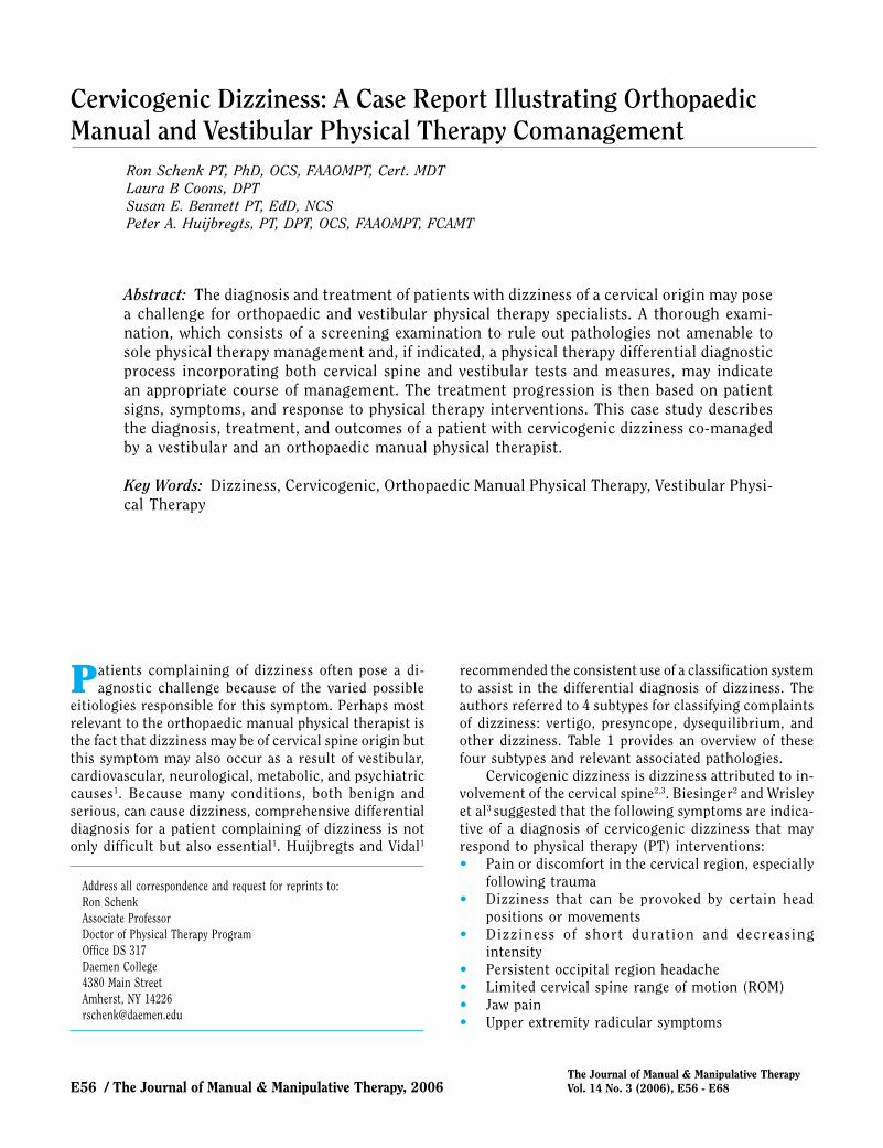
Abstract: The diagnosis and treatment of patients with dizziness of a cervical origin may pose a challenge for orthopaedic and vestibular physical therapy specialists. A thorough exami-nation, which consists of a screening examination to rule out pathologies not amenable to sole physical therapy management and, if indicated, a physical therapy differential diagnostic process incorporating both cervical spine and vestibular tests and measures, may indicate an appropriate course of management. The treatment progression is then based on patient signs, symptoms, and response to physical therapy interventions. This case study describes the diagnosis, treatment, and outcomes of a patient with cervicogenic dizziness co-managed by a vestibular and an orthopaedic manual physical therapist.
Key Words: Dizziness, Cervicogenic, Orthopaedic Manual Physical Therapy, Vestibular Physi-cal Therapy
Ron Schenk PT, PhD, OCS, FAAOMPT, Cert. MDTLaura B Coons, DPTSusan E. Bennett PT, EdD, NCSPeter A. Huijbregts, PT, DPT, OCS, FAAOMPT, FCAMT
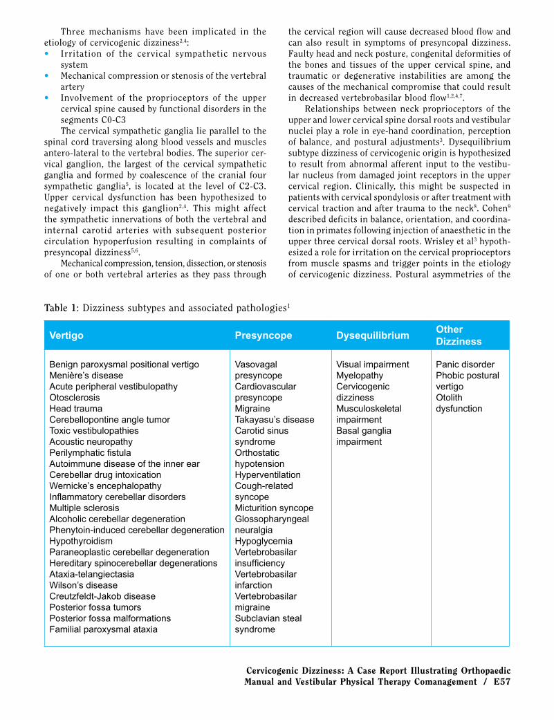
recommended the consistent use of a classification system to assist in the differential diagnosis of dizziness. The authors referred to 4 subtypes for classifying complaints of dizziness: vertigo, presyncope, dysequilibrium, and other dizziness. Table 1 provides an overview of these four subtypes and relevant associated pathologies.
Cervicogenic dizziness is dizziness attributed to in-volvement of the cervical spine2,3. Biesinger2 and Wrisley et al3 suggested that the following symptoms are indica-tive of a diagnosis of cervicogenic dizziness that may respond to physical therapy (PT) interventions:• Pain or discomfort in the cervical region, especially
following trauma• Dizziness that can be provoked by certain head
positions or movements• Dizziness of short duration and decreasing
intensity• Persistent occipital region headache• Limited cervical spine range of motion (ROM)• Jaw pain• Upper extremity radicular symptoms
The Journal of Manual & Manipulative TherapyVol. 14 No. 3 (2006), E56 - E68
Cervicogenic Dizziness: A Case Report Illustrating Orthopaedic Manual and Vestibular Physical Therapy Comanagement / E57
Three mechanisms have been implicated in the etiology of cervicogenic dizziness2,4:• Irritation of the cervical sympathetic nervous
system• Mechanical compression or stenosis of the vertebral
artery• Involvement of the proprioceptors of the upper
cervical spine caused by functional disorders in the segments C0-C3The cervical sympathetic ganglia lie parallel to the
spinal cord traversing along blood vessels and muscles antero-lateral to the vertebral bodies. The superior cer-vical ganglion, the largest of the cervical sympathetic ganglia and formed by coalescence of the cranial four sympathetic ganglia5, is located at the level of C2-C3. Upper cervical dysfunction has been hypothesized to negatively impact this ganglion2,4. This might affect the sympathetic innervations of both the vertebral and internal carotid arteries with subsequent posterior circulation hypoperfusion resulting in complaints of presyncopal dizziness5,6.
Mechanical compression, tension, dissection, or stenosis of one or both vertebral arteries as they pass through
the cervical region will cause decreased blood flow and can also result in symptoms of presyncopal dizziness. Faulty head and neck posture, congenital deformities of the bones and tissues of the upper cervical spine, and traumatic or degenerative instabilities are among the causes of the mechanical compromise that could result in decreased vertebrobasilar blood flow1,2,4,7.
Relationships between neck proprioceptors of the upper and lower cervical spine dorsal roots and vestibular nuclei play a role in eye-hand coordination, perception of balance, and postural adjustments3. Dysequilibrium subtype dizziness of cervicogenic origin is hypothesized to result from abnormal afferent input to the vestibu-lar nucleus from damaged joint receptors in the upper cervical region. Clinically, this might be suspected in patients with cervical spondylosis or after treatment with cervical traction and after trauma to the neck8. Cohen9 described deficits in balance, orientation, and coordina-tion in primates following injection of anaesthetic in the upper three cervical dorsal roots. Wrisley et al3 hypoth-esized a role for irritation on the cervical proprioceptors from muscle spasms and trigger points in the etiology of cervicogenic dizziness. Postural asymmetries of the
Table 1: Dizziness subtypes and associated pathologies1
Vertigo Presyncope DysequilibriumOther Dizziness
Benign paroxysmal positional vertigoMenière’s diseaseAcute peripheral vestibulopathyOtosclerosisHead traumaCerebellopontine angle tumorToxic vestibulopathiesAcoustic neuropathyPerilymphatic fistulaAutoimmune disease of the inner earCerebellar drug intoxicationWernicke’s encephalopathyInflammatory cerebellar disordersMultiple sclerosisAlcoholic cerebellar degenerationPhenytoin-induced cerebellar degenerationHypothyroidismParaneoplastic cerebellar degenerationHereditary spinocerebellar degenerationsAtaxia-telangiectasiaWilson’s diseaseCreutzfeldt-Jakob diseasePosterior fossa tumorsPosterior fossa malformationsFamilial paroxysmal ataxia
Vasovagal presyncopeCardiovascular presyncopeMigraineTakayasu’s diseaseCarotid sinus syndromeOrthostatic hypotensionHyperventilationCough-related syncopeMicturition syncopeGlossopharyngeal neuralgiaHypoglycemiaVertebrobasilar insufficiencyVertebrobasilar infarctionVertebrobasilar migraineSubclavian steal syndrome
Visual impairmentMyelopathyCervicogenic dizzinessMusculoskeletal impairmentBasal ganglia impairment
Panic disorderPhobic postural vertigoOtolith dysfunction
E58 / The Journal of Manual & Manipulative Therapy, 2006
head and neck might create unequal compression and tension on the articulating surfaces of the first three vertebrae, ligaments, and muscles. Faulty posture and muscle imbalances might also cause decreased ROM and produce conflicting signals with regard to head position to the central nervous system (CNS) when it compares vestibular, visual, and cervical input. Both the deep cer-vical flexor muscles and the cervical joint capsules are lined with mechanoreceptors and are hypothesized to play a role in dizziness if dysfunctional3. Brown10 stated that with strong connections between the cervical pro-prioceptors and balance function, it is understandable that injury or pathology of the neck may be associated with a sense of dizziness or dysequilibrium.
Because all of these factors may contribute to cervi-cogenic dizziness, orthopaedic manual physical therapy (OMPT) intervention may include stability exercises, postural re-education, stretching of shortened muscles, strengthening of weak muscles, and improvement of cervical spine joint play2,3,10-12. In a systematic review of the literature, Reid and Rivett13 noted that all studies of manual therapy treatment of patients with cervicogenic dizziness reported consistent post-treatment decreases in symptoms and signs of dizziness. Vestibular rehabilita-tion is sometimes a necessary adjunct to the treatment of patients with dizziness of suspected cervical origin3. Several authors have reported successful outcomes when incorporating vestibular rehabilitation exercises with OMPT in the treatment of patients with cervicogenic dizziness2,3,12,14.
The literature on PT evaluation and management of patients with cervicogenic dizziness is limited. Cer-vicogenic dizziness is a diagnosis of exclusion: When dizziness related to other conditions has been ruled out, dizziness due to either hypomobility or instability of the upper cervical spine may be considered1. This clearly illustrates the need for a screening examination for conditions causing dizziness that are not amenable to sole PT management and that, therefore, require referral for medical-surgical (co)management. It also indicates the need for a PT differential diagnosis in order to determine both appropriate further tests and subsequent interventions. The purpose of this case report is to illustrate OMPT and vestibular physical therapy co-management of a patient complaining of dizziness of cervical origin.
Case DescriptionSubject Description and History
The patient in this case report was a 33-year-old female. Her chief complaint was an 18-month history of dizziness, cervical pain, and occipital headache. Several different physicians had provided varying diagnoses. After she failed to respond to chiropractic management, an ophthalmologist diagnosed her with ocular migraines.
A neurologist diagnosed two cervical disc herniations. A neuro-opthalmologist suggested cranio-cervical pain and possible intermittent compression of one of the vertebral arteries; this specialist provided no clear explanation for the symptoms but believed they were originating from the cervical spine.
This patient was referred to PT for dizziness and a persistent occipital headache. The headache was described as constant (76-100%) and rated 6/10 at rest and 8/10 at its worst (with 10 rated as “the worst possible pain”) on the numeric pain rating scale (NPRS). The patient rated the overall impact of dizziness on function at 3/5 on a 6-point numeric rating scale (NRS, where 0 is rated as no impact and 5 as a complete disability). Williamson and Hoggart15 reported that the NPRS was a valid, reli-able, and responsive measure appropriate for use in the clinical setting. Childs et al16 found that a 2-point change on the NPRS demonstrates a minimum clinically impor-tant difference although this study was of patients with low-back and not cervical pain. The present authors are not aware of research investigating reliability, validity, and responsiveness for using a 6-point NRS to measure impact of dizziness on function.
The patient also complained of bilateral tinnitus, a “burning” sensation in the right cervical and bilateral upper trapezius region, numbness in the 4th and 5th fingers on the right hand, loss of balance with quick neck movements involving rotation, episodes of blurred vision with overhead reaching, and “silver flashes of light” in her peripheral vision. She reported no personal or family history of heart disease, diabetes, cancer, loss of vision, glaucoma, or ocular surgery. Her major functional limitations included reaching overhead, inability to work as a cosmetologist, and difficulty sleeping through the night without awakening due to neck pain.
Initial PT intervention had consisted of myofascial release and craniosacral therapy. Three months of treat-ment did not decrease symptoms, and the patient was referred to another PT facility with a diagnosis of cervical derangement and cephalalgia. Three treatments of moist heat, ultrasound to both upper trapezius muscles, and mechanical traction were unsuccessful in decreasing complaints and the patient was again referred back to the family physician.
Vestibular Physical Therapy Examination One week later, the family physician referred the
patient for vestibular rehabilitation to a board-certified PT Neurological Clinical Specialist (NCS) with specialty training in vestibular rehabilitation at a hospital-based PT outpatient clinic. At the time of referral, symptoms were unchanged from the time of the first PT referral.
Screening by way of history for cardinal signs and symptoms of vertebral artery ischaemia (including facial paraesthesiae, syncope, dysphagia, dysarthria, dysphonia, and drop attacks) was negative1. The authors are not
Cervicogenic Dizziness: A Case Report Illustrating Orthopaedic Manual and Vestibular Physical Therapy Comanagement / E59
aware of research investigating the diagnostic accuracy of these screening questions. Blood pressure was mea-sured in a seated position and immediately after rising to standing. Witting and Gallagher17 established norma-tive values for this orthostatic hypotension test: In 176 healthy subjects, systolic blood pressure decreased by 1.2 ± 9.8 mm Hg after one minute of standing preceded by five minutes of sitting. A drop in systolic blood pressure of ≥ 20 mm Hg had a specificity of 0.97 for detecting orthostatic hypotension17. In this patient, no significant change was noted. This normal blood pressure response to positional change made neurocardiogenic syncope less likely as a cause of the symptoms.
A pinwheel was used to assess conduction along sensory pathways; manual muscle tests (MMT) assessed for conduction along motor pathways. A positive response would be a lack of perception of sensation or a decrease in strength, respectively. Jepsen et al18 established inter-rater κ-values of 0.25-0.72 for upper-extremity MMT when using a dichotomous rating scale and they calculated an odds ratio (OR) of 2.5-7.7 for the presence of symptoms in the case of reduced strength on MMT, indicating that MMT may be an appropriate screening test. Numbness was present along the ulnar side of the forearm and hand including the ulnar three digits. Sensation was intact for localization. Manual muscle tests were normal.
Rapid alternating movement (diadochokinesis) of forearm supination and pronation and lower extremity toe tapping was used to screen for limb ataxia. Addi-tional limb ataxia tests included the finger-to-nose test performed with eyes closed and the heel-to-shin test. An inability to perform these tests in a coordinated fashion is considered a positive response. The patient showed no dysmetria (over or undershooting of the target) or dysdiadochokinesia. Absence of positive findings on a cranial nerve examination further diminished the likeli-hood of a contributing CNS lesion. The finger-to-nose test has poor test-retest and interrater reliability for dysmetria and tremor, but excellent reliability for time of execution19. We found no further data on reliability and validity for these limb ataxia tests.
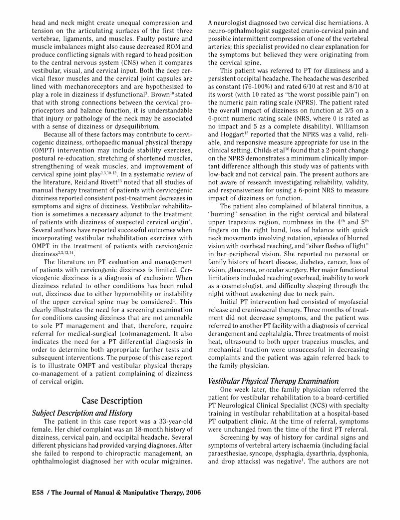
The Romberg test was used to assess the role of somatosensory feedback in balance control (Figure 1). This test is performed with the feet together and arms crossed over the chest with eyes open, then eyes closed. A positive response is an inability to maintain balance. A normal performance for a young adult is 30 sec, and a low normal score is 6 sec20. Our patient was unable to hold a sharpened Romberg position with the feet in tandem position. The Romberg test with eyes open resulted in retropulsion after 6 sec, whereas the same test with the eyes closed resulted in falling forward after 5 sec.
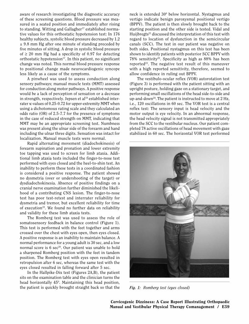
In the Hallpike-Dix test (Figures 2A,B), the patient sits on the examination table and the clinician turns the head horizontally 450. Maintaining this head position, the patient is quickly brought straight back so that the
neck is extended 300 below horizontal. Nystagmus and vertigo indicate benign paroxysmal positional vertigo (BPPV). The patient is then slowly brought back to the starting position and the other side is tested. Vidal and Huijbregts21 discussed the interpretation of this test with regard to location of dysfunction in the semicircular canals (SCC). The test in our patient was negative on both sides. Positional nystagmus on this test has been shown to identify patients with posterior SCC BPPV with 78% sensitivity22. Specificity as high as 88% has been reported23. The negative test result of this maneuver with a high reported sensitivity, therefore, seemed to allow confidence in ruling out BPPV.
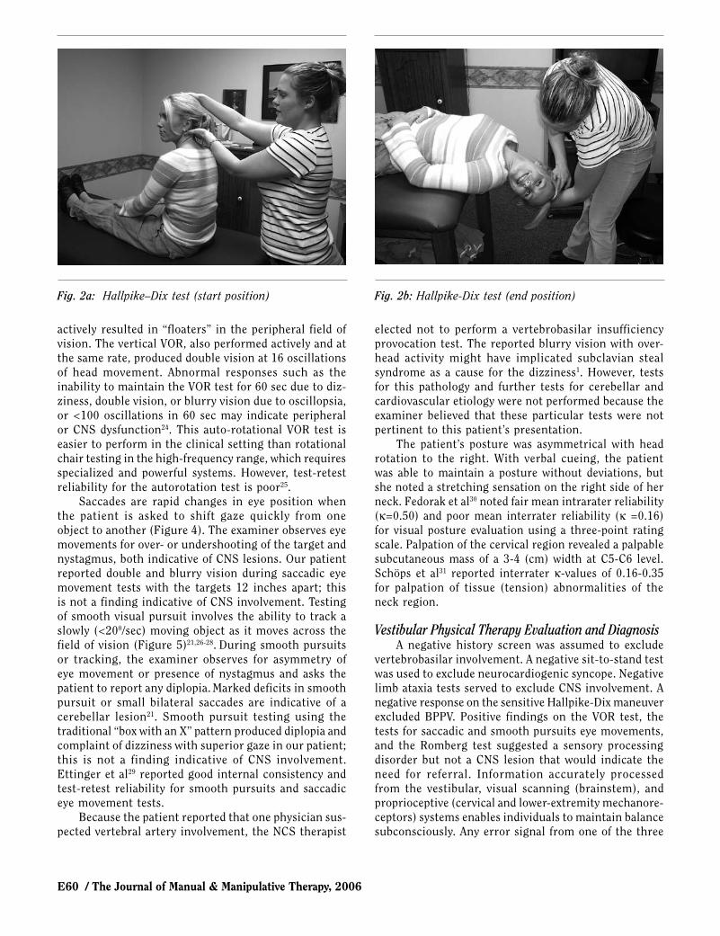
The vestibulo-ocular reflex (VOR) autorotation test (Figure 3) is performed with the patient sitting with an upright posture, holding gaze on a stationary target, and performing small oscillations of the head side-to-side and up-and-down24. The patient is instructed to move at 2 Hz, i.e., 120 oscillations in 60 sec. The VOR test is a central reflex test: The sensory input is head velocity and the motor output is eye velocity. In an abnormal response, the head velocity signal is not transmitted appropriately from the SCC to the vestibular nucleus. Our patient com-pleted 78 active oscillations of head movement with gaze stabilized in 60 sec. The horizontal VOR test performed
Fig. 1: Romberg test (eyes closed)
E60 / The Journal of Manual & Manipulative Therapy, 2006
actively resulted in “floaters” in the peripheral field of vision. The vertical VOR, also performed actively and at the same rate, produced double vision at 16 oscillations of head movement. Abnormal responses such as the inability to maintain the VOR test for 60 sec due to diz-ziness, double vision, or blurry vision due to oscillopsia, or <100 oscillations in 60 sec may indicate peripheral or CNS dysfunction24. This auto-rotational VOR test is easier to perform in the clinical setting than rotational chair testing in the high-frequency range, which requires specialized and powerful systems. However, test-retest reliability for the autorotation test is poor25.
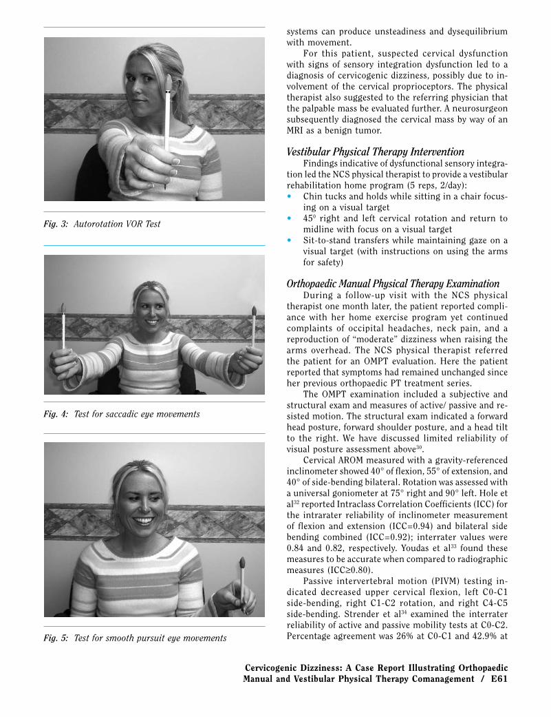
Saccades are rapid changes in eye position when the patient is asked to shift gaze quickly from one object to another (Figure 4). The examiner observes eye movements for over- or undershooting of the target and nystagmus, both indicative of CNS lesions. Our patient reported double and blurry vision during saccadic eye movement tests with the targets 12 inches apart; this is not a finding indicative of CNS involvement. Testing of smooth visual pursuit involves the ability to track a slowly (<200/sec) moving object as it moves across the field of vision (Figure 5)21,26-28. During smooth pursuits or tracking, the examiner observes for asymmetry of eye movement or presence of nystagmus and asks the patient to report any diplopia. Marked deficits in smooth pursuit or small bilateral saccades are indicative of a cerebellar lesion21. Smooth pursuit testing using the traditional “box with an X” pattern produced diplopia and complaint of dizziness with superior gaze in our patient; this is not a finding indicative of CNS involvement. Ettinger et al29 reported good internal consistency and test-retest reliability for smooth pursuits and saccadic eye movement tests.
Because the patient reported that one physician sus-pected vertebral artery involvement, the NCS therapist
elected not to perform a vertebrobasilar insufficiency provocation test. The reported blurry vision with over-head activity might have implicated subclavian steal syndrome as a cause for the dizziness1. However, tests for this pathology and further tests for cerebellar and cardiovascular etiology were not performed because the examiner believed that these particular tests were not pertinent to this patient’s presentation.
The patient’s posture was asymmetrical with head rotation to the right. With verbal cueing, the patient was able to maintain a posture without deviations, but she noted a stretching sensation on the right side of her neck. Fedorak et al30 noted fair mean intrarater reliability (κ=0.50) and poor mean interrater reliability (κ =0.16) for visual posture evaluation using a three-point rating scale. Palpation of the cervical region revealed a palpable subcutaneous mass of a 3-4 (cm) width at C5-C6 level. Schöps et al31 reported interrater κ-values of 0.16-0.35 for palpation of tissue (tension) abnormalities of the neck region.
Vestibular Physical Therapy Evaluation and DiagnosisA negative history screen was assumed to exclude
vertebrobasilar involvement. A negative sit-to-stand test was used to exclude neurocardiogenic syncope. Negative limb ataxia tests served to exclude CNS involvement. A negative response on the sensitive Hallpike-Dix maneuver excluded BPPV. Positive findings on the VOR test, the tests for saccadic and smooth pursuits eye movements, and the Romberg test suggested a sensory processing disorder but not a CNS lesion that would indicate the need for referral. Information accurately processed from the vestibular, visual scanning (brainstem), and proprioceptive (cervical and lower-extremity mechanore-ceptors) systems enables individuals to maintain balance subconsciously. Any error signal from one of the three
Fig. 2a: Hallpike–Dix test (start position) Fig. 2b: Hallpike-Dix test (end position)
Cervicogenic Dizziness: A Case Report Illustrating Orthopaedic Manual and Vestibular Physical Therapy Comanagement / E61
systems can produce unsteadiness and dysequilibrium with movement.
For this patient, suspected cervical dysfunction with signs of sensory integration dysfunction led to a diagnosis of cervicogenic dizziness, possibly due to in-volvement of the cervical proprioceptors. The physical therapist also suggested to the referring physician that the palpable mass be evaluated further. A neurosurgeon subsequently diagnosed the cervical mass by way of an MRI as a benign tumor.
Vestibular Physical Therapy InterventionFindings indicative of dysfunctional sensory integra-
tion led the NCS physical therapist to provide a vestibular rehabilitation home program (5 reps, 2/day):• Chin tucks and holds while sitting in a chair focus-
ing on a visual target• 450 right and left cervical rotation and return to
midline with focus on a visual target• Sit-to-stand transfers while maintaining gaze on a
visual target (with instructions on using the arms for safety)
Orthopaedic Manual Physical Therapy ExaminationDuring a follow-up visit with the NCS physical
therapist one month later, the patient reported compli-ance with her home exercise program yet continued complaints of occipital headaches, neck pain, and a reproduction of “moderate” dizziness when raising the arms overhead. The NCS physical therapist referred the patient for an OMPT evaluation. Here the patient reported that symptoms had remained unchanged since her previous orthopaedic PT treatment series.
The OMPT examination included a subjective and structural exam and measures of active/ passive and re-sisted motion. The structural exam indicated a forward head posture, forward shoulder posture, and a head tilt to the right. We have discussed limited reliability of visual posture assessment above30.
Cervical AROM measured with a gravity-referenced inclinometer showed 40° of flexion, 55° of extension, and 40° of side-bending bilateral. Rotation was assessed with a universal goniometer at 75° right and 90° left. Hole et al32 reported Intraclass Correlation Coefficients (ICC) for the intrarater reliability of inclinometer measurement of flexion and extension (ICC=0.94) and bilateral side bending combined (ICC=0.92); interrater values were 0.84 and 0.82, respectively. Youdas et al33 found these measures to be accurate when compared to radiographic measures (ICC≥0.80).
Passive intervertebral motion (PIVM) testing in-dicated decreased upper cervical flexion, left C0-C1 side-bending, right C1-C2 rotation, and right C4-C5 side-bending. Strender et al34 examined the interrater reliability of active and passive mobility tests at C0-C2. Percentage agreement was 26% at C0-C1 and 42.9% at
Fig. 3: Autorotation VOR Test
Fig. 4: Test for saccadic eye movements
Fig. 5: Test for smooth pursuit eye movements
E62 / The Journal of Manual & Manipulative Therapy, 2006
C1-C2. Smedmark et al35 found that interrater reliability for cervical PIVM tests similar to those used in this case report ranged from poor to moderate (κ=0.28-0.43). Huijbregts36 noted that, in general, interrater agree-ment of cervical PIVM tests only rarely exceeds poor to fair with lower values for evaluation of mobility than for pain. Ross et al37 questioned the validity of C1-C2 motion palpation due the interindividual differences in left-to-right joint asymmetry. In contrast, Jull et al38 compared a painful segmental restriction established with cervical accessory and physiological PIVM tests to pain relief on uncontrolled diagnostic blocks and reported 100% sensitivity and specificity for the manual examination. Humphreys et al39 compared similar PIVM testing against the gold standard of a radiographically confirmed congenital block vertebra and established sensitivity of 55-78% and specificity of 91-98%.
There was increased tone in the upper trapezius muscles during elevation of the upper extremities. Schöps et al31 reported poor to fair interrater reliability for as-sessing trapezius tone (κ=0.20-0.30). Lower trapezius MMT was measured at 2/5. We have discussed reliability of MMT above18. Muscle length tests indicated shortened suboccipital, sternocleidomastoid (SCM; right greater than left) and pectoralis minor muscles in our patient.
Sensation testing revealed a decreased response to sharp sensation at the C7 and C8 dermatomes on the right. Upper-limb tension testing (ULTT3) was positive on the right, indicating positive adverse neural tension of the ulnar nerve. Bertilson et al40 reported an inter-rater agreement of 92% for this test.
Foraminal closure (Spurling) testing and cervical spine compression tests were negative. Bertilson et al40 reported 65% and 73% interrater agreement for the right and left Spurling test, respectively. Wainner et al41 reported an interrater κ-value of 0.60 (95% confidence interval, CI:
0.32-0.87). Tong et al42 reported 30% sensitivity and 93% specificity for diagnosing cervical radiculopathy with the Spurling test as compared to electrodiagnostic testing. Wainner et al41 reported a sensitivity of 50% (95% CI: 27-73%) and a specificity of 86% (95% CI: 77-94%) for the Spurling test when compared to electrodiagnostic findings of radiculopathy.
Repeated cervical neck retraction with extension decreased right-sided cervical spine complaints in our patient. Dionne et al43 reported moderate interrater agreement for repeated movement assessment of the neck: Overall diagnosis yielded a κ-value of 0.55, de-rangement subcategory 0.47, and directional preference 0.46. The Romberg test performed with eyes open and eyes closed both resulted in loss of balance at 15 sec, which was an improvement from the initial test by the vestibular physical therapist.
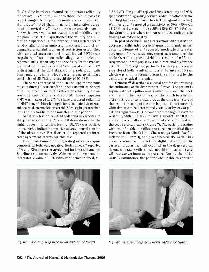
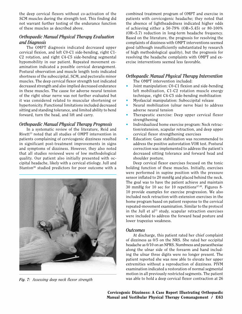
Grimmer44 described a clinical test for determining the endurance of the deep cervical flexors. The patient is supine without a pillow and is asked to retract the neck and then lift the back of head off the plinth to a height of 2 cm. Endurance is measured as the time from start of the test to the moment the chin begins to thrust forward. Chin thrust can be determined visually or by way of pal-pation (Figures 6A,B). Grimmer reported high test-retest reliability with ICC=0.92 in female subjects and 0.93 in male subjects. Falla et al45 described a strength test for the deep cervical flexors (Figure 7). The patient is supine with an inflatable, air-filled pressure sensor (Stabilizer Pressure Biofeedback Unit, Chattanooga South Pacific) inflated to 20 mmHg and placed behind the neck. This pressure sensor will detect the slight flattening of the cervical lordosis that will occur when the deep cervical flexors contract (with a head nod-like movement) and will register an increase in pressure. During the initial OMPT examination, the patient was unable to contract
Fig. 6a: Assessing deep neck flexor endurance (start) Fig. 6b: Assessing deep neck flexor endurance (finish)
Cervicogenic Dizziness: A Case Report Illustrating Orthopaedic Manual and Vestibular Physical Therapy Comanagement / E63
the deep cervical flexors without co-activation of the SCM muscles during the strength test. This finding did not warrant further testing of the endurance function of these muscles as described above.
Orthopaedic Manual Physical Therapy Evaluation and Diagnosis
The OMPT diagnosis indicated decreased upper cervical flexion, and left C0-C1 side-bending, right C1-C2 rotation, and right C4-C5 side-bending segmental hypomobility in our patient. Repeated movement ex-amination indicated a possible cervical derangement. Postural observation and muscle length tests indicated shortness of the suboccipital, SCM, and pectoralis minor muscles. The deep cervical flexor strength test indicated decreased strength and also implied decreased endurance in these muscles. The cause for adverse neural tension of the right ulnar nerve was not further evaluated but it was considered related to muscular shortening or hypertonicity. Functional limitations included decreased sitting and standing tolerance, and limited ability to bend forward, turn the head, and lift and carry.
Orthopaedic Manual Physical Therapy PrognosisIn a systematic review of the literature, Reid and
Rivett13 noted that all studies of OMPT intervention in patients complaining of cervicogenic dizziness resulted in significant post-treatment improvements in signs and symptoms of dizziness. However, they also noted that all studies reviewed were of low methodological quality. Our patient also initially presented with oc-cipital headache, likely with a cervical etiology. Jull and Stanton46 studied predictors for poor outcome with a
combined treatment program of OMPT and exercise in patients with cervicogenic headache; they noted that the absence of lightheadedness indicated higher odds of achieving either a 50-79% (OR=5.45) or 80-100% (OR=5.7) reduction in long-term headache frequency. Based on the literature, the prognosis for resolving the complaints of dizziness with OMPT interventions seemed good (although insufficiently substantiated by research of high methodological quality), but the prognosis for resolving the headache complaints with OMPT and ex-ercise interventions seemed less favorable.
Orthopaedic Manual Physical Therapy InterventionThe OMPT intervention included:
• Joint manipulation: C0-C1 flexion and side-bending left mobilization, C1-C2 rotation muscle energy technique, right C4-C5 side-bending mobilization
• Myofascial manipulation: Suboccipital release• Neural mobilization (ulnar nerve bias) to address
adverse neural tension• Therapeutic exercise: Deep upper cervical flexor
strengthening• Individualized home exercise program: Neck retrac-
tion/extension, scapular retraction, and deep upper cervical flexor strengthening exercises
• Education: Gaze stabilization was recommended to address the positive autorotation VOR test. Postural correction was implemented to address the patient’s decreased sitting tolerance and forward head and shoulder posture.Deep cervical flexor exercises focused on the tonic
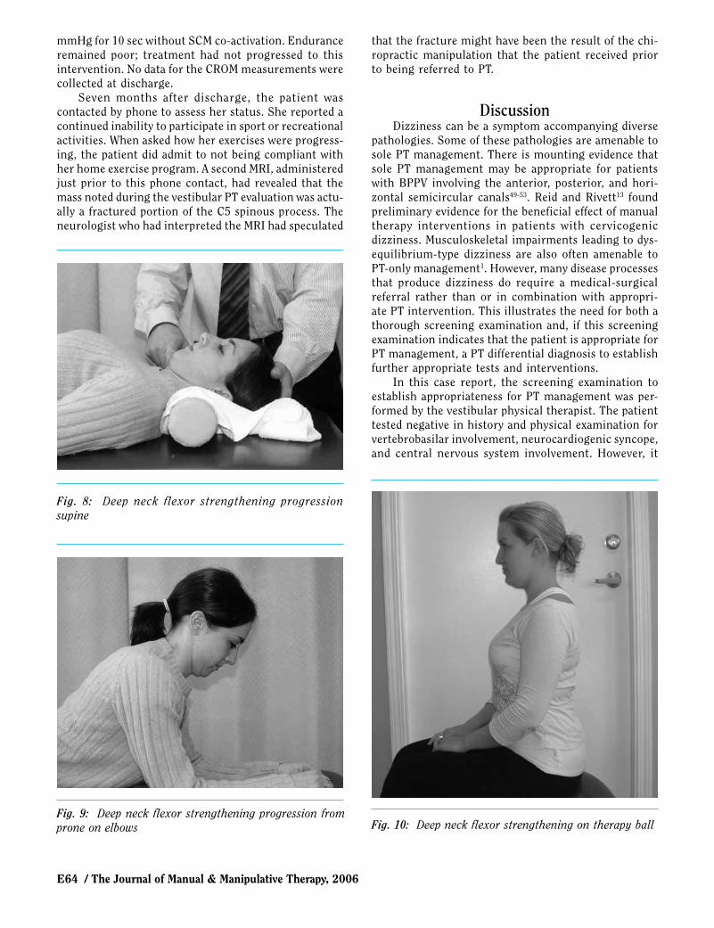
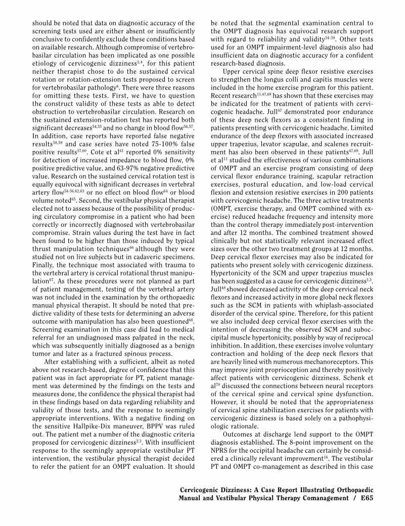
holding function of these muscles. Initially, exercises were performed in supine position with the pressure sensor inflated to 20 mmHg and placed behind the neck. The goal was to have the patient achieve and maintain 30 mmHg for 10 sec for 10 repetitions47,48. Figures 8-10 provide examples for exercise progression. We also included neck retraction with extension exercises in the home program based on patient response to the cervical repeated-movement examination. Similar to the protocol in the Jull et al11 study, scapular retraction exercises were included to address the forward head posture and lower trapezius weakness.
OutcomesAt discharge, this patient rated her chief complaint
of dizziness as 0/5 on the NRS. She rated her occipital headache as 0/10 on an NPRS. Numbness and paraesthesiae along the ulnar side of the forearm and hand includ-ing the ulnar three digits were no longer present. The patient reported she was now able to elevate her upper extremities without a reproduction of dizziness. PIVM examination indicated a restoration of normal segmental motion in all previously restricted segments. The patient was able to hold a deep cervical flexor contraction at 26 Fig. 7: Assessing deep neck flexor strength
E64 / The Journal of Manual & Manipulative Therapy, 2006
mmHg for 10 sec without SCM co-activation. Endurance remained poor; treatment had not progressed to this intervention. No data for the CROM measurements were collected at discharge.
Seven months after discharge, the patient was contacted by phone to assess her status. She reported a continued inability to participate in sport or recreational activities. When asked how her exercises were progress-ing, the patient did admit to not being compliant with her home exercise program. A second MRI, administered just prior to this phone contact, had revealed that the mass noted during the vestibular PT evaluation was actu-ally a fractured portion of the C5 spinous process. The neurologist who had interpreted the MRI had speculated
that the fracture might have been the result of the chi-ropractic manipulation that the patient received prior to being referred to PT.
DiscussionDizziness can be a symptom accompanying diverse
pathologies. Some of these pathologies are amenable to sole PT management. There is mounting evidence that sole PT management may be appropriate for patients with BPPV involving the anterior, posterior, and hori-zontal semicircular canals49-53. Reid and Rivett13 found preliminary evidence for the beneficial effect of manual therapy interventions in patients with cervicogenic dizziness. Musculoskeletal impairments leading to dys-equilibrium-type dizziness are also often amenable to PT-only management1. However, many disease processes that produce dizziness do require a medical-surgical referral rather than or in combination with appropri-ate PT intervention. This illustrates the need for both a thorough screening examination and, if this screening examination indicates that the patient is appropriate for PT management, a PT differential diagnosis to establish further appropriate tests and interventions.
In this case report, the screening examination to establish appropriateness for PT management was per-formed by the vestibular physical therapist. The patient tested negative in history and physical examination for vertebrobasilar involvement, neurocardiogenic syncope, and central nervous system involvement. However, it
Fig. 8: Deep neck flexor strengthening progression supine
Fig. 9: Deep neck flexor strengthening progression from prone on elbows Fig. 10: Deep neck flexor strengthening on therapy ball
Cervicogenic Dizziness: A Case Report Illustrating Orthopaedic Manual and Vestibular Physical Therapy Comanagement / E65
should be noted that data on diagnostic accuracy of the screening tests used are either absent or insufficiently conclusive to confidently exclude these conditions based on available research. Although compromise of vertebro-basilar circulation has been implicated as one possible etiology of cervicogenic dizziness2,4, for this patient neither therapist chose to do the sustained cervical rotation or rotation-extension tests proposed to screen for vertebrobasilar pathology6. There were three reasons for omitting these tests. First, we have to question the construct validity of these tests as able to detect obstruction to vertebrobasilar circulation. Research on the sustained extension-rotation test has reported both significant decreases54,55 and no change in blood flow56,57. In addition, case reports have reported false negative results58,59 and case series have noted 75-100% false positive results57,60. Cote et al61 reported 0% sensitivity for detection of increased impedance to blood flow, 0% positive predictive value, and 63-97% negative predictive value. Research on the sustained cervical rotation test is equally equivocal with significant decreases in vertebral artery flow54-56,62,63 or no effect on blood flow64 or blood volume noted65. Second, the vestibular physical therapist elected not to assess because of the possibility of produc-ing circulatory compromise in a patient who had been correctly or incorrectly diagnosed with vertebrobasilar compromise. Strain values during the test have in fact been found to be higher than those induced by typical thrust manipulation techniques66 although they were studied not on live subjects but in cadaveric specimens. Finally, the technique most associated with trauma to the vertebral artery is cervical rotational thrust manipu-lation67. As these procedures were not planned as part of patient management, testing of the vertebral artery was not included in the examination by the orthopaedic manual physical therapist. It should be noted that pre-dictive validity of these tests for determining an adverse outcome with manipulation has also been questioned68. Screening examination in this case did lead to medical referral for an undiagnosed mass palpated in the neck, which was subsequently initially diagnosed as a benign tumor and later as a fractured spinous process.
After establishing with a sufficient, albeit as noted above not research-based, degree of confidence that this patient was in fact appropriate for PT, patient manage-ment was determined by the findings on the tests and measures done, the confidence the physical therapist had in these findings based on data regarding reliability and validity of those tests, and the response to seemingly appropriate interventions. With a negative finding on the sensitive Hallpike-Dix maneuver, BPPV was ruled out. The patient met a number of the diagnostic criteria proposed for cervicogenic dizziness2,3. With insufficient response to the seemingly appropriate vestibular PT intervention, the vestibular physical therapist decided to refer the patient for an OMPT evaluation. It should
be noted that the segmental examination central to the OMPT diagnosis has equivocal research support with regard to reliability and validity34-39. Other tests used for an OMPT impairment-level diagnosis also had insufficient data on diagnostic accuracy for a confident research-based diagnosis.
Upper cervical spine deep flexor resistive exercises to strengthen the longus colli and capitis muscles were included in the home exercise program for this patient. Recent research11,47,69 has shown that these exercises may be indicated for the treatment of patients with cervi-cogenic headache. Jull47 demonstrated poor endurance of these deep neck flexors as a consistent finding in patients presenting with cervicogenic headache. Limited endurance of the deep flexors with associated increased upper trapezius, levator scapulae, and scalenes recruit-ment has also been observed in these patients47,69. Jull et al11 studied the effectiveness of various combinations of OMPT and an exercise program consisting of deep cervical flexor endurance training, scapular retraction exercises, postural education, and low-load cervical flexion and extension resistive exercises in 200 patients with cervicogenic headache. The three active treatments (OMPT, exercise therapy, and OMPT combined with ex-ercise) reduced headache frequency and intensity more than the control therapy immediately post-intervention and after 12 months. The combined treatment showed clinically but not statistically relevant increased effect sizes over the other two treatment groups at 12 months. Deep cervical flexor exercises may also be indicated for patients who present solely with cervicogenic dizziness. Hypertonicity of the SCM and upper trapezius muscles has been suggested as a cause for cervicogenic dizziness1,3. Jull48 showed decreased activity of the deep cervical neck flexors and increased activity in more global neck flexors such as the SCM in patients with whiplash-associated disorder of the cervical spine. Therefore, for this patient we also included deep cervical flexor exercises with the intention of decreasing the observed SCM and suboc-cipital muscle hypertonicity, possibly by way of reciprocal inhibition. In addition, these exercises involve voluntary contraction and holding of the deep neck flexors that are heavily lined with numerous mechanoreceptors. This may improve joint proprioception and thereby positively affect patients with cervicogenic dizziness. Schenk et al70 discussed the connections between neural receptors of the cervical spine and cervical spine dysfunction. However, it should be noted that the appropriateness of cervical spine stabilization exercises for patients with cervicogenic dizziness is based solely on a pathophysi-ologic rationale.
Outcomes at discharge lend support to the OMPT diagnosis established. The 8-point improvement on the NPRS for the occipital headache can certainly be consid-ered a clinically relevant improvement16. The vestibular PT and OMPT co-management as described in this case
E66 / The Journal of Manual & Manipulative Therapy, 2006
report was consistent with previously documented ef-ficacious management approaches3,4,13. As discussed above, based on available research the prognosis for resolving the complaints of dizziness13 and, to a lesser extent, the complaints of headache46 were favorable. Long-term outcomes were less favorable. It is unclear if this was the result of non-compliance or if this was related to the spinous process fracture diagnosed after PT treatment.
We recognize that this case report has a number of limitations. First, the case report format does not allow conclusions with regard to a cause-and-effect relation-ship between intervention and outcome. Second, we used an NRS to measure outcome with regard to dizziness. Psychometric properties for this use of a numeric rating scale have not been established. The use of an outcome measure such as the Dizziness Handicap Inventory with established psychometric properties would have been preferable71-73. Third, we did not substantiate a diagnosis of cervicogenic headache. The International Headache Society has provided diagnostic criteria for cervicogenic headache74:1. Pain referred from a source in the neck and per-
ceived in one or more regions of the head and/or face, fulfilling criteria 3 and 4
2. Clinical, imaging, and/or laboratory evidence of a disorder or lesion within the cervical spine or soft tissues of the neck known to be, or generally ac-cepted as, a valid cause of headache
3. Evidence that the pain can be attributed to the neck disorder or lesion based on demonstration of clini-cal signs that implicate a source of pain in the neck and/or abolition of headache following diagnostic blockade of a cervical structure or its nerve supply using placebo- or other adequate controls
4. Pain resolves within 3 months after successful treat-ment of the causative disorder or lesion
Although this diagnosis may well have applied to this
patient, a definitive diagnosis of cervicogenic headache would have provided more confidence in our application of the research on deep cervical flexor muscle exercises for this patient.
ConclusionDiagnosis and management of patients with a main
complaint of dizziness requires advanced knowledge and skills. Therefore, patients may benefit from co-management by a vestibular and orthopaedic manual physical therapist. In this case report, vestibular PT and OMPT management of a patient with cervicogenic dizziness consisting of OMPT, exercise, and education provided for a positive short-term outcome after previ-ous failed medical, chiropractic, and PT management. Long-term outcome may have been negatively affected by non-compliance.
Physical therapists involved in the diagnosis and management of patients with dizziness need to first provide a screening examination to establish if the patient is in fact appropriate for sole PT management or if referral needs to be initiated for medical-surgical (co)management. If the patient proves appropriate for PT management, further diagnostic tests and measures are required to establish a specific PT diagnosis and determine a plan of care. This case report has indicated that, to date, data on diagnostic accuracy for both screening and diagnostic tests and measures is in large part absent or insufficient to support a research-based management approach. Further research is required both on psychometric properties of the individual tests and measures and on the most effective and efficient management strategies for patients with cervicogenic dizziness. Outcome studies will require the use of reliable, valid, and responsive outcome measures and monitoring of the effect of long-term compliance with home program instructions.
REFERENCES1. Huijbregts P, Vidal P. Dizziness in orthopaedic physical therapy
practice: Classification and pathophysiology. J Manual Manipula-tive Ther 2004;12:199-214.
2. Biesinger E. Vertigo caused by disorders of the cervical vertebral column. Adv Otorhinolaryngol 1988;39:44-51.
3. Wrisley DM, Sparto PJ, Whitney SL, Furman JM. Cervicogenic dizziness: A review of diagnosis and treatment. J Orthop Sports Phys Ther 2000;30:755-766.
4. Fitz-Ritson D. Assessment of cervicogenic vertigo. J Manipulative Physiol Ther 1991;14:193-198.
5. Clemente CD, ed. Anatomy of the Human Body. 13th American ed. Baltimore, MD: Williams & Wilkins, 1984.
6. Oostendorp R. Functionele Vertebrobasilaire Insufficientie [Func-tional Vertebrobasilar Insufficiency]. PhD Thesis. Nijmegen, The Netherlands: Katholieke Universiteit Nijmegen, 1988.
7. Pettman E. Stress tests of the craniovertebral joints. In: Boyling JD, Palastanga N, eds. Grieve’s Modern Manual Therapy: The Vertebral Column. 2nd ed. Edinburgh, Scotland: Churchill Liv-ingstone, 1994.
8. Ryan MS, Cope S. Cervical vertigo. Lancet 1955;2:1355-1358.9. Cohen LA. Role of eye and neck proprioceptive mechanisms in body
orientation and motor coordination. J Neurophysiol 1961;24:1-11.10. Brown JJ. Cervical contributions to balance: Cervical vertigo. In:
Berthoz A, Vidal PP, Graf W, eds. The Head Neck Sensory Motor
Cervicogenic Dizziness: A Case Report Illustrating Orthopaedic Manual and Vestibular Physical Therapy Comanagement / E67
System. New York, NY: Oxford University Press, 1992.11. Jull G, et al. A randomized controlled trial of exercise and ma-
nipulative therapy for cervicogenic headache. Spine 2002;27:1835-1843.
12. Karlberg M, et al. Postural and symptomatic improvement after physiotherapy in patients with dizziness of suspected cervical origin. Arch Phys Med Rehabil 1996;77:874-882.
13. Reid SA, Rivett DA. Manual therapy treatment of cervicogenic dizziness: A systematic review. Man Ther 2005;10:4-13.
14. Galm R, Rittmeister M, Schmitt E. Vertigo in patients with cervi-cal spine dysfunction. Eur Spine J 1998;755-758.
15. Williamson A, Hoggart B. Pain: A review of three commonly used pain rating scales. J Clin Nursing 2005;14:798-804.
16. Childs J, Piva S, Fritz J. Responsiveness of the numeric pain rating scale in patients with low back pain. Spine 2005;30:1331-1334.
17. Witting MD, Gallagher K. Unique cutpoints for sitting-to-standing orthostatic vital signs. Am J Emerg Med 2003;21:45-47.
18. Jepsen JR, Laursen LH, Larsen AI, Hagert CG. Manual strength testing in 14 upper limb muscles: A study of interrater reliability. Acta Orthop Scand 2004;75:442-448.
19. Swaine BR, Sullivan SJ. Reliability of scores of the finger-to-nose test in adults with traumatic brain injury. Phys Ther 1993;73:71-78.
20. Hain CH, Micco AG. Cranial nerve VIII: Vestibulocochlear system. In: Goetz CG, ed. Textbook of Clinical Neurology. 2nd ed. Phila-delphia, PA: Elsevier Science, 2003:195-210.
21. Vidal P. Huijbregts P. Dizziness in orthopaedic physical therapy practice: History and physical examination. J Manual Manipula-tive Ther 2005;13:222-251.
22. Katsarkas A, Kirkham T. Paroxysmal positional vertigo: A study of 255 cases. J Otolaryngol 1978;7:320-330.
23. Hoffman RM, Einstadter D, Kroenke K. Evaluating dizziness. Am J Med 1999;107:468-478.
24. Herdman S, Schubert MC. Vestibular rehabilitation. In: O’Sullivan SB, Schmitz TJ, eds. Physical Rehabilitation: Assessment and Treatment. 4th ed. Philadelphia, PA: FA Davis Company, 2000:821-843.
25. Guyot JP, Psillas G. Test-retest reliability of vestibular autorota-tion testing in healthy subjects. Otolaryngol Head Neck Surg 1997;117:704-707.
26. Baloh RW, Honrubia V. Clinical Neurophysiology of the Vestibular System. Philadelphia, PA: FA Davis Company, 1990.
27. Demer JL. Evaluation of vestibular and oculomotor function. Otolaryngol Head Neck Surg 1995;2:16.
28. Dixon JS, Bird HA. Reproducibility along a 10 cm vertical visual analog scale. Ann Rheum Dis 1981;40:87.
29. Ettinger U, et al. Reliability of smooth pursuit, fixation and sac-cadic eye movements. Psychophysiology 2003;40:620-628.
30. Fedorak CA, Ashworth N, Marshall J, Paull H. Reliability of visual assessment of cervical and lumbar lordosis: How good are we? Spine 2003;28:1857-1859.
31. Schöps P, Pfingsten M, Siebert U. Reliabilität manualmedizinischer Untersuchungstechniken an der Halswirbelsäule. Studie zur Qual-itätssicherung in der manuellen Diagnostik [Reliability of cervical manual medicine diagnostic techniques: Study to ascertain manual diagnostic accuracy]. Z Orthop Ihre Grenzgeb 2000;138:2-7.
32. Hole DE, Cook JM, Bolton JE. Reliability and concurrent valid-
ity of two instruments for measuring cervical range of motion: Effects of age and gender. Man Ther 1995;1:36-42.
33. Youdas JW, Carey JR, Garrett TR. Reliability of measurements of cervical spine range of motion: Comparison of three methods. Phys Ther 1991;71:98-104.
34. Strender LE, Lundin M, Nell K. Interexaminer reliability in physical examination of the neck. J Manipulative Physiol Ther 1997;20:516-520.
35. Smedmark V, Wallin M, Arvidsson I. Interexaminer reliability in assessing passive intervertebral motion of the cervical spine. Man Ther 2000;5:97-101.
36. Huijbregts PA. Spinal motion palpation: A review of reliability studies. J Manual Manipulative Ther 2002;10:24-39.
37. Ross JK, Bereznick DE, McGill SM. Atlas-axis asymmetry: Implica-tions in manual palpation. Spine 1999:24:1203-1209.
38. Jull G, Bogduk N, Marsland A. The accuracy of manual diagno-sis for cervical zygapophysial joint pain syndromes. Med J Aust 1988;148:233-236.
39. Humphreys BK Delahaye M, Peterson CK. An investigation into the validity of cervical spine motion palpation using subjects with congenital block vertebrae as a gold standard. BMC Musculoskelet Disord 2004;5:19.
40. Bertilson BC, Grunnesjö M, Strender LE. Reliability of clinical tests in the assessment of patients with neck/shoulder problems: Impact of history. Spine 2003;28:2222-2231.
41. Wainner RS, et al. Reliability and diagnostic accuracy of the clinical examination and patient self-report measures for cervical radiculopathy. Spine 2003;28:52-62.
42. Tong HC, Haig AJ, Yamakawa K. The Spurling test and cervical radiculopathy. Spine 2002;27:156-159.
43. Dionne CP, Bybee RF, Tomaka J. inter-rater reliability of McK-enzie assessment for neck pain. J Manual Manipulative Ther 2005;13:180-181.
44. Grimmer K. Measuring the endurance capacity of the cervical short flexor muscle group. Aust J Physiother 1994;40:251-254.
45. Falla DL, Campbell CD, Fagan AE, Thompson DC, Jull GA. Re-lationship between craniocervical flexion range of motion and pressure change during the craniocervical flexion test. Man Ther 2003;8:92-96.
46. Jull GA, Stanton WR. Predictors of responsiveness to physiotherapy management of cervicogenic headache. Cephalalgia 2005;25:101-108.
47. Jull G, Barrett C, Magee R, Ho P. Further clinical clarification of the muscle dysfunction in cervical headache. Cephalalgia 1999;19:179-185.
48. Jull GA. Deep cervical flexor muscle dysfunction in whiplash. J Musculoskeletal Pain 2000;8:143-154.
49. Van der Velde GM. Benign paroxysmal positional vertigo. Part II: A qualitative review of non-pharmacological, conservative treat-ments and a case report presenting Epley’s “canalith repositioning procedure,” a non-invasive bedside manoeuvre for treating BPPV. J Can Chiropr Assoc 1999;43:41-49.
50. Hilton M, Pinder D. The Epley (canalith repositioning) manoeuvre for benign paroxysmal positional vertigo. The Cochrane Database of Systematic Reviews 2004, Issue 2. Art. No.: CD003162.pub2. DOI: 10.1002/14651858.CD003162.pub2.
51. Herdman SJ, Blatt PJ, Schubert MC. Vestibular rehabilitation of
E68 / The Journal of Manual & Manipulative Therapy, 2006
patients with vestibular hypofunction or with benign paroxysmal positional vertigo. Curr Opin Neurol 2000;13:39-43.
52. Pollak L, Davies RA, Luxon LL. Effectiveness of the particle repositioning maneuver in benign paroxysmal positional vertigo with and without additional vestibular pathology. Otol Neurotol 2002;23:79-83.
53. Kim YK, Shin JE, Chung JW. The effect of canalith repositioning for anterior semicircular canal canalithiasis. J Otorhinolaryngol Relat Spec 2005;67:56-60.
54. Rivett DA, Sharpless KJ, Milburn PD. Effect of premanipulative tests on vertebral artery and internal carotid artery blood flow: A pilot study. J Manipulative Physiol Ther 1999;22:368-375.
55. Yi-Kai L, Yun-Kun Z, Cai-Mo L, Shi-Zhen Z. Changes and im-plications of blood flow velocity of the vertebral artery during rotation and extension of the head. J Manipulative Physiol Ther 1999;22:91-95.
56. Arnold C, Bourassa R, Langer T, Stoneham G. Doppler studies evaluating the effect of a physical therapy screening protocol on vertebral artery bloodflow. Man Ther 2004;9:13-21.
57. Licht PB, Christensen HW, Hoilund-Carlsen PF. Is there a role for premanipulative testing before cervical manipulation? J Ma-nipulative Physiol Ther 2000;23:175-179.
58. Westaway MD, Stratford P, Symons B. False negative extension/rota-tion pre-manipulative screening test on a patient with an atretic and hypoplastic vertebral artery. Man Ther 2003;8:120-127.
59. Rivett DA, Milburn PD, Chapple C. Negative premanipulative vertebral artery testing despite complete occlusion: A case of false negativity. Man Ther 1998;3:102-107.
60. Haynes MJ. Vertebral arteries and cervical movement: Doppler ultrasound velocimetry for screening before manipulation. J Manipulative Physiol Ther 2002;25:556-567.
61. Cote P, et al. The validity of the extension-rotation test as a clini-cal screening procedure before neck manipulation: A secondary analysis. J Manipulative Physiol Ther 1996;19:159-164.
62. Nakamura K, Saku Y, Torigoe R, Ibayashi S, Fujishima M. Sonographic detection of heamodynamic changes in a case of
vertebrobasilar insufficiency. Neuroradiology 1998;40:164-166.63. Mitchell JA. Changes in vertebral artery blood flow following
normal rotation of the cervical spine. J Manipulative Physiol Ther 2003;26:347-351.
64. Haynes MJ, Cala LA, Melsom A, Mastaglia FL, Milne N, McGeachie JK. Vertebral arteries and cervical rotation: Modeling and mag-netic resonance angiography studies. J Manipulative Physiol Ther 2002;25:370-383.
65. Licht PB, Christensen HW, Hoilund-Carlsen PF. Vertebral artery volume flow in human beings. J Manipulative Physiol Ther 1999;22:363-367.
66. Symons B, Leonard T, Herzog W. Internal forces sustained by the vertebral artery during spinal manipulative therapy. J Manipula-tive Physiol Ther 2002;25:504-510.
67. Haldeman S, Kohlbeck FJ, McGregor M. Risk factors and recipitating neck movements causing vertebral artery dissection after cervical trauma and spinal manipulation. Spine 1999;24(8):785-794.
68. Thiel H, Rix G. Is it time to stop pre-manipulation testing of the cervical spine? Man Ther 2005;10:154-158.
69. Jull G. Management of cervical headache. Man Ther 1997;2:182-190.
70. Schenk RJ, Dukhon I, Guyer Z, Sommers M, Venning A, Zalewski K. An evidence-based examination of the cervical spine. Orthop Phys Ther Practice 2002;14(4):9-11.
71. Jacobson GP, Newman CW. The development of the Dizziness Handicap Inventory. Arch Otolaryngol Head Neck Surg 1990;116:424-427.
72. Jacobson GP, Newman CW, Hunter L, Blazer GK. Balance function test correlates of the Dizziness Handicap Inventory. J Am Acad Audiol 2001;2:253-260.
73. Murray K, Carroll S, Hill K. Relationship between change in balance and self-reported handicap after vestibular rehabilitation therapy. Physiother Res Int 2001;6:251-263.
74. Headache Classification Subcommittee of the International Headache Society. The International Classification of Headache Disorders. 2nd ed. Cephalalgia 2004;24(suppl 1): 1-150.
![Cervicogenic Headache - Physiopedia · Another possibility to distinguish cervicogenic headache from migraine and tension headache is the use of a Cybex dynamometry. [20] Testing](https://img.pdfslide.us/doc/110x75/5cc7f9b088c993c4398ca482/cervicogenic-headache-physiopedia-another-possibility-to-distinguish-cervicogenic.jpg)