Embed Size (px)
Citation preview

Cellular/Molecular
AP-1 Transcription Factors Mediate BDNF-PositiveFeedback Loop in Cortical Neurons
Jurgen Tuvikene, Priit Pruunsild, Ester Orav, Eli-Eelika Esvald, and Tonis TimmuskDepartment of Gene Technology, Tallinn University of Technology, 12618 Tallinn, Estonia
Brain-derived neurotrophic factor (BDNF), a member of the neurotrophin family, regulates both survival and differentiation of severalneuronal populations in the nervous system during development, as well as synaptic plasticity in the adult brain. BDNF exerts itsbiological functions through its receptor TrkB. Although the regulation of BDNF transcription by neuronal activity has been widelystudied, little is known about TrkB signaling-dependent expression of BDNF. Using rat primary cortical neuron cultures, we show that theBDNF gene is a subject to an extensive autoregulatory loop, where TrkB signaling upregulates the expression of all major BDNFtranscripts, mainly through activating MAPK pathways. Investigating the mechanisms behind this autoregulation, we found thatAP-1 transcription factors, comprising Jun and Fos family members, participate in the induction of BDNF exon I, III, and VI transcripts.AP-1 transcription factors directly upregulate the expression of exon I transcripts by binding two novel AP-1 cis-elements in promoter I.Moreover, our results show that the effect of AP-1 proteins on the activity of rat BDNF promoters III and VI is indirect, because AP-1proteins were not detected to bind the respective promoter regions by chromatin immunoprecipitation (ChIP). Collectively, we describean extensive positive feedback system in BDNF regulation, adding a new layer to the elaborate control of BDNF gene expression.
Key words: AP-1; BDNF autoregulation; BDNF-positive feedback loop; Fos; Jun; TrkB
IntroductionBrain-derived neurotrophic factor (BDNF) is a secretory proteinbelonging to the neurotrophin family (Huang and Reichardt,
2001). BDNF binds to two distinctive classes of receptors, recep-tor tyrosine kinase subfamily member TrkB and tumor necrosisfactor receptor superfamily member p75 NTR, with mature BDNFpreferentially binding to the TrkB receptor and proBDNF to thep75 NTR receptor (Lu et al., 2005). During development, BDNFfunctions as a differentiation and survival factor for various pop-ulations of neurons in the peripheral nervous system and CNS,strictly regulating the number of neurons needed for properfunctioning of the nervous system (Bibel and Barde, 2000; Huangand Reichardt, 2001; Park and Poo, 2013). In the adult brain,BDNF has an important role in long-term potentiation (LTP)and memory formation (for review, see Park and Poo, 2013).Dysregulation of BDNF expression is implicated in various neu-ropsychiatric disorders, indicating the importance of BDNF inhigher cognitive functions and mood regulation (for review, seeAutry and Monteggia, 2012).
BDNF has a complex gene structure (Timmusk et al., 1993),with the rodent BDNF gene consisting of one protein-coding 3�exon that is spliced together with one of the eight noncoding 5�
Received Sept. 8, 2015; revised Dec. 17, 2015; accepted Dec. 21, 2015.Author contributions: J.T., P.P., and T.T. designed research; J.T., P.P., E.O., and E.-E.E. performed research; J.T.,
P.P., E.O., E.-E.E., and T.T. analyzed data; J.T., P.P., and T.T. wrote the paper.This work was supported by Estonian Research Council (institutional research funding IUT19-18 and Grant
ETF8844), National R&D program “Biotechnology” (Grant AR12030), Estonian Enterprise (Grant EU27553), Norwe-gian Financial Mechanism (Grant EMP128), and Estonian Academy of Sciences. We thank Epp Vali and Maila Rahn fortechnical assistance, and Indrek Koppel for critical reading of the paper.
The authors declare no competing financial interests.Correspondence should be addressed to either Tonis Timmusk or Jurgen Tuvikene, Department of Gene Technol-
ogy, Tallinn University of Technology, Akadeemia tee 15, 12618 Tallinn, Estonia, E-mail: [email protected] [email protected].
P. Pruunsild’s present address: Department of Neurobiology, Interdisciplinary Center for Neurosciences (IZN),University of Heidelberg, 69120 Heidelberg, Germany.
E. Orav’s present address: Neuroscience Center and Department of Biosciences, University of Helsinki, 00014Helsinki, Finland.
DOI:10.1523/JNEUROSCI.3360-15.2016Copyright © 2016 the authors 0270-6474/16/361290-16$15.00/0
Significance Statement
Here, we show for the first time that in rat primary cortical neurons the expression of all major BDNF transcripts (exon I, II, III, IV,VI, and IXa transcripts) is upregulated in response to TrkB signaling, and that AP-1 transcription factors participate in theinduction of exon I, III, and VI transcripts. Moreover, we have described two novel functional AP-1 cis-elements in BDNF promoterI, responsible for the activation of the promoter in response to TrkB signaling. Our results indicate the existence of a positivefeedback loop for obtaining sufficient BDNF levels necessary for various TrkB signaling-dependent physiological outcomes inneurons.
1290 • The Journal of Neuroscience, January 27, 2016 • 36(4):1290 –1305
exons (Aid et al., 2007). In humans, two additional exons (Vh andVIIIh) have been described (Pruunsild et al., 2007). Each of the 5�exons, except the human cassette exons VIII, VIIIh, and IXb, hasits own promoter, allowing complex spatiotemporal regulationof BDNF expression in different brain regions and peripheraltissues throughout development (Aid et al., 2007; Pruunsild et al.,2007) and in response to various stimuli (Metsis et al., 1993;Karpova et al., 2010; Salerno et al., 2012; Baj et al., 2013).
AP-1 transcription factors, consisting of Fos and Jun familymembers, are immediate early genes, whose expression in theCNS is induced by various stimuli, including hypoxia, sensorystimulation, neurotransmitters, neurotrophins, and other gro-wth factors (Sheng and Greenberg, 1990; Radler-Pohl et al., 1993;Karin et al., 1997; Herdegen and Leah, 1998). AP-1 proteins havea conserved basic leucine zipper domain, where the leucine zip-per mediates protein dimerization and the basic region is respon-sible for binding to DNA (Eferl and Wagner, 2003). AP-1transcription factors preferentially bind to the AP-1 element (alsoknown as TRE, TPA-response element), with consensus se-quence TGAC/GTCA (Eferl and Wagner, 2003). As with otherbasic leucine zipper domain-containing transcription factors,AP-1 proteins can bind DNA only as dimers, and binding to DNAgreatly increases the dimer stability (Patel et al., 1994). WhereasFos family proteins (c-Fos, FosB, Fra1, Fra2) are unable to formhomodimers (Halazonetis et al., 1988; Shaulian and Karin, 2001),members of the Jun family (c-Jun, JunB, JunD) are able to formboth homodimers and heterodimers within the family, and het-erodimers with Fos family proteins or with other transcriptionfactors, such as members of the ATF family, C/EBP, and others(Herdegen and Leah, 1998; Chinenov and Kerppola, 2001).
There is evidence suggesting that AP-1 proteins play a role insynaptic plasticity and memory (for review, see Alberini, 2009).For instance, AP-1 proteins have been implicated in both mem-ory formation (Grimm et al., 1997) and consolidation of addic-tive behavior (Raivich and Behrens, 2006; Perez-Cadahía et al.,2011). AP-1 proteins also regulate neuronal survival and apopto-sis, with the outcome depending on the exact cellular context andstimulus (Herdegen and Leah, 1998).
Although the mechanisms of calcium- and neuronal activity-dependent BDNF expression have been widely studied (forreview, see West et al., 2014), little is known about TrkBsignaling-dependent regulation of BDNF gene expression, withthe main focus having been on studying the regulation of exon IVtranscripts (Yasuda et al., 2007; Zheng and Wang, 2009; Bambah-Mukku et al., 2014). Here, we show for the first time that in ratprimary cortical neurons, the expression of all major BDNF tran-scripts is induced in response to TrkB signaling, and that part ofthis positive feedback loop is dependent on AP-1 family tran-scription factors.
Materials and MethodsPrimary neuron cultures. All animal procedures were performed in com-pliance with the local ethics committee. Preparation of rat primary neu-ron cultures was performed as follows. Cortices and hippocampi of bothmale and female embryonic day (E)20 –E21 Sprague Dawley rat pupswere dissected, cells were dissociated using trypsin-EDTA (0.25% tryp-sin, 1 mM EDTA, Life Technologies) for 20 min at 37°C, washed oncewith 1� HBSS, and treated with 0.05% DNase I (Roche) solution in 1�HBSS (including 12 mM MgSO4) for 5 min at 37°C. After washing with1� HBSS again, solution of 0.1% trypsin inhibitor (AppliChem) and 1%BSA (Sigma-Aldrich) in 1� HBSS was added and cortices and hip-pocampi were triturated using flame-polished Pasteur pipette. Undisso-ciated tissue clumps were allowed to sediment by gravity for 3–5 min,supernatant was moved to a new tube and centrifuged at 200 � g for 5
min. Neurons were resuspended in prewarmed Neurobasal A med-ium (Invitrogen) containing 1� B27 supplement (Invitrogen), 1 mM
L-glutamine (PAA Laboratories), 100 U/ml penicillin (PAA Laborato-ries), and 0.1 mg/ml streptomycin (PAA Laboratories), and plated onculture plates previously coated with 0.2 mg/ml poly-L-lysine (Sigma-Aldrich) in borate buffer, pH 8.5. At 2 d in vitro (DIV), one-half of themedium was replaced with fresh medium, containing 5-fluoro-2�-deoxyuridine (10 �M final concentration, Sigma-Aldrich) to inhibit pro-liferation of glial cells.
Cloning and mutagenesis. Full-length coding sequences of AP-1 geneswere amplified from either human genomic DNA or from human cDNA,for Jun family and Fos family members, respectively, using Phusion HotStart II High-Fidelity DNA Polymerase (Thermo Scientific). For expres-sion in eukaryotic cells, the protein coding sequences of AP-1 genes werecloned into pQM vector backbone (Icosagen) after murine phosphoglyc-erate kinase 1 (PGK ) promoter (mouse genome assembly GRCm38/mm10, chromosome X region 106186727-106187231). The sequencecoding for VP16 transactivation domain was obtained from pACT ve-ctor (Promega), the sequence coding for V5-tag was obtained frompcDNA3.1 vector (Invitrogen), to make vectors coding for VP16- orV5-tagged AP-1 proteins, respectively. Plasmid encoding A-Fos was ob-tained from Addgene (plasmid 33353).
The rat and human BDNF promoter I reporter constructs and the respec-tive plasmids with mutated AP1-1 and PasRE site have been described pre-viously (Pruunsild et al., 2011). For amplifying human and rat BDNFpromoter III and VI regions from genomic DNA, Phusion Hot Start II High-Fidelity DNA polymerase (Thermo Scientific) was used. The respective pro-moter regions were cloned upstream of firefly luciferase-coding sequenceinto pGL4.15 [Luc2P/Hygro] vector (Promega).
Site-directed mutagenesis was performed using complementary prim-ers containing the desired mutation and Phusion Hot Start II DNA poly-merase. After PCR, sample was treated with 0.5 U/�l DpnI restrictionenzyme (Thermo Scientific) for at least 1 h at 37°C to digest parentalDNA template, and transformed into TOP10 competent cells (Invitro-gen). All constructs were verified by sequencing.
Treatments and RT-qPCR. At 7– 8 DIV, as indicated below each figure,primary neurons were treated with 50 ng/ml human recombinant BDNF(Peprotech). Where indicated, cells were pretreated with inhibitors of differ-ent signaling pathways for 30 min before adding BDNF to the medium. Finalconcentrations of the compounds were as follows: 0.1% DMSO (Sigma-Aldirch), 10 �M U0126 (Tocris Bioscience), 10 �M Bix02189 (Axon Med-chem), 1 �M PD184352 (Sigma-Aldrich), 25 nM Az-23 (Axon Medchem), 10�M Go6983 (Tocris Bioscience), 100 nM Wortmannin (Millipore), 1 �M
U73122 (Cayman Europe). Total RNA was isolated using RNeasy Micro orMini kit (Qiagen) according to the manufacturer’s protocol.
For human embryonic kidney HEK293 cell-line, cells grown on six-well plate were transfected using LipoD293 DNA In Vitro TransfectionReagent (SignaGen) according to the manufacturer’s instruction, using 3�g of DNA and a ratio of 1:2 DNA–LipoD293 per well. Forty-eight hoursafter transfection, cells were lysed and RNA was isolated using RNeasyMini kit (Qiagen).
First strand cDNA was synthesized from 500 to 2000 ng or 5 �g ofRNA, for primary neurons or HEK293, respectively, using oligo(dT)20
primer and Superscript III Reverse Transcriptase (Invitrogen) as recom-mended by the manufacturer. Quantitative PCR (qPCR) reactions wereperformed in triplicates using LightCycler 480 SYBR Green I MasterqPCR mix (Roche) on LightCycler 480 II Real Time PCR System(Roche). Levels of HPRT1 mRNA were used to normalize qPCR data.Before statistical analysis, normalized data were log-transformed andautoscaled using data of all time points for the respective transcripts (Fig.1A), or using data of the respective transcripts in DMSO-treated neuronsthat were left untreated or treated with BDNF for 3 h (Fig. 1 B, C). Forgraphical representation, data were backtransformed, with error barsrepresenting upper and lower limits of backtransformed mean � SEM.
Primer sequences used to amplify rat HPRT1 and rat BDNF transcripts havebeendescribedpreviously(Kairisaloetal.,2009).ForamplifyinghumanHPRT1and human BDNF (hBDNF) transcripts, the following primers were used: hu-man HPRT1 (forward: GCCAGACTTTGTTGGATTTG, reverse: CTCTCATCTTAGGCTTTGTATTTTG), hBDNF exon I (forward: AACAAGA
Tuvikene et al. • AP-1 Proteins in BDNF Autoregulation J. Neurosci., January 27, 2016 • 36(4):1290 –1305 • 1291
CACATTACCTTCCAGCAT, reverse: CTCTTCTCACCTGGTGGAACATT),hBDNF exon III (forward: TGGCTTAGAGGGTTCCCGCT, reverse:ATGGGGGCAGCCTTCATGCA), hBDNF exon IV (forward: GAAGTCTTTCCCGGAGCAGCT, reverse: ATGGGGGCAGCCTTCATGCA), andhBDNF exon VI (forward: GGTTTGTGTGGACCCCGAGTTC, reverseATGGGGGCAGCCTTCATGCA).
Western blot analysis. For Western blot analysis, primary neuronswere lysed in 1� Laemmli buffer. Proteins were separated using SDS-PAGE and transferred to PVDF membrane (Millipore) using Trans-Blot SD SemiDry Transfer Cell (Bio-Rad). Membranes were blockedin 1� PBS containing 5% skimmed milk and 0.1% Tween 20 for 1 h atroom temperature, followed by incubation with primary antibody in1� PBS containing 2% skimmed milk and 0.1% Tween 20 for 1 h atroom temperature or overnight at 4°C. Next, membranes werewashed 3 � 5 min with 0.1% Tween 20 solution in 1� PBS. Blots wereincubated with secondary antibody for 1 h at room temperature in 1�PBS containing 2% skimmed milk and 0.1% Tween 20. Membranes
were washed 3 � 5 min and chemiluminescence signal was detectedusing SuperSignal West Femto Chemiluminescence Substrate(Thermo Scientific) and ImageQuant LAS 4000 bioimager (GEHealthcare).
Antibodies against AP-1 family proteins were obtained from SantaCruz Biotechnology: c-Jun (1:5000, sc-1694x, H2808), JunB (1:5000, sc-8051x, B1611), JunD (1:5000, sc-74x, G1911), c-Fos (1:5000, sc-7202x,C3108), FosB (1:5000, sc-48x, K1811), Fra1 (1:5000, sc-605x, G1612),and Fra2 (1:1000, sc-604x, G1911). GAPDH antibody was from Milli-pore (1:5000, MAB374, lot 2506322). Stabilized Goat Anti-Mouse IgG(H�L) Peroxidase-Conjugated (32430) and Stabilized Goat Anti-RabbitIgG (H�L) Peroxidase-Conjugated (32460) secondary antibodies (1:5000 dilution) used in Western blot analysis were purchased fromThermo Scientific.
Transduction using adeno associated virus vectors. Adeno associatedvirus (AAV) virions coding EGFP or A-Fos under the control of humanSynapsin I promoter were prepared as described previously (Koppel et
mR
NA
exp
ress
ion
BDNF total
0
5
10
15
20
I II III IV VI IXa Coding
mR
NA
fold
indu
ctio
n
rBDNF transcript (5' exon)
30
90
150
210 CTRL0.5 h BDNF1 h BDNF3 h BDNF6 h BDNF
CTRL3 h BDNF
A
mR
NA
exp
ress
ion
BDNF exon I BDNF exon II C BDNF exon III
mR
NA
exp
ress
ion
BDNF exon IV BDNF exon VI BDNF exon IXa
B
C
0
2
4
6
8
10
0
2
4
6
8
10
3050
024
10
0
4
8
12
16
204060
0
2
4
80
0
1
2
3
4
5
0
2
4
6
8
******
***
***
*** ***
***
***
******* **
**
****
****
**
** ** **
**
* *
*
*
*
*
*
**
****
*** *
****
*** ***
*****
***
** * * *
**
***
** **
**** **
***** ***
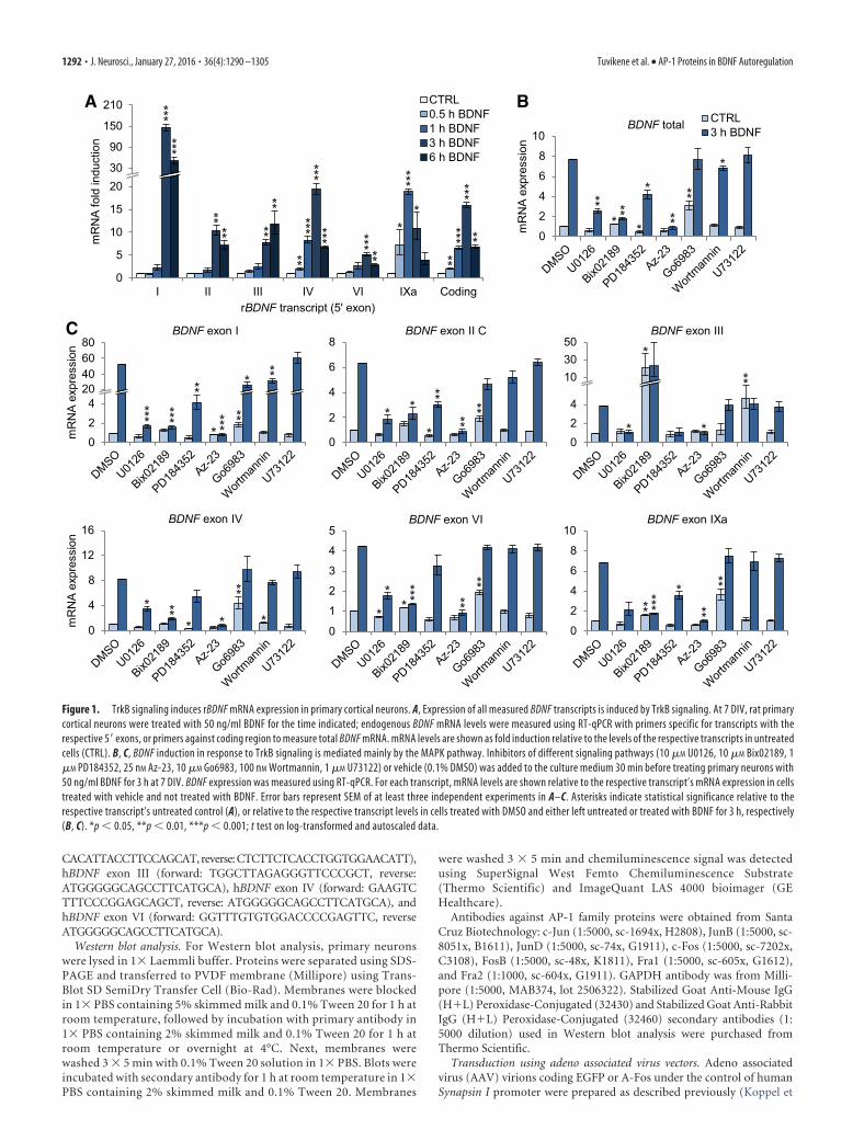
Figure 1. TrkB signaling induces rBDNF mRNA expression in primary cortical neurons. A, Expression of all measured BDNF transcripts is induced by TrkB signaling. At 7 DIV, rat primarycortical neurons were treated with 50 ng/ml BDNF for the time indicated; endogenous BDNF mRNA levels were measured using RT-qPCR with primers specific for transcripts with therespective 5� exons, or primers against coding region to measure total BDNF mRNA. mRNA levels are shown as fold induction relative to the levels of the respective transcripts in untreatedcells (CTRL). B, C, BDNF induction in response to TrkB signaling is mediated mainly by the MAPK pathway. Inhibitors of different signaling pathways (10 �M U0126, 10 �M Bix02189, 1�M PD184352, 25 nM Az-23, 10 �M Go6983, 100 nM Wortmannin, 1 �M U73122) or vehicle (0.1% DMSO) was added to the culture medium 30 min before treating primary neurons with50 ng/ml BDNF for 3 h at 7 DIV. BDNF expression was measured using RT-qPCR. For each transcript, mRNA levels are shown relative to the respective transcript’s mRNA expression in cellstreated with vehicle and not treated with BDNF. Error bars represent SEM of at least three independent experiments in A–C. Asterisks indicate statistical significance relative to therespective transcript’s untreated control (A), or relative to the respective transcript levels in cells treated with DMSO and either left untreated or treated with BDNF for 3 h, respectively(B, C). *p � 0.05, **p � 0.01, ***p � 0.001; t test on log-transformed and autoscaled data.
1292 • J. Neurosci., January 27, 2016 • 36(4):1290 –1305 Tuvikene et al. • AP-1 Proteins in BDNF Autoregulation
al., 2015). Titer of infectious particles was assessed by immunocytochem-istry using either anti-EGFP antibody (a kind gift from Andres Merits,Institute of Technology, University of Tartu, Tartu, Estonia) or anti-FLAG antibody (F1804, Sigma-Aldrich). Primary neurons were infectedwith two to three multiplicity of infection at 2 DIV, immunocytochem-istry, and Western blot analysis or BDNF treatment and RNA extractionwere performed at 8 DIV. RT-qPCR data were log-transformed and au-toscaled using data of the respective transcripts in neurons infected withAAV-EGFP. Statistical analysis was performed on autoscaled data. Forgraphical representation, data were backtransformed to linear scale, witherror bars indicating upper and lower limits of backtransformed mean �SEM.
DNA transfection and luciferase reporter assay. For luciferase reporterassay, neurons grown on 48-well plates were transfected at 6 DIV usingLipofectamine 2000 (Invitrogen) reagent according to the manufactur-er’s recommendations. Lipofectamine–DNA ratio of 2:1 was used, with0.5 �g of DNA per well. Where applicable, BDNF promoter constructand plasmids coding effector proteins or empty pQM vector werecotransfected at 1:1 ratio. For normalization, 20 ng pPGK/pGL4.83 plas-mid, expressing Renilla luciferase under the control of murine PGK pro-moter, was cotransfected per well.
At 7 DIV, neurons were treated with 50 ng/ml human recombinantBDNF (Peprotech). Cells were lysed after 8 h of BDNF treatment using1� Passive Lysis Buffer (Promega) and luciferase assays were performedusing Dual-Glo Luciferase Assay System (Promega). Luminescence wasmeasured in duplicate samples using GeniOS Pro (Tecan). For present-ing data in relative luciferase units (RLUs), background signal from un-transfected neurons was subtracted from both firefly luciferase (FFLuc)and Renilla luciferase (RLuc) signals. Background corrected FFLuc sig-nals were normalized using RLuc signals. Normalized data were log-transformed and autoscaled using data of wild-type (WT) promoterconstruct (cotransfected with empty pQM vector, where applicable). Forstatistical analysis, autoscaled data were used. For graphical representa-tion of results, means, and mean � SEM of autoscaled data were calcu-lated and backtransformed to linear scale. Error bars represent upper andlower limits of backtransformed mean � SEM.
Electrophoretic mobility shift assay. For preparation of cell lysates, neu-rons grown for 7 DIV on a 10 cm dish were washed with 1� PBS andcollected in 200 �l ice-cold sonication buffer containing 20 mM HEPES-KOH, pH 7.9, 25% glycerol, 500 mM KCl, 1.5 mM MgCl2, 0.4 mM EDTA,1 mM EGTA, 5 mM DTT, 0.5 mM PMSF, 1� cOmplete Protease InhibitorCocktail (Roche), and 1� PhosSTOP Phosphatase Inhibitor Cocktail(Roche). Lysates were then incubated on ice for 15 min, sonicated untilno viscous matter remained, and centrifuged at 4°C and 16,100 � g for 10min. Aliquots of supernatant were snap-frozen in liquid nitrogen andstored at �80°C.
Oligonucleotides (5 pmol per reaction) were labeled with T4 polynu-cleotide kinase (Thermo Scientific) using ATP �- 32P (PerkinElmer). Af-ter labeling, sense and antisense oligonucleotides were annealed inannealing buffer (50 mM NaCl, 1 mM EDTA, 0.2� PNK buffer A, 100 �ltotal volume) by placing the tubes in a 95°C water bath and allowed tocool to room temperature overnight. Annealed oligonucleotides wereseparated from unincorporated label using Sephadex G-50 (PharmaciaFine Chemicals) resin.
Electrophoretic mobility shift assay (EMSA) binding reaction con-tained �10 �g (estimated using BCA Protein Assay Kit; Pierce) crudewhole-cell protein extract (�2.25 �l lysate), 1 �g poly(dI-dC; Sigma-Aldrich), 0.1 mg/ml BSA (Thermo Scientific), and 1� binding buffer (10mM HEPES-KOH, pH 7.5, 0.5 mM EDTA, 2 mM MgCl2, 0.05% NP-40,4% Ficoll-400) in a total volume of 20 �l. Binding reactions were equil-ibrated on ice for 10 –15 min, followed by addition of 75 fmol radioactiveprobe and an additional 20 min incubation at room temperature. Forcompetition experiments, tenfold excess of unlabeled competitor wasadded 5 min before adding the probe. For supershift experiments, 1 �g ofantibody was added to the binding reaction and incubated for 1 h at roomtemperature before adding the probe. Electrophoresis was performedusing 4 –5% non-denaturing polyacrylamide gels containing 0.25� TBEand 0.01% NP-40, with 0.5� TBE as electrophoresis buffer.
ChIP. Neurons grown on 10 cm dishes were left untreated or treatedwith 50 ng/ml BDNF for 2 h at 7 DIV. Following treatment, chromatinwas crosslinked for 10 min using 1% formaldehyde, crosslinking reactionwas quenched by adding a final concentration of 200 mM glycine to themedium and incubating for 10 min. Cells were washed twice with 1�PBS and scraped together in ice-cold lysis buffer (1% SDS, 10 mM EDTA,50 mM Tris-HCl, pH 8.0, cOmplete Protease Inhibitor Cocktail, Roche).Lysates were kept on ice and sonicated to obtain DNA fragments of anaverage length �1 kbp. After sonication, lysate was centrifuged for 5 minat 16 100 g to remove insoluble material. Lysate protein content wasmeasured using BCA Protein Assay kit (Pierce). Lysate (700 –1000 �gprotein per IP) was diluted 1:9 with dilution buffer (1% Triton X-100,150 mM NaCl, 2 mM EDTA, 20 mM Tris-HCl, pH 8.0, cOmplete ProteaseInhibitor Cocktail, Roche) and samples were rotated with 5 �g pan-Fosantibody (Santa Cruz Biotechnology, sc-253x, K0110), c-Jun antibody(Santa Cruz Biotechnology, sc-1694x, H2808), or no antibody overnightat �4°C. Then, 50 �l of 50% Protein A Sepharose CL-4B (GE Health-care) slurry that had been preabsorbed with 200 �g/ml BSA and 10 �g/mlsheared salmon sperm DNA overnight at �4°C, was added to each sam-ple and rotated an additional 2– 4 h at �4°C. Sepharose-chromatin com-plexes were washed four times with wash buffer (1% Triton X-100, 0.1%SDS, 150 mM NaCl, 2 mM EDTA, 20 mM Tris-HCl, pH 8.0, cOmpleteProtease Inhibitor Cocktail, Roche), and once with final wash buffer (1%Triton X-100, 0.1% SDS, 500 mM NaCl, 2 mM EDTA, 20 mM Tris-HCl,pH 8.0, cOmplete Protease Inhibitor Cocktail, Roche). Immune com-plexes were eluted three times using 50 �l elution buffer (1% SDS, 100mM NaHCO3); eluates from the same samples were combined. Chroma-tin was de-crosslinked by incubating samples with 250 mM NaCl at 65°Covernight. DNA was purified using QIAquick PCR Purification Kit (Qia-gen). Abundanceof targetgenomicregionswasquantifiedusingqPCRwiththefollowing primers: rat BDNF (rBDNF) promoter I (forward: ACGTCCGCTGGAGACCCTTAGT, reverse: GGCAGCCTCTCTGAGCCAGTTA), rBDNFpromoter III (forward: TAGGTGAGAACCTGGGGCAA, reverse: CTTGAGCTTCCCCAACCTCG), rBDNF promoter IV (forward: ATGCAATGCCCTGGAACGGAA, reverse: CGGTGAATGGGAAAGTGGGTGG), rBDNFpromoter VI (forward: CGCTGTCTGACCAATCGAAG, reverse: GTTTCCTTCTCCAAGCCGGG), rat matrix metallopeptidase 9 (MMP9) promoter(forward: CTTTGGGCTGCCCAACACACA, reverse: GAAGCAGAATTTGCGGAGGTTTT), unrelated region (forward: TAGACCCAGGAGGGAGTTATTTAAGAG, reverse: TTGGGAATGCAATGCAGTGTGTAC). Datawere calculated as a percentage of immunoprecipitated DNA relative to the re-spective target levels in input DNA. Data were log-transformed, means andmean�SEM were calculated, and statistical analysis was performed. For graph-ical representation, data were backtransformed into linear scale with error barsrepresenting backtransformed mean � SEM.
For ChIP analysis in HEK293 cells, cells grown on 10 cm dishes weretransfected with polyethylenimine (PEI), using 10 �g DNA per 10 cmdish and DNA:PEI ratio of 1:2. Twenty-four hours after transfection,chromatin was fixed and cells were lysed as with primary neurons. Lysatemade from cells grown on one 10 cm dish was used per IP, 50 �l ofanti-V5 agarose 50% slurry (Sigma-Aldrich) that had been preabsorbedwith 200 �g/ml BSA and 10 �g/ml sheared salmon sperm DNA, was usedper sample and rotated overnight at 4°C. Washes and elution were per-formed as with ChIP analysis from lysates of primary neurons. The fol-lowing primers were used to quantify immunoprecipitated DNA regionswith qPCR: hBDNF promoter I (forward: TCACGACCTCATCGGCTGGA,reverse: GACGACTAACCTCGCTGTTT), hBDNF promoter IV (forward:CTGGTAATTCGTGCACTAGAGT, reverse: CACGAGAGGGCTCCACGGT), human metallothionein 2A (MT2A) promoter (forward: GTTCGCTGGGACTTGGAGG, reverse: ACTCGTCCCGGCTCTTTCTA), unrelated re-gion (forward: GTCATGAGGGCTCCACTCTTA, reverse: AAGGGCAAA-GAGGGCAACAGA).Datawerenormalizedtothe levelsof therespective targetin input DNA and calculated as fold induction relative to the respective levels inpQM transfected cells. Data were log-transformed and autoscaled, means andmean�SEM were calculated, and statistical analysis was performed. For graph-ical representation, data were backtransformed into linear scale with error barsrepresenting backtransformed mean � SEM.
Statistical analysis. For statistical analysis, all data were log-tran-sformed to obtain normal distribution of the data. Where noted in figure
Tuvikene et al. • AP-1 Proteins in BDNF Autoregulation J. Neurosci., January 27, 2016 • 36(4):1290 –1305 • 1293
legends, log-transformed data were autoscaled according to Willems et al.(2008) before statistical analysis, to account for variations between biologicalreplicates. As the data does not meet ANOVA’s requirement of homoscedas-ticity (due to normalization and autoscaling, the control groups have zerovariance), two-tailed unequal variance t test (Welch’s t test) was used onlog-transformed data instead of ANOVA. Only hypothesis specified a prioriwere tested for statistical significance. To preserve the power of statisticalanalysis, p values were left uncorrected for multiple comparisons as recom-mended by Rothman (1990), Feise (2002), and Streiner and Norman (2011).Differences were considered statistically significant when p � 0.05. In allexperiments, data are presented as mean � SEM.
ResultsExpression of BDNF mRNA in rat primary cortical neurons isinduced in response to TrkB signalingTo investigate BDNF gene autoregulation and to determine thetemporal pattern of TrkB signaling-induced BDNF mRNA tran-scription, we used rat primary cortical neuron cultures. At 7 DIV,neurons were either left untreated or treated with 50 ng/ml BDNFfor 30 min to 6 h, after which the levels of different BDNF tran-scripts were measured using RT-qPCR (Fig. 1A).
Our results showed that the expression of BDNF mRNA wasstrongly induced in response to BDNF treatment of primary neu-rons (Fig. 1A), with a �2-fold induction seen already at 30 minand a peak induction of �16-fold at 3 h of BDNF treatment, afterwhich the levels started to decline. Next, we measured the expres-sion of different BDNF transcripts and found that the expressionof exon I, II, III, IV, VI, and IXa transcripts was induced aftertreating primary neurons with BDNF. The expression of exon Vtranscripts was too low to measure reliably.
The induction of exon IV transcripts was similar to that oftotal BDNF mRNA, which is in accordance with the fact that exonIV-containing transcripts are the most abundant BDNF tran-scripts in the rat cerebral cortex (Timmusk et al., 1994; Aid et al.,2007), thus comprising the majority of total BDNF mRNA mea-sured. A statistically significant �2-fold increase in the expres-sion of exon IV transcripts was seen at 30 min, and a maximuminduction of �19-fold was detected after 3 h of BDNF treatment.
The overall temporal pattern of induction for exon I, II (II C,longest splice variant), and VI transcripts was similar, with aslight (�2-fold) but statistically insignificant induction after 1 hof treatment with BDNF, and a statistically significant maximuminduction at 3 h of stimulation. The highest induction in responseto BDNF was seen for exon I-containing transcripts, with a peakof �145-fold increase at 3 h. A moderate induction was seen forexon II transcripts (�10-fold) and a low induction for exon VItranscripts (�5-fold) at 3 h time point. Interestingly, the levels ofexon III-containing transcripts continued to rise, albeit statisti-cally insignificantly (p 0.18 for 6 h vs 3 h time point) even after3 h of treatment, reaching �12-fold induction at 6 h.
Notably, the induction of exon IXa-containing transcriptsshowed a faster temporal pattern than other BDNF transcripts; astrong �7-fold induction was seen already after 30 min of treat-ment, with a peak induction of �19-fold at 1 h, after which thelevels of exon IXa transcripts started to decline.
Next, we decided to check which signaling pathways were re-sponsible for the TrkB signaling-dependent BDNF mRNA induc-tion. It is known that TrkB signaling activates three major pathways:MAPK cascade, PI-3K and AKT pathway, and PLC�1-dependentactivation of PKC and intracellular calcium stores (Reichardt, 2006).Therefore, we applied inhibitors of these pathways 30 min beforetreating neurons for 3 h with BDNF. RNA was extracted and theexpression levels of different BDNF transcripts were measured usingRT-qPCR (Fig. 1B,C). We found that Az-23, a potent Trk inhibitor
(Thress et al., 2009), abolished the BDNF-dependent BDNF mRNAinduction, indicating that TrkB signaling, and not signaling throughp75NTR, was responsible for the induction.
Using MAPK cascade inhibitors U0126 (MEK1 and MEK2inhibitor), Bix02189 (ERK5 inhibitor), and PD184352 (ERK1/2inhibitor), we found that both ERK5 and ERK1/2 pathwayscontribute to the TrkB signaling-induced expression of BDNFmRNA. Pretreating neurons with U0126 decreased the BDNF-induced levels of all measured transcripts. Similar effect was seenfor Bix02189, with the exception of exon III transcripts, for whichBix02189 strongly increased both the basal and the induced levels(both to �30-fold compared with vehicle-treated neurons notstimulated with BDNF). The robust increase of exon III mRNAlevels by the ERK5 inhibitor Bix02189 is possibly accountable toan off-target effect, as similar results were not seen when inhibit-ing both ERK1/2 and ERK5 with U0126. Similarly to U0126treatment, PD184352, an ERK1/2-specific inhibitor, caused a sta-tistically significant decrease in the BDNF-induced levels of exonI, II, and IXa transcripts.
Inhibiting PI-3K with Wortmannin decreased the BDNF-induced levels of total BDNF mRNA by �10% (Fig. 1B). At the levelof different transcripts, the BDNF-induced expression of exonI-containing transcripts was decreased by 40% compared with exonI mRNA levels in vehicle-treated neurons stimulated with BDNF(Fig. 1C). Interestingly, Wortmannin increased the basal, but not theBDNF-induced levels of exon III transcripts. The expression of otherBDNF transcripts was not affected by inhibiting PI-3K. Togetherwith the results obtained using Bix02189, this indicates that the reg-ulation of exon III transcript basal expression is significantly differ-ent from that of other BDNF transcripts.
Applying PKC inhibitor Go6983 to the media did not havesignificant effect on the TrkB signaling-induced levels of totalBDNF mRNA (Fig. 1B). Nevertheless, Go6983 decreased the in-duction of exon I-containing transcripts by �50%, but did nothave a statistically significant effect on the BDNF-induced levelsof other transcripts (Fig. 1C). Notably, Go6983 increased thebasal levels of all transcripts except exon III transcripts. However,U73122, a PLC inhibitor, did not have an effect on the expressionof any BDNF transcripts neither at 1 �M (Fig. 1C) nor 2.5 �M
concentration (data not shown). As inhibiting PLC upstream ofPKC failed to reproduce the effect of Go6983 on the basal levels ofBDNF expression, the upregulation by Go6983 can probably beattributed to an off-target effect.
Collectively, rat BDNF expression is strongly induced by ap-plying BDNF to cultured primary neurons, indicating the exis-tence of a positive feedback loop in the regulation of BDNFexpression, and this feedback loop is mainly regulated by theactivation of MAPK cascades downstream of TrkB receptor.
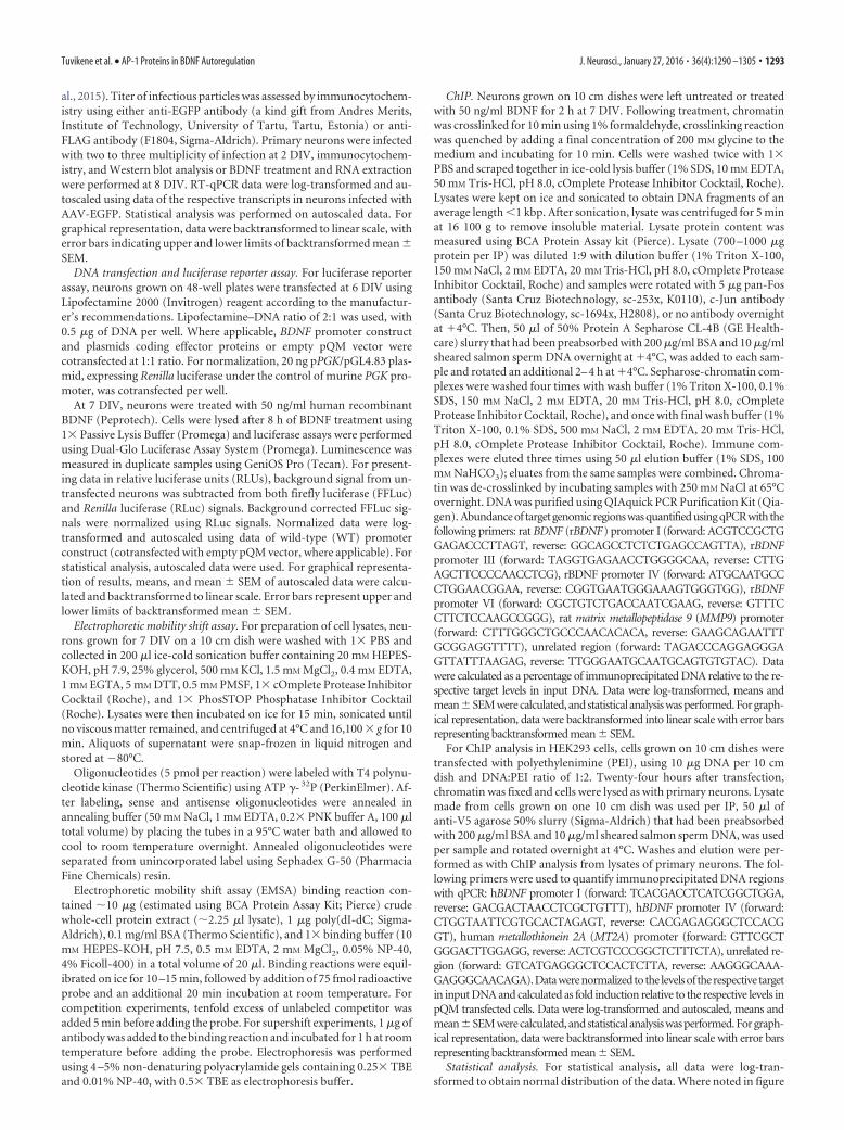
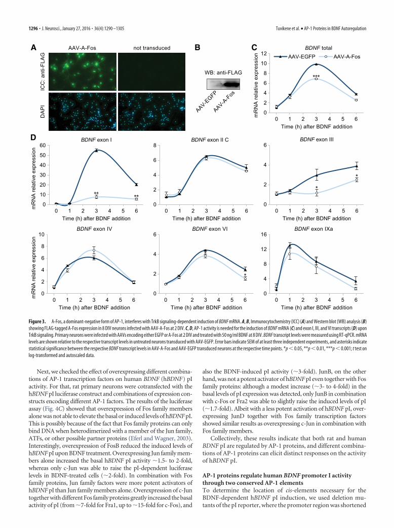
TrkB signaling-dependent induction of rat BDNF exon I, III,and VI transcripts requires AP-1 proteinsThere is evidence that BDNF stimulates AP-1 binding and AP-1-dependent transcriptional activity in neurons (Gaiddon et al., 1996;Okamoto et al., 2003). Therefore, we decided to investigate the pos-sible role of AP-1 transcription factors in the regulation of BDNFexpression by TrkB signaling. By using RT-qPCR and Western blotanalysis, we determined that the expression of mRNA and also pro-tein of all AP-1 members was induced in response to TrkB signalingin our cortical neuron cultures (Fig. 2A,B).
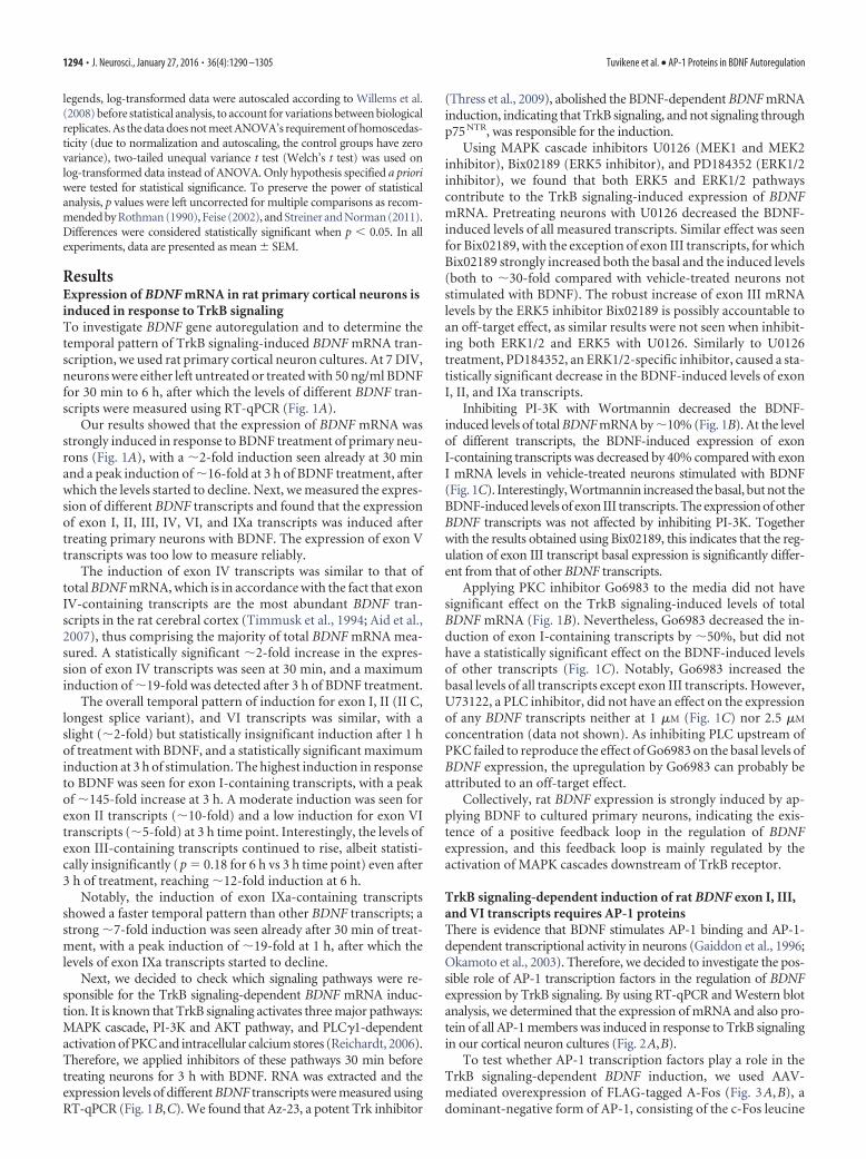
To test whether AP-1 transcription factors play a role in theTrkB signaling-dependent BDNF induction, we used AAV-mediated overexpression of FLAG-tagged A-Fos (Fig. 3A,B), adominant-negative form of AP-1, consisting of the c-Fos leucine
1294 • J. Neurosci., January 27, 2016 • 36(4):1290 –1305 Tuvikene et al. • AP-1 Proteins in BDNF Autoregulation
zipper domain with an acidic amphipathic extension appendedto the N-terminus (Olive et al., 1997). Infected neurons weretreated with BDNF at 8 DIV for different time periods, and BDNFtranscript levels were measured using RT-qPCR. We found thatoverexpressing A-Fos did not change the basal level of BDNFmRNA expression (Fig. 3C). However, A-Fos overexpression de-creased the induced level of total BDNF mRNA by �30% at 3 h ofBDNF treatment (from 9.8- to 6.9-fold induction compared withEGFP-expressing neurons not treated with BDNF).
At the level of alternative transcripts, A-Fos overexpressiondid not change the basal expression of any BDNF transcript, buteffectively reduced the BDNF-dependent induction of exonI-containing transcripts by 86% and 73% at 3 and 6 h time point,respectively (Fig. 3D). However, the induction of exon I tran-scripts was not completely abolished by the overexpression ofA-Fos, with a �7.6-fold induction remaining at 3 h time pointcompared with untreated neurons overexpressing EGFP. Thisindicates that the induction of exon I transcripts might also beregulated by other transcription factors in addition to AP-1, orthat the remaining induction of exon I transcripts occurs in theminority of cells (�10%) that were not infected. A-Fos also di-minished the TrkB signaling-dependent induction of exon III-containing transcripts, with the induction being completelyabolished at 3 h time point, and reduced by 36% at 6 h treatmentwith BDNF, and reduced the induced levels of transcript VI by�18 and �40% at 3 and 6 h BDNF treatment, respectively. The
induction of exon II, IV, and IXa transcripts was not changed byoverexpressing A-Fos, indicating that the integrity of TrkB sig-naling itself was not compromised. Collectively, these resultsindicate that AP-1 activity is needed for the TrkB signaling-dependent transcription of BDNF exon I, III, and VI transcripts,but not for their basal expression.
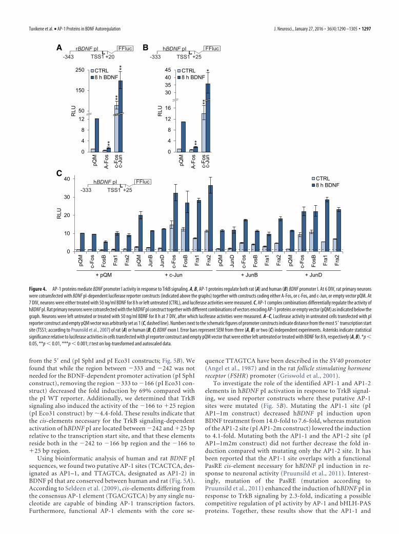
Induction of BDNF promoter I in response to TrkB signalingis mediated by AP-1 proteinsAs BDNF exon I-containing transcripts showed the strongest induc-tion in response to BDNF treatment, we first decided to elucidate themechanism behind this induction. For this, we transfected plasmidscontaining the human or rat BDNF promoter I (pI) regions in frontof luciferase coding sequence, together with either A-Fos, or c-Fosand c-Jun overexpression vectors into rat primary neurons. Neuronswere treated with BDNF for 8 h to induce TrkB signaling, after whichcells were lysed and luciferase activities were measured. According toour reporter assays, both rat (Fig. 4A) and human (Fig. 4B) BDNF pIactivity were upregulated by BDNF treatment (�12.2- and �10.9-fold induction, respectively). Cotransfecting A-Fos together with thereporter constructs effectively decreased the induced levels ofpromoter activity for both rat and human BDNF pI, by 88 and83%, respectively. Overexpressing AP-1 proteins c-Jun andc-Fos increased the promoter activity of human and rat BDNFpI in both unstimulated and BDNF-treated cells (Fig. 4 A, B).
GAPDH
Fra1
Fra2
JunB
c-Jun
FosB
JunD
c-Fos
35
48
35
48
35
48
35
48
35
0 0,5 1 2 3 4
35
4848
63
35
48
0 0,5 1 2 3 4
BDNF (h)
BDNF (h)
BA
0
20
40
60
80
0 1 2 3 4 5 6
c-Fos
0
50
100
150
200
250
0 1 2 3 4 5 6
FosB
0
5
10
15
20
0 1 2 3 4 5 6
Fra1
0
10
20
30
0 1 2 3 4 5 6
Fra2
0
2
4
6
8
0 1 2 3 4 5 6
c-Jun
0
10
20
30
40
50
0 1 2 3 4 5 6
JunB
0
1
2
3
4
0 1 2 3 4 5 6
JunD
Time (h) after BDNF addition
Time (h) after BDNF addition
Time (h) after BDNF addition
kDa
kDa
mR
NA
fold
indu
ctio
nm
RN
A fo
ld in
duct
ion
mR
NA
fold
indu
ctio
n
Figure 2. TrkB signaling induces expression of AP-1 family members in rat primary cortical neurons. A, RT-qPCR analysis of AP-1 family mRNA levels after treating primary neurons with 50 ng/mlBDNF. mRNA levels are shown as fold induction relative to the levels of the respective transcripts in untreated cells. Error bars represent SEM of three independent experiments. B, Western blotanalysis of neurons treated with 50 ng/ml BDNF for the indicated time at 7 DIV showing that the expression of all the members of the AP-1 family is induced upon TrkB signaling. Bands correspondingto different AP-1 proteins or GAPDH are shown with arrows.
Tuvikene et al. • AP-1 Proteins in BDNF Autoregulation J. Neurosci., January 27, 2016 • 36(4):1290 –1305 • 1295
Next, we checked the effect of overexpressing different combina-tions of AP-1 transcription factors on human BDNF (hBDNF) pIactivity. For that, rat primary neurons were cotransfected with thehBDNF pI luciferase construct and combinations of expression con-structs encoding different AP-1 factors. The results of the luciferaseassay (Fig. 4C) showed that overexpression of Fos family membersalone was not able to elevate the basal or induced levels of hBDNF pI.This is possibly because of the fact that Fos family proteins can onlybind DNA when heterodimerized with a member of the Jun family,ATFs, or other possible partner proteins (Eferl and Wagner, 2003).Interestingly, overexpression of FosB reduced the induced levels ofhBDNF pI upon BDNF treatment. Overexpressing Jun family mem-bers alone increased the basal hBDNF pI activity �1.5- to 2-fold,whereas only c-Jun was able to raise the pI-dependent luciferaselevels in BDNF-treated cells (�2-fold). In combination with Fosfamily proteins, Jun family factors were more potent activators ofhBDNF pI than Jun family members alone. Overexpression of c-Juntogether with different Fos family proteins greatly increased the basalactivity of pI (from �7-fold for Fra1, up to �15-fold for c-Fos), and
also the BDNF-induced pI activity (�3-fold). JunB, on the otherhand, was not a potent activator of hBDNF pI even together with Fosfamily proteins: although a modest increase (�3- to 4-fold) in thebasal levels of pI expression was detected, only JunB in combinationwith c-Fos or Fra2 was able to slightly raise the induced levels of pI(�1.7-fold). Albeit with a less potent activation of hBDNF pI, over-expressing JunD together with Fos family transcription factorsshowed similar results as overexpressing c-Jun in combination withFos family members.
Collectively, these results indicate that both rat and humanBDNF pI are regulated by AP-1 proteins, and different combina-tions of AP-1 proteins can elicit distinct responses on the activityof hBDNF pI.
AP-1 proteins regulate human BDNF promoter I activitythrough two conserved AP-1 elementsTo determine the location of cis-elements necessary for theBDNF-dependent hBDNF pI induction, we used deletion mu-tants of the pI reporter, where the promoter region was shortened
0
2
4
6
8
0 1 2 3 4 5 6Time (h) after BDNF addition
BDNF exon II C
Time (h) after BDNF addition
BDNF exon VI
0
2
4
6
8
10
12
0 1 2 3 4 5 6mR
NA
rela
tive
expr
essi
on
Time (h) after BDNF addition
BDNF total
0
4
8
12
16
0 1 2 3 4 5 6Time (h) after BDNF addition
BDNF exon IXa
0
2
4
6
8
10
0 1 2 3 4 5 6
noisserpxe evitaler A
NR
m
Time (h) after BDNF addition
BDNF exon IV
AAV-EGFP
AAV-A-F
os
WB: anti-FLAG
0
10
20
30
40
50
60
0 1 2 3 4 5 6
noisserpxe evitaler A
NR
m
Time (h) after BDNF addition
BDNF exon I
AAV-A-Fos not transducedIC
C: a
nti-F
LAG
DA
PI
0
2
4
6
0 1 2 3 4 5 6Time (h) after BDNF addition
BDNF exon III
0
2
4
6
0 1 2 3 4 5 6
A B C
D
AAV-EGFP AAV-A-Fos
***
** **
*
*
*
*
Figure 3. A-Fos, a dominant-negative form of AP-1, interferes with TrkB signaling-dependent induction of BDNF mRNA. A, B, Immunocytochemistry (ICC) (A) and Western blot (WB) analysis (B)showing FLAG-tagged A-Fos expression in 8 DIV neurons infected with AAV-A-Fos at 2 DIV. C, D, AP-1 activity is needed for the induction of BDNF mRNA (C) and exon I, III, and VI transcripts (D) uponTrkB signaling. Primary neurons were infected with AAVs encoding either EGFP or A-Fos at 2 DIV and treated with 50 ng/ml BDNF at 8 DIV. BDNF transcript levels were measured using RT-qPCR. mRNAlevels are shown relative to the respective transcript levels in untreated neurons transduced with AAV-EGFP. Error bars indicate SEM of at least three independent experiments, and asterisks indicatestatistical significance between the respective BDNF transcript levels in AAV-A-Fos and AAV-EGFP transduced neurons at the respective time points. *p � 0.05, **p � 0.01, ***p � 0.001; t test onlog-transformed and autoscaled data.
1296 • J. Neurosci., January 27, 2016 • 36(4):1290 –1305 Tuvikene et al. • AP-1 Proteins in BDNF Autoregulation
from the 5� end (pI SphI and pI Eco31 constructs; Fig. 5B). Wefound that while the region between �333 and �242 was notneeded for the BDNF-dependent promoter activation (pI SphIconstruct), removing the region �333 to �166 (pI Eco31 con-struct) decreased the fold induction by 69% compared withthe pI WT reporter. Additionally, we determined that TrkBsignaling also induced the activity of the �166 to �25 region(pI Eco31 construct) by �4.4-fold. These results indicate thatthe cis-elements necessary for the TrkB signaling-dependentactivation of hBDNF pI are located between �242 and �25 bprelative to the transcription start site, and that these elementsreside both in the �242 to �166 bp region and the �166 to�25 bp region.
Using bioinformatic analysis of human and rat BDNF pIsequences, we found two putative AP-1 sites (TCACTCA, des-ignated as AP1–1, and TTAGTCA, designated as AP1-2) inBDNF pI that are conserved between human and rat (Fig. 5A).According to Seldeen et al. (2009), cis-elements differing fromthe consensus AP-1 element (TGAC/GTCA) by any single nu-cleotide are capable of binding AP-1 transcription factors.Furthermore, functional AP-1 elements with the core se-
quence TTAGTCA have been described in the SV40 promoter(Angel et al., 1987) and in the rat follicle stimulating hormonereceptor (FSHR) promoter (Griswold et al., 2001).
To investigate the role of the identified AP1-1 and AP1-2elements in hBDNF pI activation in response to TrkB signal-ing, we used reporter constructs where these putative AP-1sites were mutated (Fig. 5B). Mutating the AP1-1 site (pIAP1–1m construct) decreased hBDNF pI induction uponBDNF treatment from 14.0-fold to 7.6-fold, whereas mutationof the AP1-2 site (pI AP1-2m construct) lowered the inductionto 4.1-fold. Mutating both the AP1-1 and the AP1-2 site (pIAP1–1m2m construct) did not further decrease the fold in-duction compared with mutating only the AP1-2 site. It hasbeen reported that the AP1-1 site overlaps with a functionalPasRE cis-element necessary for hBDNF pI induction in re-sponse to neuronal activity (Pruunsild et al., 2011). Interest-ingly, mutation of the PasRE (mutation according toPruunsild et al., 2011) enhanced the induction of hBDNF pI inresponse to TrkB signaling by 2.3-fold, indicating a possiblecompetitive regulation of pI activity by AP-1 and bHLH-PASproteins. Together, these results show that the AP1-1 and
A-343 +20
FFlucrBDNF pI
0
4
8
1250
150
250
pQM
A-Fo
s
RLU
c-Fo
sc-
Jun
CTRL8 h BDNF
B-333 +25TSS1
FFluchBDNF pI
0
4
8
12
16
30354045
pQM
A-Fo
s
c-Fo
sc-
Jun
RLU
CTRL8 h BDNF
-333 +25TSS1
TSS1
FFluchBDNF pIC
CTRL8 h BDNF
c-Fo
s
FosB
Fra1
Fra2
JunB
JunD
c-Fo
s
FosB
Fra1
Fra2
JunD
c-Fo
s
FosB
Fra1
Fra2
c-Fo
s
FosB
Fra1
Fra2
+ c-Jun + JunB + JunD+ pQM
pQM
pQM
pQM
pQM
0
10
20
30
40
RLU
**
**
**
**
**
*
Figure 4. AP-1 proteins mediate BDNF promoter I activity in response to TrkB signaling. A, B, AP-1 proteins regulate both rat (A) and human (B) BDNF promoter I. At 6 DIV, rat primary neuronswere cotransfected with BDNF pI-dependent luciferase reporter constructs (indicated above the graphs) together with constructs coding either A-Fos, or c-Fos, and c-Jun, or empty vector pQM. At7 DIV, neurons were either treated with 50 ng/ml BDNF for 8 h or left untreated (CTRL), and luciferase activities were measured. C, AP-1 complex combinations differentially regulate the activity ofhBDNF pI. Rat primary neurons were cotransfected with the hBDNF pI construct together with different combinations of vectors encoding AP-1 proteins or empty vector (pQM) as indicated below thegraph. Neurons were left untreated or treated with 50 ng/ml BDNF for 8 h at 7 DIV, after which luciferase activities were measured. A–C, Luciferase activity in untreated cells transfected with pIreporter construct and empty pQM vector was arbitrarily set as 1 (C, dashed line). Numbers next to the schematic figures of promoter constructs indicate distance from the most 5� transcription startsite (TSS1; according to Pruunsild et al., 2007) of rat (A) or human (B, C) BDNF exon I. Error bars represent SEM from three (A, B) or two (C) independent experiments. Asterisks indicate statisticalsignificance relative to luciferase activities in cells transfected with pI reporter construct and empty pQM vector that were either left untreated or treated with BDNF for 8 h, respectively (A, B). *p �0.05, **p � 0.01, ***p � 0.001; t test on log-transformed and autoscaled data.
Tuvikene et al. • AP-1 Proteins in BDNF Autoregulation J. Neurosci., January 27, 2016 • 36(4):1290 –1305 • 1297
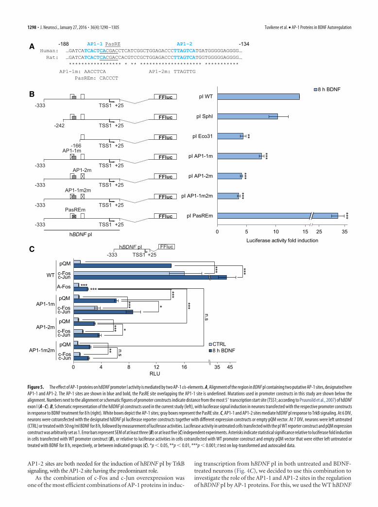
AP1-2 sites are both needed for the induction of hBDNF pI by TrkBsignaling, with the AP1-2 site having the predominant role.
As the combination of c-Fos and c-Jun overexpression wasone of the most efficient combinations of AP-1 proteins in induc-
ing transcription from hBDNF pI in both untreated and BDNF-treated neurons (Fig. 4C), we decided to use this combination toinvestigate the role of the AP1-1 and AP1-2 sites in the regulationof hBDNF pI by AP-1 proteins. For this, we used the WT hBDNF
35 45
WT
AP1-1m
AP1-2m
AP1-1m2m
pQM
c-Fosc-Jun
A-Fos
pQM
c-Fosc-Jun
pQM
c-Fosc-Jun
pQM
c-Fosc-Jun
0 4 8 12 16RLU
-333 +25TSS1FFluchBDNF pI
n.s
n.s
C
CTRL8 h BDNF
-333 +25TSS1FFluc
-242 +25TSS1FFluc
AP1-1m
-333 +25TSS1FFluc
AP1-2m
-333 +25TSS1FFluc
AP1-1m2m
-333 +25TSS1FFluc
PasREm
-333 +25TSS1FFluc
-166 +25TSS1FFluc
pI WT
pI SphI
pI Eco31
pI AP1-1m
pI AP1-2m
pI AP1-1m2m
pI PasREm
0 5 10 15 25 35
Luciferase activity fold inductionhBDNF pI
B8 h BDNF
AP1-1 PasRE AP1-2 Human: … GATCATCACTCACGACCTCATCGGCTGGAGACCCTTAGTCATGATGGGGGAGGGG… Rat:
… GATCATCACTCACGACCACGTCCGCTGGAGACCCTTAGTCATGGTGGGGGAGGGG… ***************** * ** ******************** ***********
-188 -134
AP1-1m: AACCTCA AP1-2m: TTAGTTGPasREm: CACCCT
A
***
******
***
*****
***
***
***
***
*** *
*
**
******
Figure 5. The effect of AP-1 proteins on hBDNF promoter I activity is mediated by two AP-1 cis-elements. A, Alignment of the region in BDNF pI containing two putative AP-1 sites, designated hereAP1-1 and AP1-2. The AP-1 sites are shown in blue and bold, the PasRE site overlapping the AP1-1 site is underlined. Mutations used in promoter constructs in this study are shown below thealignment. Numbers next to the alignment or schematic figures of promoter constructs indicate distance from the most 5� transcription start site (TSS1; according to Pruunsild et al., 2007) of hBDNFexon I (A–C). B, Schematic representation of the hBDNF pI constructs used in the current study (left), with luciferase signal induction in neurons transfected with the respective promoter constructsin response to BDNF treatment for 8 h (right). White boxes depict the AP-1 sites; gray boxes represent the PasRE site. C, AP1-1 and AP1-2 sites mediate hBDNF pI response to TrkB signaling. At 6 DIV,neurons were cotransfected with the designated hBDNF pI luciferase reporter constructs together with different expression constructs or empty pQM vector. At 7 DIV, neurons were left untreated(CTRL) or treated with 50 ng/ml BDNF for 8 h, followed by measurement of luciferase activities. Luciferase activity in untreated cells transfected with the pI WT reporter construct and pQM expressionconstruct was arbitrarily set as 1. Error bars represent SEM of at least three (B) or at least five (C) independent experiments. Asterisks indicate statistical significance relative to luciferase fold inductionin cells transfected with WT promoter construct (B), or relative to luciferase activities in cells cotransfected with WT promoter construct and empty pQM vector that were either left untreated ortreated with BDNF for 8 h, respectively, or between indicated groups (C). *p � 0.05, **p � 0.01, ***p � 0.001; t test on log-transformed and autoscaled data.
1298 • J. Neurosci., January 27, 2016 • 36(4):1290 –1305 Tuvikene et al. • AP-1 Proteins in BDNF Autoregulation
pI promoter construct, or constructs with mutated AP-1 sites,together with overexpression of c-Fos and c-Jun in rat primaryneurons (Fig. 5C). Overexpressing c-Fos/c-Jun together with theWT hBDNF pI construct raised both the basal (�16-fold) and theBDNF-induced expression levels (�3-fold) of hBDNF pI (Fig.5C). Mutating either of the AP1-1 or AP1-2 elements decreasedthe effect of c-Fos/c-Jun overexpression on pI activity upon TrkBsignaling, but did not completely remove it. When both sites weremutated, the BDNF-induced levels of pI activity were notchanged by AP-1 protein overexpression. However, mutatingboth the AP1-1 and the AP1-2 site did not completely abolish theeffect of AP-1 protein overexpression on the basal promoter ac-tivity levels, as AP-1 protein overexpression was able to raise thepI AP1–1m2m reporter activity �1.5-fold. Mutating both theAP1-1 and the AP1-2 site together, but not either of the sitesalone, decreased the BDNF-induced activity of pI to the samelevel as in the case of overexpressing A-Fos together with the WThBDNF pI construct (�2.1 and �2.4-fold induction, respec-tively, compared with the WT promoter activity in untreated cellscotransfected with empty pQM vector), indicating that the twoAP-1 sites described in this study are the main cis-acting elementsresponsible for the AP-1-mediated activation of hBDNF pI uponTrkB signaling.
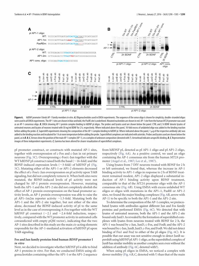
Fos and Jun family proteins bind human BDNF promoter Iin vitroNext, we decided to investigate whether hBDNF pI is able to bindAP-1 proteins in vitro. For that, we conducted EMSA using oli-gonucleotides containing either the AP1-1 or the AP1-2 sequence
from hBDNF pI, denoted as pI AP1-1 oligo and pI AP1-2 oligo,respectively (Fig. 6A). As a positive control, we used an oligocontaining the AP-1 consensus site from the human MT2A pro-moter (Angel et al., 1987; Lee et al., 1987).
Using lysates from 7 DIV neurons treated with BDNF for 2 hor left untreated, we found that, whereas the increase in AP-1binding activity to AP1-1 oligo in response to 2 h of BDNF treat-ment remained modest, AP1-2 oligo displayed a substantial in-duction of AP-1 binding activity upon BDNF treatment,comparable to that of the MT2A promoter oligo with the AP-1consensus site (Fig. 6B). Using EMSA with excess unlabeled WToligos or oligos with mutations in the AP1–1, PasRE or AP1-2sites, we found the major binding complexes (Fig. 6B, arrow withAP-1) to be specific to both the AP1-1 and the AP1-2 site.
To determine the composition of the AP-1 complex, we preincu-bated lysates with antibodies against different Jun and Fos familyproteins and performed EMSA (Fig. 6C). We detected that withlysates of untreated neurons, both the AP1-1 and the AP1-2 sitebound only JunD. As revealed by the formation of supershifted com-plexes with lysates from neurons treated with BDNF for 2 h, theAP1-1 was bound by c-Jun, JunD, c-Fos, and FosB; and the AP1-2was bound by c-Jun, JunB, JunD, c-Fos, and FosB. We did not detectbinding of Fra1 and Fra2 to either of the pI oligos (Fig. 6C). It ispossible that our assay was not sensitive enough to detect JunB su-pershift using hBDNF pI AP1-1 oligo, as the supershifted complex ofJunB has similar mobility as another complex seen even without theaddition of antibody (Fig. 6C, denoted with?).
When using pI oligos in EMSA, we noticed a complex withslower mobility (Fig. 6B,C, denoted with ?) than that of the main
10x
WT
10x A
P1-1
m10
x Pa
sREm
10x A
P-1
cons
.
10x
WT
10x A
P1-2
m10
x AP1
con
s.
AP-1?
CTRL 2h BDNF
CTRL 2h BDNF
CTRL
2h B
DNF
pI AP1-1 oligo pI AP1-2 oligo AP1 cons.oligo
*
*
*
*
**
*
2h BDNFCTRLpI AP1-1 oligo
no a
bc-
Jun
abJu
nB a
bJu
nD a
bc-
Fos
ab
FosB
ab
Fra1
ab
Fra2
ab
no a
bc-
Jun
abJu
nB a
bJu
nD a
bc-
Fos
abFo
sB a
bFr
a1 a
bFr
a2 a
b
* **
**
2h BDNFCTRLpI AP1-2 oligo
no a
bc-
Jun
abJu
nB a
bJu
nD a
bc-
Fos
abFo
sB a
bFr
a1 a
bFr
a2 a
bno
ab
c-Ju
n ab
JunB
ab
JunD
ab
c-Fo
s ab
FosB
ab
Fra1
ab
Fra2
ab
pI AP1-1 oligo:
WT: TCCCCATTTGATCATCACTCACGACCTCATCGGCT AP1-1m: TCCCCATTTGATCAAACCTCACGACCTCATCGGCT
PasREm: TCCCCATTTGATCATCACTCACCCTCTCATCGGCT
pI AP1-2 oligo: WT: GCTGGAGACCCTTAGTCATGATGGGGGAGGGGGA
AP1-2m: GCTGGAGACCCTTAGTTGTGATGGGGGAGGGGGA
CCGCAAGTGACTCAGCGCGGG
A B
C
AP1 cons. oligo:
?AP-1
Figure 6. hBDNF promoter I binds AP-1 family members in vitro. A, Oligonucleotides used in EMSA experiments. The sequence of the sense oligo is shown for simplicity; double-stranded oligoswere used in all EMSA experiments. The AP-1 sites are shown in blue and bold, the PasRE site is underlined. Mutated nucleotides are shown in red. AP-1 site from the human MT2A promoter was usedas an AP-1 consensus oligo. B, EMSA showing AP-1 protein complex binding to hBDNF pI oligos. The probes and lysates used are shown below the panel. CTRL and 2 h BDNF denote lysates ofuntreated neurons and lysates of neurons treated with 50 ng/ml BDNF for 2 h, respectively. Where indicated above the panel, 10-fold excess of unlabeled oligo was added to the binding reactionbefore adding the probe. C, Supershift experiments showing the composition of the AP-1 complex binding to hBDNF pI. Where indicated above the panel, 1 �g of the respective antibody (ab) wasadded to the binding reaction and incubated for 1 h at room temperature before adding the probe. Supershifted complexes are indicated with asterisks. Probes and lysates used are shown below thepanel, as in B. B, C, Arrows show the position of the main AP-1 complex (AP-1), or a complex of unknown composition (denoted with ?). Arrowhead indicates unspecific binding. B, C, Representativeimages of three independent experiments. C, Gamma has been altered for clearer visualization of supershifted complexes.
Tuvikene et al. • AP-1 Proteins in BDNF Autoregulation J. Neurosci., January 27, 2016 • 36(4):1290 –1305 • 1299
AP-1 complex. Furthermore, a complex with similar mobilitywas also seen when using AP-1 consensus oligo as the probe.Competition with unlabeled oligos indicated that this complexbinds to the AP1-2 site, but is not specific for the AP1-1 site (Fig.6B). Of note, we saw a decrease in binding of this complex insupershift experiments using pI AP1-1 oligo when the untreatedneuronal lysate was incubated with c-Jun and JunD antibody,and when lysate from BDNF-treated neurons was incubated withc-Jun antibody (Fig. 6C). Even though according to the compe-tition experiments the complex was not AP1–1-specific, this in-dicates that Jun family proteins might be involved. When using pIAP1-2 oligo as the probe, we could not determine a clear patternin the decrease in binding of the slower-mobility complex insupershift experiments. We propose that the complex consists ofJun family homodimers, Jun family proteins together with ATFs,or other possible Jun heterodimerization partners.
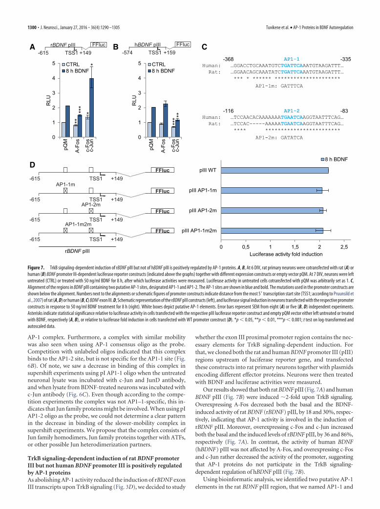
TrkB signaling-dependent induction of rat BDNF promoterIII but not human BDNF promoter III is positively regulatedby AP-1 proteinsAs abolishing AP-1 activity reduced the induction of rBDNF exonIII transcripts upon TrkB signaling (Fig. 3D), we decided to study
whether the exon III proximal promoter region contains the nec-essary elements for TrkB signaling-dependent induction. Forthat, we cloned both the rat and human BDNF promoter III (pIII)regions upstream of luciferase reporter gene, and transfectedthese constructs into rat primary neurons together with plasmidsencoding different effector proteins. Neurons were then treatedwith BDNF and luciferase activities were measured.
Our results showed that both rat BDNF pIII (Fig. 7A) and humanBDNF pIII (Fig. 7B) were induced �2-fold upon TrkB signaling.Overexpressing A-Fos decreased both the basal and the BDNF-induced activity of rat BDNF (rBDNF) pIII, by 18 and 30%, respec-tively, indicating that AP-1 activity is involved in the induction ofrBDNF pIII. Moreover, overexpressing c-Fos and c-Jun increasedboth the basal and the induced levels of rBDNF pIII, by 36 and 86%,respectively (Fig. 7A). In contrast, the activity of human BDNF(hBDNF) pIII was not affected by A-Fos, and overexpressing c-Fosand c-Jun rather decreased the activity of the promoter, suggestingthat AP-1 proteins do not participate in the TrkB signaling-dependent regulation of hBDNF pIII (Fig. 7B).
Using bioinformatic analysis, we identified two putative AP-1elements in the rat BDNF pIII region, that we named AP1-1 and
***
*****
***
*
-615 +149TSS1FFlucrBDNF pIII
-574 +159TSS1FFluchBDNF pIIIA B
AP1-1 Human: …GGACCTGCAAATGTCTGATTCAAATGTAAGATTT… Rat: …GGAACAGCAAATATCTGATTCAAATGTAAGATTT… *** * ****** *********************
AP1-2 Human: …TCCAACACAAAAAAATGAATCAAGGTAATTTCAG… Rat: …TCCAC-----AAAAATGAATCAAGGTAATTTCAG… **** ************************
C-368 -335
-116 -83
AP1-1m: GATTTCA
AP1-2m: GATATCA
D
0 0,5 1 1,5 2 2,5
pIII WT
pIII AP1-1m
pIII AP1-2m
pIII AP1-1m2m
Luciferase activity fold induction
-615 +149TSS1FFluc
rBDNF pIII
AP1-1m2m
-615 +149TSS1FFluc
AP1-2m
-615 +149TSS1FFluc
AP1-1m
-615 +149TSS1FFluc
RLU
CTRL8 h BDNF
0
1
2
3
4
5pQ
M
A-Fo
s
c-Fo
sc-
Jun
CTRL8 h BDNF
0
1
2
3
4
5
pQM
A-Fo
s
c-Fo
sc-
Jun
RLU
8 h BDNF
Figure 7. TrkB signaling-dependent induction of rBDNF pIII but not of hBDNF pIII is positively regulated by AP-1 proteins. A, B, At 6 DIV, rat primary neurons were cotransfected with rat (A) orhuman (B) BDNF promoter III-dependent luciferase reporter constructs (indicated above the graphs) together with different expression constructs or empty vector pQM. At 7 DIV, neurons were leftuntreated (CTRL) or treated with 50 ng/ml BDNF for 8 h, after which luciferase activities were measured. Luciferase activity in untreated cells cotransfected with pQM was arbitrarily set as 1. C,Alignment of the regions in BDNF pIII containing two putative AP-1 sites, designated AP1-1 and AP1-2. The AP-1 sites are shown in blue and bold. The mutations used in the promoter constructs areshown below the alignment. Numbers next to the alignments or schematic figures of promoter constructs indicate distance from the most 5� transcription start site (TSS1; according to Pruunsild etal., 2007) of rat (A, D) or human (B, C) BDNF exon III. D, Schematic representation of the rBDNF pIII constructs (left), and luciferase signal induction in neurons transfected with the respective promoterconstructs in response to 50 ng/ml BDNF treatment for 8 h (right). White boxes depict putative AP-1 elements. Error bars represent SEM from eight (A) or five (B, D) independent experiments.Asterisks indicate statistical significance relative to luciferase activity in cells transfected with the respective pIII luciferase reporter construct and empty pQM vector either left untreated or treatedwith BDNF, respectively (A, B), or relative to luciferase fold induction in cells transfected with WT promoter construct (D). *p � 0.05, **p � 0.01, ***p � 0.001; t test on log-transformed andautoscaled data.
1300 • J. Neurosci., January 27, 2016 • 36(4):1290 –1305 Tuvikene et al. • AP-1 Proteins in BDNF Autoregulation
AP1-2 (Fig. 7C). Of note, both of these sites are conserved be-tween human and rat. To check whether these AP-1 elements areresponsible for the TrkB signaling-dependent induction ofrBDNF pIII, we used luciferase reporter constructs where thesesites alone (pIII AP1–1m and pIII AP1-2m constructs) or to-gether (pIII AP1–1m2m construct) were mutated (Fig. 7D). Us-ing reporter assay, we found that neither mutating these sitesalone nor in combination had effect on the induction of rBDNFpIII in response to TrkB signaling (Fig. 7D).
Together, these results indicate that rat and human BDNF pIIIare differentially regulated in response to TrkB signaling, and thatrBDNF pIII does not seem to contain functional AP-1 sites, sug-gesting that the effect of AP-1 proteins on rBDNF pIII activity isindirect.
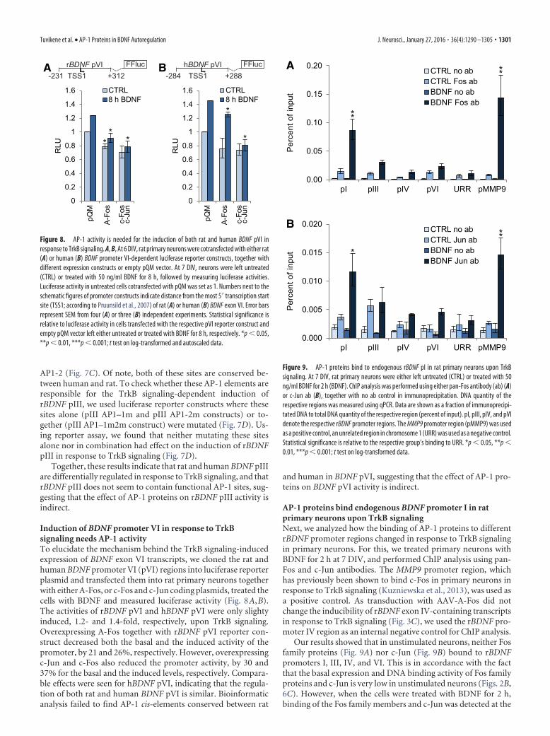
Induction of BDNF promoter VI in response to TrkBsignaling needs AP-1 activityTo elucidate the mechanism behind the TrkB signaling-inducedexpression of BDNF exon VI transcripts, we cloned the rat andhuman BDNF promoter VI (pVI) regions into luciferase reporterplasmid and transfected them into rat primary neurons togetherwith either A-Fos, or c-Fos and c-Jun coding plasmids, treated thecells with BDNF and measured luciferase activity (Fig. 8A,B).The activities of rBDNF pVI and hBDNF pVI were only slightyinduced, 1.2- and 1.4-fold, respectively, upon TrkB signaling.Overexpressing A-Fos together with rBDNF pVI reporter con-struct decreased both the basal and the induced activity of thepromoter, by 21 and 26%, respectively. However, overexpressingc-Jun and c-Fos also reduced the promoter activity, by 30 and37% for the basal and the induced levels, respectively. Compara-ble effects were seen for hBDNF pVI, indicating that the regula-tion of both rat and human BDNF pVI is similar. Bioinformaticanalysis failed to find AP-1 cis-elements conserved between rat
and human in BDNF pVI, suggesting that the effect of AP-1 pro-teins on BDNF pVI activity is indirect.
AP-1 proteins bind endogenous BDNF promoter I in ratprimary neurons upon TrkB signalingNext, we analyzed how the binding of AP-1 proteins to differentrBDNF promoter regions changed in response to TrkB signalingin primary neurons. For this, we treated primary neurons withBDNF for 2 h at 7 DIV, and performed ChIP analysis using pan-Fos and c-Jun antibodies. The MMP9 promoter region, whichhas previously been shown to bind c-Fos in primary neurons inresponse to TrkB signaling (Kuzniewska et al., 2013), was used asa positive control. As transduction with AAV-A-Fos did notchange the inducibility of rBDNF exon IV-containing transcriptsin response to TrkB signaling (Fig. 3C), we used the rBDNF pro-moter IV region as an internal negative control for ChIP analysis.
Our results showed that in unstimulated neurons, neither Fosfamily proteins (Fig. 9A) nor c-Jun (Fig. 9B) bound to rBDNFpromoters I, III, IV, and VI. This is in accordance with the factthat the basal expression and DNA binding activity of Fos familyproteins and c-Jun is very low in unstimulated neurons (Figs. 2B,6C). However, when the cells were treated with BDNF for 2 h,binding of the Fos family members and c-Jun was detected at the
pQM
A-Fo
s
c-Fo
sc-
Jun
*
*
1.6
0
0.2
0.4
0.6
0.8
1
1.2
1.4
RLU
CTRL8 h BDNF
-231 +312TSS1FFlucrBDNF pVI
-284 +288TSS1FFluchBDNF pVIA B
pQM
A-Fo
s
c-Fo
sc-
Jun
***
RLU
CTRL8 h BDNF
0
0.2
0.4
0.6
0.8
1
1.2
1.4
1.6
Figure 8. AP-1 activity is needed for the induction of both rat and human BDNF pVI inresponse to TrkB signaling. A, B, At 6 DIV, rat primary neurons were cotransfected with either rat(A) or human (B) BDNF promoter VI-dependent luciferase reporter constructs, together withdifferent expression constructs or empty pQM vector. At 7 DIV, neurons were left untreated(CTRL) or treated with 50 ng/ml BDNF for 8 h, followed by measuring luciferase activities.Luciferase activity in untreated cells cotransfected with pQM was set as 1. Numbers next to theschematic figures of promoter constructs indicate distance from the most 5� transcription startsite (TSS1; according to Pruunsild et al., 2007) of rat (A) or human (B) BDNF exon VI. Error barsrepresent SEM from four (A) or three (B) independent experiments. Statistical significance isrelative to luciferase activity in cells transfected with the respective pVI reporter construct andempty pQM vector left either untreated or treated with BDNF for 8 h, respectively. *p � 0.05,**p � 0.01, ***p � 0.001; t test on log-transformed and autoscaled data.
Per
cent
of i
nput
Per
cent
of i
nput
0.00
0.05
0.10
0.15
0.20
pI pIII pIV pVI URR pMMP9
0.000
0.005
0.010
0.015
0.020
pI pIII pIV pVI URR pMMP9
CTRL no abCTRL Fos abBDNF no abBDNF Fos ab
CTRL no abCTRL Jun abBDNF no abBDNF Jun ab
A
B
**
**
***
Figure 9. AP-1 proteins bind to endogenous rBDNF pI in rat primary neurons upon TrkBsignaling. At 7 DIV, rat primary neurons were either left untreated (CTRL) or treated with 50ng/ml BDNF for 2 h (BDNF). ChIP analysis was performed using either pan-Fos antibody (ab) (A)or c-Jun ab (B), together with no ab control in immunoprecipitation. DNA quantity of therespective regions was measured using qPCR. Data are shown as a fraction of immunoprecipi-tated DNA to total DNA quantity of the respective region (percent of input). pI, pIII, pIV, and pVIdenote the respective rBDNF promoter regions. The MMP9 promoter region (pMMP9) was usedas a positive control, an unrelated region in chromosome 1 (URR) was used as a negative control.Statistical significance is relative to the respective group’s binding to URR. *p � 0.05, **p �0.01, ***p � 0.001; t test on log-transformed data.
Tuvikene et al. • AP-1 Proteins in BDNF Autoregulation J. Neurosci., January 27, 2016 • 36(4):1290 –1305 • 1301
rBDNF pI region (Fig. 9). The extent of this binding was compa-rable to that of the MMP9 promoter region used as a positivecontrol. No significant binding of AP-1 proteins was seen forrBDNF promoters III, IV, and VI.
Collectively, these results indicate that AP-1 proteins directlyregulate the activity of rBDNF pI, whereas the effect of AP-1proteins on rBDNF pIII and pVI activity is indirect, possiblyacting through different transcription factors that are induced byAP-1 proteins.
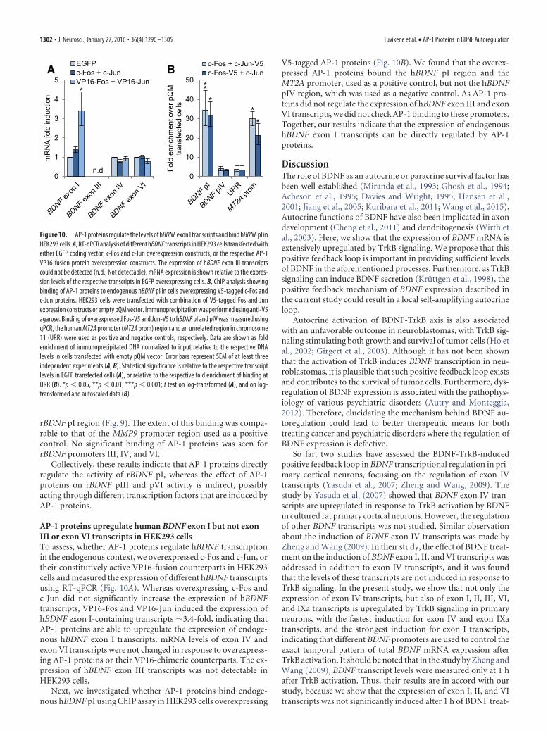
AP-1 proteins upregulate human BDNF exon I but not exonIII or exon VI transcripts in HEK293 cellsTo assess, whether AP-1 proteins regulate hBDNF transcriptionin the endogenous context, we overexpressed c-Fos and c-Jun, ortheir constitutively active VP16-fusion counterparts in HEK293cells and measured the expression of different hBDNF transcriptsusing RT-qPCR (Fig. 10A). Whereas overexpressing c-Fos andc-Jun did not significantly increase the expression of hBDNFtranscripts, VP16-Fos and VP16-Jun induced the expression ofhBDNF exon I-containing transcripts �3.4-fold, indicating thatAP-1 proteins are able to upregulate the expression of endoge-nous hBDNF exon I transcripts. mRNA levels of exon IV andexon VI transcripts were not changed in response to overexpress-ing AP-1 proteins or their VP16-chimeric counterparts. The ex-pression of hBDNF exon III transcripts was not detectable inHEK293 cells.
Next, we investigated whether AP-1 proteins bind endoge-nous hBDNF pI using ChIP assay in HEK293 cells overexpressing
V5-tagged AP-1 proteins (Fig. 10B). We found that the overex-pressed AP-1 proteins bound the hBDNF pI region and theMT2A promoter, used as a positive control, but not the hBDNFpIV region, which was used as a negative control. As AP-1 pro-teins did not regulate the expression of hBDNF exon III and exonVI transcripts, we did not check AP-1 binding to these promoters.Together, our results indicate that the expression of endogenoushBDNF exon I transcripts can be directly regulated by AP-1proteins.
DiscussionThe role of BDNF as an autocrine or paracrine survival factor hasbeen well established (Miranda et al., 1993; Ghosh et al., 1994;Acheson et al., 1995; Davies and Wright, 1995; Hansen et al.,2001; Jiang et al., 2005; Kuribara et al., 2011; Wang et al., 2015).Autocrine functions of BDNF have also been implicated in axondevelopment (Cheng et al., 2011) and dendritogenesis (Wirth etal., 2003). Here, we show that the expression of BDNF mRNA isextensively upregulated by TrkB signaling. We propose that thispositive feedback loop is important in providing sufficient levelsof BDNF in the aforementioned processes. Furthermore, as TrkBsignaling can induce BDNF secretion (Kruttgen et al., 1998), thepositive feedback mechanism of BDNF expression described inthe current study could result in a local self-amplifying autocrineloop.
Autocrine activation of BDNF-TrkB axis is also associatedwith an unfavorable outcome in neuroblastomas, with TrkB sig-naling stimulating both growth and survival of tumor cells (Ho etal., 2002; Girgert et al., 2003). Although it has not been shownthat the activation of TrkB induces BDNF transcription in neu-roblastomas, it is plausible that such positive feedback loop existsand contributes to the survival of tumor cells. Furthermore, dys-regulation of BDNF expression is associated with the pathophys-iology of various psychiatric disorders (Autry and Monteggia,2012). Therefore, elucidating the mechanism behind BDNF au-toregulation could lead to better therapeutic means for bothtreating cancer and psychiatric disorders where the regulation ofBDNF expression is defective.
So far, two studies have assessed the BDNF-TrkB-inducedpositive feedback loop in BDNF transcriptional regulation in pri-mary cortical neurons, focusing on the regulation of exon IVtranscripts (Yasuda et al., 2007; Zheng and Wang, 2009). Thestudy by Yasuda et al. (2007) showed that BDNF exon IV tran-scripts are upregulated in response to TrkB activation by BDNFin cultured rat primary cortical neurons. However, the regulationof other BDNF transcripts was not studied. Similar observationabout the induction of BDNF exon IV transcripts was made byZheng and Wang (2009). In their study, the effect of BDNF treat-ment on the induction of BDNF exon I, II, and VI transcripts wasaddressed in addition to exon IV transcripts, and it was foundthat the levels of these transcripts are not induced in response toTrkB signaling. In the present study, we show that not only theexpression of exon IV transcripts, but also of exon I, II, III, VI,and IXa transcripts is upregulated by TrkB signaling in primaryneurons, with the fastest induction for exon IV and exon IXatranscripts, and the strongest induction for exon I transcripts,indicating that different BDNF promoters are used to control theexact temporal pattern of total BDNF mRNA expression afterTrkB activation. It should be noted that in the study by Zheng andWang (2009), BDNF transcript levels were measured only at 1 hafter TrkB activation. Thus, their results are in accord with ourstudy, because we show that the expression of exon I, II, and VItranscripts was not significantly induced after 1 h of BDNF treat-
0
10
20
30
40
50
Fold
enr
ichm
ent o
ver p
QM
tra
nsfe
cted
cel
ls0
1
2
3
4
5
BDNF exon
I
BDNF exon
III
BDNF exon
IV
BDNF pI
BDNF pIV
URR
MT2A pr
om
n.d
BDNF exon
VI
EGFPc-Fos + c-JunVP16-Fos + VP16-Jun
c-Fos + c-Jun-V5c-Fos-V5 + c-JunA B
mR
NA
fold
indu
ctio
n
**
**
**
Figure 10. AP-1 proteins regulate the levels of hBDNF exon I transcripts and bind hBDNF pI inHEK293 cells. A, RT-qPCR analysis of different hBDNF transcripts in HEK293 cells transfected witheither EGFP coding vector, c-Fos and c-Jun overexpression constructs, or the respective AP-1VP16-fusion protein overexpression constructs. The expression of hBDNF exon III transcriptscould not be detected (n.d., Not detectable). mRNA expression is shown relative to the expres-sion levels of the respective transcripts in EGFP overexpressing cells. B, ChIP analysis showingbinding of AP-1 proteins to endogenous hBDNF pI in cells overexpressing V5-tagged c-Fos andc-Jun proteins. HEK293 cells were transfected with combination of V5-tagged Fos and Junexpression constructs or empty pQM vector. Immunoprecipitation was performed using anti-V5agarose. Binding of overexpressed Fos-V5 and Jun-V5 to hBDNF pI and pIV was measured usingqPCR, the human MT2A promoter (MT2A prom) region and an unrelated region in chromosome11 (URR) were used as positive and negative controls, respectively. Data are shown as foldenrichment of immunoprecipitated DNA normalized to input relative to the respective DNAlevels in cells transfected with empty pQM vector. Error bars represent SEM of at least threeindependent experiments (A, B). Statistical significance is relative to the respective transcriptlevels in EGFP transfected cells (A), or relative to the respective fold enrichment of binding atURR (B). *p � 0.05, **p � 0.01, ***p � 0.001; t test on log-transformed (A), and on log-transformed and autoscaled data (B).
1302 • J. Neurosci., January 27, 2016 • 36(4):1290 –1305 Tuvikene et al. • AP-1 Proteins in BDNF Autoregulation
ment, whereas exon IV transcripts displayed a moderate �8-foldinduction. Furthermore, we show that at later time points thelevels of exon I, II, and VI transcripts are also increased in re-sponse to TrkB signaling. As it has been reported that the expres-sion of BDNF exon IV transcripts can be induced as animmediate-early gene in response to neuronal activity (Lauter-born et al., 1996; Tao et al., 1998), it is possible that the fastinduction of exon IV and IXa transcripts upon TrkB signalingcould be independent of protein synthesis. It is plausible that invivo, the BDNF transcripts that are induced first in response toTrkB signaling provide enough BDNF to start and sustain theBDNF autocrine loop, which in turn would induce the expressionof other, more slowly inducible BDNF transcripts (eg, exon I, II,III transcripts), further prolonging the TrkB signaling necessaryfor various physiological outcomes.
The effect of TrkB signaling on the regulation of BDNF tran-scription has also been shown in vivo: infusion of BDNF into thedentate gyrus of adult anesthetized rats triggers stable LTP, anactivity-dependent synaptic plasticity, and upregulates bothBDNF exon IV-containing transcripts and total BDNF mRNAlevels (Wibrand et al., 2006). Furthermore, it has been reportedthat C/EBP�-mediated BDNF-positive feedback loop regulatingBDNF exon IV mRNA expression in the hippocampus is impor-tant for memory consolidation in vivo (Bambah-Mukku et al.,2014), indicating that BDNF autoregulation is critical for hip-pocampus-dependent memory functions. We propose, in thelight of our results obtained using primary cortical neuron cul-tures, that not only the induction of exon IV transcripts, but alsoof other BDNF transcripts could be important for memory-related functions. Moreover, our results suggest that in additionto the role of BDNF-positive feedback loop in the hippocampus,such regulatory mechanisms might be involved in proper func-tioning and/or development of the cortex.
It is well proven that BDNF potentiates synaptic transmissionthat is connected to neuronal membrane depolarization (Lu,2003; Park and Poo, 2013). As the expression of BDNF is alsoactivated by membrane depolarization and calcium influx intoneurons (Park and Poo, 2013), one might argue that the effectsseen in this study are a result of increased synaptic activity, andnot caused by direct regulation of BDNF gene expression by TrkBsignaling. However, it has been reported that TrkB signaling butnot neuronal activity activates ERK5 pathway in neurons (Ca-vanaugh et al., 2001). By using pharmacological inhibitors, weshow that the BDNF-positive feedback loop in primary corticalneurons is conveyed through both ERK1/2 and ERK5 pathways,suggesting that the induction of BDNF is at least partially con-veyed through different pathways upon TrkB signaling and neu-ronal activity. Furthermore, it has been described that uponneuronal activity, hBDNF pI is activated by the depolarization-induced NPAS4 transcription factor through a conserved PasREelement in BDNF pI (Pruunsild et al., 2011). Here, we show thatthis PasRE element is not involved in the TrkB signaling-mediated activation of hBDNF pI. Moreover, it has been reportedthat in primary culture of cortical neurons, synapses are not yetfunctional at 7 DIV (Basarsky et al., 1994; Chiappalone et al.,2006; Cohen et al., 2008). Therefore, it is highly unlikely that theinduction of BDNF in response to TrkB signaling in our studywas due to an increase in neuronal activity.
Although the cis-elements responsible for the neuronaldepolarization-dependent BDNF transcription have been widelystudied (West et al., 2001; Greer and Greenberg, 2008; Pruunsildet al., 2011), the cis-elements and trans-acting factors regulatingBDNF expression in response to TrkB signaling have been poorly
characterized. Here, we show that AP-1 family transcription fac-tors are necessary for the upregulation of BDNF exon I, III, andVI transcripts in response to TrkB signaling. Moreover, we haveidentified two functional AP-1 cis-elements in hBDNF pI, that areconserved between human and rat. Importantly, we have previ-ously shown that the AP1-1 cis-element in hBDNF pI is not nec-essary for hBDNF pI activation in response to depolarization(Pruunsild et al., 2011). Here, we show that the AP1-1 elementmediates TrkB signaling-dependent activation of hBDNF pI. Fur-thermore, as stated previously, the neuronal activity-responsivePasRE element in hBDNF pI (Pruunsild et al., 2011) does notcontribute to the BDNF-dependent promoter activation. Rather,mutating the PasRE site increased the promoter’s inducibility inresponse to TrkB signaling, possibly indicating complex interplayand competitive inhibition between TrkB signaling and neuronalactivity-dependent induction of hBDNF pI. Together, these re-sults indicate that the regulation of BDNF expression by neuronalactivity and TrkB signaling can be conveyed through differentcis-elements and trans-acting factors, further adding complexityto the elaborate regulation of BDNF gene expression.
AP-1 transcription factors have been implicated in the regu-lation of BDNF transcription by other groups. Compared withwild-type mice, the induction of BDNF in response to kainate-evoked neuronal activity was reduced in the hippocampi of post-natal forebrain neuron conditional c-Fos knock-out mice both atmRNA (Dong et al., 2006) and protein levels (Zhang et al., 2002).No change in basal BDNF levels was seen in those knock-out mice(Zhang et al., 2002). It is plausible that the lower BDNF induc-ibility by kainate treatment seen in the c-Fos knock-out mice isdue to faulty BDNF autoregulation, since our results indicate thatAP-1 activity is needed for the TrkB signaling-dependent induc-tion of BDNF exon I, III, and VI transcripts. The dysregulation ofBDNF expression in the c-Fos knock-out mice could partially becaused by subeffective use of the two novel AP-1 sites in BDNF pIidentified in our study. Moreover, in agreement with our results,overexpressing constitutively active c-Fos in cultured hippocam-pal neurons increases BDNF mRNA levels (Benito et al., 2011),further indicating that AP-1 proteins are involved in the regula-tion of BDNF expression.
In conclusion, we have shown that in primary culturedcortical neurons, the BDNF gene is a subject to an extensiveautoregulatory-positive feedback loop, where TrkB signaling in-duces the expression of all major BDNF transcripts. Elucidatingthe mechanism behind this phenomenon, we found that the up-regulation of exon I, III, and VI transcripts is dependent on AP-1family transcription factors, with a direct effect on BDNF pro-moter I and an indirect effect on promoters III and VI. Finally, wehave identified two conserved functional AP-1 sites in the BDNFpromoter I region necessary for the induction of exon I tran-scripts in response to TrkB signaling. This novel knowledge ofBDNF autoregulation and its mechanisms provides new insightsinto the complex regulation of BDNF gene expression.
ReferencesAcheson A, Conover JC, Fandl JP, DeChiara TM, Russell M, Thadani A,
Squinto SP, Yancopoulos GD, Lindsay RM (1995) A BDNF autocrineloop in adult sensory neurons prevents cell death. Nature 374:450 – 453.CrossRef Medline
Aid T, Kazantseva A, Piirsoo M, Palm K, Timmusk T (2007) Mouse and ratBDNF gene structure and expression revisited. J Neurosci Res 85:525–535. CrossRef Medline
Alberini CM (2009) Transcription factors in long-term memory and synap-tic plasticity. Physiol Rev 89:121–145. CrossRef Medline
Angel P, Imagawa M, Chiu R, Stein B, Imbra RJ, Rahmsdorf HJ, Jonat C,
Tuvikene et al. • AP-1 Proteins in BDNF Autoregulation J. Neurosci., January 27, 2016 • 36(4):1290 –1305 • 1303
Herrlich P, Karin M (1987) Phorbol ester-inducible genes contain acommon cis element recognized by a TPA-modulated trans-acting factor.Cell 49:729 –739. CrossRef Medline
Autry AE, Monteggia LM (2012) Brain-derived neurotrophic factor and neuropsy-chiatric disorders. Pharmacol Rev 64:238–258. CrossRef Medline
Baj G, Del Turco D, Schlaudraff J, Torelli L, Deller T, Tongiorgi E (2013)Regulation of the spatial code for BDNF mRNA isoforms in the rat hip-pocampus following pilocarpine-treatment: a systematic analysis usinglaser microdissection and quantitative real-time PCR. Hippocampus 23:413– 423. CrossRef Medline
Bambah-Mukku D, Travaglia A, Chen DY, Pollonini G, Alberini CM (2014)A positive autoregulatory BDNF feedback loop via C/EBP� mediates hip-pocampal memory consolidation. J Neurosci 34:12547–12559. CrossRefMedline
Basarsky TA, Parpura V, Haydon PG (1994) Hippocampal synaptogenesisin cell culture: developmental time course of synapse formation, calciuminflux, and synaptic protein distribution. J Neurosci 14:6402– 6411.Medline
Benito E, Valor LM, Jimenez-Minchan M, Huber W, Barco A (2011) cAMPresponse element-binding protein is a primary hub of activity-drivenneuronal gene expression. J Neurosci 31:18237–18250. CrossRef Medline
Bibel M, Barde YA (2000) Neurotrophins: key regulators of cell fate and cellshape in the vertebrate nervous system. Genes Dev 14:2919 –2937.CrossRef Medline
Cavanaugh JE, Ham J, Hetman M, Poser S, Yan C, Xia Z (2001) Differentialregulation of mitogen-activated protein kinases ERK1/2 and ERK5 byneurotrophins, neuronal activity, and cAMP in neurons. J Neurosci 21:434 – 443. Medline
Cheng PL, Song AH, Wong YH, Wang S, Zhang X, Poo MM (2011) Self-amplifying autocrine actions of BDNF in axon development. Proc NatlAcad Sci U S A 108:18430 –18435. CrossRef Medline
Chiappalone M, Bove M, Vato A, Tedesco M, Martinoia S (2006) Dissoci-ated cortical networks show spontaneously correlated activity patternsduring in vitro development. Brain Res 1093:41–53. CrossRef Medline
Chinenov Y, Kerppola TK (2001) Close encounters of many kinds: Fos-Juninteractions that mediate transcription regulatory specificity. Oncogene20:2438 –2452. CrossRef Medline
Cohen E, Ivenshitz M, Amor-Baroukh V, Greenberger V, Segal M (2008)Determinants of spontaneous activity in networks of cultured hippocam-pus. Brain Res 1235:21–30. CrossRef Medline
Davies AM, Wright EM (1995) Neurotrophic factors: neurotrophin auto-crine loops. Curr Biol 5:723–726. CrossRef Medline
Dong M, Wu Y, Fan Y, Xu M, Zhang J (2006) c-fos modulates brain-derivedneurotrophic factor mRNA expression in mouse hippocampal CA3 anddentate gyrus neurons. Neurosci Lett 400:177–180. CrossRef Medline
Eferl R, Wagner EF (2003) AP-1: a double-edged sword in tumorigenesis.Nat Rev Cancer 3:859 – 868. CrossRef Medline
Feise RJ (2002) Do multiple outcome measures require p-value adjustment?BMC Med Res Methodol 2:8. CrossRef Medline
Gaiddon C, Loeffler JP, Larmet Y (1996) Brain-derived neurotrophic factorstimulates AP-1 and cyclic AMP-responsive element dependent tran-scriptional activity in central nervous system neurons. J Neurochem 66:2279 –2286. Medline
Ghosh A, Carnahan J, Greenberg ME (1994) Requirement for BDNF inactivity-dependent survival of cortical neurons. Science 263:1618 –1623.CrossRef Medline
Girgert R, Wittrock J, Pfister S, Schweizer P (2003) Farnesyltransferase in-hibitor FTI-277 prevents autocrine growth stimulation of neuroblastomaby BDNF. J Cancer Res Clin Oncol 129:227–233. Medline
Greer PL, Greenberg ME (2008) From synapse to nucleus: calcium-dependent gene transcription in the control of synapse development andfunction. Neuron 59:846 – 860. CrossRef Medline
Grimm R, Schicknick H, Riede I, Gundelfinger ED, Herdegen T, ZuschratterW, Tischmeyer W (1997) Suppression of c-fos induction in rat brainimpairs retention of a brightness discrimination reaction. Learn Mem3:402– 413. CrossRef Medline
Griswold MD, Kim JS, Tribley WA (2001) Mechanisms involved in the ho-mologous down-regulation of transcription of the follicle-stimulatinghormone receptor gene in Sertoli cells. Mol Cell Endocrinol 173:95–107.CrossRef Medline
Halazonetis TD, Georgopoulos K, Greenberg ME, Leder P (1988) c-Jun
dimerizes with itself and with c-Fos, forming complexes of different DNAbinding affinities. Cell 55:917–924. CrossRef Medline
Hansen MR, Zha XM, Bok J, Green SH (2001) Multiple distinct signal path-ways, including an autocrine neurotrophic mechanism, contribute to thesurvival-promoting effect of depolarization on spiral ganglion neurons invitro. J Neurosci 21:2256 –2267. Medline
Herdegen T, Leah JD (1998) Inducible and constitutive transcription fac-tors in the mammalian nervous system: control of gene expression by Jun,Fos and Krox, and CREB/ATF proteins. Brain Res Brain Res Rev 28:370 – 490. CrossRef Medline
Ho R, Eggert A, Hishiki T, Minturn JE, Ikegaki N, Foster P, Camoratto AM,Evans AE, Brodeur GM (2002) Resistance to chemotherapy mediated byTrkB in neuroblastomas. Cancer Res 62:6462– 6466. Medline
Huang EJ, Reichardt LF (2001) Neurotrophins: roles in neuronal develop-ment and function. Annu Rev Neurosci 24:677–736. CrossRef Medline
Jiang X, Tian F, Mearow K, Okagaki P, Lipsky RH, Marini AM (2005) Theexcitoprotective effect of N-methyl-D-aspartate receptors is mediated by abrain-derived neurotrophic factor autocrine loop in cultured hippocam-pal neurons. J Neurochem 94:713–722. CrossRef Medline
Kairisalo M, Korhonen L, Sepp M, Pruunsild P, Kukkonen JP, Kivinen J,Timmusk T, Blomgren K, Lindholm D (2009) NF-�B-dependent regu-lation of brain-derived neurotrophic factor in hippocampal neurons byX-linked inhibitor of apoptosis protein. Eur J Neurosci 30:958 –966.CrossRef Medline
Karin M, Liu Zg, Zandi E (1997) AP-1 function and regulation. Curr OpinCell Biol 9:240 –246. CrossRef Medline
Karpova NN, Rantamaki T, Di Lieto A, Lindemann L, Hoener MC, Castren E(2010) Darkness reduces BDNF expression in the visual cortex and in-duces repressive chromatin remodeling at the BDNF gene in both hip-pocampus and visual cortex. Cell Mol Neurobiol 30:1117–1123. CrossRefMedline
Koppel I, Tuvikene J, Lekk I, Timmusk T (2015) Efficient use of a transla-tion start codon in BDNF exon I. J Neurochem 134:1015–1025. CrossRefMedline
Kruttgen A, Moller JC, Heymach JV Jr, Shooter EM (1998) Neurotrophinsinduce release of neurotrophins by the regulated secretory pathway. ProcNatl Acad Sci U S A 95:9614 –9619. CrossRef Medline
Kuribara M, Hess MW, Cazorla M, Roubos EW, Scheenen WJ, Jenks BG(2011) Brain-derived neurotrophic factor stimulates growth of pituitarymelanotrope cells in an autocrine way. Gen Comp Endocrinol 170:156 –161. CrossRef Medline
Kuzniewska B, Rejmak E, Malik AR, Jaworski J, Kaczmarek L, Kalita K(2013) Brain-derived neurotrophic factor induces matrix metallopro-teinase-9 expression in neurons via serum response factor/c-Fos pathway.Mol Cell Biol 33:2149 –2162. CrossRef Medline
Lauterborn JC, Rivera S, Stinis CT, Hayes VY, Isackson PJ, Gall CM (1996) Differ-ential effects of protein synthesis inhibition on the activity-dependent expressionof BDNF transcripts: evidence for immediate-early gene responses from specificpromoters. J Neurosci 16:7428–7436. Medline
Lee W, Mitchell P, Tjian R (1987) Purified transcription factor AP-1 inter-acts with TPA-inducible enhancer elements. Cell 49:741–752. CrossRefMedline
Lu B (2003) BDNF and activity-dependent synaptic modulation. LearnMem 10:86 –98. CrossRef Medline
Lu B, Pang PT, Woo NH (2005) The yin and yang of neurotrophin action.Nat Rev Neurosci 6:603– 614. CrossRef Medline
Metsis M, Timmusk T, Arenas E, Persson H (1993) Differential usage ofmultiple brain-derived neurotrophic factor promoters in the rat brainfollowing neuronal activation. Proc Natl Acad Sci U S A 90:8802– 8806.CrossRef Medline
Miranda RC, Sohrabji F, Toran-Allerand CD (1993) Neuronal colocaliza-tion of mRNAs for neurotrophins and their receptors in the developingcentral nervous system suggests a potential for autocrine interactions.Proc Natl Acad Sci U S A 90:6439 – 6443. CrossRef Medline
Okamoto H, Shino Y, Hashimoto K, Kumakiri C, Shimizu E, Shirasawa H,Iyo M (2003) Dynamic changes in AP-1 transcription factor DNA bind-ing activity in rat brain following administration of antidepressant ami-triptyline and brain-derived neurotrophic factor. Neuropharmacology45:251–259. CrossRef Medline
Olive M, Krylov D, Echlin DR, Gardner K, Taparowsky E, Vinson C (1997)A dominant negative to activation protein-1 (AP1) that abolishes DNA
1304 • J. Neurosci., January 27, 2016 • 36(4):1290 –1305 Tuvikene et al. • AP-1 Proteins in BDNF Autoregulation

binding and inhibits oncogenesis. J Biol Chem 272:18586 –18594.CrossRef Medline
Park H, Poo MM (2013) Neurotrophin regulation of neural circuit devel-opment and function. Nat Rev Neurosci 14:7–23. CrossRef Medline
Patel LR, Curran T, Kerppola TK (1994) Energy transfer analysis of Fos-Jundimerization and DNA binding. Proc Natl Acad Sci U S A 91:7360 –7364.CrossRef Medline
Perez-Cadahía B, Drobic B, Davie JR (2011) Activation and function ofimmediate-early genes in the nervous system. Biochem Cell Biol 89:61–73. CrossRef Medline
Pruunsild P, Kazantseva A, Aid T, Palm K, Timmusk T (2007) Dissectingthe human BDNF locus: bidirectional transcription, complex splicing,and multiple promoters. Genomics 90:397– 406. CrossRef Medline
Pruunsild P, Sepp M, Orav E, Koppel I, Timmusk T (2011) Identification ofcis-elements and transcription factors regulating neuronal activity-dependent transcription of human BDNF gene. J Neurosci 31:3295–3308.CrossRef Medline
Radler-Pohl A, Gebel S, Sachsenmaier C, Konig H, Kramer M, Oehler T,Streile M, Ponta H, Rapp U, Rahmsdorf HJ (1993) The activation andactivity control of AP-1 (fos/jun). Ann N Y Acad Sci 684:127–148.CrossRef Medline
Raivich G, Behrens A (2006) Role of the AP-1 transcription factor c-Junin developing, adult and injured brain. Prog Neurobiol 78:347–363.CrossRef Medline
Reichardt LF (2006) Neurotrophin-regulated signalling pathways. PhilosTrans R Soc Lond B Biol Sci 361:1545–1564. CrossRef Medline
Rothman KJ (1990) No adjustments are needed for multiple comparisons.Epidemiology 1:43– 46. CrossRef Medline
Salerno KM, Jing X, Diges CM, Cornuet PK, Glorioso JC, Albers KM (2012)Sox11 modulates brain-derived neurotrophic factor expression in anexon promoter-specific manner. J Neurosci Res 90:1011–1019. CrossRefMedline
Seldeen KL, McDonald CB, Deegan BJ, Farooq A (2009) Single nucleotidevariants of the TGACTCA motif modulate energetics and orientation ofbinding of the Jun-Fos heterodimeric transcription factor. Biochemistry48:1975–1983. CrossRef Medline
Shaulian E, Karin M (2001) AP-1 in cell proliferation and survival. Onco-gene 20:2390 –2400. CrossRef Medline
Sheng M, Greenberg ME (1990) The regulation and function of c-fos andother immediate early genes in the nervous system. Neuron 4:477– 485.CrossRef Medline
Streiner DL, Norman GR (2011) Correction for multiple testing: is there aresolution? Chest 140:16 –18. CrossRef Medline
Tao X, Finkbeiner S, Arnold DB, Shaywitz AJ, Greenberg ME (1998) Ca2�
influx regulates BDNF transcription by a CREB family transcriptionfactor-dependent mechanism. Neuron 20:709 –726. CrossRef Medline
Thress K, Macintyre T, Wang H, Whitston D, Liu ZY, Hoffmann E, Wang T,Brown JL, Webster K, Omer C, Zage PE, Zeng L, Zweidler-McKay PA(2009) Identification and preclinical characterization of AZ-23, a novel,selective, and orally bioavailable inhibitor of the Trk kinase pathway. MolCancer Ther 8:1818 –1827. CrossRef Medline
Timmusk T, Palm K, Metsis M, Reintam T, Paalme V, Saarma M, Persson H(1993) Multiple promoters direct tissue-specific expression of the ratBDNF gene. Neuron 10:475– 489. CrossRef Medline
Timmusk T, Belluardo N, Persson H, Metsis M (1994) Developmentalregulation of brain-derived neurotrophic factor messenger RNAs tran-scribed from different promoters in the rat brain. Neuroscience 60:287–291. CrossRef Medline
Wang L, Chang X, She L, Xu D, Huang W, Poo MM (2015) Autocrine actionof BDNF on dendrite development of adult-born hippocampal neurons.J Neurosci 35:8384 – 8393. CrossRef Medline
West AE, Chen WG, Dalva MB, Dolmetsch RE, Kornhauser JM, Shaywitz AJ,Takasu MA, Tao X, Greenberg ME (2001) Calcium regulation of neuro-nal gene expression. Proc Natl Acad Sci U S A 98:11024 –11031. CrossRefMedline
West AE, Pruunsild P, Timmusk T (2014) Neurotrophins: transcriptionand translation. Handb Exp Pharmacol 220:67–100. CrossRef Medline
Wibrand K, Messaoudi E, Håvik B, Steenslid V, Løvlie R, Steen VM, Bram-ham CR (2006) Identification of genes co-upregulated with Arc duringBDNF-induced long-term potentiation in adult rat dentate gyrus in vivo.Eur J Neurosci 23:1501–1511. CrossRef Medline
Willems E, Leyns L, Vandesompele J (2008) Standardization of real-timePCR gene expression data from independent biological replicates. AnalBiochem 379:127–129. CrossRef Medline
Wirth MJ, Brun A, Grabert J, Patz S, Wahle P (2003) Accelerated dendriticdevelopment of rat cortical pyramidal cells and interneurons after biolis-tic transfection with BDNF and NT4/5. Development 130:5827–5838.CrossRef Medline
Yasuda M, Fukuchi M, Tabuchi A, Kawahara M, Tsuneki H, Azuma Y, ChibaY, Tsuda M (2007) Robust stimulation of TrkB induces delayed in-creases in BDNF and Arc mRNA expressions in cultured rat corticalneurons via distinct mechanisms. J Neurochem 103:626 – 636. CrossRefMedline
Zhang J, Zhang D, McQuade JS, Behbehani M, Tsien JZ, Xu M (2002) c-fosregulates neuronal excitability and survival. Nat Genet 30:416 – 420.CrossRef Medline
Zheng F, Wang H (2009) NMDA-mediated and self-induced bdnf exon IVtranscriptions are differentially regulated in cultured cortical neurons.Neurochem Int 54:385–392. CrossRef Medline
Tuvikene et al. • AP-1 Proteins in BDNF Autoregulation J. Neurosci., January 27, 2016 • 36(4):1290 –1305 • 1305