Embed Size (px)
Citation preview

Contents lists available at ScienceDirect
Journal of Magnetism and Magnetic Materials
journal homepage: www.elsevier.com/locate/jmmm
Cellular uptake of magnetite nanoparticles enhanced by NdFeB magnets instaggered arrangement
Yi-Ching Lua, Fan-Yu Changa, Shu-Ju Tub, Jyh-Ping Chenc, Yunn-Hwa Maa,d,⁎
a Department of Physiology and Pharmacology & Healthy Aging Research Center, Guishan, Taoyuan City 33302, Taiwan, ROCb Department of Medical Imaging and Radiological Sciences, Chang Gung University, Guishan, Taoyuan City 33302, Taiwan, ROCc Department of Chemical and Materials Engineering, Chang Gung University, Guishan, Taoyuan City 33302, Taiwan, ROCd Department of Neurology, Chang Gung Memorial Hospital, Guishan, Taoyuan City 33305, Taiwan, ROC
A R T I C L E I N F O
Keywords:Magnetic nanoparticlesCellular uptakeMagnetCultureHomogeneity
A B S T R A C T
Magnetic force may greatly enhance uptake of magnetic nanoparticles (MNPs) by cultured cells; however, theeffects of non-uniformity of magnetic field/ magnetic gradient on MNP internalization in culture has not beenelucidated. Cellular uptake of polyacrylic acid coated-MNP by LN229 cells was measured with cylindrical NdFeBmagnets arranged in a staggered pattern. The magnetic field generated by placing a magnet underneath (H-field) elicited a homogenous distribution of MNPs on the cells in culture; whereas the field without magnetunderneath (L-field) resulted in MNP distribution along the edge of the wells. Cell-associated MNP (MNPcell)appeared to be magnetic field- and concentration-dependent. In H-field, MNPcell reached plateau withinone hour of exposure to MNP with only one-min application of the magnetic force in the beginning ofincubation; continuous presence of the magnet for 2 h did not further increase MNPcell, suggesting thatmagnetic force-induced uptake may be primarily contributed to enhanced MNP sedimentation. Although MNPdistribution was much inhomogeneous in L-field, averaged MNPcell in the L-field may reach as high as 80% ofthat in H-field during 1–6 h incubation, suggesting high capacity of MNP internalization. In addition, nosignificant difference was observed in MNPcell analyzed by flow cytometry with the application of H-field ofstaggered plate vs. filled magnet plate. Therefore, biological variation may dominate MNP internalization evenunder relatively uniformed magnetic field; whereas non-uniformed magnetic field may serve as a model fortumor targeting with MNPs in vivo.
1. Introduction
Magnetic nanoparticles (MNPs) with iron oxide core and polymercoating have been extensively studied and demonstrated with manyadvantages including easy synthesis, inoffensive toxicity [1], andreactive surface that can be readily modified with excellent biocompat-ibility [2–4]. There has been spanning a wild range of MNPs in bio-applications that include drug delivery [2–5], gene transfection [6–10],hyperthermia [4,5,11,12], magnetic separation [5], as contrast mediain magnetic resonance imaging [2,12–14], and as sensors for metabo-lites and other biomolecules [12]. In these applications, the magneticproperties of MNPs are crucial, which exert a rapid magnetization inresponse to an external magnetic field, and lose the magnetic respon-siveness while removal of the magnetic field, known as superparamag-netism [15]. When cultured cells are exposed to MNP under theinfluence of an applied magnetic field, the superparamagnetism ofMNPs allows enhancement of MNP sedimentation and enhanced
cellular internalization [16,17]. It is assumed that an enhancedsedimentation may mediate the effects of enhanced internalization.
A variety of cultured cells have been demonstrated to uptake MNPs,including primary [18] cells and many tumor cells [18–21]. Cellularinternalization of nanoparticles is primarily via endocytosis pathways[15,22]. Several parameters including size, shape, and surface char-acteristics of nanoparticles may determine the endocytotic mechanismsinvolved [15,22]. Based on the size of the uptaken cargos, endocytosismay be divided into two categories, phagocytosis and pinocytosis[22,23]. Uptake of particles with nanometer size range may bemediated by pinocytosis involving different molecules including cla-thrin or caveolae [22]. Surface characteristics of nanoparticles, such ascharge, critically affect how nanoparticles interact with proteins andcells in culture and in circulation [22–24]. Therefore, cellular uptake ofnanoparticles may be influenced by different surface coating withpolymers [22]. For instance, coating the nanoparticles with polyethy-lene glycol (PEG) may attenuate protein adsorption and consequently,
http://dx.doi.org/10.1016/j.jmmm.2016.11.010Received 25 June 2016; Received in revised form 19 October 2016; Accepted 1 November 2016
⁎ Corresponding author at: Department of Physiology and Pharmacology, Chang Gung University, 259 Wen-Hua 1st Road, Kwei-Shan, Tao-Yuan 333, Taiwan, ROC.E-mail address: [email protected] (Y.-H. Ma).
Journal of Magnetism and Magnetic Materials 427 (2017) 71–80
Available online 05 November 20160304-8853/ © 2016 Elsevier B.V. All rights reserved.
MARK

reduce cellular uptake of nanoparticles [24–26].It has been demonstrated that magnetic flux density may affect
MNP internalized in culture [18,21,26–28] by augmenting interactionamong MNPs, and enhancing interaction with plasma membrane.Magnetic field aligns the magnetic moment of MNPs and createsattractive dipole–dipole interaction between MNPs, leading to MNPaggregation [27] and attracting MNPs toward the surface of the magnet[29]. Application of magnetic force in culture has been conducted byplacement of permanent magnet underneath the culture plate[10,16,19,21,26,29], which creates a force on MNP clusters, drawsMNPs in contact with the cell surface, and thus increases MNPinternalization [16,18,28]. Such effects of magnetic force on MNPinternalization may be applied in gene transfection [5,7–10] ortargeted drug delivery [3–5,12]. However, it is not clear whethermagnetic force may further enhance MNP uptake in addition toenhance sedimentation.
Cultured cells may be exposed to magnetic field that are generatedby an array of permanent magnets, which are commercially available[16] or home-made with NdFeB magnets [10,19,21,26]. Previousstudies have demonstrated using rectangular block of NdFeB magnetunder 6- or 24-well plate [10] or cylindrical magnets in 96-well plate[6,10], which significantly increase the transfection efficiency by 2 tothousands fold [10]. However, the magnetic flux density and magneticgradient of the individual magnet may be influenced by adjacentmagnets in a compact arrangement under a culture plate with multiplewells, resulting in relatively non-uniformed magnetic field gradients[29], which may result in inhomogeneous distribution of MNPs andsubsequently, high variation of MNP internalization in cell populationof each well [10], and thus hinder further analysis of individual cells bytechniques such as flow cytometry. Alternatively, cells may be seeded inevery second well to avoid such problem [21]. Previous simulationsuggested non-uniformity of the magnetic field generated by themagnet array with alternating magnetization [29]. However, it is notknown whether magnet array with staggered arrangement may pro-duce a relatively uniformed magnetic field to avoid potentially hetero-geneous distribution of MNPs in culture.
We thus characterized MNP uptake by tumor cells under arelatively high magnetic field generated by a staggered arrangementof the NdFeB magnets and ask whether continuous presence ofmagnetic force may enhance uptake in addition to enhance sedimenta-tion, and whether the cells in the wells without a magnet underneathmay respond to the magnets underneath adjacent wells. In this study,internalization of MNP with polyacrylic acid (PAA) coating wasdetermined, which has been demonstrated to serve as an effectivecarrier system in magnetic targeting with good stability and bioavail-ability [30,31]. Such anionic polymer has been used to stabilize MNP inwater-based suspensions by providing electrostatic and steric repulsionagainst particle aggregation [31,32]. It has been demonstrated thatPAA-coated MNP may be readily internalized via endocytosis within asshort as 15 min [33] by different cells in culture [34,35]. Our resultsdemonstrated that even under relatively high magnetic field providedby staggered arrangement of NdFeB magnets underneath, variation ofMNP uptake may still occur, and that magnet-induced sedimentationmay be the major mechanism in uptake enhancement by the NdFeBmagnet placed underneath.
2. Materials and methods
2.1. Materials
Superparamagnetic nanoparticles with polyacrylic acid coating(fluidMAG-PAS; 200 nm) were purchased from Chemicell (Berlin,Germany). Fetal bovine serum (FBS) was purchased from PAALaboratories GmbH (Pasching Austria). Trypsin-EDTA, Dulbecco'sModified Eagle Medium (DMEM) was purchased from Gibco BRL(Grand Island, NY). Penicillin/streptomycin/amphotericin B was pur-
chased from Upstate (Lake Placid, NY, USA). Hydrochloric acid (HCl),ammonium persulphate (APS), potassium thiocyanate (KSCN), 4-(2-hydroxyethyl)−1-piperazineethanesulfonic acid (HEPES), 3-(4,5-Dimethylthiazol-2-yl)-2,5-diphenyl-tetrazolium bromide (MTT) werepurchased from Sigma (St. Louis, MO). Dimethyl sulfoxide waspurchased from Bioman Scientific Co. (Fairfield, OH). The magneticplates in staggered pattern (6 well plate: MTR-06, 24 well plate: MTR-24) were obtained from MagQu, Taiwan, ROC.
2.2. Cell culture
Human glioma cell line LN229 were obtained from American TypeCulture Collection (Manassas, VA), and were cultured in DMEMsupplemented with 10% FBS and 1% penicillin/streptomycin/ampho-tericin B mixture. The cells were maintained at 37 °C in an incubatorsupplied with 5% CO2.
2.3. Application of magnetic force on cultured cells
The cells were cultured in every well of 6- or 24-well plates to 90%confluence for further study. The magnetic plate with cylindricalNdFeB magnet array in staggered arrangement (Fig. 1) was placedunderneath the cell culture plate. Cells grown in the wells with themagnet placed underneath were subjected to relatively high anduniformed magnetic dragging force of ~4 kGauss with variation lessthan 10% (H magnetic field, Table 1). In contrast, cells grown in thewells with no magnet underneath were subjected to relatively low andnon-uniformed magnetic force (L magnetic field, Table 1). In someexperiments, a home-made magnetic plate with 24 pieces of cylindricalNdFeB magnet with a diameter of 1.8 cm arranged to provide amagnetic field of 3.4 kG at the center of each well was applied. Themagnetic field intensity of magnets was measured by a hand-heldGauss meter (FW Bell 5180, Sypris Test and Measurement, FW Bell,FL) equipped with a transverse probe.
2.4. Dark field microscopy
Cells were cultured on round cover slips (22 mm diameter;Assistent, Glaswarenfabrik Karl Hecht, Germany) that was placed inthe wells of 6-well plates prior to be exposed to MNP. Cells were thanincubated with PAA-coated MNPs (80 μg/well) under magnetic fieldfor 2 h, followed by washing with PBS, fixing with 4% paraformalde-hyde and mounting on the slide glass for imaging. Images of cells withMNP uptake was captured with light scattering microscope OlympusIX71 equipped with CytoViva Adapter (CytoViva Advanced DarkfieldIllumination System; Aetos Technologies, Inc., Auburn, AL).
2.5. MNP assay
The cells were cultured in every well of 6- or 24-well plates to 90%confluence. Cells were than incubated with MNPs for 1–6 h. The cellswere digested with trypsin and the cell suspension was subjected to HCl(2.1% v/v) and incubated at 60 °C for 3 h. Ammonium persulphate(95 μg/ml) was then added to convert ferrous ion to ferric ion, followedby addition of potassium thiocyanate (87 mM), allowing formation ofiron-thiocynate complex. Iron content was determined at OD490 with amicroplate reader (Victor 3™ Multilabel Plate Reader, PerkinElmer,MA, USA). A calibration curve with serial dilution of known concen-trations (w/v) of MNPs was prepared to analyze the amount of MNPuptake.
2.6. Flow cytometry analysis
In response to MNP uptake, cells were characterized with flowcytometry for determination of FSC (forward scatter) and SSC (sidescatter), which correlates with the cell volume and the complexicity of
Y.-C. Lu et al. Journal of Magnetism and Magnetic Materials 427 (2017) 71–80
72

cytosolic structure, respectively. Briefly, cells were cultured in 24-wellplates and incubated with PAA-coated MNPs. After incubation for 2 h,cells were washed twice with ice-cold PBS prior to trypsinization foranalysis. After centrifugation, the resulting cell pellet was resuspendedin PBS, and subjected to analysis using flow cytometer (BDFACSCalibur™, NJ, USA). The FSC (forward scatter) and SSC (sidescatter) distribution were gated based on normal cell conditionsacquired from a control group without MNP. Side scatter (SSC) is
generally thought to be an indicative of the granularity/complexity ofthe cells while forward scatter (FSC) provides information on theoverall size of cells.
2.7. MTT assay
After 2 h incubation with various MNP concentrations, the mediumwas removed and washed with PBS, followed by incubation with MTT(0.5 mg/ml) at 37 °C for 1 h. The dark blue formazan crystalsgenerated by the mitochondrial dehydrogenase were dissolved withdimethyl sulfoxide followed by measurement of OD540.
2.8. Statistical analysis
Values are expressed as mean ± SE. Effects of drugs were examinedby Student t-test, 2-way analysis of variance (ANOVA) followed byDuncan's post hoc test when appropriate. Statistical significance wasdeclared as a p value of < 0.05.
3. Results
Effects of magnetic field on cellular uptake of MNPs were observedby dark field microscopy. Fig. 2 illustrates representative results ofMNP uptake in H-field in the 6-well plate for one min (m) vs. 2 h (M).
Fig. 1. Magnetic field designed for 6-well (A) and 24-well (B) culture plates. Cylindrical NdFeB magnets were placed in wells (shaded area) that exerted high and relative uniformedmagnetic field (H), whereas the wells without placement of the magnet were only subjected to adjacent magnets with lower and relative non-uniformed magnetic field (L). Magnetic fieldwas measured at each spotted sites (C & D) with a Gauss meter. The MNP distribution on single layer of cultured cells in medium without phenol red for observation of MNPdistribution under the influence of H- & L-field for 10 min was demonstrated in photos (D).
Table 1Magnetic field (kGauss) at specific sites (a-f) of 6- or 24-well plates as designated inFigs. 1C & D. The measurements were conducted with (H) or without (L) an NdFeBmagnet underneath. Data are presented as mean ± SE; numbers of each measurement atdifferent but equivalent spots in the same plate are indicated in parenthesis.
Magnetic field sites 6-well 24-well
H a 4.0 ± 0.1 (3) 4.07 ± 0.21 (12)b 3.9 ± 0.1 (6) 3.81 ± 0.31 (10)
L c 0.5 ± 0.2 (3) 0.35 ± 0.16 (8)d 1.0 ± 0.1 (7) 0.55 ± 0.21 (10)e 0.9 ± 0.04 (4) 0.67 ± 0.15 (7)f 0.41 (1) 0.26 ± 0.10 (8)g 0.17 (2) 0.019 (2)
Y.-C. Lu et al. Journal of Magnetism and Magnetic Materials 427 (2017) 71–80
73
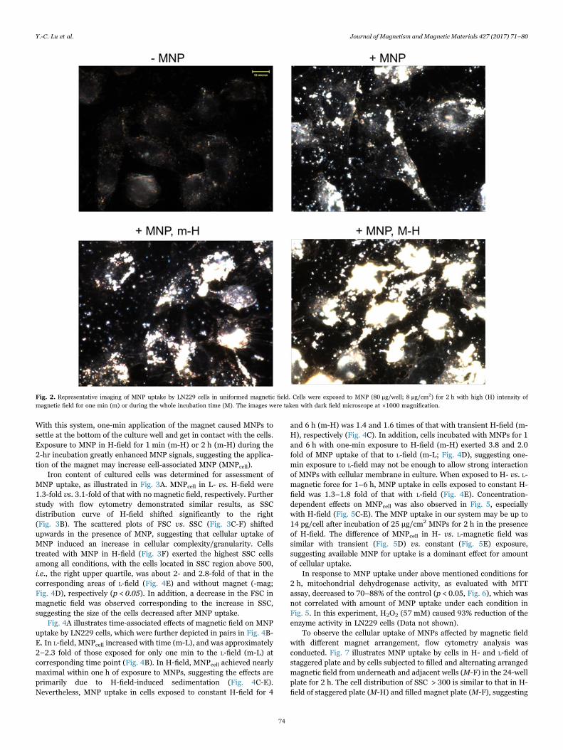
With this system, one-min application of the magnet caused MNPs tosettle at the bottom of the culture well and get in contact with the cells.Exposure to MNP in H-field for 1 min (m-H) or 2 h (m-H) during the2-hr incubation greatly enhanced MNP signals, suggesting the applica-tion of the magnet may increase cell-associated MNP (MNPcell).
Iron content of cultured cells was determined for assessment ofMNP uptake, as illustrated in Fig. 3A. MNPcell in L- vs. H-field were1.3-fold vs. 3.1-fold of that with no magnetic field, respectively. Furtherstudy with flow cytometry demonstrated similar results, as SSCdistribution curve of H-field shifted significantly to the right(Fig. 3B). The scattered plots of FSC vs. SSC (Fig. 3C-F) shiftedupwards in the presence of MNP, suggesting that cellular uptake ofMNP induced an increase in cellular complexity/granularity. Cellstreated with MNP in H-field (Fig. 3F) exerted the highest SSC cellsamong all conditions, with the cells located in SSC region above 500,i.e., the right upper quartile, was about 2- and 2.8-fold of that in thecorresponding areas of L-field (Fig. 4E) and without magnet (-mag;Fig. 4D), respectively (p < 0.05). In addition, a decrease in the FSC inmagnetic field was observed corresponding to the increase in SSC,suggesting the size of the cells decreased after MNP uptake.
Fig. 4A illustrates time-associated effects of magnetic field on MNPuptake by LN229 cells, which were further depicted in pairs in Fig. 4B-E. In L-field, MNPcell increased with time (m-L), and was approximately2–2.3 fold of those exposed for only one min to the L-field (m-L) atcorresponding time point (Fig. 4B). In H-field, MNPcell achieved nearlymaximal within one h of exposure to MNPs, suggesting the effects areprimarily due to H-field-induced sedimentation (Fig. 4C-E).Nevertheless, MNP uptake in cells exposed to constant H-field for 4
and 6 h (m-H) was 1.4 and 1.6 times of that with transient H-field (m-H), respectively (Fig. 4C). In addition, cells incubated with MNPs for 1and 6 h with one-min exposure to H-field (m-H) exerted 3.8 and 2.0fold of MNP uptake of that to L-field (m-L; Fig. 4D), suggesting one-min exposure to L-field may not be enough to allow strong interactionof MNPs with cellular membrane in culture. When exposed to H- vs. L-magnetic force for 1–6 h, MNP uptake in cells exposed to constant H-field was 1.3–1.8 fold of that with L-field (Fig. 4E). Concentration-dependent effects on MNPcell was also observed in Fig. 5, especiallywith H-field (Fig. 5C-E). The MNP uptake in our system may be up to14 pg/cell after incubation of 25 μg/cm2 MNPs for 2 h in the presenceof H-field. The difference of MNPcell in H- vs. L-magnetic field wassimilar with transient (Fig. 5D) vs. constant (Fig. 5E) exposure,suggesting available MNP for uptake is a dominant effect for amountof cellular uptake.
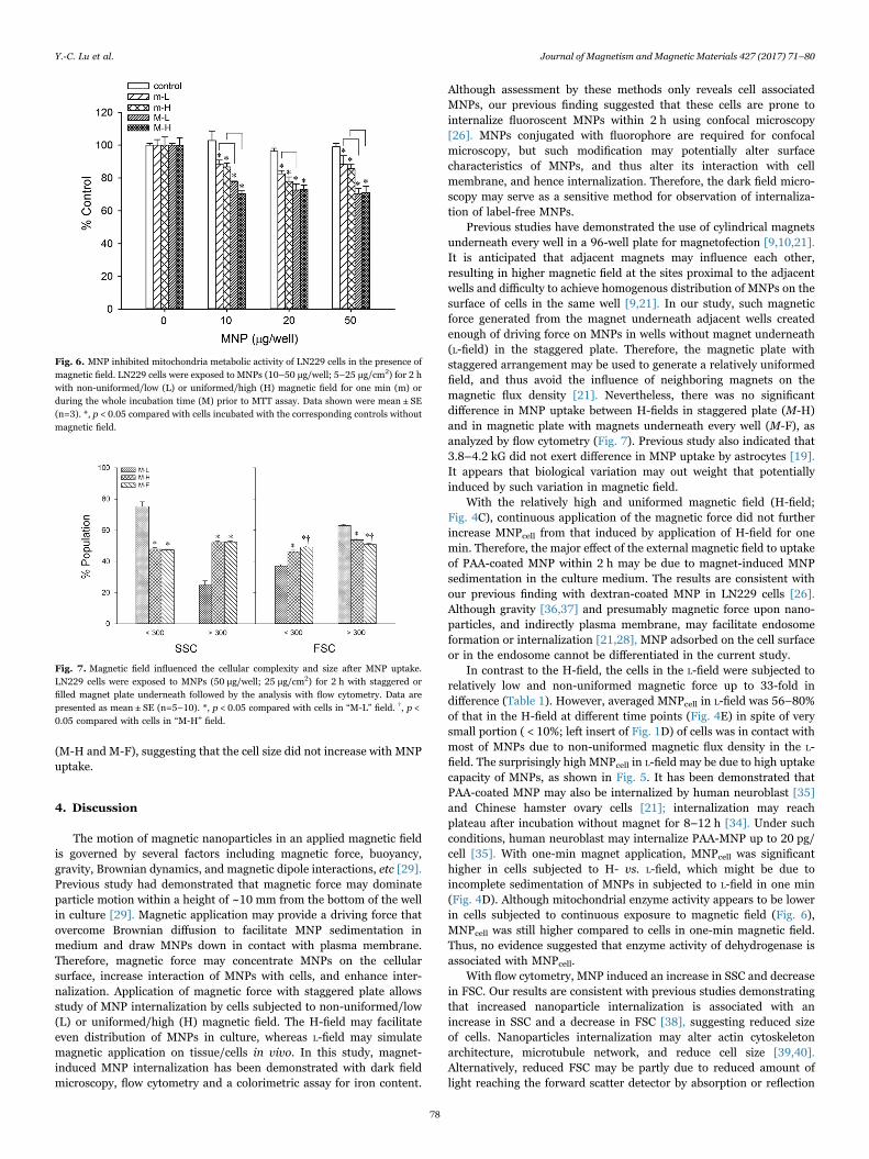
In response to MNP uptake under above mentioned conditions for2 h, mitochondrial dehydrogenase activity, as evaluated with MTTassay, decreased to 70–88% of the control (p < 0.05, Fig. 6), which wasnot correlated with amount of MNP uptake under each condition inFig. 5. In this experiment, H2O2 (57 mM) caused 93% reduction of theenzyme activity in LN229 cells (Data not shown).
To observe the cellular uptake of MNPs affected by magnetic fieldwith different magnet arrangement, flow cytometry analysis wasconducted. Fig. 7 illustrates MNP uptake by cells in H- and L-field ofstaggered plate and by cells subjected to filled and alternating arrangedmagnetic field from underneath and adjacent wells (M-F) in the 24-wellplate for 2 h. The cell distribution of SSC > 300 is similar to that in H-field of staggered plate (M-H) and filled magnet plate (M-F), suggesting
Fig. 2. Representative imaging of MNP uptake by LN229 cells in uniformed magnetic field. Cells were exposed to MNP (80 μg/well; 8 μg/cm2) for 2 h with high (H) intensity ofmagnetic field for one min (m) or during the whole incubation time (M). The images were taken with dark field microscope at ×1000 magnification.
Y.-C. Lu et al. Journal of Magnetism and Magnetic Materials 427 (2017) 71–80
74

that the MNPcell increased in the presence of the magnet, despite of thearrangement of magnets; whereas the fraction of SSC > 300 in M-Lgroup was approximately 50% of that in other groups. In addition, thecells located in SSC > 600 region was about 3-fold with H-field (M-H or
M-F) vs. L-field (M-L), suggesting the magnet underneath H-wells maysignificantly increase the MNPcell in a small population of cells in Lwells, despite the uniformity variation of the magnetic field. Thefraction of FSC > 300 in L-field (M-L) was 1.2-fold of that in H-field
Fig. 3. Inhomogeneous MNP Uptake under Magnetic Field. LN229 cells were incubated with MNP (200 μg/well; 20.8 μg/cm2) for 2 h with magnet of non-uniformed/low (L) oruniformed/high (H) intensity and then immediately processed for iron assay (A) and flow cytometry (B-F). Data shown were mean ± SE. Numbers indicat % cell number of defined area(n=3). *, p < 0.05 compared with control group without magnet; †, p < 0.05 compared with the L group.
Y.-C. Lu et al. Journal of Magnetism and Magnetic Materials 427 (2017) 71–80
75

Fig. 4. Time-dependent MNP uptake by LN229 cells. LN229 cells were exposed to MNPs (10 μg/well; 5 μg/cm2) for different time periods with non-uniformed/low (L) or uniformed/high (H) magnet field for one minute (m) or during the incubation time as indicated (M). Data shown were mean ± SE (n=3). *, p < 0.05 compared with corresponding m group; †, p <0.05 compared with corresponding L group.
Y.-C. Lu et al. Journal of Magnetism and Magnetic Materials 427 (2017) 71–80
76

Fig. 5. Concentration-dependent MNP uptake by LN229 cells. LN229 cells were exposed to MNPs (10–50 μg/well; 5–25 μg/cm2) for 2 h with magnet of non-uniformed/low (L) oruniformed/high (H) intensity for one min (m) or during the whole incubation time (M). Data shown were mean ± SE (n=3). *, p < 0.05 compared with corresponding m group; †, p < 0.05compared with corresponding L group.
Y.-C. Lu et al. Journal of Magnetism and Magnetic Materials 427 (2017) 71–80
77
(M-H and M-F), suggesting that the cell size did not increase with MNPuptake.
4. Discussion
The motion of magnetic nanoparticles in an applied magnetic fieldis governed by several factors including magnetic force, buoyancy,gravity, Brownian dynamics, and magnetic dipole interactions, etc [29].Previous study had demonstrated that magnetic force may dominateparticle motion within a height of ~10 mm from the bottom of the wellin culture [29]. Magnetic application may provide a driving force thatovercome Brownian diffusion to facilitate MNP sedimentation inmedium and draw MNPs down in contact with plasma membrane.Therefore, magnetic force may concentrate MNPs on the cellularsurface, increase interaction of MNPs with cells, and enhance inter-nalization. Application of magnetic force with staggered plate allowsstudy of MNP internalization by cells subjected to non-uniformed/low(L) or uniformed/high (H) magnetic field. The H-field may facilitateeven distribution of MNPs in culture, whereas L-field may simulatemagnetic application on tissue/cells in vivo. In this study, magnet-induced MNP internalization has been demonstrated with dark fieldmicroscopy, flow cytometry and a colorimetric assay for iron content.
Although assessment by these methods only reveals cell associatedMNPs, our previous finding suggested that these cells are prone tointernalize fluoroscent MNPs within 2 h using confocal microscopy[26]. MNPs conjugated with fluorophore are required for confocalmicroscopy, but such modification may potentially alter surfacecharacteristics of MNPs, and thus alter its interaction with cellmembrane, and hence internalization. Therefore, the dark field micro-scopy may serve as a sensitive method for observation of internaliza-tion of label-free MNPs.
Previous studies have demonstrated the use of cylindrical magnetsunderneath every well in a 96-well plate for magnetofection [9,10,21].It is anticipated that adjacent magnets may influence each other,resulting in higher magnetic field at the sites proximal to the adjacentwells and difficulty to achieve homogenous distribution of MNPs on thesurface of cells in the same well [9,21]. In our study, such magneticforce generated from the magnet underneath adjacent wells createdenough of driving force on MNPs in wells without magnet underneath(L-field) in the staggered plate. Therefore, the magnetic plate withstaggered arrangement may be used to generate a relatively uniformedfield, and thus avoid the influence of neighboring magnets on themagnetic flux density [21]. Nevertheless, there was no significantdifference in MNP uptake between H-fields in staggered plate (M-H)and in magnetic plate with magnets underneath every well (M-F), asanalyzed by flow cytometry (Fig. 7). Previous study also indicated that3.8–4.2 kG did not exert difference in MNP uptake by astrocytes [19].It appears that biological variation may out weight that potentiallyinduced by such variation in magnetic field.
With the relatively high and uniformed magnetic field (H-field;Fig. 4C), continuous application of the magnetic force did not furtherincrease MNPcell from that induced by application of H-field for onemin. Therefore, the major effect of the external magnetic field to uptakeof PAA-coated MNP within 2 h may be due to magnet-induced MNPsedimentation in the culture medium. The results are consistent withour previous finding with dextran-coated MNP in LN229 cells [26].Although gravity [36,37] and presumably magnetic force upon nano-particles, and indirectly plasma membrane, may facilitate endosomeformation or internalization [21,28], MNP adsorbed on the cell surfaceor in the endosome cannot be differentiated in the current study.
In contrast to the H-field, the cells in the L-field were subjected torelatively low and non-uniformed magnetic force up to 33-fold indifference (Table 1). However, averaged MNPcell in L-field was 56–80%of that in the H-field at different time points (Fig. 4E) in spite of verysmall portion ( < 10%; left insert of Fig. 1D) of cells was in contact withmost of MNPs due to non-uniformed magnetic flux density in the L-field. The surprisingly high MNPcell in L-field may be due to high uptakecapacity of MNPs, as shown in Fig. 5. It has been demonstrated thatPAA-coated MNP may also be internalized by human neuroblast [35]and Chinese hamster ovary cells [21]; internalization may reachplateau after incubation without magnet for 8–12 h [34]. Under suchconditions, human neuroblast may internalize PAA-MNP up to 20 pg/cell [35]. With one-min magnet application, MNPcell was significanthigher in cells subjected to H- vs. L-field, which might be due toincomplete sedimentation of MNPs in subjected to L-field in one min(Fig. 4D). Although mitochondrial enzyme activity appears to be lowerin cells subjected to continuous exposure to magnetic field (Fig. 6),MNPcell was still higher compared to cells in one-min magnetic field.Thus, no evidence suggested that enzyme activity of dehydrogenase isassociated with MNPcell.
With flow cytometry, MNP induced an increase in SSC and decreasein FSC. Our results are consistent with previous studies demonstratingthat increased nanoparticle internalization is associated with anincrease in SSC and a decrease in FSC [38], suggesting reduced sizeof cells. Nanoparticles internalization may alter actin cytoskeletonarchitecture, microtubule network, and reduce cell size [39,40].Alternatively, reduced FSC may be partly due to reduced amount oflight reaching the forward scatter detector by absorption or reflection
Fig. 6. MNP inhibited mitochondria metabolic activity of LN229 cells in the presence ofmagnetic field. LN229 cells were exposed to MNPs (10–50 μg/well; 5–25 μg/cm2) for 2 hwith non-uniformed/low (L) or uniformed/high (H) magnetic field for one min (m) orduring the whole incubation time (M) prior to MTT assay. Data shown were mean ± SE(n=3). *, p < 0.05 compared with cells incubated with the corresponding controls withoutmagnetic field.
Fig. 7. Magnetic field influenced the cellular complexity and size after MNP uptake.LN229 cells were exposed to MNPs (50 μg/well; 25 μg/cm2) for 2 h with staggered orfilled magnet plate underneath followed by the analysis with flow cytometry. Data arepresented as mean ± SE (n=5–10). *, p < 0.05 compared with cells in “M-L” field. †, p <0.05 compared with cells in “M-H” field.
Y.-C. Lu et al. Journal of Magnetism and Magnetic Materials 427 (2017) 71–80
78

of light by the particles [38,41].Although increased SSC was observed with MNP administration
and application of magnetic field, variation of SSC distributionsuggested variation of MNP internalization in cell population studied.Recent studies have demonstrated that nanoparticle internalizationmay differ between cell cycle phases [42,43], with the highest nano-particle uptake in the G2/M phase and lowest in G0/G1 phase.Therefore, subpopulations of cells in different cell cycle phases maybe responsible for the diversity in MNPcell observed in our study.Nevertheless, similar effects were observed in cells cultured in the H-field of both plates. Therefore, MNP internalization in H-field ofstaggered plate cannot be differentiated from that of the filled magnetplate due to biological variation.
Manipulation of magnetic field in vitro may serve as a modelsystem for application of magnetic field in vivo to achieve targetdelivery of genes/drugs. It is well known that the abnormal bloodvessels that synthesized by tumor-induced angiogenesis exert largepores ranging between 380 and 780 nm in diameter [44,45], allowingnanoparticles to reach the perivascular space and accumulate in thetumor interstitium via enhanced permeability and retention effect [45].Recent studies have demonstrated that under the influence of anexternal magnetic force, MNP may serve as a promising platform todeliver therapeutic cargos to targeted site such as solid tumor or brain[46–48]. It is anticipated that magnetic targeting allows MNP retentionin the vessels around the tumor, and subsequently tumor interstitial,the inhomogeneous uptake in the tumor is expected. Controlling drugrelease in the extracellular space may ensure more exposure of tumorcells to the drugs, and thus better therapeutic effect. Our resultssuggested that magnetic field may enhance overall MNP uptake to asimilar degree regardless of the uniformity of magnetic field.
5. Conclusion
Our study suggests that enhanced sedimentation may be the majormechanism underlies the effects of magnetic force on MNP internaliza-tion; spatial separation of the magnets under the cultured cells may beconsidered if biological variation was reduced, such as in synchronizedcells. The finding supports utilization of all wells on the culture plate instudy of magnetic influence on cultured cells to reduce cost andincrease efficiency in such experiments.
Acknowledgement
This work was supported by grants from Ministry of Science andTechnology, Taiwan (NSC 100–2120-M-182-001-), National HealthResearch Institute, Taiwan (NHRI-EX9909937EI), Healthy AgingResearch Program at Chang Gung University (EMRPD1E1651), andChang Gung Memorial Hospital (BMRP432). The authors thank Dr.Shieh-Yueh Yang and Dr. Hsin-Hsien Chen at MagQu Co., Ltd. formagnetic field measurement.
References
[1] N. Lewinski, V. Colvin, R. Drezek, Cytotoxicity of nanoparticles, Small 4 (2008)26–49.
[2] O. Veiseh, J.W. Gunn, M. Zhang, Design and fabrication of magnetic nanoparticlesfor targeted drug delivery and imaging, Adv. Drug Deliv. Rev. 62 (2010) 284–304.
[3] M. Namdeo, et al., Magnetic nanoparticles for drug delivery applications, J.Nanosci. Nanotechnol. 8 (2008) 3247–3271.
[4] E. Duguet, S. Vasseur, S. Mornet, J.M. Devoisselle, Magnetic nanoparticles andtheir applications in medicine, Nanomed. (Lond.) 1 (2006) 157–168.
[5] A.K. Gupta, M. Gupta, Synthesis and surface engineering of iron oxide nanopar-ticles for biomedical applications, Biomaterials 26 (2005) 3995–4021.
[6] U. Schillinger, et al., Advances in magnetofection—magnetically guided nucleic aciddelivery, J. Magn. Magn. Mater. 293 (2005) 501–508.
[7] J. Dobson, Gene therapy progress and prospects: magnetic nanoparticle-based
Gene delivery, Gene Ther. 13 (2006) 283–287.[8] C. Plank, O. Zelphati, O. Mykhaylyk, Magnetically enhanced nucleic acid delivery.
Ten years of magnetofection-Progress and prospects, Adv. Drug Deliv. Rev. 63(2011) 1300–1331.
[9] C. Plank, et al., The magnetofection method: Using magnetic force to enhance genedelivery, Biol. Chem. 384 (2003) 737–747.
[10] F. Scherer, et al., Magnetofection: enhancing and targeting gene delivery bymagnetic force in vitro and in vivo, Gene Ther. 9 (2002) 102–109.
[11] B. Thiesen, A. Jordan, Clinical applications of magnetic nanoparticles forhyperthermia, Int. J. Hyperth. 24 (2008) 467–474.
[12] V.I. Shubayev, T.R. Pisanic 2nd, S. Jin, Magnetic nanoparticles for theragnostics,Adv. Drug Deliv. Rev. 61 (2009) 467–477.
[13] C. Sun, J.S. Lee, M. Zhang, Magnetic nanoparticles in MR imaging and drugdelivery, Adv. Drug Deliv. Rev. 60 (2008) 1252–1265.
[14] C. Corot, P. Robert, J.M. Idee, M. Port, Recent advances in iron oxide nanocrystaltechnology for medical imaging, Adv. Drug Deliv. Rev. 58 (2006) 1471–1504.
[15] Wahajuddin, S. Arora, Superparamagnetic iron oxide nanoparticles: magneticnanoplatforms as drug carriers, Int. J. Nanomed. 7 (2012) 3445–3471.
[16] T. Dejardin, et al., Influence of both a static magnetic field and penetratin onmagnetic nanoparticle delivery into fibroblasts, Nanomed. (Lond.) 6 (2011)1719–1731.
[17] C. Dahmani, et al., Rotational magnetic pulses enhance the magnetofectionefficiency in vitro in adherent and suspension cells, J. Magn. Magn. Mater. 332(2013) 163–171.
[18] C. MacDonald, K. Barbee, B. Polyak, Force dependent internalization of magneticnanoparticles results in highly loaded endothelial cells for use as potential therapydelivery vectors, Pharm. Res. 29 (2012) 1270–1281.
[19] M.C. Lamkowsky, M. Geppert, M.M. Schmidt, R. Dringen, Magnetic field-inducedacceleration of the accumulation of magnetic iron oxide nanoparticles by culturedbrain astrocytes, J. Biomed. Mater. Res. Part A 100 (2012) 323–334.
[20] Q. Liu, J. Zhang, W. Xia, H. Gu, Magnetic field enhanced cell uptake efficiency ofmagnetic silica mesoporous nanoparticles, Nanoscale 4 (2012) 3415–3421.
[21] S. Prijic, et al., Increased cellular uptake of biocompatible superparamagnetic ironoxide nanoparticles into malignant cells by an external magnetic field, J. Membr.Biol. 236 (2010) 167–179.
[22] I. Canton, G. Battaglia, Endocytosis at the nanoscale, Chem. Soc. Rev. 41 (2012)2718–2739.
[23] F. Zhao, et al., Cellular uptake, intracellular trafficking, and cytotoxicity ofnanomaterials, Small 7 (2011) 1322–1337.
[24] S. Mishra, P. Webster, M.E. Davis, PEGylation significantly affects cellular uptakeand intracellular trafficking of non-viral gene delivery particles, Eur. J. Cell Biol. 83(2004) 97–111.
[25] B. Pelaz, et al., Surface functionalization of nanoparticles with polyethylene glycol:effects on protein adsorption and cellular uptake, ACS Nano 9 (2015) 6996–7008.
[26] Y.C. Lu, et al., Augmented cellular uptake of nanoparticles using tea catechins:effect of surface modification on nanoparticle-cell interaction, Nanoscale 6 (2014)10297–10306.
[27] D. Fayol, N. Luciani, L. Lartigue, F. Gazeau, C. Wilhelm, Managing magneticnanoparticle aggregation and cellular uptake: a precondition for efficient stem-celldifferentiation and MRI tracking, Adv. Healthc. Mater. 2 (2013) 313–325.
[28] C.A. Smith, et al., The effect of static magnetic fields and tat peptides on cellular andnuclear uptake of magnetic nanoparticles, Biomaterials 31 (2010) 4392–4400.
[29] E.P. Furlani, X. Xue, Field, force and transport analysis for magnetic particle-basedgene delivery, Microfluid. Nanofluidics 13 (2012) 589–602.
[30] Y.H. Ma, et al., Magnetically targeted thrombolysis with recombinant tissueplasminogen activator bound to polyacrylic acid-coated nanoparticles, Biomaterials30 (2009) 3343–3351.
[31] V.B.B. Mojca Pavlin, Stability of nanoparticle suspensions in different biologicallyrelevant media, Dig. J. Nanomater. Biostruct. 7 (2012) 1389–1400.
[32] C.L. Lin, C.F. Lee, W.Y. Chiu, Preparation and properties of poly(acrylic acid)oligomer stabilized superparamagnetic ferrofluid, J. Colloid Interface Sci. 291(2005) 411–420.
[33] V.B. Bregar, J. Lojk, V. Šuštar, P. Veranic, M. Pavlin, Visualization of internaliza-tion of functionalized cobalt ferrite nanoparticles and their intracellular fate, Int. J.Nanomed. 8 (2013) 919–931.
[34] J. Lojk, et al., Cell type-specific response to high intracellular loading of polyacrylicacid-coated magnetic nanoparticles, Int. J. Nanomed. 10 (2015) 1449–1462.
[35] M.P. Calatayud, et al., The effect of surface charge of functionalized Fe3O4
nanoparticles on protein adsorption and cell uptake, Biomaterials 35 (2014)6389–6399.
[36] E.C. Cho, Q. Zhang, Y. Xia, The effect of sedimentation and diffusion on cellularuptake of gold nanoparticles, Nat. Nanotechnol. 6 (2011) 385–391.
[37] T. Zhu, Z. Jiang, Y. Ma, Adsorption of nanoparticles and nanoparticle aggregates onmembrane under gravity, Appl. Phys. Lett. 102 (2013) 153109.
[38] R.M. Zucker, E.J. Massaro, K.M. Sanders, L.L. Degn, W.K. Boyes, Detection of TiO2
nanoparticles in cells by flow cytometry, Cytom. A 77A (2010) 677–685.[39] S.J.H. Soenen, N. Nuytten, S.F. De Meyer, S.C. De Smedt, M. De Cuyper, High
intracellular iron oxide nanoparticle concentrations affect cellular cytoskeleton andfocal adhesion kinase-mediated signaling, Small 6 (2010) 832–842.
[40] S.J.H. Soenen, et al., The role of nanoparticle concentration-dependent inductionof cellular stress in the internalization of non-toxic cationic magnetoliposomes,Biomaterials 30 (2009) 6803–6813.
[41] A. Kumar, A.K. Pandey, S.S. Singh, R. Shanker, A. Dhawan, A flow cytometricmethod to assess nanoparticle uptake in bacteria, Cytom. A 79A (2011) 707–712.
[42] A.H. Abouzeid, V.P. Torchilin, The role of cell cycle in the efficiency and activity ofcancer nanomedicines, Expert Opin. Drug Deliv. 10 (2013) 775–786.
[43] J.A. Kim, C. Aberg, A. Salvati, K.A. Dawson, Role of cell cycle on the cellular uptake
Y.-C. Lu et al. Journal of Magnetism and Magnetic Materials 427 (2017) 71–80
79

and dilution of nanoparticles in a cell population, Nat. Nano 7 (2012) 62–68.[44] I. Brigger, C. Dubernet, P. Couvreur, Nanoparticles in cancer therapy and diagnosis,
Adv. Drug Deliv. Rev. 54 (2002) 631–651.[45] N. Bertrand, J. Wu, X. Xu, N. Kamaly, O.C. Farokhzad, Cancer nanotechnology: the
impact of passive and active targeting in the era of modern cancer biology, Adv.Drug Deliv. Rev. 66 (2014) 2–25.
[46] D. Singh, McMillan, M. JoEllyn, A.V. Kabanov, M. Sokolsky-Papkov,
H.E. Gendelman, Bench-to-bedside translation of magnetic nanoparticles,Nanomed. (Lond.) 9 (2014) 501–516.
[47] M.S. Muthu, D.T. Leong, L. Mei, S.S. Feng, Nanotheranostics - application andfurther development of nanomedicine strategies for advanced theranostics,Theranostics 4 (2014) 660–677.
[48] L.S. del Burgo, R.M. Hernandez, G. Orive, J.L. Pedraz, Nanotherapeutic approachesfor brain cancer management, Nanomedicine 10 (2014) 905–919.
Y.-C. Lu et al. Journal of Magnetism and Magnetic Materials 427 (2017) 71–80
80


![Magnetic nanoparticles supported ionic liquids for lipase ...sourcedb.ipe.cas.cn/zw/lwlb/200908/P020090901287922534554.pdf · nanoparticles [3–5]. The magnetite-loaded enzymes are](https://img.pdfslide.us/doc/110x75/5f36f13cb95d7d6ff46da159/magnetic-nanoparticles-supported-ionic-liquids-for-lipase-nanoparticles-3a5.jpg)