Embed Size (px)
Citation preview

Cell Structures and Functions
Cells 2
Honors BiologyMilton High School
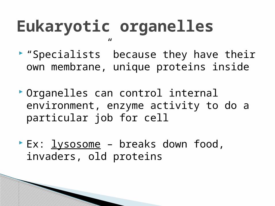
Eukaryotic organelles
“Specialists” because they have their own membrane, unique proteins inside
Organelles can control internal environment, enzyme activity to do a particular job for cell
Ex: lysosome – breaks down food, invaders, old proteins

Recall that most cell processes involve chains of enzymes
Organelles pack enzymes together so reactions happen efficiently
Organelles only found in eukaryotic cells … perhaps allowed cells to become larger over time
ONLY eukaryotic cells have potential to be multicellular (no truly multicellular bacteria)
Why have organelles?

Two major groups of workers:
1) Protein production◦ endoplasmic reticulum◦ Golgi apparatus◦ (also, vesicles and the cytoskeleton)
2) Energy transformation◦ mitochondrion◦ chloroplast
3) Also the vacuole and lysosome
Our organelle discussion

Recall – where is code for building proteins?
Recall – what structure assembles proteins?
For prokaryotic cells, end of story (protein is built and ready to go)
For eukaryotic cells, protein is often modified by organelles
Protein production and shipping

Eukaryotic protein producers
(endoplasmicreticulum)
DNA
ribosome(little red dot)

Eukaryotic protein productionDNA
ribosome
code is copied, sent to
endoplasmic reticulum
puts together amino acids, gives to
Golgi apparatus
more about this in genetics (ch. 16)
modification of protein
final destination
final modifications, tagged for shipping

Eukaryotic protein producers
(endoplasmicreticulum)
DNA
ribosome(little red dot)

Cells are NOT just bowls of jelly
Proteins travel along cytoskeleton “roads”
Carried inside a vesicle (piece of membrane) by a motor protein
Getting from place to place

Depends on the protein
1. Cytoplasm
2. Cell membrane
3. Organelle membrane
4. Inside organelle
5. Secreted outside cell
What is the “final destination”?

Mitochondrion – converts food energy into ATP energy
Chloroplast – converts sunlight energy into food energy
Energy transformation in cells
= cell respiration
= photosynthesis
By the way, prokaryotes can still do both, they just use their cell membrane

Vacuole – storage (food, water, other compounds)
(especially large in plant cells)
Lysosome – breakdown (lyse = breaking up) – can be pathogens, food, old proteins (recycling amino acids)
Other organelles

We’ll compare plant vs. animal, eventually we’ll look at protist, fungi cells also
Comparing eukaryotic cells
plant cell animal cell
large central vacuole
chloroplasts
cell wall

Cell wall – structural support for cell
NOT the same as a cell membrane
ALL cells have a cell membrane
SOME cells have a cell wall (NOT animals)
Cell wall (vs. cell membrane)

There is no such thing as a “typical” animal / plant cell
Cells are very diverse in shape / function around an organism
And by the way

We’ve discussed membranes broadly
Our goal now:
◦ how a membrane is built to do its job,
◦ how particles move in and out of cells,
◦ how a membrane helps to maintain homeostasis
Membranes in more detail

2 major components of a membrane
1) Phospholipid bilayer (purple)
2) Proteins (blue)
Real membranes are liquidy (not motionless)
Membrane structure

Polarity leads to self-assembly in a bilayer
Water on both sides attracts polar heads, repels nonpolar tails
Component 1: phospholipids
Polar head
Nonpolar tails
Polar heads
Polar heads
Nonpolar tails
Polar water inside cell
Polar water outside cell

Purpose of bilayer: block most particles from getting through
Effectively blocks polar particles
Small, nonpolar molecules can still cross (ex: O2, CO2)
Component 1: phospholipids
Polar heads
Polar heads
Nonpolar tails
Polar water inside cell
Polar water outside cell

First, we’ll focus on transport proteins
Like enzymes, have specific shape to let specific particle pass through membrane
Component 2: proteins

Phospholipid bilayer blocks almost all particles from entering or leaving
Multiple types of transport proteins select what particles cross
= membrane’s ability to control movement
Overall membrane

Enzymes
Receptor proteins – receive signals from outside, pass along message
Marker proteins – identify type of cell
Other membrane protein types

Before discussing movement around membranes, let’s discuss particle movement more broadly
Individual particles move randomly, though the overall group moves very predictably
Particles ALWAYS spread out, or move from high concentration toward low concentration
Particle movement

Natural movement is for particles to move from high low concentration
Each particle type can be considered independently
Particle movement and membranes

Random movement continues, but eventually no net change because particles already spread out = dynamic equilibrium
This assumes particles can cross membrane … if not, then no change occurs
Particle movement and membranes

If cell allows particles to move naturally, = passive transport
Different types of passive transport depending on how particle gets through membrane
1. Diffusion2. Facilitated diffusion3. Osmosis
Particle movement and membranes

Particle can cross directly through phospholipid bilayer
Small, nonpolar molecules (O2, CO2)
Diffusion

Particle can only move through a transport protein (polar particle)
Osmosis is the facilitated diffusion of water specifically
Facilitated diffusion

Even if particles cannot move across membranes, they influence the movement of water
Any particle dissolved in watery solution = solute
Overall solute level influences water movement
Key: water follows the solute
Water movement

Water follows the solute
Water movement

Hypotonic – solute level is lower
Hypertonic – solute level is higher
Isotonic – solute level is equal
Water movement terms

Water follows the solute
Water movement
Inside: hypertonicOutside: hypotonic
Inside: hypotonicOutside: hypertonic
Inside: isotonicOutside: isotonic

Animalcell
Hypotonic environment
Isotonic environment
Hypertonic environment
Plantcell

Always movement from high low concentration
Always leads to equilibrium (equal concentrations inside and outside cell)
Often, cells do NOT want this equilibrium – they need to counteract passive transport
Passive transport summary

Transport proteins pushing particles against their natural direction (from low high concentration)
Requires ATP energy
Active transport

If active and passive transport = each other, then no overall change occurs
This is cell homeostasis – using energy to maintain the right, unequal conditions
A balancing act

Macromolecules are usually too large to fit in transport proteins
A piece of membrane folds in or out, forming vesicle around particle
Endocytosis = coming in
Exocytosis = going out
Transporting large particles

Here, we’ll study how cells make exact copies
Why?
1. Growth
2. Replacing damaged cells
3. Reproduction (not everybody – not humans)
Making new cells

Why not grow by increasing cell size?
Growing by increasing cell number
Conclusion: diffusion occurs faster in smaller cells (nutrients in, wastes out)

Goal: Get a copy of DNA to the new cell
Basic steps:
1. Copy DNA
2. Pack and split up DNA copies
3. Split up cytoplasm into two separate cells
Making a new cell copy
= replication (more in genetics unit)

Very simple – called binary fission
Much less DNA than eukaryotes, so only packs up into one chromosome
Prokaryotic cell division

Much more complex cells, so more DNA to organize (multiple chromosomes)
Eukaryotic cell division
Eukaryotic organism
# of chromosomes in each body cell

46 chromosomes in human cells note: doesn’t look like this in nucleus

actually one chromosome tied together with its exact copy
DNA’s two different forms 1) fully packed up = chromosome form –
ONLY appears during cell division 2) unpacked = thin chromatin –
DNA code can be read to make proteins

involves mitosis – more complex chromosome organization
Eukaryotic cell division

Interphase
Mitosis
Cytokinesis
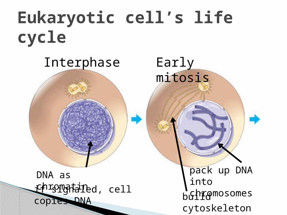
Eukaryotic cell’s life cycle
normal cell activity – most cells here
DNA in thin chromatin form
if cell signaled to divide, DNA is copied
DNA packed up into chromosomes (copies tied together)
Xs lined up in 1 line, split up
DNA unpacks back into chromatin
2 cells separate, both return to interphase
Eukaryotic cell’s life cycle
DNA as chromatin
Interphase
if signaled, cell copies DNA
Early mitosis
pack up DNA into chromosomes
build cytoskeleton fibers to pull Xs

Eukaryotic cell’s life cycle
Mitosis
One line of Xs

Eukaryotic cell’s life cycle
MitosisLate mitosis / cytokinesis
DNA unpacks back to chromatin
cytokinesisstarting
both cells return to interphase

Binary fission or mitosis:
2 cells that are genetic copies of original cell(if DNA is copied correctly)
End results

Normal cells
Know when to divide(they are signaled)
Know when to stop dividing
Cell division is regulated

Body cells with messed up signaling pathways
Continue to divide out of control
Tumor = ball of dividing cells
Cancer cells

Cells need resources to survive long-term
Cancerous tumors starve normal body cells, lead to eventual death
What makes cancer benign? Malignant? We don’t know
Why is cancer a problem?

Cancer is complicated

Damaged DNA code = damaged signal pathways possible cancer
What damages DNA?
High energy waves (UV rays, X-rays)
Chemicals (in cigarette smoke, ex)
Poor diet
What causes cancer?

Benign tumors = local surgery
Malignant tumors that have spread = chemotherapy (targets ALL dividing cells)
How do we treat cancer?