Embed Size (px)
Citation preview
Algological Studies 100 95-105 Stuttgart, December 2000
Cell wall development, microfibril and pyrenoidstructure in type strains of Chlorella vulgaris,
C. kessleri, C sorokiniana compared withC luteoviridis (Trebouxiophyceae, Chlorophyta)
By YVONNE NEMcovA and TOMAs KALINA
Department of Botany, Charles University, Prague, Czech Republic
With 14 figures in the text
Abstract: Ultrastructural examination of three glucosamine-type Chlorella species (C.vulgaris var. vulgaris, C. kessleri, C. sorokiniana), forming a related group in phylogenetictrees inferred from 18S rRNA gene sequences (FRIEDL 1995, Huss et al. 1999), revealed asimilar cell ultrastructure but some differences in early and later stages of the cell walldevelopment. All species mentioned above contain the monosaccharide glucosamine asthe main constituent of the rigid cell wall (TAKEDA 1991, 1993a, 1993b). A thin electron-dense layer is the first visible' structure covering the young daughter protoplasts of C.vulgaris and C. sorokiniana. Layered microfibrils can be observed in cross-sections of adultcell walls. Remnants of the broken maternal cell walls (MCW) persist in a culture medium.In C. kessleri the initial electrondense layer was not found. The cell wall is hardly visible, norrricrofibrillar structure was detected. No MCW remnants were found in the medium.Negatively stained microfibrils of all the three species obtained by IN NaOH and 2MTFAA treatment are straight or slightly bent. The pyrenoid is transversed by two thy-lakoids. The rigid cell wall of C. luteoviridis is composed of glucose and mannose (TAKEDA1991, 1993a, 1993b). However some ultrastructural features of C. luteoviridis resemble thatof glucosamine-type cWorellas (the thin electrondense layer covering the young daughterprotoplasts, microfibrillar structure of the adult cell wall visible on cross-sections, MCWremnants persisting in a medium). Microfibrils do not form a net, they are kinked andflexuous. C. luteoviridis differs from glucosamine-type species in the pyrenoid structure (thepyrenoid is bisected by four or two thylakoids). Thickness of microfibrils in all studiedspecies is about 5 nm.
Key words: Trebouxiophyceae, Chlorophyta, Chlorella vulgaris, C. kessleri, C. soro-kiniana, C. luteoviridis, rigid cell wall, microfibrils, pyrenoid structure.
Introduction
A taxonomy of the genus Chlorella BEIJERINCK based on light microscopy
observations was published by FOTT & N ov AKOV A (1969). In this monograph the
0342-112310010136-001 $ 2.75@ 2000 E. Schweizerbart.sche Verlagsbuchhandlung, D-70176 Stuttgart
Algological Studies 100 = Arch. Hydrobiol. Suppl. 136

96 Y. NEMcovA and T. KALINA
authors established the nomenclatoric types (iconotYpes and type cultures) anddocumented the phenoplasticity of eight species and four varieties. After numer-ous chemotaxonomical investigations of KESSLER and his co-workers (KESSLER1992, KESSLER & Huss 1992) it became evident that the genus Chlorella repre-sents taxonomically heterogeneous assembly of simple unicells. KALINA &PUNCOCHARov A (1987) and ANDREYEVA (1998) restricted the list of Chlorellaspecies only to those, which do not contain the algenan layer in their cell walls. Anew insight into the taxonomy of coccoid green algae was achieved by employ-ment of 18S rRNA gene sequence data. Taxa of Chlorella were dispersed over twoclasses: the Chlorophyceae and the Trebouxiophyceae (FRIEDL 1995, Huss et al.1999). The class Trebouxiophyceae has been established by FRIEDL (1995).HANAGATA & CHIHARA (1997) proposed that only the species of Chlorella
vulgaris-group should be kept in the genus Chlorella (Trebouxiophyceae). Theexistence of a distinct group containing C. vulgaris BEIJERINCK, C. lobophoraANDREYEVA, C. sorokiniana SHIHlRA et KRAUSS and C. kessleri FOTT et NovA-KovA has been also resolved in the 18S rRNA phylogeny (Huss et al. 1999).TAKEDA (1991, 1993a, 1993b) proposed the monosaccharide composition ofrigidcell walls as a taxonomical marker. He found that the rigid cell walls of C. vulgaris,C. sorokiniana and C. kessleri consisted of glucosamine, while other Chlorella
species (concerning C. luteoviridis) possessed glucose and mannose as amainconstituent of the rigid cell wall. KAPAUN & REISSER (1995) studied electron-microscopic preparations of rigid cell walls of symbiotic Chlorella Phi strain,which belongs to the Chlorella vulgaris /sorokiniana cluster (Huss et al. 1999,Reisser & WrnowsKI 1992). The rigid cell wall of this strain is composed of themonosaccharide glucosamine (KAPAUN et al. 1992). In both freeze-etched andnegatively stained samples KAPAUN & REISSER (1995) revealed microfibrils ofdiameter about 5 Dm. They investigated the rigid cell wall by sugar analysis, infrared spectroscopy, lectin binding, enzymatic degradation, X-ray diffraction andconcluded that it was composed of the polysaccharide glycosaminoglycan, whichcan be regarded as a chitin-like glycan. The authors assumed the same nature ofmicrofibrils in related species with glucosamine rigid cell wall (C. kessleri, C.sorokiniana and C. vulgaris). Another character indicating a closer evolutionaryrelationship of species with glucosamine cell walls is the identical pyrenoidstructure. The thylakoid that penetrates into the pyrenoid matrix, is uniformlydouble-layered (IKEDA & TAKEDA 1995).
The purpose of this study is to compare an early stage of the cell walldevelopment, the structure of extracted microfibrils and the pyrenoid morpholo-gy in type strains of three glucosamine-type species of Chlorella: C. vulgaris var.
vulgaris, C. kessleri and C. sorokiniana. Ultrastructure features mentioned aboveare compared with C. luteoviridis, which shows certain resemblance to glucosa-
mine-type species. The microfibrillar structure of a Chlorella-like species withglucose and mannose as a main constituent of the rigid cell wall has not beenstudied yet.
Development and structure in type strains of Chlorella 97
Material and methods
Chlorella type strains were obtained from the Culture Collection of Algae at theDepartment of Botany, Faculty of Natural Science, Charles University in Prague(CAUP): C. vulgaris var. vulgaris strain H1955 (identical with SAG 211-11b); c.kessleri strain H1901 (identical with SAG 211-11 g); c. sorokiniana strain H1957(identical with SAG 211-8k) and C. luteoviridis strain H1906 (identical with SAG211-2a). Chlorella cultures were grown in 60 mI glass tubes filled with BOLD'SBasal Medium (STEIN 1975), aerated with filtered air under continuos illumina-tion oflight intensity 100 mW: cm-2. Cells were cultivated at 25 °C for 6 days.
Preparation of the ultrathin sections. Actively growing cells were harvestedby centrifugation, fiXed with 2% (w/v) glutaraldehyde in 0.05 M phosphate buffer(pH 6.9) at room temperature overnight, and postfixed with 1% OSO4 in 0.05 Mphosphate buffer for 2 hours at 12 °C and with 1% uranyl acetate in methanolovernight. Cells were dehydrated in graded concentration of ethanol, thantransferred into butanol and embedded in SPURR'S low viscosity epoxy medium(SPURR 1969). Sections were cut with a diamond knife by an Ultracut microtomeand collected on a Formvar-coated copper grid. Sections were poststained withuranyl acetate and bismuth oxynitrate.
Preparation of the cell walls. Algal cells ( ca. 1 mI of packed cell volume) weremechanically broken by vortexing (on a Griffin & George Ltd. homogenizer) in aplastic 7 mI tube in the presence of glass beads (0.5 mm diameter, Sigma). Cellwalls were gathered by successive centrifugation (1500 r.p.m. for 5 min. -
sediment was under interest, cell organelles and diluted material were elimi-nated; 800 r. p. m. for 1 min. -supernatant was under interest, unbroken cells andcell conglomerates were eliminated; 4200 r.p.m. -broken cell walls were har-vested). Cell walls were washed with double destilled water by repeated centri-
fugation.Extraction of the cell walls. Cell wall preparations were extracted according to
KAPAUN & REISSER (1995) by alkaline treatment (1 N NaOH, 20 min, 100 °C) and,after washing with water, with 2 M trifluoroacetic acid (TFAA, 2 h, 110 °C). Rigidcell wall material was diluted and dropped onto Formvar-coated copper grids.
Negative staining was achieved by 2% uranyl acetate, after 5 min the liquid wascarefully removed. All samples were examined with a transmission electronmicroscope Philips 300.
Results
Cell wall
The cell walls of the tree species (C. vulgaris var. vulgaris, C. sorokiniana and Cluteoviridis) differ in its development, in electron density after contrasting solu-
98 Y. NEMcovA andT. KALINA
99Development and structure in type strains of Ch/ore//a
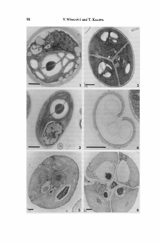
tion application, and in the ability to persist in the medium after autosporedeliberation. A thin electrondense layer as the first visible structure of the newlyformed cell wall was observed in autosporangia of C. vulgaris var. vulgaris, C.sorokiniana and C. luteoviridis (Figs 2, 6, 8). The cell wall thickness depends on thegrowth rate. In the intensively growing culture, the cell walls remain thinner.Layered microfibrils are visible on a cross-section of adult cell walls in all speciesnamed above (Figs I, 4, 7). A cell wall structure of c. kessleri is hardly visible notonly in young autospores but as well in adult cells, because of low contrast.Contrast of the cell wall was not increased even after additional staining with 20/0
potassium permanganate solution. No microfibrillar structure was detected evenin a maternal cell wall (MCW) of the autosporangium (Figs 5, 6). MCW remnants
persist in the culture medium of C vulgaris var. vulgaris, C. sorokiniana and Cluteoviridis (Figs 2, 4, 7).
pyrenoid
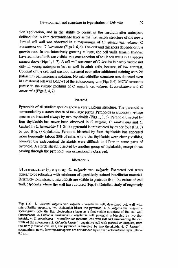
pyrenoids of all studied species show a very uniform structure. The pyrenoid issurrounded by a starch sheath of two large plates. pyrenoids in glucosamine-typespecies are bisected always by two thylakoids (Figs I, 3, 5). pyrenoid bisected byfour thylakoids has never been observed in C. vulgaris, C sorokiniana and C.kessleri. In C. luteoviridis 211-2a the pyrenoid is transversed by either four (Fig. 7)or two (Fig. 8) thylakoids. pyrenoid bisected by four thylakoids has appearedmore frequently (about 8()0/o of cells, where the thylakoids were clearly visible),however the independent thylakoids were difficult to follow in same parts ofpyrenoid. A starch sheath bisected by another group of thylakoids, except thosepassing through the pyrenoid, was occassionally observed.
Microfibrils
Glucosamine-type group C. vulgaris var. vulgaris. Extracted cell wallsappear to be reticulate with minimum of a positively stained interfibrilar material.
Relatively long straight microfibrils are visible to protrude from the extracted cellwall, especially where the wall has ruptured (Fig. 9). Detailed study of negatively
Figs 1-6. I. Chlorella vulgaris var. vulgaris -vegetative cell, developed cell wall withmicrofibrillar structure, two thylakoids bisect the pyrenoid. 2. C. vulgaris var. vulgaris -sporangium, note the thin electrodense layer as a first visible structure of the cell wall(arrowhead). 3. Chlorella sorokiniana -vegetative cell, pyrenoid is bisected by two thy-lakoids. 4. C. sorokiniana -microfibrillar maternal cell wall (MCW) surrounding the cellwalls of the autospores. 5. Chlorella kessleri -vegetative cell with parietal chloroplast, notethe hardly visible cell wall, the pyrenoid is bisected by two thylakoids. 6. C. kessleri -sporangium, newly forming autospores are not divided by a thin electrondense layer. [Bar =0.5 um.l
100 Y. NEMcovA and To KALINA
Figs 7-10. 7. Chlorella luteoviridis -vegetatiVe cell, MCW remnants persist in the medium,the pyrenoid is bisected by four thylakoids. 8. C. luteoviridis -vegetative cell, two thylakoidsbisect the pyrenoid. Figs 9-10. Extracted cell walls. 9. C. vulgaris var. vulgaris, reticulatestructure of an empty cell wall, note long protruding microfibrils (arrowhead). 10. C.luteoviridis -empty cell walls with a fuzzy surface and a large amount of a positively stainedinterfibrilar material. [Bar = 0.5 ~.]
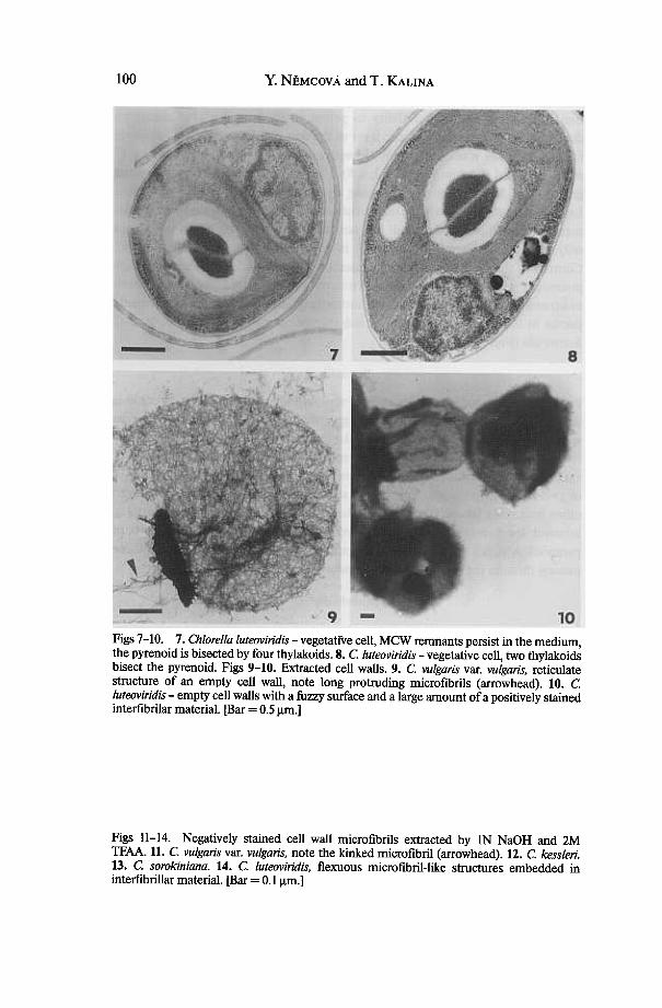
Figs 11-14. Negatively stained cell wall microfibrils extracted by IN NaOH and 2MTFAA. 11. C. vu/garis var. vu/garis, note the kinked microfibril (arrowhead). 12. C. kess/eri.13. C. sorokiniana. 14. C. /uteoviridis, flexuous microfibril-like structures embedded ininterfibrillar material. [Bar = 0.1 Jlm.]
Development and structure in type strains of Chlorella 101
Y. NEMcovA and T. KALINA
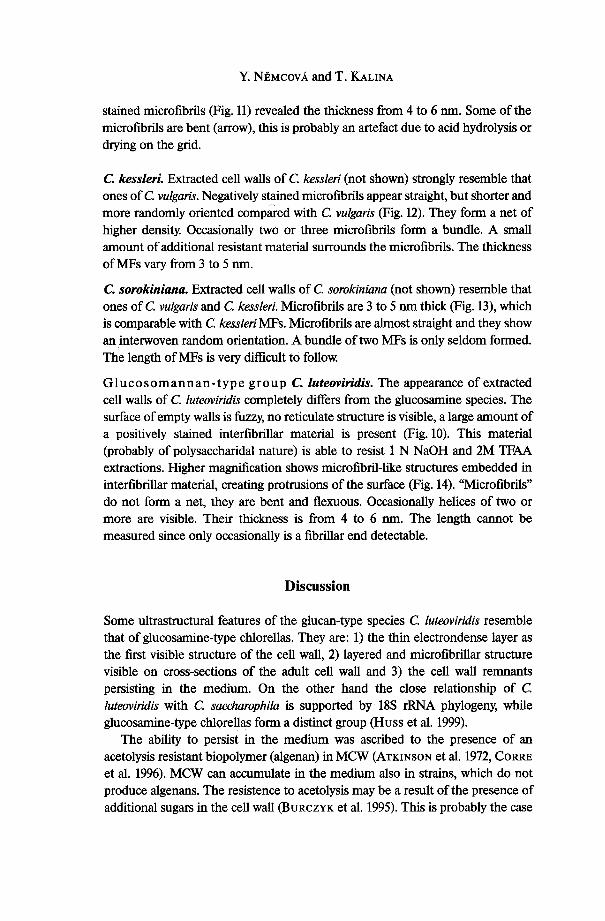
stained microfibrils (Fig.ll) revealed the thickness from 4 to 6 nm. Some of themicrofibrils are bent (arrow), this is probably an artefact due to acid hydrolysis ordrying on the grid.
c. kessleri. Extracted cell walls of C. kessleri (not shown) strongly resemble thatones of c. vulgaris. Negatively stained microfibrils appear straight, but shorter andmore randomly oriented compared with c. vulgaris (Fig. 12). They form a net ofhigher density, Occasionally two or three microfibrils form a bundle. A smallamount of additional resistant material surrounds the microfibrils. The thicknessofMFs vary from 3 to 5 nm.
C. sorokiniana. Extracted cell walls of c. sorokiniana (not shown) resemble thatones of c. vulgaris and C. kessleri. Microfibrils are 3 to 5 nm thick (Fig. 13), whichis comparable with C. kessleri MFs. Microfibrils are almost straight and they showan interwoven random orientation. A bundle of two MFs is only seldom formed.The length ofMFs is very difficult to follow;
Glucosomannan-type group C. luteo1liridis. The appearance of extractedcell walls of c. luteo1liridis completely differs from the glucosarnine species. Thesurface of empty walls is fuzzy, no reticulate structure is visible, a large amount ofa positively stained interfibrillar material is present (Fig. 10). This material(probably ofpolysaccharidal nature) is able to resist 1 N NaOH and 2M TFAAextractions. Higher magnification shows microfibril-like structures embedded ininterfibrillar material, creating protrusions of the surface (Fig. 14). "Microfibrils"do not form a net, they are bent and flexuous. Occasionally helices of two ormore are visible. Their thickness is from 4 to 6 nm. The length cannot bemeasured since only occasionally is a fibrillar end detectable.
Discussion
Some ultrastructural features of the glucan-type species C. luteoviridis resemblethat of glucosamine-type chlorellas. They are: I) the thin electrondense layer asthe first visible structure of the cell wall, 2) layered and microfibrillar structurevisible on cross-sections of the adult cell wall and 3) the cell wall remnantspersisting in the medium. On the other hand the close relationship of Cluteoviridis with C. saccharophila is supported by 18S rRNA phylogeny, whileglucosamine-type chlorellas form a distinct group (Buss et al. 1999).
The ability to persist in the medium was ascribed to the presence of anacetolysis resistant biopolymer (algenan) in MCW (ATKINSON et al. 1972, CORREet al. 1996). MCW can accumulate in the medium also in strains, which do notproduce algenans. The resistence to acetolysis may be a result of the presence ofadditional sugars in the cell wall (BURCZYK et al. 1995). This is probably the case
Development and structure in type strains of Chlorella 103
of C. vulgaris var. vulgaris and C sorokiniana. It is speculative whether theresistance might be caused by chitin-like glycans.
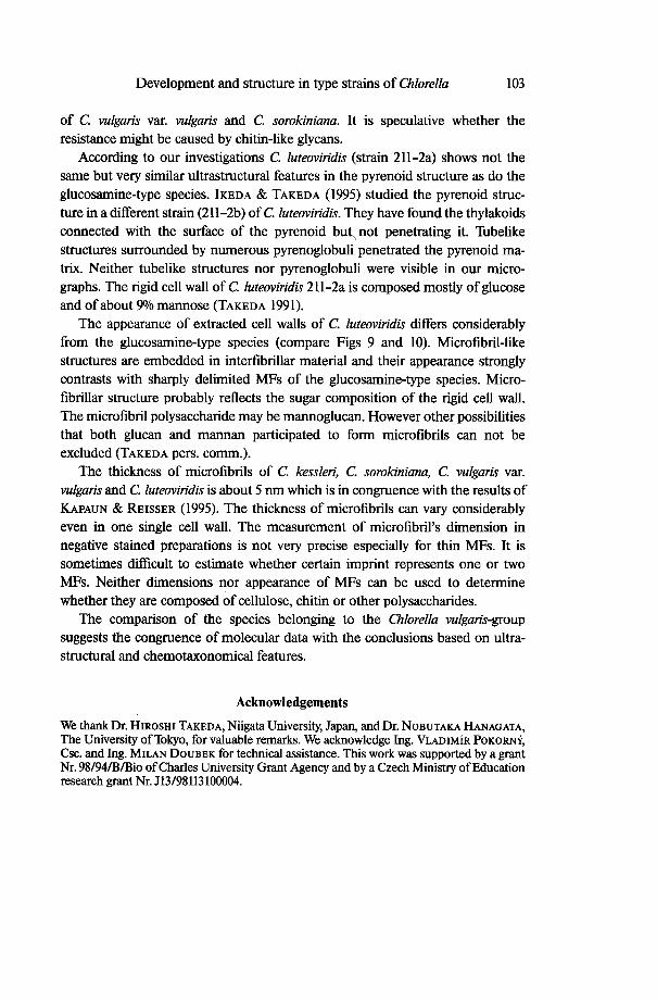
According to our investigations C. luteoviridis (strain 211-2a) shows not thesame but very similar ultrastructural features in the pyrenoid structure as do theglucosamine-type species. IKEDA & TAKEDA (1995) studied the pyrenoid struc-ture in a different strain (211-2b) ofC. luteoviridis. They have found the thylakoidsconnected with the surface of the pyrenoid but, not penetrating it. Tubelikestructures surrounded by numerous pyrenoglobuli penetrated the pyrenoid ma-trix. Neither tubelike structures nor pyrenoglobuli were visible in our micro-graphs. The rigid cell wall of C luteoviridis 211-2a is composed mostly of glucoseand of about 90/0 mannose (TAKEDA 1991).
The appearance of extracted cell walls of C. luteoviridis differs considerablyfrom the glucosamine-type species (compare Figs 9 and 10). Microfibril-likestructures are embedded in interfibrillar material and their appearance stronglycontrasts with sharply delimited MFs of the glucosamine-type species. Micro-fibrillar structure probably reflects the sugar composition of the rigid cell wall.The microfibril polysaccharide may be mannoglucan. However other possibilitiesthat both glucan and mannan participated to form microfibrils can not beexcluded (TAKEDA pers. comm.).
The thickness of microfibrils of C. kessleri, C. sorokiniana, C. vulgaris var.vulgaris and C. luteoviridis is about 5 nm which is in congruence with the results ofKAPAUN & REISSER (1995). The thickness of microfibrils can vary considerablyeven in one single cell wall. The measurement of microfibril's dimension innegative stained preparations is not very precise especially for thin MFs. It issometimes difficult to estimate whether certain imprint represents one or twoMFs. Neither dimensions nor appearance of MFs can be used to determinewhether they are composed of cellulose, chitin or other polysaccharides.
The comparison of the species belonging to the Chlorella vulgaris-groupsuggests the congruence of molecular data with the conclusions based on ultra-structural and chemotaxonomical features.
Acknowledgements
We thank Dr. HIROSHI TAKEDA, Niigata University, Japan, and Dr. NOBUTAKA HANAGATA,The University of Tokyo, for valuable remarks. We acknowledge Ing. VLADIMiR POKORNY,Csc. and Ing. MILAN DOUBEK for technical assistance. This work was supported by a grantNr. 981941BIBio ofCharles University Grant Agency and bya Czech Ministry of Educationresearch grant Nr. J13198113100004.
y, NEMcovA and T. KALINA104
References
ANDREYEVA, V.M. (1998): Pochvennye i aerofilnye zelenye vodorosli (Chlorophyta: Tetra-sporales, Chlorococcales, Chlorosarcinales). Terestrial and aerophylic green algae(Chlorophyta: Tetrasporales, Chlorococcales, Chlorosarcinales). -349 pp., Nauka,
Sankt-Petersburg.ATKINSON, A. W.; GUNNING, B.E. & JOHN, P.C.L. (1972): Sporopollenin in the cell wall
of Ollorella and other algae; ultrastructure, chemistry and incorporation of 14C-acetate,studied in synchronous cultures. -Planta 107: 1-32.
BURCZYK, J.; TERMINSKA-PABIS, K. & SMIETANA, B. (1995): Cell wall neutral sugarcomposition of chlorococcalean algae formin-g; and not forming acetolysis resistantbiopolymer. -Phytochemistry 38(4): 837-841.
CORRE, G.; TEMPLIER, J.; LARGEAU, C.; RUSSEAU, B. &BERKALoFF, C. (1996). Influenceof cell wall composition on the resistance of two Chlorella species (Chlorophyta) todetergents. -J. Phycol. 32: 584-590.
FOTT, B. & NovAKovA, M. (1969): A monograph of the genus Chlorella. The fresh waterspecies. -In: FOTT, B. (ed.): Studies in phycology; p. 10-74, Academia, Prague.
FRIEDL, T. (1995): Inferring taxonomic position and testing genus level assignments incoccoid green algae: a phylogenetic analysis of 18S ribosomal RNA sequences fromDictyochloropsis reticulata and from members of the genus Mynnecia (Chlorophyta,Trebouxiophyceae cl. nov.). -J. Phycol. 31: 632-639.
RANAGATA, N. & CHIHARA, M. (1997): Concordance between phylogenetic data andultrastructural features in the classification of Chlorella and related taxa. -Phycologia36(Suppl.): 38.
Russ, V.A.R.; FRANK, C.; RARTMANN, R.C.; RIRMER, M.; KLOBOUCEK, A.; SEIDEL,B.M.; WENZELER, P. & KESSLER, E. (1999): Biochemical taxonomy and molecularphylogeny of the genus Chlorella (Chlorophyta). -J. Phycol. 35: 587-598.
IKEDA, T. & TAKEDA, R. (1995): Species-specific differences of pyrenoids in Ollorella-(Chlorophyta). -J. Phycol. 31: 813-818.
KALINA, T. & PUNCOCHARovA, M. (1987): Taxonomy of the subfamily ScotiellocystoideaeFOTT 1976 (Chlorellaceae, Chlorophyceae). -Arch. Rydrobiol./Suppl. 73, AlgologicalStudies 45: 473-521.
KAPAuN, E.; Loos, E. & REISSER, W. (1992): Cell wall composition of virus-sensitivesymbiotic Chlorella species. -Phytochemistry 31: 3103-3104.
KAPAuN, E. & REISSER, W. (1995): A chitin-like glycan in the cell wall of a Ollorella sp.(Chlorococcales, Chlorophyceae). -Planta 197: 577-582.
KESSLER, E. (1992): Chlorella-biochemische Taxonomie einer fiir Forschung und Bio-technologie wichtigen Gattung einzelliger Griinalgen. -Naturwissenschaften 79:260-265.
KESSLER, E. & Russ, V.A.R. (1992): Comparative physiology and biochemistry andtaxonomic assignment of the Chlorella (Chlorophyceae) strains of the Culture Collec-tion of the UniversitY of Texas at Austin. -J. Phycol. 28: 550-553.
REISSER, W. & WIDOW SKI, M. (1992): Taxonomy of eukaryotic algae endosymbiotic infreshwater associations. -In: REISSER, W. (ed.): Algae and symbiosis, p. 21-40,Biopress Limited, Bristol.
SPURR, A.R. (1969): A low viscositY epoxy resin embedding medium for electron micros-copy. -J. Ultrastruc. Res. 26: 31-43.
STEIN, J. (1975): Handbook of phycological methods. Culture methods and growthmeasurements. -447 pp., Cambridge UniversitY Press.
TAKEDA, R. (1991): Sugar composition of the cell wall and the taxonomy of Chlorella(Chlorophyceae). -J. Phycol. 27: 224-32.
-(1993a). Chemical composition of cell walls as a taxonomical marker. -J. Plant Res.106: 195-200.
-(1993b): Taxonomical assignment of chlorococcal algae from their cell wall composi-tion. -Phytochemistry 34: 1053-1055.
Development and structure in type strains of Chlorella 105
Manuscript received September, 29, 1999, accepted January, 24,2000.
The authors' address:
Mgr. YVONNE NEMCOvA,Doc. RNDr. TOMAs KALINA, CSc.,Department of BotanyCharles University Prague,Ben8.tska 2,CZ- 12801 Praha 2, Czech Republic.