Embed Size (px)
Citation preview
Cell wall constrains lateral diffusion of plantplasma-membrane proteinsAlexandre Martinièrea, Irene Lavagia, Gayathri Nageswarana, Daniel J. Rolfeb, Lilly Maneta-Peyretc, Doan-Trung Luud,Stanley W. Botchwayb, Stephen E. D. Webbb, Sebastien Mongrandc, Christophe Maureld, Marisa L. Martin-Fernandezb,Jürgen Kleine-Vehne, Jirí Frimlf, Patrick Moreauc, and John Runionsa,1
aDepartment of Biological and Medical Sciences, Oxford Brookes University, Oxford OX3 0BP, United Kingdom; bCentral Laser Facility, Research Complex atHarwell, Science and Technology Facilities Council, Rutherford Appleton Laboratory, Oxfordshire OX11 0QX, United Kingdom; cLaboratoire de BiogenèseMembranaire, Unité Mixte de Recherche 5200 Centre National de la Recherche Scientifique, Université Bordeaux Segalen, 33076 Bordeaux, France;dLaboratoire de Biochimie et Physiologie Moléculaire des Plantes, Institut de Biologie Intégrative des Plantes, Unité Mixte de Recherche 5004, Centre Nationalde la Recherche Scientifique/Unité Mixte de Recherche 0386 Institut National de la Recherche Agronomique, 34060 Montpellier, France; eDepartment ofApplied Genetics and Cell Biology, University of Natural Resources and Life Sciences, 1190 Vienna, Austria; and fDepartment of Plant Biotechnology andGenetics, Ghent University, 9052 Ghent, Belgium
Edited by Daniel J. Cosgrove, Pennsylvania State University, University Park, PA, and approved May 16, 2012 (received for review February 3, 2012)
A cell membrane can be considered a liquid-phase plane in whichlipids and proteins theoretically are free to diffuse. Numerousreports, however, describe retardeddiffusion ofmembraneproteinsin animal cells. This anomalous diffusion results from a combinationof structuring factors including protein–protein interactions, cyto-skeleton corralling, and lipid organization into microdomains. Inplant cells, plasma-membrane (PM) proteins have been describedas relatively immobile, but the control mechanisms that structurethe PM have not been studied. Here, we use fluorescence recoveryafter photobleaching to estimate mobility of a set of minimal PMproteins. These proteins consist only of a PM-anchoring domainfused to a fluorescent protein, but their mobilities remained limited,as is the case for many full-length proteins. Neither the cytoskeletonnor membrane microdomain structure was involved in constrainingthe diffusion of these proteins. The cell wall, however, was shownto have a crucial role in immobilizing PM proteins. In addition, bysingle-molecule fluorescence imaging we confirmed that the pat-tern of cellulose deposition in the cell wall affects the trajectoryand speedof PMprotein diffusion. Regulation of PMprotein dynam-ics by the plant cell wall can be interpreted as a mechanism forregulating protein interactions in processes such as trafficking andsignal transduction.
Proteins within membranes play significant roles in signalperception and transduction, solute partitioning, and secre-
tion. Accordingly, more than 25% of the proteome of higherplants is predicted to be membrane-associated proteins (1, 2).Proteins diffuse within the plane of a membrane through
thermal agitation. Each protein diffusing freely (3) has a diffu-sion constant that is dependent on the protein’s hydrodynamicradius and the viscosity of the membrane and surrounding me-dium (4). In a hypothetical uniform membrane, proteins wouldbe distributed randomly. However, biological membranes arespatially complex, with regions of protein and lipid concentra-tion. Numerous reports describe retarded diffusion of membraneproteins (5–8) because of structuring factors such as protein–protein interactions (9), cytoskeleton corralling (10), and lipidorganization into nanodomains (11). Membrane nanostructuringis crucial for protein–protein interactions and can either segre-gate or colocalize membrane proteins, thus optimizing proteininteractions in processes such as trafficking and signal trans-duction (12).Like yeast and animal cells, plant cells have a subcompart-
mentalized plasma membrane (PM). Membrane rafts (reviewedin ref. 13) have been demonstrated in plant PMs by proteomicson detergent-insoluble membranes (DIMs). DIMs are enrichedin signaling, stress response, cellular trafficking, and cell-wallmetabolism proteins (14–16). The Chlorella kessleri hexon-protonsymporter HUP1 and Solanum tuberosum remorin StREM1.3have been visualized in clusters within the PM (17, 18), and theclustering localization pattern of HUP1 is disrupted in mutant
yeast lines lacking typical ergosterol and sphingolipid micro-domains (17). The physiological role of plant PM substructuringhas been demonstrated in several studies. For instance, in thesterol mutant cyclopropylsterol isomerase1-1 (cpi1.1) of Arabi-dopsis thaliana, the asymmetric localization of PIN2 and, hence,polar auxin transport are perturbed (19). In polarized cells suchas pollen tubes, perfusion with the sterol-binding toxin filipin notonly perturbs membrane microdomain structure but also alterscalcium gradients, production of reactive oxygen species, andnormal cell elongation (20).The relationship between membrane subcompartmentaliza-
tion and protein diffusion has not been studied in detail, how-ever, and only a few reports have quantified protein diffusion inplant-cell membranes. In the case of PM proteins, such as KAT1,PMA2 H+ATPase, PIN2, PIP2;1, BOR1, NIP5;1, and AtFH1,only a small fraction of the protein pool is mobile (19, 21–23). Incontrast, endoplasmic reticulum-associated proteins, nuclearmembrane proteins, and tonoplast-associated proteins diffusemore freely within the membrane (24–26). Consequently, we areled to believe that the plant cell has specific properties thatconstrain PM protein diffusion.Here, we studied PM protein diffusion in plant cells to un-
derstand better PM structure and function. Protein mobility wasquantified using fluorescence recovery after photobleaching(FRAP) for a set of 13 plant PM proteins fused to fluorescentproteins. Then we developed a set of modified PM proteins thatwe term “minimal” because only the membrane-interacting or-spanning domains are present. Minimal PM proteins weredesigned to reduce the effect of protein interactions on diffusionand showed that DIM association and cytoskeleton have verylittle effect on protein mobility. However, PM proteins thatnormally are almost immobile become mobile when the cell wallis absent or when the distance between PM and cell wall is in-creased. Then cell-wall interaction with PM proteins was con-firmed by single-molecule tracking using total internal reflectionfluorescence (TIRF) microscopy. Our results show that the cellwall constrains protein diffusion, especially for proteins withlarger extracellular domains, even in the absence of bindinginteractions between proteins and cell-wall components.
Author contributions: A.M., S.W.B., S.M., C.M., M.L.M.-F., P.M., and J.R. designed research;A.M., I.L., G.N., D.J.R., L.M.-P., D.-T.L., S.E.D.W., and J.R. performed research; D.J.R., D.-T.L.,C.M., J.K.-V., J.F., P.M., and J.R. contributed new reagents/analytic tools; A.M., D.J.R., P.M.,and J.R. analyzed data; and A.M. and J.R. wrote the paper.
The authors declare no conflict of interest.
This article is a PNAS Direct Submission.
See Commentary on page 12274.1To whom correspondence should be addressed. E-mail: [email protected].
This article contains supporting information online at www.pnas.org/lookup/suppl/doi:10.1073/pnas.1202040109/-/DCSupplemental.
www.pnas.org/cgi/doi/10.1073/pnas.1202040109 PNAS | July 31, 2012 | vol. 109 | no. 31 | 12805–12810
PLANTBIOLO
GY
SEECO
MMEN
TARY
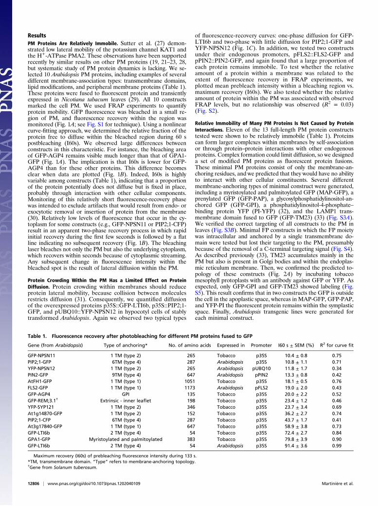
ResultsPM Proteins Are Relatively Immobile. Sutter et al. (27) demon-strated low lateral mobility of the potassium channel KAT1 andthe H+-ATPase PMA2. These observations have been supportedrecently by similar results on other PM proteins (19, 21–23, 28,but systematic study of PM protein dynamics is lacking. We se-lected 10 Arabidopsis PM proteins, including examples of severaldifferent membrane-association types: transmembrane domains,lipid modifications, and peripheral membrane proteins (Table 1).These proteins were fused to fluorescent protein and transientlyexpressed in Nicotiana tabacum leaves (29). All 10 constructsmarked the cell PM. We used FRAP experiments to quantifyprotein mobility. GFP fluorescence was bleached in a small re-gion of PM, and fluorescence recovery within the region wasmonitored (Fig. 1A; see Fig. S1 for technique). Using a nonlinearcurve-fitting approach, we determined the relative fraction of theprotein free to diffuse within the bleached region during 60 spostbleaching (I60s). We observed large differences betweenconstructs in this characteristic. For instance, the bleaching areaof GFP-AGP4 remains visible much longer than that of GPA1-GFP (Fig. 1A). The implication is that I60s is lower for GFP-AGP4 than for these other proteins. This difference becomesclear when data are plotted (Fig. 1B). Indeed, I60s is highlyvariable among constructs (Table 1), indicating that a proportionof the protein potentially does not diffuse but is fixed in place,probably through interaction with other cellular components.Monitoring of this relatively short fluorescence-recovery phasewas intended to exclude artifacts that would result from endo- orexocytotic removal or insertion of protein from the membrane(30). Relatively low levels of fluorescence that occur in the cy-toplasm for some constructs (e.g., GFP-NPSN11 or PIP2;1-CFP)result in an apparent two-phase recovery process in which rapidinitial recovery during the first few seconds is followed by a flatline indicating no subsequent recovery (Fig. 1B). The bleachinglaser bleaches not only the PM but also the underlying cytoplasm,which recovers within seconds because of cytoplasmic streaming.Any subsequent change in fluorescence intensity within thebleached spot is the result of lateral diffusion within the PM.
Protein Crowding Within the PM Has a Limited Effect on ProteinDiffusion. Protein crowding within membranes should reduceprotein lateral mobility, because collision between moleculesrestricts diffusion (31). Consequently, we quantified diffusionof the overexpressed proteins p35S::GFP-LTI6b, p35S::PIP2;1-GFP, and pUBQ10::YFP-NPSN12 in hypocotyl cells of stablytransformed Arabidopsis. Again we observed two typical types
of fluorescence-recovery curves: one-phase diffusion for GFP-LTI6b and two-phase with little diffusion for PIP2;1-GFP andYFP-NPSN12 (Fig. 1C). In addition, we tested two constructsunder their endogenous promoters, pFLS2::FLS2-GFP andpPIN2::PIN2-GFP, and again found that a large proportion ofeach protein remains immobile. To test whether the relativeamount of a protein within a membrane was related to theextent of fluorescence recovery in FRAP experiments, weplotted mean prebleach intensity within a bleaching region vs.maximum recovery (I60s). We also tested whether the relativeamount of protein within the PM was associated with observedFRAP levels, but no relationship was observed (R2 = 0.03)(Fig. S2).
Relative Immobility of Many PM Proteins Is Not Caused by ProteinInteractions. Eleven of the 13 full-length PM protein constructstested were shown to be relatively immobile (Table 1). Proteinscan form larger complexes within membranes by self-associationor through protein–protein interactions with other endogenousproteins. Complex formation could limit diffusion, so we designeda set of modified PM proteins as fluorescent protein fusions.These minimal PM proteins consist of only the membrane-an-choring residues, and we predicted that they would have no abilityto interact with other cellular constituents. Several differentmembrane-anchoring types of minimal construct were generated,including a myristoylated and palmitoylated GFP (MAP-GFP), aprenylated GFP (GFP-PAP), a glycosylphosphatidylinositol-an-chored GFP (GFP-GPI), a phosphatidylinositol-4-phosphate–binding protein YFP (PI-YFP) (32), and the LAMP1 trans-membrane domain fused to GFP (GFP-TM23) (33) (Fig. S3A).We verified the correct targeting of all constructs to the PM inleaves (Fig. S3B). Minimal FP constructs in which the FP moietywas intracellular and anchored by a single transmembrane do-main were tested but lost their targeting to the PM, presumablybecause of the removal of a C-terminal targeting signal (Fig. S4).As described previously (33), TM23 accumulates mainly in thePM but also is present in Golgi bodies and within the endoplas-mic reticulum membrane. Then, we confirmed the predicted to-pology of these constructs (Fig. 2A) by incubating tobaccomesophyll protoplasts with an antibody against GFP or YFP. Asexpected, only GFP-GPI and GFP-TM23 showed labeling (Fig.S5). This result confirms that in two constructs the GFP is outsidethe cell in the apoplastic space, whereas in MAP-GFP, GFP-PAP,and YFP-PI the fluorescent protein remains within the symplasticspace. Finally, Arabidopsis transgenic lines were generated foreach minimal construct.
Table 1. Fluorescence recovery after photobleaching for different PM proteins fused to GFP
Gene (from Arabidopsis) Type of anchoring* No. of amino acids Expressed in Promoter I60 s ± SEM (%) R2 for curve fit
GFP-NPSN11 1 TM (type 2) 265 Tobacco p35S 10.4 ± 0.8 0.75PIP2;1-GFP 6TM (type 4) 287 Arabidopsis p35S 10.8 ± 1.1 0.71YFP-NPSN12 1 TM (type 2) 265 Arabidopsis pUBQ10 11.8 ± 1.7 0.34PIN2-GFP 9TM (type 4) 647 Arabidopsis pPIN2 13.3 ± 0.8 0.42AtFH1-GFP 1 TM (type 1) 1051 Tobacco p35S 18.1 ± 0.5 0.76FLS2-GFP 1 TM (type 1) 1173 Arabidopsis pFLS2 19.0 ± 2.0 0.43GFP-AGP4 GPI 135 Tobacco p35S 20.0 ± 2.2 0.52GFP-REM;3.1† Extrinsic - inner leaflet 198 Tobacco p35S 23.4 ± 1.2 0.46YFP-SYP121 1 TM (type 2) 346 Tobacco p35S 23.7 ± 3.4 0.69At1g14870-GFP 1 TM (type 2) 152 Tobacco p35S 36.2 ± 2.7 0.74PIP2;1-CFP 6TM (type 4) 287 Tobacco p35S 43.7 ± 1.7 0.41At3g17840-GFP 1 TM (type 1) 647 Tobacco p35S 58.9 ± 3.8 0.73GFP-LTI6b 2 TM (type 4) 54 Tobacco p35S 72.4 ± 2.7 0.84GPA1-GFP Myristoylated and palmitoylated 383 Tobacco p35S 79.8 ± 3.9 0.90GFP-LTI6b 2 TM (type 4) 54 Arabidopsis p35S 91.4 ± 3.6 0.99
Maximum recovery (I60s) of prebleaching fluorescence intensity during 133 s.*TM, transmembrane domain. ”Type” refers to membrane-anchoring topology.†Gene from Solanum tuberosum.
12806 | www.pnas.org/cgi/doi/10.1073/pnas.1202040109 Martinière et al.
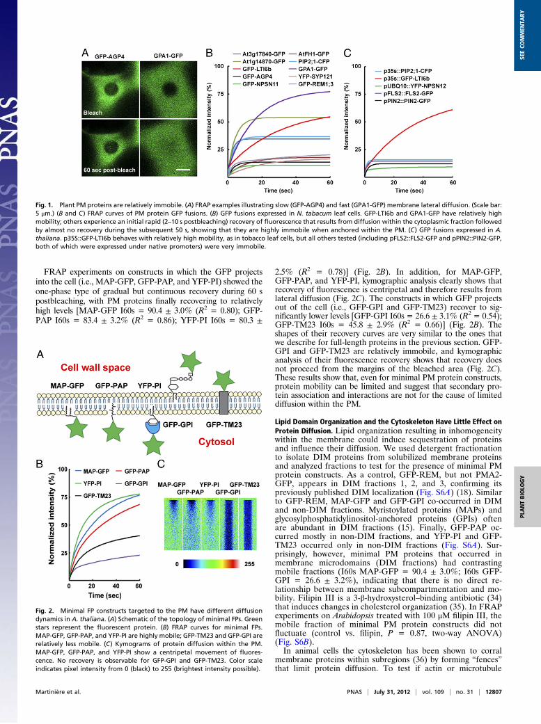
FRAP experiments on constructs in which the GFP projectsinto the cell (i.e., MAP-GFP, GFP-PAP, and YFP-PI) showed theone-phase type of gradual but continuous recovery during 60 spostbleaching, with PM proteins finally recovering to relativelyhigh levels [MAP-GFP I60s = 90.4 ± 3.0% (R2 = 0.80); GFP-PAP I60s = 83.4 ± 3.2% (R2 = 0.86); YFP-PI I60s = 80.3 ±
2.5% (R2 = 0.78)] (Fig. 2B). In addition, for MAP-GFP,GFP-PAP, and YFP-PI, kymographic analysis clearly shows thatrecovery of fluorescence is centripetal and therefore results fromlateral diffusion (Fig. 2C). The constructs in which GFP projectsout of the cell (i.e., GFP-GPI and GFP-TM23) recover to sig-nificantly lower levels [GFP-GPI I60s = 26.6 ± 3.1% (R2 = 0.54);GFP-TM23 I60s = 45.8 ± 2.9% (R2 = 0.66)] (Fig. 2B). Theshapes of their recovery curves are very similar to the ones thatwe describe for full-length proteins in the previous section. GFP-GPI and GFP-TM23 are relatively immobile, and kymographicanalysis of their fluorescence recovery shows that recovery doesnot proceed from the margins of the bleached area (Fig. 2C).These results show that, even for minimal PM protein constructs,protein mobility can be limited and suggest that secondary pro-tein association and interactions are not for the cause of limiteddiffusion within the PM.
Lipid Domain Organization and the Cytoskeleton Have Little Effect onProtein Diffusion. Lipid organization resulting in inhomogeneitywithin the membrane could induce sequestration of proteinsand influence their diffusion. We used detergent fractionationto isolate DIM proteins from solubilized membrane proteinsand analyzed fractions to test for the presence of minimal PMprotein constructs. As a control, GFP-REM, but not PMA2-GFP, appears in DIM fractions 1, 2, and 3, confirming itspreviously published DIM localization (Fig. S6A) (18). Similarto GFP-REM, MAP-GFP and GFP-GPI co-occurred in DIMand non-DIM fractions. Myristoylated proteins (MAPs) andglycosylphosphatidylinositol-anchored proteins (GPIs) oftenare abundant in DIM fractions (15). Finally, GFP-PAP oc-curred mostly in non-DIM fractions, and YFP-PI and GFP-TM23 occurred only in non-DIM fractions (Fig. S6A). Sur-prisingly, however, minimal PM proteins that occurred inmembrane microdomains (DIM fractions) had contrastingmobile fractions (I60s MAP-GFP = 90.4 ± 3.0%; I60s GFP-GPI = 26.6 ± 3.2%), indicating that there is no direct re-lationship between membrane subcompartmentation and mo-bility. Filipin III is a 3-β-hydroxysterol–binding antibiotic (34)that induces changes in cholesterol organization (35). In FRAPexperiments on Arabidopsis treated with 100 μM filipin III, themobile fraction of minimal PM protein constructs did notfluctuate (control vs. filipin, P = 0.87, two-way ANOVA)(Fig. S6B).In animal cells the cytoskeleton has been shown to corral
membrane proteins within subregions (36) by forming “fences”that limit protein diffusion. To test if actin or microtubule
Fig. 1. Plant PM proteins are relatively immobile. (A) FRAP examples illustrating slow (GFP-AGP4) and fast (GPA1-GFP) membrane lateral diffusion. (Scale bar:5 μm.) (B and C) FRAP curves of PM protein GFP fusions. (B) GFP fusions expressed in N. tabacum leaf cells. GFP-LTI6b and GPA1-GFP have relatively highmobility; others experience an initial rapid (2–10 s postbleaching) recovery of fluorescence that results from diffusion within the cytoplasmic fraction followedby almost no recovery during the subsequent 50 s, showing that they are highly immobile when anchored within the PM. (C) GFP fusions expressed in A.thaliana. p35S::GFP-LTI6b behaves with relatively high mobility, as in tobacco leaf cells, but all others tested (including pFLS2::FLS2-GFP and pPIN2::PIN2-GFP,both of which were expressed under native promoters) were very immobile.
Fig. 2. Minimal FP constructs targeted to the PM have different diffusiondynamics in A. thaliana. (A) Schematic of the topology of minimal FPs. Greenstars represent the fluorescent protein. (B) FRAP curves for minimal FPs.MAP-GFP, GFP-PAP, and YFP-PI are highly mobile; GFP-TM23 and GFP-GPI arerelatively less mobile. (C) Kymograms of protein diffusion within the PM.MAP-GFP, GFP-PAP, and YFP-PI show a centripetal movement of fluores-cence. No recovery is observable for GFP-GPI and GFP-TM23. Color scaleindicates pixel intensity from 0 (black) to 255 (brightest intensity possible).
Martinière et al. PNAS | July 31, 2012 | vol. 109 | no. 31 | 12807
PLANTBIOLO
GY
SEECO
MMEN
TARY
cytoskeletons might exert a similar influence on membrane-protein mobility in plant cells, we incubated minimal GFP-expressing Arabidopsis seedlings with either cytochalasin D ororyzalin to depolymerize actin microfilaments or microtubules,respectively. No increase in mobile fraction for these constructswas observed (Fig. S6C). Consequently, we believe that the cy-toskeleton is not responsible for the relative immobility of GFP-GPI or GFP-TM23.
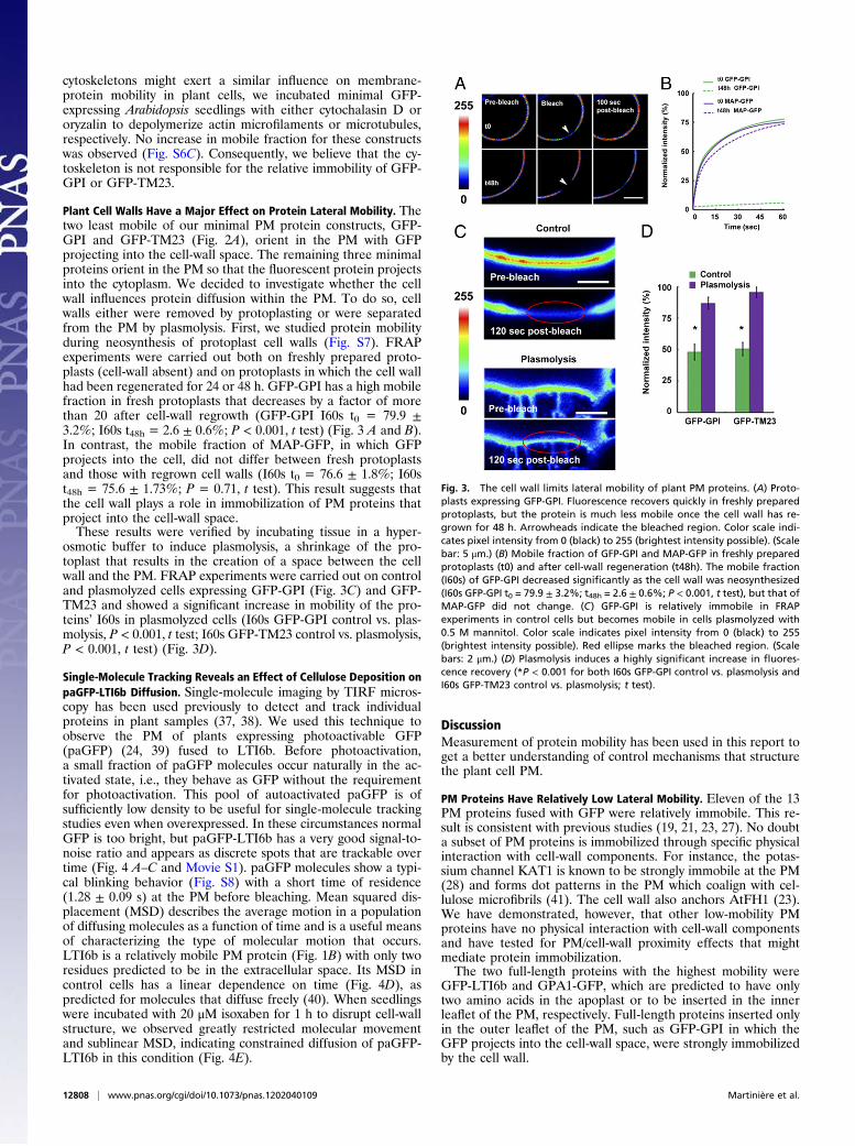
Plant Cell Walls Have a Major Effect on Protein Lateral Mobility. Thetwo least mobile of our minimal PM protein constructs, GFP-GPI and GFP-TM23 (Fig. 2A), orient in the PM with GFPprojecting into the cell-wall space. The remaining three minimalproteins orient in the PM so that the fluorescent protein projectsinto the cytoplasm. We decided to investigate whether the cellwall influences protein diffusion within the PM. To do so, cellwalls either were removed by protoplasting or were separatedfrom the PM by plasmolysis. First, we studied protein mobilityduring neosynthesis of protoplast cell walls (Fig. S7). FRAPexperiments were carried out both on freshly prepared proto-plasts (cell-wall absent) and on protoplasts in which the cell wallhad been regenerated for 24 or 48 h. GFP-GPI has a high mobilefraction in fresh protoplasts that decreases by a factor of morethan 20 after cell-wall regrowth (GFP-GPI I60s t0 = 79.9 ±3.2%; I60s t48h = 2.6 ± 0.6%; P < 0.001, t test) (Fig. 3 A and B).In contrast, the mobile fraction of MAP-GFP, in which GFPprojects into the cell, did not differ between fresh protoplastsand those with regrown cell walls (I60s t0 = 76.6 ± 1.8%; I60st48h = 75.6 ± 1.73%; P = 0.71, t test). This result suggests thatthe cell wall plays a role in immobilization of PM proteins thatproject into the cell-wall space.These results were verified by incubating tissue in a hyper-
osmotic buffer to induce plasmolysis, a shrinkage of the pro-toplast that results in the creation of a space between the cellwall and the PM. FRAP experiments were carried out on controland plasmolyzed cells expressing GFP-GPI (Fig. 3C) and GFP-TM23 and showed a significant increase in mobility of the pro-teins’ I60s in plasmolyzed cells (I60s GFP-GPI control vs. plas-molysis, P < 0.001, t test; I60s GFP-TM23 control vs. plasmolysis,P < 0.001, t test) (Fig. 3D).
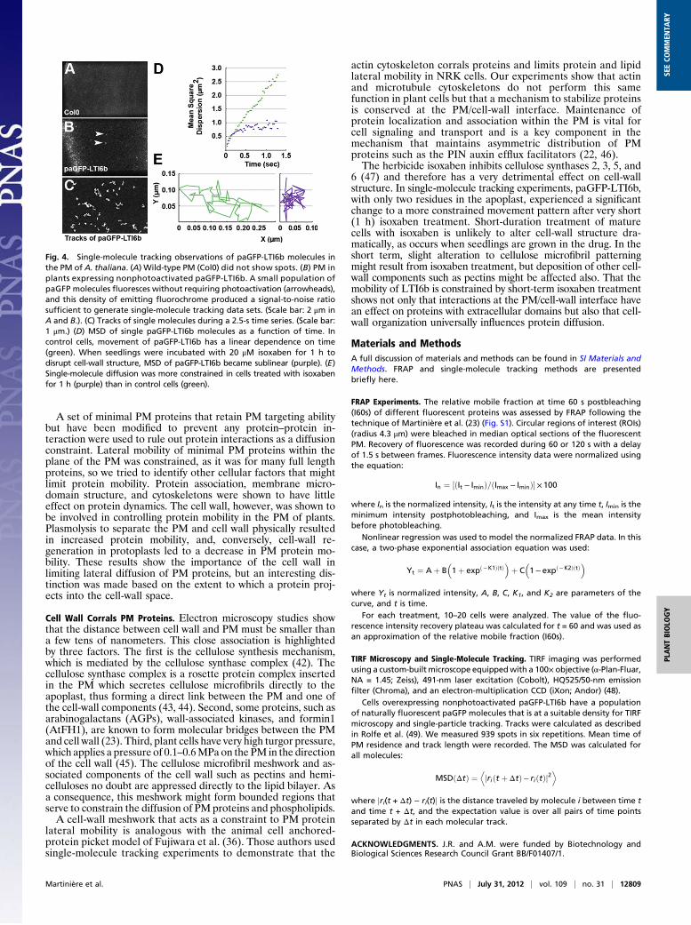
Single-Molecule Tracking Reveals an Effect of Cellulose Deposition onpaGFP-LTI6b Diffusion. Single-molecule imaging by TIRF micros-copy has been used previously to detect and track individualproteins in plant samples (37, 38). We used this technique toobserve the PM of plants expressing photoactivable GFP(paGFP) (24, 39) fused to LTI6b. Before photoactivation,a small fraction of paGFP molecules occur naturally in the ac-tivated state, i.e., they behave as GFP without the requirementfor photoactivation. This pool of autoactivated paGFP is ofsufficiently low density to be useful for single-molecule trackingstudies even when overexpressed. In these circumstances normalGFP is too bright, but paGFP-LTI6b has a very good signal-to-noise ratio and appears as discrete spots that are trackable overtime (Fig. 4 A–C and Movie S1). paGFP molecules show a typi-cal blinking behavior (Fig. S8) with a short time of residence(1.28 ± 0.09 s) at the PM before bleaching. Mean squared dis-placement (MSD) describes the average motion in a populationof diffusing molecules as a function of time and is a useful meansof characterizing the type of molecular motion that occurs.LTI6b is a relatively mobile PM protein (Fig. 1B) with only tworesidues predicted to be in the extracellular space. Its MSD incontrol cells has a linear dependence on time (Fig. 4D), aspredicted for molecules that diffuse freely (40). When seedlingswere incubated with 20 μM isoxaben for 1 h to disrupt cell-wallstructure, we observed greatly restricted molecular movementand sublinear MSD, indicating constrained diffusion of paGFP-LTI6b in this condition (Fig. 4E).
DiscussionMeasurement of protein mobility has been used in this report toget a better understanding of control mechanisms that structurethe plant cell PM.
PM Proteins Have Relatively Low Lateral Mobility. Eleven of the 13PM proteins fused with GFP were relatively immobile. This re-sult is consistent with previous studies (19, 21, 23, 27). No doubta subset of PM proteins is immobilized through specific physicalinteraction with cell-wall components. For instance, the potas-sium channel KAT1 is known to be strongly immobile at the PM(28) and forms dot patterns in the PM which coalign with cel-lulose microfibrils (41). The cell wall also anchors AtFH1 (23).We have demonstrated, however, that other low-mobility PMproteins have no physical interaction with cell-wall componentsand have tested for PM/cell-wall proximity effects that mightmediate protein immobilization.The two full-length proteins with the highest mobility were
GFP-LTI6b and GPA1-GFP, which are predicted to have onlytwo amino acids in the apoplast or to be inserted in the innerleaflet of the PM, respectively. Full-length proteins inserted onlyin the outer leaflet of the PM, such as GFP-GPI in which theGFP projects into the cell-wall space, were strongly immobilizedby the cell wall.
Fig. 3. The cell wall limits lateral mobility of plant PM proteins. (A) Proto-plasts expressing GFP-GPI. Fluorescence recovers quickly in freshly preparedprotoplasts, but the protein is much less mobile once the cell wall has re-grown for 48 h. Arrowheads indicate the bleached region. Color scale indi-cates pixel intensity from 0 (black) to 255 (brightest intensity possible). (Scalebar: 5 μm.) (B) Mobile fraction of GFP-GPI and MAP-GFP in freshly preparedprotoplasts (t0) and after cell-wall regeneration (t48h). The mobile fraction(I60s) of GFP-GPI decreased significantly as the cell wall was neosynthesized(I60s GFP-GPI t0 = 79.9 ± 3.2%; t48h = 2.6 ± 0.6%; P < 0.001, t test), but that ofMAP-GFP did not change. (C) GFP-GPI is relatively immobile in FRAPexperiments in control cells but becomes mobile in cells plasmolyzed with0.5 M mannitol. Color scale indicates pixel intensity from 0 (black) to 255(brightest intensity possible). Red ellipse marks the bleached region. (Scalebars: 2 μm.) (D) Plasmolysis induces a highly significant increase in fluores-cence recovery (*P < 0.001 for both I60s GFP-GPI control vs. plasmolysis andI60s GFP-TM23 control vs. plasmolysis; t test).
12808 | www.pnas.org/cgi/doi/10.1073/pnas.1202040109 Martinière et al.
A set of minimal PM proteins that retain PM targeting abilitybut have been modified to prevent any protein–protein in-teraction were used to rule out protein interactions as a diffusionconstraint. Lateral mobility of minimal PM proteins within theplane of the PM was constrained, as it was for many full lengthproteins, so we tried to identify other cellular factors that mightlimit protein mobility. Protein association, membrane micro-domain structure, and cytoskeletons were shown to have littleeffect on protein dynamics. The cell wall, however, was shown tobe involved in controlling protein mobility in the PM of plants.Plasmolysis to separate the PM and cell wall physically resultedin increased protein mobility, and, conversely, cell-wall re-generation in protoplasts led to a decrease in PM protein mo-bility. These results show the importance of the cell wall inlimiting lateral diffusion of PM proteins, but an interesting dis-tinction was made based on the extent to which a protein proj-ects into the cell-wall space.
Cell Wall Corrals PM Proteins. Electron microscopy studies showthat the distance between cell wall and PM must be smaller thana few tens of nanometers. This close association is highlightedby three factors. The first is the cellulose synthesis mechanism,which is mediated by the cellulose synthase complex (42). Thecellulose synthase complex is a rosette protein complex insertedin the PM which secretes cellulose microfibrils directly to theapoplast, thus forming a direct link between the PM and one ofthe cell-wall components (43, 44). Second, some proteins, such asarabinogalactans (AGPs), wall-associated kinases, and formin1(AtFH1), are known to form molecular bridges between the PMand cell wall (23). Third, plant cells have very high turgor pressure,which applies a pressure of 0.1–0.6MPa on the PM in the directionof the cell wall (45). The cellulose microfibril meshwork and as-sociated components of the cell wall such as pectins and hemi-celluloses no doubt are appressed directly to the lipid bilayer. Asa consequence, this meshwork might form bounded regions thatserve to constrain the diffusion of PM proteins and phospholipids.A cell-wall meshwork that acts as a constraint to PM protein
lateral mobility is analogous with the animal cell anchored-protein picket model of Fujiwara et al. (36). Those authors usedsingle-molecule tracking experiments to demonstrate that the
actin cytoskeleton corrals proteins and limits protein and lipidlateral mobility in NRK cells. Our experiments show that actinand microtubule cytoskeletons do not perform this samefunction in plant cells but that a mechanism to stabilize proteinsis conserved at the PM/cell-wall interface. Maintenance ofprotein localization and association within the PM is vital forcell signaling and transport and is a key component in themechanism that maintains asymmetric distribution of PMproteins such as the PIN auxin efflux facilitators (22, 46).The herbicide isoxaben inhibits cellulose synthases 2, 3, 5, and
6 (47) and therefore has a very detrimental effect on cell-wallstructure. In single-molecule tracking experiments, paGFP-LTI6b,with only two residues in the apoplast, experienced a significantchange to a more constrained movement pattern after very short(1 h) isoxaben treatment. Short-duration treatment of maturecells with isoxaben is unlikely to alter cell-wall structure dra-matically, as occurs when seedlings are grown in the drug. In theshort term, slight alteration to cellulose microfibril patterningmight result from isoxaben treatment, but deposition of other cell-wall components such as pectins might be affected also. That themobility of LTI6b is constrained by short-term isoxaben treatmentshows not only that interactions at the PM/cell-wall interface havean effect on proteins with extracellular domains but also that cell-wall organization universally influences protein diffusion.
Materials and MethodsA full discussion of materials and methods can be found in SI Materials andMethods. FRAP and single-molecule tracking methods are presentedbriefly here.
FRAP Experiments. The relative mobile fraction at time 60 s postbleaching(I60s) of different fluorescent proteins was assessed by FRAP following thetechnique of Martinière et al. (23) (Fig. S1). Circular regions of interest (ROIs)(radius 4.3 μm) were bleached in median optical sections of the fluorescentPM. Recovery of fluorescence was recorded during 60 or 120 s with a delayof 1.5 s between frames. Fluorescence intensity data were normalized usingthe equation:
In ¼ ½ðIt − IminÞ=ðImax − IminÞ�×100
where In is the normalized intensity, It is the intensity at any time t, Imin is theminimum intensity postphotobleaching, and Imax is the mean intensitybefore photobleaching.
Nonlinear regression was used to model the normalized FRAP data. In thiscase, a two-phase exponential association equation was used:
Yt ¼ Aþ B�1þ expð−K1ÞðtÞ
�þ C
�1−expð−K2ÞðtÞ
�
where Yt is normalized intensity, A, B, C, K1, and K2 are parameters of thecurve, and t is time.
For each treatment, 10–20 cells were analyzed. The value of the fluo-rescence intensity recovery plateau was calculated for t = 60 and was used asan approximation of the relative mobile fraction (I60s).
TIRF Microscopy and Single-Molecule Tracking. TIRF imaging was performedusing a custom-builtmicroscope equippedwith a 100× objective (α-Plan-Fluar,NA = 1.45; Zeiss), 491-nm laser excitation (Cobolt), HQ525/50-nm emissionfilter (Chroma), and an electron-multiplication CCD (iXon; Andor) (48).
Cells overexpressing nonphotoactivated paGFP-LTI6b have a populationof naturally fluorescent paGFP molecules that is at a suitable density for TIRFmicroscopy and single-particle tracking. Tracks were calculated as describedin Rolfe et al. (49). We measured 939 spots in six repetitions. Mean time ofPM residence and track length were recorded. The MSD was calculated forall molecules:
MSDðΔtÞ ¼ jriðt þ ΔtÞ− riðtÞj2D E
where jri(t + Δt) − ri(t)j is the distance traveled by molecule i between time tand time t + Δt, and the expectation value is over all pairs of time pointsseparated by Δt in each molecular track.
ACKNOWLEDGMENTS. J.R. and A.M. were funded by Biotechnology andBiological Sciences Research Council Grant BB/F01407/1.
Fig. 4. Single-molecule tracking observations of paGFP-LTI6b molecules inthe PM of A. thaliana. (A) Wild-type PM (Col0) did not show spots. (B) PM inplants expressing nonphotoactivated paGFP-LTI6b. A small population ofpaGFP molecules fluoresces without requiring photoactivation (arrowheads),and this density of emitting fluorochrome produced a signal-to-noise ratiosufficient to generate single-molecule tracking data sets. (Scale bar: 2 μm inA and B.). (C) Tracks of single molecules during a 2.5-s time series. (Scale bar:1 μm.) (D) MSD of single paGFP-LTI6b molecules as a function of time. Incontrol cells, movement of paGFP-LTI6b has a linear dependence on time(green). When seedlings were incubated with 20 μM isoxaben for 1 h todisrupt cell-wall structure, MSD of paGFP-LTI6b became sublinear (purple). (E)Single-molecule diffusion was more constrained in cells treated with isoxabenfor 1 h (purple) than in control cells (green).
Martinière et al. PNAS | July 31, 2012 | vol. 109 | no. 31 | 12809
PLANTBIOLO
GY
SEECO
MMEN
TARY
1. Schwacke R, et al. (2003) ARAMEMNON, a novel database for Arabidopsis integralmembrane proteins. Plant Physiol 131:16–26.
2. Engelman DM (2005) Membranes are more mosaic than fluid. Nature 438:578–580.3. Singer SJ, Nicolson GL (1972) The fluid mosaic model of the structure of cell mem-
branes. Science 175:720–731.4. Saffman PG, Delbrück M (1975) Brownian motion in biological membranes. Proc Natl
Acad Sci USA 72:3111–3113.5. Thompson NL, Axelrod D (1980) Reduced lateral mobility of a fluorescent lipid probe
in cholesterol-depleted erythrocyte membrane. Biochim Biophys Acta 597:155–165.6. Yechiel E, Edidin M (1987) Micrometer-scale domains in fibroblast plasma mem-
branes. J Cell Biol 105:755–760.7. Kwik J, et al. (2003) Membrane cholesterol, lateral mobility, and the phosphatidyli-
nositol 4,5-bisphosphate-dependent organization of cell actin. Proc Natl Acad Sci USA100:13964–13969.
8. Lenne PF, et al. (2006) Dynamic molecular confinement in the plasma membrane bymicrodomains and the cytoskeleton meshwork. EMBO J 25:3245–3256.
9. Sieber JJ, et al. (2007) Anatomy and dynamics of a supramolecular membrane proteincluster. Science 317:1072–1076.
10. Kusumi A, et al. (2005) Paradigm shift of the plasma membrane concept from thetwo-dimensional continuum fluid to the partitioned fluid: High-speed single-mole-cule tracking of membrane molecules. Annu Rev Biophys Biomol Struct 34:351–378.
11. Simons K, Ikonen E (1997) Functional rafts in cell membranes. Nature 387:569–572.12. Owen DM, Williamson D, Rentero C, Gaus K (2009) Quantitative microscopy: Protein
dynamics and membrane organisation. Traffic 10:962–971.13. Mongrand S, Stanislas T, Bayer EM, Lherminier J, Simon-Plas F (2010) Membrane rafts
in plant cells. Trends Plant Sci 15:656–663.14. Borner GH, et al. (2005) Analysis of detergent-resistant membranes in Arabidopsis.
Evidence for plasma membrane lipid rafts. Plant Physiol 137:104–116.15. Morel J, et al. (2006) Proteomics of plant detergent-resistant membranes. Mol Cell
Proteomics 5:1396–1411.16. Lefebvre B, et al. (2007) Characterization of lipid rafts from Medicago truncatula root
plasma membranes: A proteomic study reveals the presence of a raft-associated redoxsystem. Plant Physiol 144:402–418.
17. Grossmann G, Opekarova M, Novakova L, Stolz J, Tanner W (2006) Lipid raft-basedmembrane compartmentation of a plant transport protein expressed in Saccharo-myces cerevisiae. Eukaryot Cell 5:945–953.
18. Raffaele S, et al. (2009) Remorin, a solanaceae protein resident in membrane rafts andplasmodesmata, impairs potato virus X movement. Plant Cell 21:1541–1555.
19. Men S, et al. (2008) Sterol-dependent endocytosis mediates post-cytokinetic acquisi-tion of PIN2 auxin efflux carrier polarity. Nat Cell Biol 10:237–244.
20. Liu P, et al. (2009) Lipid microdomain polarization is required for NADPH oxidase-dependent ROS signaling in Picea meyeri pollen tube tip growth. Plant J 60:303–313.
21. Takano J, et al. (2010) Polar localization and degradation of Arabidopsis borontransporters through distinct trafficking pathways. Proc Natl Acad Sci USA 107:5220–5225.
22. Feraru E, et al. (2011) PIN polarity maintenance by the cell wall in Arabidopsis. CurrBiol 21:338–343.
23. Martinière A, Gayral P, Hawes C, Runions J (2011) Building bridges: Formin1 of Ara-bidopsis forms a connection between the cell wall and the actin cytoskeleton. Plant J66:354–365.
24. Runions J, Brach T, Kühner S, Hawes C (2006) Photoactivation of GFP reveals proteindynamics within the endoplasmic reticulum membrane. J Exp Bot 57:43–50.
25. Graumann K, Runions J, Evans DE (2010) Characterization of SUN-domain proteins atthe higher plant nuclear envelope. Plant J 61:134–144.
26. Sparkes I, Runions J, Hawes C, Griffing L (2009) Movement and remodeling of theendoplasmic reticulum in nondividing cells of tobacco leaves. Plant Cell 21:3937–3949.
27. Sutter JU, Campanoni P, Tyrrell M, Blatt MR (2006) Selective mobility and sensitivity toSNAREs is exhibited by the Arabidopsis KAT1 K+ channel at the plasma membrane.Plant Cell 18:935–954.
28. Sorieul M, Santoni V, Maurel C, Luu DT (2011) Mechanisms and effects of retention of
over-expressed aquaporin AtPIP2;1 in the endoplasmic reticulum. Traffic 12:473–482.29. Sparkes IA, Runions J, Kearns A, Hawes C (2006) Rapid, transient expression of fluo-
rescent fusion proteins in tobacco plants and generation of stably transformed plants.
Nat Protoc 1:2019–2025.30. Luu DT, et al. (2012) Fluorescence recovery after photobleaching reveals high cycling
dynamics of plasma membrane aquaporins in Arabidopsis roots under salt stress.
Plant J 69:894–905.31. Umenishi F, Verbavatz JM, Verkman AS (2000) cAMP regulated membrane diffusion
of a green fluorescent protein-aquaporin 2 chimera. Biophys J 78:1024–1035.32. Vermeer JE, et al. (2009) Imaging phosphatidylinositol 4-phosphate dynamics in living
plant cells. Plant J 57:356–372.33. Brandizzi F, et al. (2002) The destination for single-pass membrane proteins is influ-
enced markedly by the length of the hydrophobic domain. Plant Cell 14:1077–1092.34. Grebe M, et al. (2003) Arabidopsis sterol endocytosis involves actin-mediated traf-
ficking via ARA6-positive early endosomes. Curr Biol 13:1378–1387.35. Bonneau L, et al. (2010) Plasma membrane sterol complexation, generated by fil-
ipin, triggers signaling responses in tobacco cells. Biochim Biophys Acta 1798:
2150–2159.36. Fujiwara T, Ritchie K, Murakoshi H, Jacobson K, Kusumi A (2002) Phospholipids
undergo hop diffusion in compartmentalized cell membrane. J Cell Biol 157:
1071–1081.37. Li X, et al. (2011) Single-molecule analysis of PIP2;1 dynamics and partitioning reveals
multiple modes of Arabidopsis plasma membrane aquaporin regulation. Plant Cell 23:
3780–3797.38. Vizcay-Barrena G, Webb SE, Martin-Fernandez ML, Wilson ZA (2011) Subcellular and
single-molecule imaging of plant fluorescent proteins using total internal reflection
fluorescence microscopy (TIRFM). J Exp Bot 62:5419–5428.39. Patterson GH, Lippincott-Schwartz J (2002) A photoactivatable GFP for selective
photolabeling of proteins and cells. Science 297:1873–1877.40. Holtzer L, Schmidt T (2010) The tracking of individual molecules in cells and tissues.
Single Particle Tracking and Single Molecule Energy Transfer, eds Brauchle C, Lamb D,
Michaelis J (Wiley-VCH, Weinheim, Germany).41. Homann U,Meckel T, Hewing J, HüttMT, Hurst AC (2007) Distinctfluorescent pattern of
KAT1:GFP in the plasma membrane of Vicia faba guard cells. Eur J Cell Biol 86:489–500.42. Carpita NC (2011) Update on mechanisms of plant cell wall biosynthesis: How plants
make cellulose and other (1->4)-β-D-glycans. Plant Physiol 155:171–184.43. Giddings TH, Jr., Brower DL, Staehelin LA (1980) Visualization of particle complexes in
the plasma membrane of Micrasterias denticulata associated with the formation of
cellulose fibrils in primary and secondary cell walls. J Cell Biol 84:327–339.44. Mueller SC, Brown RM, Jr. (1980) Evidence for an intramembrane component asso-
ciated with a cellulose microfibril-synthesizing complex in higher plants. J Cell Biol 84:
315–326.45. Wang L, Hukin D, Pritchard J, Thomas C (2006) Comparison of plant cell turgor
pressure measurement by pressure probe and micromanipulation. Biotechnol Lett 28:
1147–1150.46. Kleine-Vehn J, et al. (2011) Recycling, clustering, and endocytosis jointly maintain PIN
auxin carrier polarity at the plasma membrane. Mol Syst Biol 7:540.47. Bischoff V, Cookson SJ, Wu S, Scheible WR (2009) Thaxtomin A affects CESA-complex
density, expression of cell wall genes, cell wall composition, and causes ectopic lig-
nification in Arabidopsis thaliana seedlings. J Exp Bot 60:955–965.48. Clarke DT, et al. (2011) Optics clustered to output unique solutions: A multi-laser
facility for combined single molecule and ensemble microscopy. Review of Scientific
Instruments 82, 093705.49. Rolfe DJ, et al. (2011) Automated multidimensional single molecule fluorescence
microscopy feature detection and tracking. Eur Biophys J 40:1167–1186.
12810 | www.pnas.org/cgi/doi/10.1073/pnas.1202040109 Martinière et al.