Embed Size (px)
Citation preview
Cell, Vol. 97, 621–633, May 28, 1999, Copyright ©1999 by Cell Press
Relocalization of Telomeric Ku and SIR Proteinsin Response to DNA Strand Breaks in Yeast
for religation (reviewed by Haber, 1998). The third is acomplex of Xrcc4 and Ligase IV, both of which are es-sential for the religation step following DSB repair.
Sophie G. Martin,* Thierry Laroche,*Noriyuki Suka,† Michael Grunstein,†and Susan M. Gasser*‡
*Swiss Institute for Experimental Cancer Research The mammalian Ku70 and Ku86 proteins form a het-erodimer that binds the ends of double-stranded DNACh. des Boveresses 155
CH-1066 Epalinges with high affinity (reviewed in Dynan and Yoo, 1998).Their role in both DSB repair and V(D)J recombinationSwitzerland
†Department of Biological Chemistry requires DNA-protein kinase (DNA-PK), which is thoughtto be targeted to sites of damage by Ku (Lieber et al.,UCLA School of Medicine and
Molecular Biology Institute 1997). Consistent with this role, mice lacking DNA-PK,or either Ku subunit, are sensitive to ionizing radiation,University of California
Los Angeles, California 90095 impaired for growth, and have severe combined immu-nodeficiency. Intriguingly, primary fibroblasts isolatedfrom Ku-deficient mice also have phenotypes of prema-ture senescence (Nussenzweig et al., 1996; Gu et al.,Summary1997). In yeast, DSBs are most frequently repaired bythe RAD52-dependent homologous recombination path-Telomeric TG-rich repeats and their associated pro-way. However, rad52 strains lacking yKu70p or yKu80pteins protect the termini of eukaryotic chromosomes(encoded by HDF1 and HDF2, respectively) are highlyfrom end-to-end fusions. Associated with the capsensitive to irradiation (Siede et al., 1996), and strainsstructure at yeast telomeres is a subtelomeric domainlacking yKu alone are deficient in end-joining reactionsof heterochromatin, containing the silent information(Mages et al., 1996).regulator (SIR) complex. The Ku70/80 heterodimer
Recently it has been shown that Rad52� strains lack-(yKu) is associated both with the chromosome ending either HDF gene have defects in telomere mainte-and with subtelomeric chromatin. Surprisingly, bothnance. Notably, the terminal TG1-3 repeat is shorteryKu and the chromatin-associated Rap1 and SIR pro-(Porter et al., 1996; Boulton and Jackson, 1998), stretchesteins are released from telomeres in a RAD9-depen-of single-strand DNA accumulate at chromosomal endsdent response to DNA damage. yKu is recruited rapidly(Gravel et al., 1998), and yeast telomeres are delocalizedto double-strand cuts, while low levels of SIR proteinsfrom the nuclear periphery (Laroche et al., 1998). Con-are detected near cleavage sites at later time points.comitantly, telomere position effect (TPE), which confersConsistently, yKu- or SIR-deficient strains are hyper-transcriptional repression on genes in subtelomeric re-sensitive to DNA-damaging agents. The release of yKugions, drops by 103-fold. Genetic analyses indicate thatfrom telomeric chromatin may allow efficient scanninghdf mutants exacerbate the growth and telomere main-of the genome for DNA strand breaks.tenance defects resulting from mutation of either telo-merase (est2) or a protein that binds the single-strandtelomeric DNA (cdc13; Gravel et al., 1998; Nugent et al.,Introduction1998; Polotnianka et al., 1998). Moreover, hdf mutantsaggravate defects provoked by the loss of the Mre11/The telomere is a unique structure that serves to protect
the termini of linear chromosomes from fusions and deg- Rad50/Xrs2 complex, which may process the chromo-some end creating the 3� overhang necessary for telo-radation. Other ends in DNA, such as breaks caused by
ionizing radiation or other DNA-damaging agents, must merase function. These data are consistent with theproposal that yKu helps cap the telomere and/or regu-be repaired to ensure that every chromosomal fragment
is linked to a single centromere for equal distribution at lates 3� end processing enzymes.The fact that hdf mutants are defective in both telo-mitosis. The induction of a single double-strand break
(DSB) will cause cells to arrest in the cell cycle at the mere maintenance and DSB repair suggests the directDNA damage checkpoint until the break is repaired (San- involvement of yKu at both sites. However, if true, yKudell and Zakian, 1993). binding must be able to produce two opposite results:
One universally conserved mechanism by which cells at double-strand breaks, DNA ends are processed andrepair DSB is a process referred to as nonhomologous religated, while in the case of telomere maintenance,end joining (NHEJ). This involves three well-character- end-to-end ligation is specifically suppressed. At pres-ized protein complexes. One is the Ku heterodimer, ent, it is unknown whether yKu binds directly at bothwhich in mammalian cells associates with the ATM- sites of action, what distinguishes the two complexes,related DNA-stimulated protein kinase (DNA-PK; re- and whether yKu is regulated by the damage responseviewed in Critchlow and Jackson, 1998). The second is pathway.the Mre11/Rad50/Xrs2 complex, which has endo- and Here, we examine the fate of yKu and the nucleosome-exonucleolytic activities thought to process the break binding silent information regulator (SIR) complex (re-
viewed by Shore, 1998) under conditions of inducedDNA damage. Our results support a model in which‡ To whom correspondence should be addressed (e-mail: sgasser@
eliot.unil.ch). telomeric heterochromatin serves as a reservoir for
Cell622
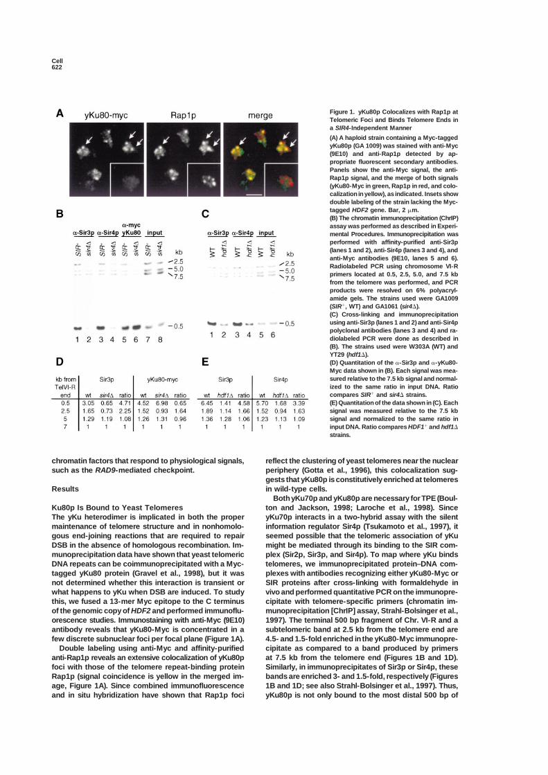
Figure 1. yKu80p Colocalizes with Rap1p atTelomeric Foci and Binds Telomere Ends ina SIR4-Independent Manner
(A) A haploid strain containing a Myc-taggedyKu80p (GA 1009) was stained with anti-Myc(9E10) and anti-Rap1p detected by ap-propriate fluorescent secondary antibodies.Panels show the anti-Myc signal, the anti-Rap1p signal, and the merge of both signals(yKu80-Myc in green, Rap1p in red, and colo-calization in yellow), as indicated. Insets showdouble labeling of the strain lacking the Myc-tagged HDF2 gene. Bar, 2 �m.(B) The chromatin immunoprecipitation (ChrIP)assay was performed as described in Experi-mental Procedures. Immunoprecipitation wasperformed with affinity-purified anti-Sir3p(lanes 1 and 2), anti-Sir4p (lanes 3 and 4), andanti-Myc antibodies (9E10, lanes 5 and 6).Radiolabeled PCR using chromosome VI-Rprimers located at 0.5, 2.5, 5.0, and 7.5 kbfrom the telomere was performed, and PCRproducts were resolved on 6% polyacryl-amide gels. The strains used were GA1009(SIR�, WT) and GA1061 (sir4�).(C) Cross-linking and immunoprecipitationusing anti-Sir3p (lanes 1 and 2) and anti-Sir4ppolyclonal antibodies (lanes 3 and 4) and ra-diolabeled PCR were done as described in(B). The strains used were W303A (WT) andYT29 (hdf1�).(D) Quantitation of the �-Sir3p and �-yKu80-Myc data shown in (B). Each signal was mea-sured relative to the 7.5 kb signal and normal-ized to the same ratio in input DNA. Ratiocompares SIR� and sir4� strains.(E) Quantitation of the data shown in (C). Eachsignal was measured relative to the 7.5 kbsignal and normalized to the same ratio ininput DNA. Ratio compares HDF1� and hdf1�
strains.
chromatin factors that respond to physiological signals, reflect the clustering of yeast telomeres near the nuclearperiphery (Gotta et al., 1996), this colocalization sug-such as the RAD9-mediated checkpoint.gests that yKu80p is constitutively enriched at telomeresin wild-type cells.Results
Both yKu70p and yKu80p are necessary for TPE (Boul-ton and Jackson, 1998; Laroche et al., 1998). SinceKu80p Is Bound to Yeast Telomeres
The yKu heterodimer is implicated in both the proper yKu70p interacts in a two-hybrid assay with the silentinformation regulator Sir4p (Tsukamoto et al., 1997), itmaintenance of telomere structure and in nonhomolo-
gous end-joining reactions that are required to repair seemed possible that the telomeric association of yKumight be mediated through its binding to the SIR com-DSB in the absence of homologous recombination. Im-
munoprecipitation data have shown that yeast telomeric plex (Sir2p, Sir3p, and Sir4p). To map where yKu bindstelomeres, we immunoprecipitated protein–DNA com-DNA repeats can be coimmunoprecipitated with a Myc-
tagged yKu80 protein (Gravel et al., 1998), but it was plexes with antibodies recognizing either yKu80-Myc orSIR proteins after cross-linking with formaldehyde innot determined whether this interaction is transient or
what happens to yKu when DSB are induced. To study vivo and performed quantitative PCR on the immunopre-cipitate with telomere-specific primers (chromatin im-this, we fused a 13-mer Myc epitope to the C terminus
of the genomic copy of HDF2 and performed immunoflu- munoprecipitation [ChrIP] assay, Strahl-Bolsinger et al.,1997). The terminal 500 bp fragment of Chr. VI-R and aorescence studies. Immunostaining with anti-Myc (9E10)
antibody reveals that yKu80-Myc is concentrated in a subtelomeric band at 2.5 kb from the telomere end are4.5- and 1.5-fold enriched in the yKu80-Myc immunopre-few discrete subnuclear foci per focal plane (Figure 1A).
Double labeling using anti-Myc and affinity-purified cipitate as compared to a band produced by primersat 7.5 kb from the telomere end (Figures 1B and 1D).anti-Rap1p reveals an extensive colocalization of yKu80p
foci with those of the telomere repeat-binding protein Similarly, in immunoprecipitates of Sir3p or Sir4p, thesebands are enriched 3- and 1.5-fold, respectively (FiguresRap1p (signal coincidence is yellow in the merged im-
age, Figure 1A). Since combined immunofluorescence 1B and 1D; see also Strahl-Bolsinger et al., 1997). Thus,yKu80p is not only bound to the most distal 500 bp ofand in situ hybridization have shown that Rap1p foci

Telomeric Response to DNA Damage623
Figure 2. GBD-yKu70p and GBD-yKu80p Nu-cleate Silencing in the Absence of a Silencer
(A) Targeted repression is monitored instrains that carry URA3 adjacent to eightGal4p-binding sites integrated at the HML lo-cus, flanked by both the E and I silencers(E-I, white; Ce134), only the E silencer (E-�i,dashed; Ce76), or neither (�e-�i, black;Ce77). These strains were transformed withvectors encoding either yKu80p, yKu70p,Sir2p, or Orc1p fused in-frame to the Gal4DNA-binding domain (GBD).(B) The influence of targeted fusion proteins(GBD-yKu70p, GBD-yKu80p, GBD-Sir2p, orGBD-Orc1p) on silencing of the URA3 genewas monitored as described in ExperimentalProcedures, as compared to a strain express-ing only the GBD (pAS2, vector). The meanand standard deviation were calculated forratios of cells growing on �FOA and �FOA,for eight independent colonies of each trans-formant (see bar graph; the x axis representsthe efficiency of URA3 repression in log scale.Less than 3 in 105 cells with vector alone growwhen one or both silencers are deleted orin a sir4::HIS3 deletion background (�e-�i,sir4�).
Chr. VI-R, but it appears to spread, like Rap1p and SIR Constructs with deleted silencers thus allow us to testwhether GBD-fusion proteins can establish repression,complexes, along the repressed subtelomeric region.
By performing this same assay in a strain deficient like the positive controls of Sir2p and Orc1p fused toGBD.for SIR4 (sir4�), we find that the association of yKu80-
Myc with the more internal sequence is SIR dependent, The colony-forming assay on 5-FOA reveals that theGBD-yKu70p is a potent nucleator of repression evenwhile its binding to the terminal fragment is not (Figures
1B and 1D). As expected, no association of Sir3p with in the absence of silencer elements (Figure 2B), silencingURA3 expression to within 2-fold of the maximal leveleither telomeric or subtelomeric fragments can be de-
tected in the sir4� strain. Similarly, Sir3p and Sir4p bind detected in the two-silencer construct (Figure 2B). In-deed, the GBD-yKu70 fusion nucleates repression moretelomeric sequences poorly in an hdf1� strain (levels
are 3- to 5-fold lower than wild-type, Figures 1C and efficiently than GBD-Orc1p or GBD-Sir2p on the con-struct lacking silencers, while on the construct con-1E), consistent with the loss of TPE reported for hdf1
and hdf2 strains (Laroche et al., 1998). Thus, although taining one silencer, all three GBD fusions are equallyefficient (Figure 2B). GBD-yKu80p silences as efficientlyyKu has a SIR-independent association with the terminal
fragment of the chromosome, it associates with re- as GBD-Ku70p on the E-�i reporter, but it is less efficienton a reporter lacking both silencers, correlating with thepressed chromatin in a SIR4-dependent manner, much
like Rap1p (Strahl-Bolsinger et al., 1997). fact that GBD-yKu80p only partially complements anhdf2 deletion, while GBD-yKu70p fully complements anhdf1 null (data not shown). In all cases yKu-mediatedyKu Can Nucleate Silencing in the Absence
of Silencer Elements repression is SIR dependent, as no growth is detectedon 5-FOA in the absence of Sir4p (see sir4�, Figure 2B).The loss of silencing that correlates with hdf mutations
suggests that yKu might help recruit the SIR complex Furthermore, GBD-yKu70p-mediated silencing requiresintact yKu80p (data not shown). It is unlikely that GBD-to subtelomeric chromatin. Moreover, it has been pro-
posed that yKu targets Sir proteins to double-strand yKu represses by tethering the reporter to the nuclearenvelope (Andrulis et al., 1998), since membrane teth-breaks, where these latter would perform an as-yet-
undefined role in repair (Jackson, 1997; Tsukamoto et ering does not enhance repression in the absence ofsilencers.al., 1997). To test whether yKu alone can nucleate SIR-
mediated repression, we have tethered it by fusion to theGal4p DNA-binding domain (GBD) to an internal URA3 Sir Mutants Are Hypersensitive
to DNA-Damaging Agentsreporter placed downstream of a cluster of eight Gal4precognition sites. The reporter was inserted at HML, Although hdf mutants have major telomere phenotypes,
the most conserved role of the Ku heterodimer is inflanked either by both HML E and I silencers, by onlythe E silencer, or by neither silencer (Figure 2A, E-I, E-�i, nonhomologous end joining during the DSB repair (re-
viewed in Lieber et al., 1997), just as the primary role ofor �e-�i). The silencer-flanked URA3 reporter (E-I) isalmost completely repressed in the presence of the GBD the SIR complex is to confer position-dependent repres-
sion on mating type genes. Yet, SIR proteins have alsoalone (vector, Figure 2B), while deletion of one or bothsilencers results in nearly complete derepression (i.e., been implicated in the NHEJ reactions monitored by the
circularization of linear plasmid DNA (Tsukamoto et al.,lack of growth, as URA3 expression is toxic on 5-FOA).
Cell624
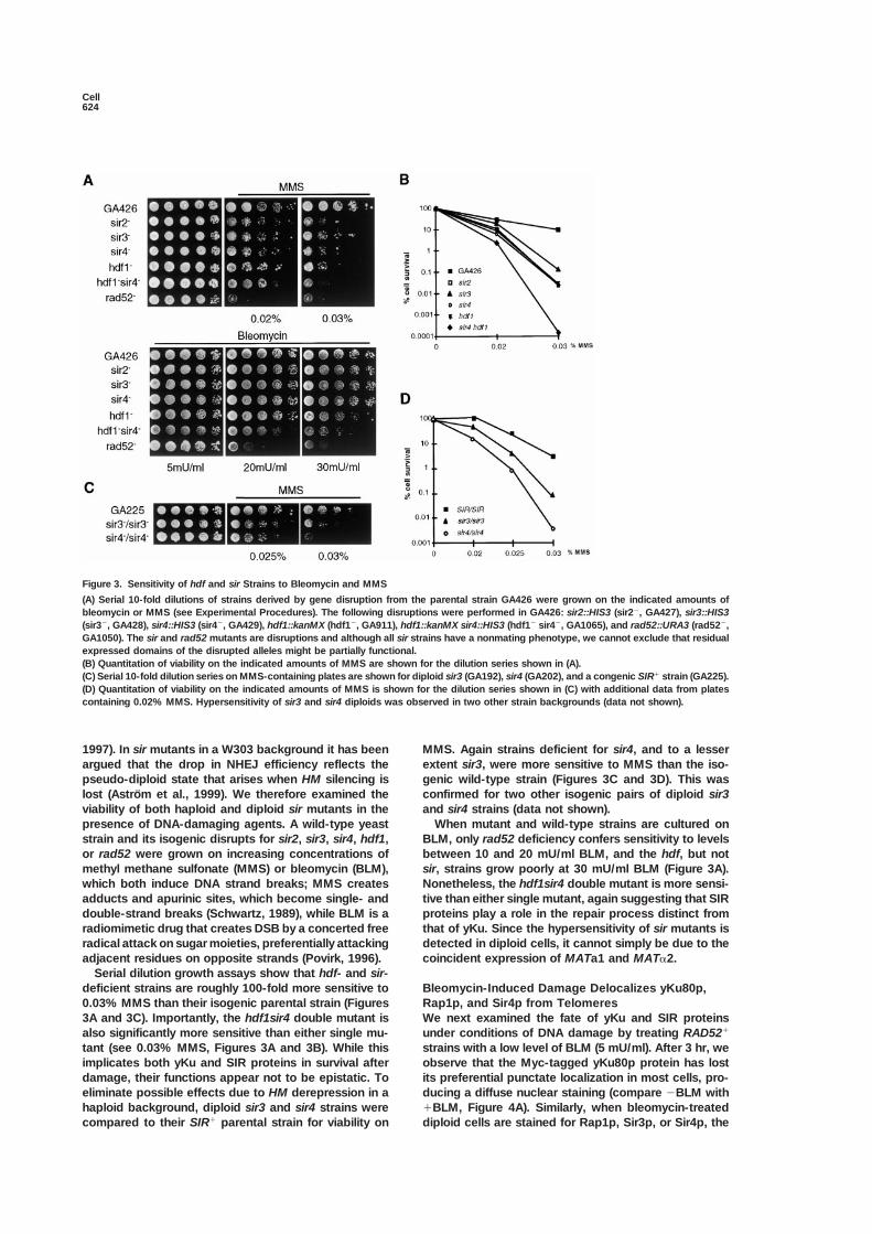
Figure 3. Sensitivity of hdf and sir Strains to Bleomycin and MMS
(A) Serial 10-fold dilutions of strains derived by gene disruption from the parental strain GA426 were grown on the indicated amounts ofbleomycin or MMS (see Experimental Procedures). The following disruptions were performed in GA426: sir2::HIS3 (sir2�, GA427), sir3::HIS3(sir3�, GA428), sir4::HIS3 (sir4�, GA429), hdf1::kanMX (hdf1�, GA911), hdf1::kanMX sir4::HIS3 (hdf1� sir4�, GA1065), and rad52::URA3 (rad52�,GA1050). The sir and rad52 mutants are disruptions and although all sir strains have a nonmating phenotype, we cannot exclude that residualexpressed domains of the disrupted alleles might be partially functional.(B) Quantitation of viability on the indicated amounts of MMS are shown for the dilution series shown in (A).(C) Serial 10-fold dilution series on MMS-containing plates are shown for diploid sir3 (GA192), sir4 (GA202), and a congenic SIR� strain (GA225).(D) Quantitation of viability on the indicated amounts of MMS is shown for the dilution series shown in (C) with additional data from platescontaining 0.02% MMS. Hypersensitivity of sir3 and sir4 diploids was observed in two other strain backgrounds (data not shown).
1997). In sir mutants in a W303 background it has been MMS. Again strains deficient for sir4, and to a lesserextent sir3, were more sensitive to MMS than the iso-argued that the drop in NHEJ efficiency reflects the
pseudo-diploid state that arises when HM silencing is genic wild-type strain (Figures 3C and 3D). This wasconfirmed for two other isogenic pairs of diploid sir3lost (Astrom et al., 1999). We therefore examined the
viability of both haploid and diploid sir mutants in the and sir4 strains (data not shown).When mutant and wild-type strains are cultured onpresence of DNA-damaging agents. A wild-type yeast
strain and its isogenic disrupts for sir2, sir3, sir4, hdf1, BLM, only rad52 deficiency confers sensitivity to levelsbetween 10 and 20 mU/ml BLM, and the hdf, but notor rad52 were grown on increasing concentrations of
methyl methane sulfonate (MMS) or bleomycin (BLM), sir, strains grow poorly at 30 mU/ml BLM (Figure 3A).Nonetheless, the hdf1sir4 double mutant is more sensi-which both induce DNA strand breaks; MMS creates
adducts and apurinic sites, which become single- and tive than either single mutant, again suggesting that SIRproteins play a role in the repair process distinct fromdouble-strand breaks (Schwartz, 1989), while BLM is a
radiomimetic drug that creates DSB by a concerted free that of yKu. Since the hypersensitivity of sir mutants isdetected in diploid cells, it cannot simply be due to theradical attack on sugar moieties, preferentially attacking
adjacent residues on opposite strands (Povirk, 1996). coincident expression of MATa1 and MAT�2.Serial dilution growth assays show that hdf- and sir-
deficient strains are roughly 100-fold more sensitive to Bleomycin-Induced Damage Delocalizes yKu80p,Rap1p, and Sir4p from Telomeres0.03% MMS than their isogenic parental strain (Figures
3A and 3C). Importantly, the hdf1sir4 double mutant is We next examined the fate of yKu and SIR proteinsunder conditions of DNA damage by treating RAD52�also significantly more sensitive than either single mu-
tant (see 0.03% MMS, Figures 3A and 3B). While this strains with a low level of BLM (5 mU/ml). After 3 hr, weobserve that the Myc-tagged yKu80p protein has lostimplicates both yKu and SIR proteins in survival after
damage, their functions appear not to be epistatic. To its preferential punctate localization in most cells, pro-ducing a diffuse nuclear staining (compare �BLM witheliminate possible effects due to HM derepression in a
haploid background, diploid sir3 and sir4 strains were �BLM, Figure 4A). Similarly, when bleomycin-treateddiploid cells are stained for Rap1p, Sir3p, or Sir4p, thecompared to their SIR� parental strain for viability on
Telomeric Response to DNA Damage625
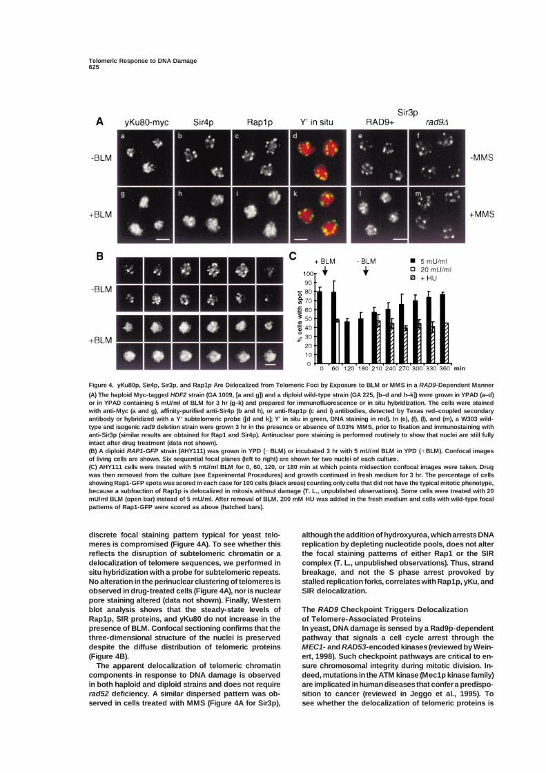
Figure 4. yKu80p, Sir4p, Sir3p, and Rap1p Are Delocalized from Telomeric Foci by Exposure to BLM or MMS in a RAD9-Dependent Manner
(A) The haploid Myc-tagged HDF2 strain (GA 1009, [a and g]) and a diploid wild-type strain (GA 225, [b–d and h–k]) were grown in YPAD (a–d)or in YPAD containing 5 mU/ml of BLM for 3 hr (g–k) and prepared for immunofluorescence or in situ hybridization. The cells were stainedwith anti-Myc (a and g), affinity-purified anti-Sir4p (b and h), or anti-Rap1p (c and i) antibodies, detected by Texas red–coupled secondaryantibody or hybridized with a Y’ subtelomeric probe ([d and k]; Y’ in situ in green, DNA staining in red). In (e), (f), (l), and (m), a W303 wild-type and isogenic rad9 deletion strain were grown 3 hr in the presence or absence of 0.03% MMS, prior to fixation and immunostaining withanti-Sir3p (similar results are obtained for Rap1 and Sir4p). Antinuclear pore staining is performed routinely to show that nuclei are still fullyintact after drug treatment (data not shown).(B) A diploid RAP1-GFP strain (AHY111) was grown in YPD (�BLM) or incubated 3 hr with 5 mU/ml BLM in YPD (�BLM). Confocal imagesof living cells are shown. Six sequential focal planes (left to right) are shown for two nuclei of each culture.(C) AHY111 cells were treated with 5 mU/ml BLM for 0, 60, 120, or 180 min at which points midsection confocal images were taken. Drugwas then removed from the culture (see Experimental Procedures) and growth continued in fresh medium for 3 hr. The percentage of cellsshowing Rap1-GFP spots was scored in each case for 100 cells (black areas) counting only cells that did not have the typical mitotic phenotype,because a subfraction of Rap1p is delocalized in mitosis without damage (T. L., unpublished observations). Some cells were treated with 20mU/ml BLM (open bar) instead of 5 mU/ml. After removal of BLM, 200 mM HU was added in the fresh medium and cells with wild-type focalpatterns of Rap1-GFP were scored as above (hatched bars).
discrete focal staining pattern typical for yeast telo- although the addition of hydroxyurea, which arrests DNAreplication by depleting nucleotide pools, does not altermeres is compromised (Figure 4A). To see whether this
reflects the disruption of subtelomeric chromatin or a the focal staining patterns of either Rap1 or the SIRcomplex (T. L., unpublished observations). Thus, stranddelocalization of telomere sequences, we performed in
situ hybridization with a probe for subtelomeric repeats. breakage, and not the S phase arrest provoked bystalled replication forks, correlates with Rap1p, yKu, andNo alteration in the perinuclear clustering of telomeres is
observed in drug-treated cells (Figure 4A), nor is nuclear SIR delocalization.pore staining altered (data not shown). Finally, Westernblot analysis shows that the steady-state levels of The RAD9 Checkpoint Triggers Delocalization
of Telomere-Associated ProteinsRap1p, SIR proteins, and yKu80 do not increase in thepresence of BLM. Confocal sectioning confirms that the In yeast, DNA damage is sensed by a Rad9p-dependent
pathway that signals a cell cycle arrest through thethree-dimensional structure of the nuclei is preserveddespite the diffuse distribution of telomeric proteins MEC1- and RAD53-encoded kinases (reviewed by Wein-
ert, 1998). Such checkpoint pathways are critical to en-(Figure 4B).The apparent delocalization of telomeric chromatin sure chromosomal integrity during mitotic division. In-
deed, mutations in the ATM kinase (Mec1p kinase family)components in response to DNA damage is observedin both haploid and diploid strains and does not require are implicated in human diseases that confer a predispo-
sition to cancer (reviewed in Jeggo et al., 1995). Torad52 deficiency. A similar dispersed pattern was ob-served in cells treated with MMS (Figure 4A for Sir3p), see whether the delocalization of telomeric proteins is

Cell626
triggered by elements of the checkpoint signaling path- 1993). To see whether a single DSB elicits the redistribu-tion of subtelomeric chromatin components, we madeway, we induced DNA damage in a rad9-deficient strain
and monitored Rap1 and SIR protein localization. In use of a yeast strain bearing an inducible HO endonucle-ase and a unique recognition site for this enzyme at thecontrast to the isogenic wild-type strain, Rap1p and SIR
proteins remain in foci in the rad9 mutant after treatment MAT� locus (gift of S. E. Lee and J. Haber; Figure 5A).Growth on galactose-containing medium induces ex-with 0.03% MMS (Figure 4A and data not shown); on
bleomycin, the rad9 strain also shows less delocaliza- pression of the HO endonuclease, and cleavage at theMAT� locus is essentially complete after 1 hr inductiontion (data not shown).(data not shown, Lee et al., 1998). Deletion of both HMloci ensures that this cut can only be repaired by the
Restoration of Rap1p Foci Requires Passage Ku-dependent end-joining pathway.through S Phase Between 2 and 4 hr after induction of the HO endonu-To test whether the damage-induced telomeric re- clease, the Myc-tagged yKu80p no longer shows asponse is reversible and to rule out artefacts such as punctate pattern of staining (Figure 5B). Between 4 toepitope masking, we have monitored the delocalization 6 hr after the shift to galactose, we see that Rap1p,of Rap1p in a diploid strain carrying two integrated cop- Sir3p, and Sir4p also assume a similar, diffuse localiza-ies of a RAP1-GFP fusion (AHY111). This fusion is fully tion (shown for SIRs, Figure 5B). Indeed, double stainingfunctional and complements the rap1 null allele (Hayashi for Sir3 and yKu80 at earlier time points reveals a major-et al., 1998). In these cells we can monitor Rap1 delocal- ity of the cells with a diffuse yKu80 staining, surroundedization live by direct GFP epifluorescence. by the typical punctate pattern of telomere-bound Sir3p
A series of six 0.3 �m optical sections (from left to (yKu80 is green, Sir3p red, Figure 5B). SIR delocalizationright, Figure 4B) shows the nucleus of a living cell, grown is not observed in the isogenic strain that lacks the HOin the absence or presence of 5 mU/ml BLM. Consistent endonuclease (see -HO inset), and nuclear pore stainingwith the immunofluorescence results, bright foci of confirms the integrity of nuclear structure (bottom pan-Rap1-GFP are visible primarily at the nuclear periphery els, Figure 5B). The fact that a single DSB is sufficientin untreated cells, whereas we frequently observe a dif- to provoke this reorganization demonstrates that thefuse Rap1-GFP fluorescence after drug addition. The response is not a mass action or titration effect, consis-delocalization phenotype was quantified on an unsyn- tent with its RAD9 dependence.chronized culture, omitting the cells in M phase (dumb-bell morphology with dividing nucleus), since we have
yKu Is Recruited to Sites ofobserved a mitosis-specific delocalization of Rap1p thatDouble-Strand Cleavageis independent of DNA damage (T. L. and S. M. G.,To quantitate the delocalization elicited by DNA dam-unpublished data). Less than 50% of the nonmetaphaseage, we performed the ChrIP assay using antibodiescells retain discrete Rap1-GFP focal sites in an equato-recognizing yKu80-Myc or SIR proteins, in the presencerial section of the cell after 2 hr in the presence of 5and absence of the induced HO endonuclease cleavage.mU/ml BLM (Figure 4C). At higher doses (20 mU/ml)The data are presented as the ratio of a telomere VIdelocalization is more extensive and more rapid. Redis-R-proximal PCR product (600 bp away from the TG re-tribution of Rap1p is reversible, and normal perinuclearpeats, Figure 6A) to a nontelomeric control fragmentRap1p foci are restored by 3 hr after removal of the drug(450 bp from within the SMC2 gene), which serves as(Figure 4C).an internal standard (neither yKu nor SIR proteins bindElsewhere we have shown that the delocalization ofSMC2 specifically). The Tel/SMC ratio is normalized toRap1p from telomeric foci correlates primarily with itsthat detected in the input DNA. In Figure 6A, we showdisplacement from subtelomeric chromatin, and notthat by 4 hr and 6 hr after induction of HO endonuclease,from high-affinity binding sites in the TG-rich repeatthe enrichment of SIR proteins on the Tel fragment drops(Gotta et al., 1996; Strahl-Bolsinger et al., 1997). Thus,by a factor of two. This result confirms that SIR proteinswe suspected that the delay in the reformation of Rap1pdelocalize from subtelomeric chromatin, as seen by im-foci after DNA damage might reflect passage throughmunofluorescence. The ChrIP analysis further indicatesS phase, which was previously shown to be necessarythat a significant fraction of the SIR complex remainsto restore SIR-mediated repression after shifting a sir3ts
bound to the telomeres even after 6 hr of persistentstrain to permissive temperature (Miller and Nasmyth,damage. Similarly, comparative Tel/SMC PCR on yKu801984). Indeed, if HU is added after removal of BLM, theimmunoprecipitates indicates a 2- to 3-fold reductionnumber of cells with a dispersed Rap1p pattern remainsin telomere-associated yKu80 upon induction of the HOconstant over the subsequent 3 hr (Figure 4C), sug-cut (Figure 6A).gesting again that replication may be necessary to rees-
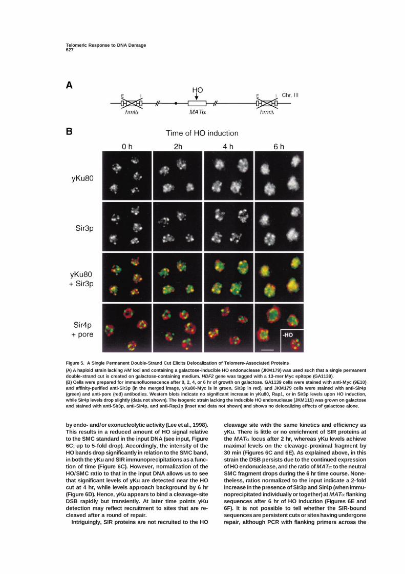
We next checked whether yKu or the SIR proteins aretablish the focal Rap1p pattern that correlates with TPE.recruited to the site of HO cleavage within the MAT�Alternatively, the HU-induced checkpoint may specifi-locus, using primers that amplify fragments at varyingcally inhibit foci reformation.distances from the HO cleavage site (Figure 6B). By 30min after induction of the endonuclease, we detect astrong enhancement of yKu80 at the HO cut, with theA Single Double-Strand DNA Cleavage Elicits
a Telomeric Response to DNA Damage highest enrichment observed for the fragment closestto the cleavage site (HO2/SMC ratio increases 5-foldA unique double-strand lesion, such as that provoked
by the HO endonuclease, is sufficient to arrest progres- after 30 min; Figures 6C and 6D). At later time points (4and 6 hr), quantitation becomes complicated by the factsion of the yeast cell cycle, as part of the RAD9-depen-
dent response to DNA damage (Sandell and Zakian, that the region around the incision becomes recessed
Telomeric Response to DNA Damage627
Figure 5. A Single Permanent Double-Strand Cut Elicits Delocalization of Telomere-Associated Proteins
(A) A haploid strain lacking HM loci and containing a galactose-inducible HO endonuclease (JKM179) was used such that a single permanentdouble-strand cut is created on galactose-containing medium. HDF2 gene was tagged with a 13-mer Myc epitope (GA1139).(B) Cells were prepared for immunofluorescence after 0, 2, 4, or 6 hr of growth on galactose. GA1139 cells were stained with anti-Myc (9E10)and affinity-purified anti-Sir3p (in the merged image, yKu80-Myc is in green, Sir3p in red), and JKM179 cells were stained with anti-Sir4p(green) and anti-pore (red) antibodies. Western blots indicate no significant increase in yKu80, Rap1, or in Sir3p levels upon HO induction,while Sir4p levels drop slightly (data not shown). The isogenic strain lacking the inducible HO endonuclease (JKM115) was grown on galactoseand stained with anti-Sir3p, anti-Sir4p, and anti-Rap1p (inset and data not shown) and shows no delocalizing effects of galactose alone.
by endo- and/or exonucleolytic activity (Lee et al., 1998). cleavage site with the same kinetics and efficiency asyKu. There is little or no enrichment of SIR proteins atThis results in a reduced amount of HO signal relative
to the SMC standard in the input DNA (see input, Figure the MAT� locus after 2 hr, whereas yKu levels achievemaximal levels on the cleavage-proximal fragment by6C; up to 5-fold drop). Accordingly, the intensity of the
HO bands drop significantly in relation to the SMC band, 30 min (Figures 6C and 6E). As explained above, in thisstrain the DSB persists due to the continued expressionin both the yKu and SIR immunoprecipitations as a func-
tion of time (Figure 6C). However, normalization of the of HO endonuclease, and the ratio of MAT� to the neutralSMC fragment drops during the 6 hr time course. None-HO/SMC ratio to that in the input DNA allows us to see
that significant levels of yKu are detected near the HO theless, ratios normalized to the input indicate a 2-foldincrease in the presence of Sir3p and Sir4p (when immu-cut at 4 hr, while levels approach background by 6 hr
(Figure 6D). Hence, yKu appears to bind a cleavage-site noprecipitated individually or together) at MAT� flankingsequences after 6 hr of HO induction (Figures 6E andDSB rapidly but transiently. At later time points yKu
detection may reflect recruitment to sites that are re- 6F). It is not possible to tell whether the SIR-boundsequences are persistent cuts or sites having undergonecleaved after a round of repair.
Intriguingly, SIR proteins are not recruited to the HO repair, although PCR with flanking primers across the

Cell628
Figure 6. yKu80 and Sir3/4 Proteins Are Re-cruited with Different Kinetics to a DNAStrand Break
(A) HO endonuclease was induced by growthon galactose for 0, 2, 4, or 6 hr in GA1139and JKM179. ChrIP assay (see ExperimentalProcedures) was performed, immunoprecipi-tating GA1139 DNA with 9E10 (anti-Myc) andJKM179 DNA with affinity-purified anti-Sir3pand anti-Sir4p IgG (mixed). After DNA purifi-cation, PCR was performed with primers fora unique sequence near the chromosomalend of VI-R, and for the SMC2 gene (see Ex-perimental Procedures). The ratio of the telo-mere signal to the SMC2 signal obtained inthe IP samples normalized to this ratio in theINPUT DNA samples is shown. The amountof Sir3/4 bound to telomeres decreases tohalf the initial value after 6 hr in both strains.(B) Scheme of the MAT� locus showing thepairs of primers used to analyze the recruit-ment of yKu80 and Sir proteins to the uniqueHO-cleavage site.(C) GA1139 strain was prepared as in (A), ex-cept that an additional 30 min time point ongalactose was prepared and cell extractswere sonicated such that the average DNAfragment size was 2 kb. PCR was performedin parallel on INPUT and yKu80-Myc immuno-precipitated DNA with the mixtures of primersdescribed above. After 4 hr of induction, theHO-proximal signals decrease in the INPUT,due to DSB processing, while the SMC signalremains constant.(D) Quantitation of the data shown in (C), pre-sented as the ratio of HO-proximal signals toSMC2 in the IP normalized to the same ratioin the corresponding INPUT.
(E) JKM179 cells were prepared as described in (A) and immunoprecipitated with a mixture of antibodies against Sir3p and Sir4p. PCR wasidentical to that in (C). Although the HO-proximal signals decrease in the INPUT after 4–6 hr due to processing at the DSB, the HO-proximalsignals decrease less in the anti-SIR IP, indicating SIR enrichment near the HO-cut site. Two exposures of the 6 hr time point are shown todemonstrate this point.(F) Quantitation of the data shown in (E), presented as the ratio of HO-proximal signals to SMC2 in the IP normalized to the same ratio in thecorresponding INPUT. Quantitation of a similar experiment using an isogenic strain lacking GAL-HO indicates that galactose alone does notaffect SIR localization (first lane).
HO cleavage site indicates that only 6% are intact at 4 subtelomeric repression as compared to internal silenc-ing, under conditions of induced DNA damage. Our re-or 6 hr after HO induction. A strain lacking GAL-HO
shows no increase of SIR binding at MAT� after 6 hr on porter strain carries both a telomere-proximal URA3 re-porter and a silencer-flanked ADE2 gene inserted at thegalactose (Figure 6F).
Since roughly half the SIR complexes are released LYS2 locus, situated 242 kb away from the telomere ofChr. 2 (Figure 7A). Under normal conditions the silencer-from telomeres after induction of DNA cleavage, the
increase in the recovery of HO-bound Sir proteins might flanked internal reporter is expressed, because telo-meres sequester SIR proteins at perinuclear foci (Mailletsimply reflect a higher background of random SIR pro-
tein binding, or possibly SIR binding nucleated by an et al., 1996). In the presence and absence of BLM, wescore for growth on 5-FOA, which directly monitors theinternal Rap1p site. However, the levels of Sir3p or Sir4p
associated with the SMC fragment and with a fragment efficiency of telomeric repression (Figure 7B). At 20 or30 mU/ml BLM, we see a decrease in growth on 5-FOA,from the HIS4 promoter that contains a Rap1-binding
site are constant even after 6 hr of HO endonuclease indicating a decrease in TPE (Figure 7B). When the dropin viability (see left panel, Figure 7B) is accounted for,expression (data not shown). Thus, the increase in SIR
detection at MAT� appears to be significant. Strikingly, TPE is reduced from 42% to 7%, or 6-fold. Derepressioncould also be detected using a telomere-proximal ADE2at early time points where yKu is most highly enriched
at the HO cut, we see no significant increase in the reporter (data not shown), ruling out URA3- or FOA-specific effects. Both assays are consistent with thebinding of SIRs.partial delocalization of SIR complexes observed byChrIP during HO induction.DNA Damage Compromises TPE, Displacing
Repression-Competent SIR Complexes To determine whether the displaced SIR proteins arestill competent for repression, we monitored repressionAs a functional assay for the delocalization of telomeric
chromatin components, we monitored the efficiency of of the internal silencer-flanked ADE2 reporter in the

Telomeric Response to DNA Damage629
Discussion
Double-strand breaks are highly deleterious to the integ-rity of the genome, and improper processing or repairof DSB can lead to gene rearrangements, deletions, oraneuploidy. Indeed, the eukaryotic cell has developedelaborate means to eliminate DSB without loss of infor-mation, through homologous recombination and/or di-rect end-joining reactions. Both pathways require anarrest in cell cycle progression until the chromosome isrepaired, which is imposed by the DNA damage check-point (Weinert, 1998).
Telomeric Domains Respond to the DNADamage Checkpoint SignalIn this paper, we document a surprising redistributionof yeast telomeric and subtelomeric chromatin compo-nents in response to DNA damage. Three independentassays, namely immunofluorescence, chromatin immu-noprecipitation, and transcriptional repression, confirmthat a significant fraction of the histone-binding SIRcomplex is displaced from subtelomeric sequences,where it is normally sequestered in perinuclear foci. Thedelocalization of SIR proteins occurs in response toMMS, bleomycin, and following the induction of a singledouble-strand cut mediated by the HO endonuclease.Irradiation by UV (Mills et al., 1999 [this issue of Cell])and treatment with hydroxyurea do not have equivalentFigure 7. DNA Damage Impairs TPE While Improving Internal Si-
lencer-Mediated Repression effects, suggesting that the response is specific for DNA(A) To monitor TPE and internal silencing, the haploid LM11 strain strand breaks. Importantly, the relocalization of SIR pro-containing a telomere-proximal URA3 gene ( TELVIIL::URA3), and an teins is abrogated by deletion of RAD9, implicating theHML E and I silencer-flanked ADE2 gene inserted at the LYS2 locus DNA damage checkpoint in the response.(lys2::E-ADE2-I) were used.
In addition to the SIR complex, subpopulations of the(B) Serial dilutions of LM11 were plated on complete medium withDNA-binding Rap1p and yKu proteins are released uponor without 5-FOA with increasing concentrations of BLM, as indi-induction of a single DSB. The delocalization of Rap1pcated. The inhibition of growth on BLM is taken into consideration
in the calculation of the percentage of colonies that repress URA3 could have been expected, since immunofluorescence(FOA resistance, indicated at right). The mean and standard devia- and chromatin cross-linking assays identified a subpop-tion are calculated from six independent experiments. ulation of Rap1p that binds telomeres in a SIR-depen-(C) Colonies growing on 20 mU/ml BLM accumulate red pigment, dent manner (Palladino et al., 1993; Gotta et al., 1996;as they repress the silencer-flanked internal ADE2 reporter gene
Strahl-Bolsinger et al., 1997). We show here that yKu(�BLM; top left). No red colonies were scored in LM11 with a sir4propagates inward along Chr. VI-R to roughly the samedisruption (�BLM, sir4�; top right; GA1056), when ADE2 lacks adja-extent as SIR and Rap1p, suggesting that the releasecent silencers (�BLM; bottom right; GA187), or in absence of BLM
(bottom left). of yKu may reflect its loss from subtelomeric regionsand not from the more distal TG repeats (Figure 1). Theseobservations provide support for the model that subtelo-
presence of bleomycin. Indeed, we observe an accumu- meric heterochromatin serves as a reservoir for proteinslation of red pigment in these colonies, which is not that are released to act at other nuclear sites in responsepresent in the absence of damage (Figure 7C, upper left to physiological signals. In the case of DSB, one mightcorner). This repression requires the intact SIR complex, argue that a checkpoint-induced release of yKu ensuresthe presence of the flanking silencer elements, and cor- sufficient protein for a rapid scanning of the genome torelates with the bleomycin-induced release of SIR pro- minimize inappropriate recombination or fusion events.teins from telomeric foci (Figure 7C). Thus, we conclude The subtelomeric reservoir model is based on thethat a significant fraction of the displaced SIR proteins finding that internal, silencer-flanked reporters (includ-are competent to form repressed chromatin and can be ing reporters at the HM loci) compete with TPE for therecruited by silencers to internal chromosomal sites. limiting pool of Sir3p and Sir4p (Maillet et al., 1996;
In summary, our data suggest that DNA damage elicits Marcand et al., 1996). rDNA repeats also compete witha change in telomeric heterochromatin, although internal telomeres for limiting amounts of Sir2p (Cockell et al.,silencer-mediated repression remains intact. A check- 1998). However, until this study, the only evidence indi-point-induced change in SIR-yKu interaction would pro- cating that SIR relocalization might be physiologicallyvide a mechanism to release yKu and SIR proteins from relevant was the observation that Sir3p and Sir4p mi-subtelomeric sequences. The modified yKu would then grate to the nucleolus in aging yeast cells and in mutantsbe free to scan the damaged genome for DSB and may that extend their average maximal number of divisionsunder these conditions preferentially interact with Mre11 (Kennedy et al., 1997). In the case of aging, the accumu-
lation of excised rDNA circles may recruit Sir3p andand the Ligase IV complex.
Cell630
Sir4p from telomeres, provoking a concomitant loss of 1996). Mutation of the yeast equivalent of the mamma-lian CAF1 (CAC1) intriguingly shows a derepression ofTPE. From the data shown here, we propose that signal-
ing through the DNA damage checkpoint elicits the re- TPE and hypersensitivity to DNA damage (Enomoto etal., 1997; Kaufman et al., 1997). Since DSB repair in-lease of yKu and SIR proteins from complexes associ-
ated with subtelomeric heterochromatin. volves extensive resynthesis of DNA and hence nucleo-some assembly, we propose that the recruitment of SIRcomplexes reflects the assembly of a repressed chro-
Potential Roles for SIR Protein Release matin state following DNA repair. It is not known whetherin DSB Repair CAF1 or another complex assembles nucleosomes afterWhy release a subfraction of SIR complexes in response NHEJ reactions, yet this pathway would be consistentto DNA damage? It is possible that SIR protein delocal- with the kinetics of SIR association at cleavage sitesization is a meaningless side effect of the release of and the well-characterized interaction of SIR proteinsyKu, which is known to play a direct role in DSB repair. with nucleosomes. Such a mechanism might preventTwo results argue against this. First, Mills et al. (1999) the spurious activation of genes as a result of DNAand Tsukamoto et al. (1997) have demonstrated that repair. It will be interesting to test whether SIR proteinssir-deficient strains are significantly less efficient in an are still targeted to the HO cut in a cac1 deletion strain.NHEJ assay than isogenic SIR� strains. Second, weshow that sir-deficient haploid and diploid strains are Adapting to a DSBreproducibly hypersensitive to MMS. BLM sensitivity is In view of the physical similarity of DSB and telomericless pronounced for sir mutants than for strains lacking termini, such damage presents a unique challenge toyKu, and yet the impairment to growth is additive in the the cell’s repair pathway. It has been demonstrated thatdouble mutant, suggesting that HDF1 and SIR4 are both cells with irreparable damage downregulate Rad9p afterinvolved, but are not epistatic, in some aspect of DSB 6–8 hr and proceed through the cell cycle in a processrepair. called adaptation (Sandell and Zakian, 1993; Toczyski
et al., 1997; Lee et al., 1998). Since the timing of the SIRprotein recruitment to an irreparable HO cleavage siteSIR Proteins Are Recruited to an HO Cleavagecorrelates with this event, it is possible that the bindingSite with Different Kinetics Than yKuof SIRs may help protect the unrepaired end from furtherEarlier studies have proposed that yKu targets the SIRdegradation, as the cells proceed through mitosis (San-complex to sites of damage to protect flanking DNAdell and Zakian, 1993). Thus, the unexpected check-from the transcriptional apparatus (Jackson, 1997).point-elicited release of telomere-associated chromatinHowever, in view of the kinetics of protein release fromfactors may reflect their involvement in adaptation totelomeres and of yKu and SIR protein association at thepersistent DNA damage, a phenomenon implicated inHO cut, we suggest a somewhat different scenario. Themammalian oncogenesis.maximal level of yKu recruitment (30 min after HO induc-
tion) persists until 4 hr, yet by 6 hr yKu is no longerExperimental Proceduressignificantly enriched at MAT�. In the case of SIR pro-
teins, association near the cleavage site at 2 hr is negli-Media
gible (�1.2-fold), but increases continually, reaching Yeast strains were grown using standard conditions (Rose et al.,�2-fold at 6 hr after HO induction. These dissimilar kinet- 1990). Bleomycin (Sigma Chemicals, Buchs, Switzerland) was di-
luted to 10 U/ml in H2O. For plates containing BLM, 5-FOA, or MMS,ics do not support the idea that yKu directly targets SIRdrugs were added to cooled media. For immunofluorescence stud-complexes to sites of DSB. Consistently, we have seenies, 5 mU/ml BLM, 0.03% MMS, or 200 mM hydroxyurea was addedthat although GBD-yKu is extremely efficient at nucleat-for 3 hr prior to fixation unless otherwise stated. For HO endonucle-ing SIR-mediated silencing at internal sites under normalase induction, strains were grown on rich medium containing 3%
growth conditions (Figure 2), yKu-mediated repression glycerol, 2% lactic acid, and 0.05% glucose, prior to adding 2%is compromised on MMS (data not shown). This is not galactose.due to inactivation of the SIR complex, since GBD-Sir2p
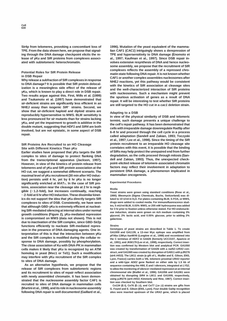
Strainsfunctions efficiently to nucleate SIR-mediated repres-Genotypes of yeast strains are described in Table 1. To createsion in the presence of DNA-damaging agents. One in-GA1009 and GA1139, a 13-mer Myc epitope was amplified fromterpretation of this is that the interaction between yKupFA6a-13Myc-kanMX6 (Longtine et al., 1998) and recombined into
and the SIR complex is modified during the cellular re- the C terminus of HDF2 in GA426 (formerly UCC3107, Aparicio etsponse to DNA damage, possibly by phosphorylation. al., 1991), and JKM179 (Lee et al., 1998), respectively. Correct inser-
tion was confirmed by Western blot and analytical PCR. GA1050The close association of Ku with DNA-PK in mammalianwas created by transformation of GA426 with a rad52::URA3 con-cells makes it likely that yKu is recognized by an ATMstruct, and GA1065 was created by disruption of GA911 with pJR276homolog in yeast (Mec1 or Tel1). Such a modification(sir4::HIS3). The LM11 strain (a gift of L. Maillet and E. Gilson, ENS,may interfere with yKu recruitment of the SIR complexLyon, France) carries both a VIIL telomere-proximal URA3 reporter
to sites of DNA damage. and a wild-type ADE2 gene flanked on either side by 1.5 kb ofAs an alternative hypothesis, we propose that the sequence containing the HML E and I silencers, integrated at LYS2,
to allow the monitoring of silencer-mediated repression at an internalrelease of SIR complexes from subtelomeric regionschromosomal site (Maillet et al., 1996). GA1056 and GA1061 wereand its recruitment to sites of repair reflect associationobtained by disrupting SIR4 in LM11 and GA1009, respectively,with newly assembled chromatin. It has been demon-using pJR276 (sir4::HIS3; Kimmerly and Rine, 1987). Correct inser-strated that the nucleosome assembly factor CAF1 istion was checked by Southern blot.
recruited to sites of DNA damage in mammalian cells Ce134 (E-I), Ce76 (E-�i), and Ce77 (�e-�i) strains are gifts from(Martini et al., 1998), and its role in nucleosome assembly G. Fourel and E. Gilson (ENS, Lyon). Four double Gal4p recognition
sites were inserted upstream of the URA3 gene, which is placedfollowing DNA repair is well documented (Gaillard et al.,
Telomeric Response to DNA Damage631
Table 1. Yeast Strains Used in This Study
Name Genotype Reference
GA192 MATa/MAT� ade2-1/ADE2 his3/his3 leu2-3/LEU2 lys2-6/LYS2 trp1/TRP1 ura3-1/ura3-52 Gotta et al., 1996can1/CAN1 sir3::TRP1/sir3::LYS2
GA202 MATa/MAT� ade2-1/ADE2 his3/his3 leu2-3/LEU2 lys2-6/LYS2 trp1/trp1 ura3-1/ura3-52 Gotta et al., 1996can1/CAN1 sir4::HIS3/sir4::HIS3
GA225 MATa/MAT� ade2-1/ADE2 his3/his3 leu2-3/LEU2 trp1/TRP1 ura3-1/ura3-52 can1/CAN1 Gotta et al., 1996GA426 MATa ade2::hisG his3-11 leu2 trp1 ura3-52 can1::hisG VR::ADE2-TEL H. Renauld, unpublished dataGA427 GA426 sir2::HIS3 H. Renauld, unpublished dataGA428 GA426 sir3::HIS3 H. Renauld, unpublished dataGA429 GA426 sir4::HIS3 H. Renauld, unpublished dataGA911 GA426 hdf1::kanMX Laroche et al., 1998GA1065 GA426 hdf1::kanMX sir4::HIS3 This studyGA1050 GA426 rad52::URA3 This studyGA1009 GA426 HDF2-myc(::kanMX4) This studyGA1061 GA426 HDF2-myc(::kanMX4) sir4::HIS3 This studyW303a MATa ade2-1 leu2-3,112 his3-11,15 trp1 ura3-1 can1-100YT29 W303a hdf1::kanMX4 Laroche et al., 1998S114 W303a rad9::LEU2 D. Shore, unpublished dataAHY111 MATa/MAT� arg4/arg4 leu2::hisG/leu2::hisG ura3/ura3 ho::LYS2/ho::LYS2 Hayashi et al., 1998
RAP1-GFP-LEU2::rap1/RAP1-GFP-LEU2::rap1JKM115 MAT� ade1 leu2-3,112 lys5 trp1::hisG ura3-52 hml::ADE1 hmr::ADE1 Lee et al., 1998JKM179 JKM115 ade3::GALHO Lee et al., 1998GA1139 JKM115 ade3::GALHO HDF2-myc(::kanMX4) This studyLM11 MAT� ade2-1 his3-11,115 leu2-3,112 trp1-1 ura3-1 can1-100 TelVII::URA3 L. Maillet and E. Gilson,
lys2::HMLEI::ADE2 unpublished dataGA1056 LM11 sir4::HIS3 This studyCe134 MATa his3� leu2-3,112 trp1 ura3-52 gal2 hml::E-GalUAS2-URA3-I G. Fourel and E. Gilson,
unpublished dataCe76 MATa his3� leu2-3,112 trp1 ura3-52 gal2 hml::E-GalUAS2-URA3-�i G. Fourel and E. Gilson,
unpublished dataCe77 MATa his3� leu2-3,112 trp1 ura3-52 gal2 hml::�e-GalUAS2-URA3-�i G. Fourel and E. Gilson,
unpublished data
between the HML E and I silencers, and deletions in Ce76 and Ce77 are specific for the 3 kb region downstream of the HO cleavage sitein MAT� (SG420: 5�-AAGTGCTCCCAATAGGCG GTTTCTC-3� andremove the full silencers (Boscheron et al., 1996).SG421: 5�-TTGATGCCGAGGACG AAGTAGAGGA-3�), producing a560 bp fragment (HO3), and for regions 0.6 kb and 2 kb upstreamPlasmids
The pGBT9-ORC1 plasmid encodes the first 238 aa of Orc1p fused from the HO cleavage site (SG422 5�-AGTATGCTGGATTTAAACTCATCTGTGATTTGTGG-3� and SG122 5�-GCAGGCTTCGAAGTAAAin-frame to the Gal4p-binding domain (GBD, gift of C. Boscheron
and E. Gilson, ENS, Lyon). The pGBT9-SIR2 plasmid contains the CATATTGTGAATGTCG-3�, producing a 350 bp fragment, HO2;SG418 5�-CTTGTATTAGACGAGGGACGGAGTG-3� and SG419entire SIR2 gene fused in-frame to GBD (gift of M. Cockell, ISREC).
pGBD-yKu fusions were created by cloning HDF1 and HDF2 genes 5�-ACAGAGGGTCACAGCACTACTACAG-3�, producing a 290 bpfragment, HO1). The Chr VI-R telomere primers (SG355: 5�-CACCGCamplified by PCR into pAS2 (2 �m-TRP1; Clontech). pGBD-HDF1
fully complements an hdf1 deletion for TPE and viability at 37C, CAAGCTTCCAATATCACG-3� and SG356: 5�-GGAGGCATTATGGCTTTGTTACGC-3�) produce a 400 bp fragment located 0.6 kb fromwhereas pGBD-HDF2 complements a hdf2 null for temperature sen-
sitivity. the TG repeat, and primers from SMC2 (SG139: 5�-AAGAGAAACTTTAGTCAAAACATGGG-3� and SG135: 5�-CCATCACATTATACTAACTACGG-3�) produce the 450 bp internal standard. PCR products wereImmunofluorescence and In Situ Hybridization
Published methods and antibodies were used for immunofluores- resolved by electrophoresis and assayed by densitometry usingAIDA (Fuji) or Phosphorimager, as indicated.cence, Y��TG1–3 repeat in situ hybridization, and confocal imaging
(Gotta et al., 1996). All rabbit sera were affinity purified prior touse. Other antibodies were as follows: anti-Myc (9E10), anti-p62 Silencing and Viability Assays
The expression of the telomere-proximal URA3 or the GAL-URA3monoclonal antibody (Mab414; Berkeley Antibody Corp.), DTAF- orTexas red–conjugated secondary antibody (Jackson ImmunoRe- construct at HML was monitored by determining the fraction of cells
capable of growth after 3–5 days at 30C on 0.1% 5-FOA-containingsearch Laboratories), Cy5-coupled reagents (Milan Analytica), andfluorescein-derivatized sheep anti-digoxigenin (DIG) F(ab) frag- medium, which allows growth of cells that fail to express URA3
(Gottschling et al., 1990). Mean and standard deviation were calcu-ments (Roche Diagnostics). Secondary antibodies are preadsorbedagainst fixed yeast spheroplasts prior to use. lated from multiple titrations of at least four independent colonies.
For the ADE2 reporter, cells were grown on medium with limitingadenine.ChrIP
The extraction and sonication of formaldehyde-fixed yeast cellswere done essentially as described in Strahl-Bolsinger et al. (1997). AcknowledgmentsFor telomere analyses (Figure 1), the average size of sonicated DNAwas �500 bp, and PCR primers were identical to those described We acknowledge invaluable help from M. Tsai-Pflugfelder (ISREC)
for the ChrIP assay. We are grateful to E. Gilson, L. Maillet, G. Fourel,(Strahl-Bolsinger et al., 1997). For analyses of the HO cleavage site,the average size fragment was �2 kb due to less extensive sonica- C. Boscheron (all ENS, Lyon, France), and H. Renauld and M. Cockell
(ISREC) for unpublished plasmids and strains. We thank J. Habertion. Immunoprecipitation was performed with precoated Dyna-beads overnight at 4C. An aliquot of each sample was not immuno- and D. Shore for published yeast strains, J. Lingner and members
of the Gasser laboratory for helpful discussions, and K. Mills andprecipitated (Input). Input and immunoprecipitated samples wereincubated for 6 hr at 65C to revert the formaldehyde cross-linking, L. Guarente for sharing information prior to submission. The Gasser
laboratory thanks the Swiss National Science Foundation, Humanprior to DNA purification and semiquantitative PCR. Primers used
Cell632
Frontier Science Program, and the Swiss Cancer League for support, Murthy, M., Pak, S.M., Laroche, T., Gasser, S.M., and Guarente, L.(1997). Redistribution of silencing proteins from telomeres to theand the Grunstein laboratory acknowledges the Human Frontier
Science Program and a grant from National Institutes of Health. nucleolus is associated with extension in life span in S. cerevisiae.Cell 89, 381–391.
Kimmerly, W.J., and Rine, J. (1987). Replication and segregation ofReceived March 8, 1999; revised April 23, 1999.plasmids containing cis-acting regulatory sites of silent mating-typegenes in Saccharomyces cerevisiae are controlled by the SIR genes.ReferencesMol. Cell. Biol. 7, 4225–4237.
Laroche, T., Martin, S.G., Gotta, M., Gorham, H.C., Pryde, F.E., Louis,Andrulis, A.D., Neiman, A.M., Zappulla, D.C., and Sternglanz, R.(1998). Perinuclear localization of chromatin facilitates transcrip- E.J., and Gasser, S.M. (1998). Mutation of yeast Ku genes disrupts
the subnuclear organization of telomeres. Curr. Biol. 8, 653–656.tional silencing. Nature 394, 592–595.
Aparicio, O.M., Billington, B.L., and Gottschling, D.E. (1991). Modifi- Lee, S.E., Moore, J.K., Holmes, A., Umezu, K., Kolodner, R.D., andHaber, J.E. (1998). Saccharomyces Ku70, mre11/rad50 and RPAers of position effect are shared between telomeric and silent mat-
ing-type loci in S. cerevisiae. Cell 66, 1279–1287. proteins regulate adaptation to G2/M arrest after DNA damage. Cell94, 399–409.Astrom, S.U., Okamura, S.M., and Rine, J. (1999). Yeast cell-type
regulation of DNA repair. Nature 397, 310. Lieber, M.R., Grawunder, U., Wu, X., and Yaneva, M. (1997). Tyingloose ends: roles of Ku and DNA-dependent protein kinase in theBoscheron, C., Maillet, L., Marcand, S., Tsai-Pflugfelder, M., Gasser,repair of double-strand breaks. Curr. Opin. Genet. Dev. 7, 99–104.S.M., and Gilson, E. (1996). Cooperation at a distance between
silencers and proto-silencers at the yeast HML locus. EMBO J. 15, Longtine, M.S., McKenzie, A., III, Demarini, D.J., Shah, N.G., Wach,A., Brachat, A., Philippsen, P., and Pringle, J.R. (1998). Additional2184–2195.modules for versatile and economical PCR-based gene deletion andBoulton, S.J., and Jackson, S.P. (1998). Components of the Ku-modification in Saccharomyces cerevisiae. Yeast 14, 953–961.dependent non-homologous end-joining pathway are involved inMages, G.J., Feldmann, H.M., and Winnacker, E.L. (1996). Involve-telomeric length maintenance and telomeric silencing. EMBO J. 17,ment of the Saccharomyces cerevisiae HDF1 gene in DNA double-1819–1828.strand break repair and recombination. J. Biol. Chem. 271, 7910–Cockell, M., Gotta, M., Palladino, F., Martin, S.G., and Gasser, S.M.7915.(1998). Targeting Sir proteins to nuclear domains: a general mecha-Maillet, L., Boscheron, C., Gotta, M., Marcand, S., Gilson, E., andnism for transcriptional repression. Cold Spring Harb. Symp. Quant.Gasser, S.M. (1996). Evidence for silencing compartments withinBiol. 63, 401–412.the yeast nucleus: a role for telomere proximity and Sir proteinCritchlow, S.E., and Jackson, S.P. (1998). DNA end-joining: fromconcentration in silencer-mediated repression. Genes Dev. 10,yeast to man. Trends Biochem. Sci. 23, 394–398.1796–1811.
Dynan, W.S., and Yoo, S. (1998). Interaction of Ku protein and DNA-Marcand, S., Buck, S.W., Moretti, P., Gilson, E., and Shore, D. (1996).dependent protein kinase catalytic subunit with nucleic acids. Nu-Silencing of genes at nontelomeric sites in yeast is controlled bycleic Acids Res. 26, 1551–1559.sequestration of silencing factors at telomeres by Rap 1 protein.
Enomoto, S., McCune-Zierath, P.D., Gerami-Nejad, M., Sanders, Genes Dev. 10, 1297–1309.M.A., and Berman, J. (1997). RLF2, a subunit of yeast chromatin
Martini, E., Roche, D.M., Marheineke, K., Verreault, A., and Almouzni,assembly factor-I, is required for telomeric chromatin function inG. (1998). Recruitment of phosphorylated chromatin assembly factorvivo. Genes Dev. 11, 358–370.1 to chromatin after UV irradiation of human cells. J. Cell Biol. 143,
Gaillard, P.H., Martini, E.M., Kaufman, P.D., Stillman, B., Moustacchi, 563–575.E., and Almouzni, G. (1996). Chromatin assembly coupled to DNA
Miller, A.M., and Nasmyth, K.A. (1984). Role of DNA replication in therepair: a new role for chromatin assembly factor I. Cell 86, 887–896.repression of silent mating type loci in yeast. Nature 312, 247–251.
Gotta, M., Laroche, T., Formenton, A., Maillet, L., Scherthan, H., andMills, K.D., Sinclair, D.A., and Guarente, L. (1999). MEC1-dependent
Gasser, S.M. (1996). The clustering of telomeres and colocalizationredistribution of the Sir3 silencing protein from telomeres to DNA
with Rap1, Sir3 and Sir4 proteins in wild-type Saccharomyces cere-double-strand breaks. Cell 97, this issue, 609–620.
visiae. J. Cell Biol. 134, 1349–1363.Nugent, C.I., Bosco, G., Ross, L.O., Evans, S.K., Salinger, A.P.,
Gottschling, D.E., Aparicio, O.M., Billington, B.L., and Zakian, V.A.Moore, J.K., Haber, J.E., and Lundblad, V. (1998). Telomere mainte-
(1990). Position effect at S. cerevisiae telomeres: reversible repres-nance is dependent on activities required for end repair of double-
sion of Pol II transcription. Cell 63, 751–762.strand breaks. Curr. Biol. 8, 657–660.
Gravel, S., Larrivee, M., Labrecque, P., and Wellinger, R.J. (1998). Nussenzweig, A., Chen, C., da Costa Soares, V., Sanchez, M., Sokol,Yeast Ku as a regulator of chromosomal DNA end structure. Science K., Nussenzweig, M.C., and Li, G.C. (1996). Requirement for Ku80280, 741–744. in growth and immunoglobulin V(D)J recombination. Nature 382,Gu, Y., Seidl, K.J., Rathbun, G.A., Zhu, C., Manis, J.P., van der Stoep, 551–555.N., Davidson, L., Cheng, H.L., Sekiguchi, J.M., Frank, K., et al. (1997). Palladino, F., Laroche, T., Gilson, E., Axelrod, A., Pillus, L., andGrowth retardation and leaky SCID phenotype of Ku70-deficient Gasser, S.M. (1993). SIR3 and SIR4 proteins are required for themice. Immunity 7, 653–665. positioning and integrity of yeast telomeres. Cell 75, 543–555.Haber, J.E. (1998). The many interfaces of Mre11. Cell 95, 583–586. Polotnianka, R.M., Li, J., and Lustig, A.J. (1998). The yeast Ku hetero-Hayashi, A., Ogawa, H., Kohno, K., Gasser, S.M., and Hiraoka, Y. dimer is essential for protection of the telomere against nucleolytic(1998). Meiotic behaviours of chromosomes and microtubules in and recombinational activities. Curr. Biol. 8, 831–834.budding yeast: relocalization of centromeres and telomeres during Porter, S.E., Greenwell, P.W., Ritchie, K.B., and Petes, T.D. (1996).meiotic prophase. Genes Cells 3, 587–601. The DNA-binding protein Hdf1p (a putative Ku homologue) is re-Jackson, S.P. (1997). Genomic stability: silencing and DNA repair quired for maintaining normal telomere length in Saccharomycesconnect. Nature 388, 829–830. cerevisiae. Nucleic Acids Res. 24, 582–585.Jeggo, P.A., Taccioli, G.E., and Jackson, S.P. (1995). Menage a Povirk, L.F. (1996). DNA damage and mutagenesis by radiomimetictrois: double strand break repair, V(D)J recombination and DNA-PK. DNA-cleaving agents: bleomycin, neocarzinostatin and other enedi-Bioessays 17, 949–957. ynes. Mutat. Res. 355, 71–89.
Kaufman, P.D., Kobayashi, R., and Stillman, B. (1997). Ultraviolet Rose, M.D., Winston, F., and Hieter, P. (1990). Methods in Yeastradiation sensitivity and reduction of telomeric silencing in Saccha- Genetics (Cold Spring Harbor, NY: Cold Spring Harbor Laboratoryromyces cerevisiae cells lacking chromatin assembly factor-I. Press).Genes Dev. 11, 345–357. Sandell, L.L., and Zakian, V.A. (1993). Loss of a yeast telomere:
arrest, recovery, and chromosome loss. Cell 75, 729–739.Kennedy, B.K., Gotta, M., Sinclair, D.A., Mills, K., McNabb, D.S.,
Telomeric Response to DNA Damage633
Schwartz, J.L. (1989). Monofunctional alkylating agent-inducedS-phase-dependent DNA damage. Mutat. Res. 216, 111–118.
Shore, D. (1998). Telomeres—unsticky ends. Science 281, 1818–1819.
Siede, W., Friedl, A.A., Dianova, I., Eckardt-Schupp, F., andFriedberg, E.C. (1996). The Saccharomyces cerevisiae Ku autoanti-gen homologue affects radiosensitivity only in the absence of homol-ogous recombination. Genetics 142, 91–102.
Strahl-Bolsinger, S., Hecht, A., Kunheng, L., and Grunstein, M.(1997). SIR2 and SIR4 interactions differ in core and extended telo-meric heterochromatin in yeast. Genes Dev. 11, 83–93.
Toczyski, D.P., Galgoczy, D.J., and Hartwell, L.H. (1997). CDC5 andCKII control adaptation to the yeast DNA damage checkpoint. Cell90, 1097–1106.
Tsukamoto, Y., Kato, J., and Ikeda, H. (1997). Silencing factors par-ticipate in DNA repair and recombination in S. cerevisiae. Nature388, 900–903.
Weinert, T. (1998). DNA damage checkpoints update: getting molec-ular. Curr. Opin. Genet. Dev. 8, 185–193.


![Antimicrobial and antioxidant activities of Saccharomyces cerevisiae IFST062013… · 2017. 8. 26. · teins, stimulation of immunoglobulin A [12], acquisition and elimination of](https://img.pdfslide.us/doc/110x75/613de7b72809574f586e42e0/antimicrobial-and-antioxidant-activities-of-saccharomyces-cerevisiae-ifst062013.jpg)