Embed Size (px)
Citation preview
Cell, Vol. 100, 57–70, January 7, 2000, Copyright ©2000 by Cell Press
The Hallmarks of Cancer Review
evolve progressively from normalcy via a series of pre-Douglas Hanahan* and Robert A. Weinberg†
*Department of Biochemistry and Biophysics and malignant states into invasive cancers (Foulds, 1954).These observations have been rendered more con-Hormone Research Institute
University of California at San Francisco crete by a large body of work indicating that the ge-nomes of tumor cells are invariably altered at multipleSan Francisco, California 94143
†Whitehead Institute for Biomedical Research and sites, having suffered disruption through lesions as sub-tle as point mutations and as obvious as changes inDepartment of Biology
Massachusetts Institute of Technology chromosome complement (e.g., Kinzler and Vogelstein,1996). Transformation of cultured cells is itself aCambridge, Massachusetts 02142multistep process: rodent cells require at least two intro-duced genetic changes before they acquire tumorigeniccompetence, while their human counterparts are moreAfter a quarter century of rapid advances, cancer re-difficult to transform (Hahn et al., 1999). Transgenicsearch has generated a rich and complex body of knowl-models of tumorigenesis have repeatedly supported theedge, revealing cancer to be a disease involving dy-conclusion that tumorigenesis in mice involves multiplenamic changes in the genome. The foundation has beenrate-limiting steps (Bergers et al., 1998; see Oncogene,set in the discovery of mutations that produce onco-1999, R. DePinho and T. E. Jacks, volume 18[38], pp.genes with dominant gain of function and tumor sup-5248–5362). Taken together, observations of humanpressor genes with recessive loss of function; bothcancers and animal models argue that tumor develop-classes of cancer genes have been identified throughment proceeds via a process formally analogous to Dar-their alteration in human and animal cancer cells andwinian evolution, in which a succession of geneticby their elicitation of cancer phenotypes in experimentalchanges, each conferring one or another type of growthmodels (Bishop and Weinberg, 1996).advantage, leads to the progressive conversion of nor-Some would argue that the search for the origin andmal human cells into cancer cells (Foulds, 1954; Nowell,treatment of this disease will continue over the next1976).quarter century in much the same manner as it has in
the recent past, by adding further layers of complexityto a scientific literature that is already complex almost An Enumeration of the Traitsbeyond measure. But we anticipate otherwise: those The barriers to development of cancer are embodiedresearching the cancer problem will be practicing a dra- in a teleology: cancer cells have defects in regulatorymatically different type of science than we have experi- circuits that govern normal cell proliferation and homeo-enced over the past 25 years. Surelymuch of this change stasis. There are more than 100 distinct types of cancer,will be apparent at the technical level. But ultimately, and subtypes of tumors can be found within specificthe more fundamental change will be conceptual. organs. This complexity provokes a number of ques-We foresee cancer research developing into a logical tions. How many distinct regulatory circuits within each
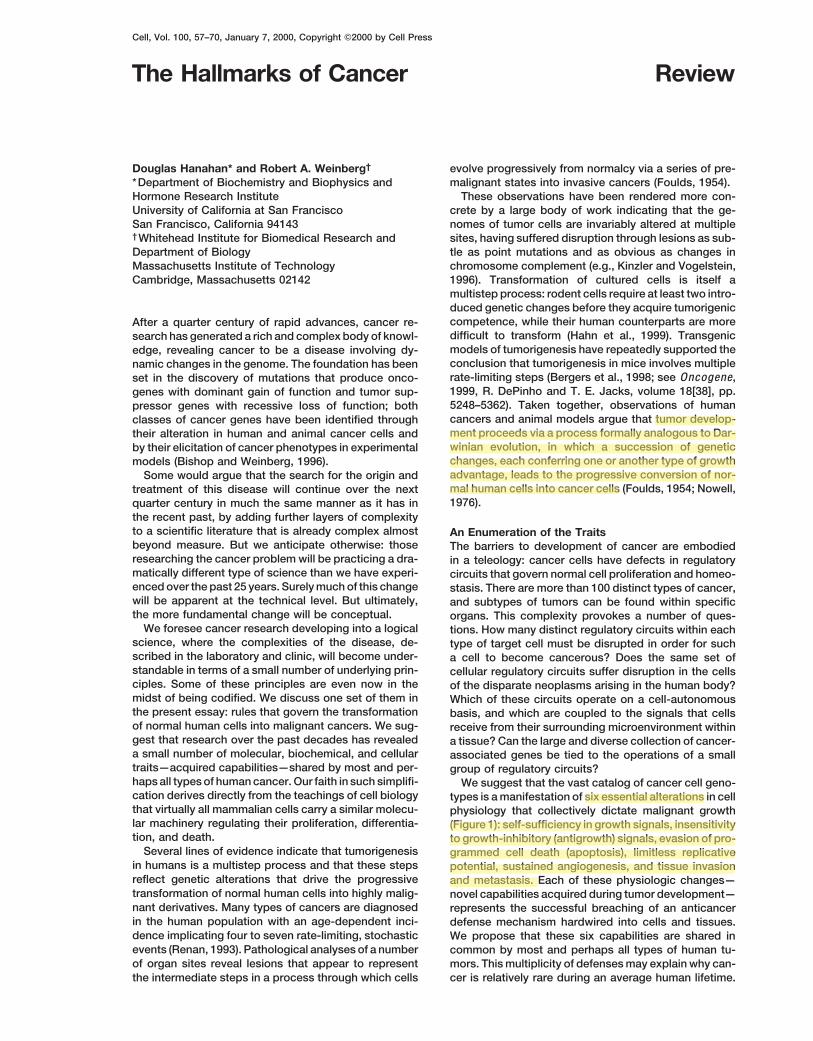
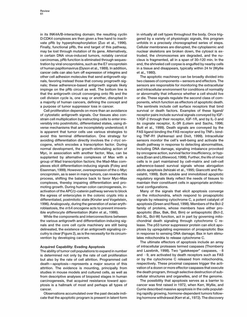
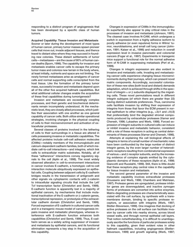
science, where the complexities of the disease, de- type of target cell must be disrupted in order for suchscribed in the laboratory and clinic, will become under- a cell to become cancerous? Does the same set ofstandable in terms of a small number of underlying prin- cellular regulatory circuits suffer disruption in the cellsciples. Some of these principles are even now in the of the disparate neoplasms arising in the human body?midst of being codified. We discuss one set of them in Which of these circuits operate on a cell-autonomousthe present essay: rules that govern the transformation basis, and which are coupled to the signals that cellsof normal human cells into malignant cancers. We sug- receive from their surrounding microenvironment withingest that research over the past decades has revealed a tissue? Can the large and diverse collection of cancer-a small number of molecular, biochemical, and cellular associated genes be tied to the operations of a smalltraits—acquired capabilities—shared by most and per- group of regulatory circuits?hapsall typesof humancancer. Our faith in such simplifi- We suggest that the vast catalog of cancer cell geno-cation derives directly from the teachings of cell biology types is amanifestation of six essential alterations in cellthat virtually all mammalian cells carry a similar molecu- physiology that collectively dictate malignant growthlar machinery regulating their proliferation, differentia- (Figure 1): self-sufficiency in growth signals, insensitivitytion, and death. to growth-inhibitory (antigrowth) signals, evasion of pro-Several lines of evidence indicate that tumorigenesis grammed cell death (apoptosis), limitless replicative
in humans is a multistep process and that these steps potential, sustained angiogenesis, and tissue invasionreflect genetic alterations that drive the progressive and metastasis. Each of these physiologic changes—transformation of normal human cells into highly malig- novel capabilities acquired during tumor development—nant derivatives. Many types of cancers are diagnosed represents the successful breaching of an anticancerin the human population with an age-dependent inci- defense mechanism hardwired into cells and tissues.dence implicating four to seven rate-limiting, stochastic We propose that these six capabilities are shared inevents (Renan, 1993). Pathological analyses of a number common by most and perhaps all types of human tu-of organ sites reveal lesions that appear to represent mors. Thismultiplicity of defensesmay explain why can-
cer is relatively rare during an average human lifetime.the intermediate steps in a process through which cells
Cell58
AcquiredGSautonomywas the first of the six capabili-ties to be clearly defined by cancer researchers, in largepart because of the prevalence of dominant oncogenesthat have been found to modulate it. Three commonmolecular strategies for achieving autonomy are evi-dent, involving alteration of extracellular growth signals,of transcellular transducers of those signals, or of intra-cellular circuits that translate those signals into action.While most soluble mitogenic growth factors (GFs) aremade by one cell type in order to stimulate proliferationof another—the process of heterotypic signaling—manycancer cells acquire the ability to synthesize GFs towhich they are responsive, creating a positive feedbacksignaling loop often termed autocrine stimulation (Fediet al., 1997). Clearly, themanufacture of a GFby a cancercell obviates dependence on GFs from other cells withinthe tissue. The production of PDGF (platelet-derivedgrowth factor) and TGF� (tumor growth factor �) byglioblastomas and sarcomas, respectively, are two illus-trative examples (Fedi et al., 1997).The cell surface receptors that transduce growth-
stimulatory signals into the cell interior are themselvestargets of deregulation during tumor pathogenesis. GFreceptors, often carrying tyrosine kinase activities intheir cytoplasmic domains, are overexpressed in manycancers. Receptor overexpression may enable the can-cer cell to become hyperresponsive to ambient levelsFigure 1. Acquired Capabilities of Cancerof GF that normally would not trigger proliferation (FediWe suggest that most if not all cancers have acquired the same setet al., 1997). For example, the epidermal GF receptorof functional capabilities during their development, albeit through
various mechanistic strategies. (EGF-R/erb B) is upregulated in stomach, brain, andbreast tumors, while the HER2/neu receptor is overex-pressed in stomach and mammary carcinomas (Slamonet al., 1987; Yarden andUllrich, 1988). Additionally, grossWe describe each capability in turn below, illustrate withoverexpression of GF receptors can elicit ligand-inde-a few examples its functional importance, and indicatependent signaling (DiFiore et al., 1987). Ligand-indepen-strategies by which it is acquired in human cancers.dent signaling can also be achieved through structuralalteration of receptors; for example, truncated versions
Acquired Capability: Self-Sufficiencyof the EGF receptor lacking much of its cytoplasmic
in Growth Signals domain fire constitutively (Fedi et al., 1997).Normal cells require mitogenic growth signals (GS) be- Cancer cells can also switch the types of extracellularfore they can move from a quiescent state into an active matrix receptors (integrins) they express, favoring onesproliferative state. These signals are transmitted into the that transmit progrowth signals (Lukashev and Werb,cell by transmembrane receptors that bind distinctive 1998; Giancotti andRuoslahti, 1999). These bifunctional,classes of signaling molecules: diffusible growth fac- heterodimeric cell surface receptors physically link cellstors, extracellular matrix components, and cell-to-cell to extracellular superstructures knownas the extracellu-adhesion/interaction molecules. To our knowledge, no lar matrix (ECM). Successful binding to specific moietiestype of normal cell can proliferate in the absence of of the ECM enables the integrin receptors to transducesuch stimulatory signals. Many of the oncogenes in the signals into the cytoplasm that influence cell behavior,cancer catalog act by mimicking normal growth signal- ranging from quiescence in normal tissue to motility,ing in one way or another. resistance to apoptosis, and entrance into the activeDependence on growth signaling is apparent when cell cycle. Conversely, the failure of integrins to forge
propagating normal cells in culture, which typically pro- these extracellular links can impair cell motility, induceliferate only when supplied with appropriate diffusible apoptosis, or cause cell cycle arrest (Giancotti and Ru-mitogenic factors and a proper substratum for their inte- oslahti, 1999). Both ligand-activated GF receptors andgrins. Such behavior contrasts strongly with that of tu- progrowth integrins engaged to extracellular matrixmor cells, which invariably show a greatly reduced components can activate the SOS-Ras-Raf-MAP kinasedependenceonexogenousgrowth stimulation. Thecon- pathway (Aplin et al., 1998; Giancotti and Ruoslahti,clusion is that tumor cells generate many of their own 1999).growth signals, thereby reducing their dependence on Themost complexmechanismsof acquiredGSauton-stimulation from their normal tissue microenvironment. omy derive from alterations in components of the down-This liberation from dependence on exogenously de- stream cytoplasmic circuitry that receives and pro-rived signals disrupts a critically important homeostatic cesses the signals emitted by ligand-activated GFmechanism that normally operates to ensure a proper receptors and integrins. The SOS-Ras-Raf-MAPK cas-
cade plays a central role here. In about 25% of humanbehavior of the various cell types within a tissue.
Review59
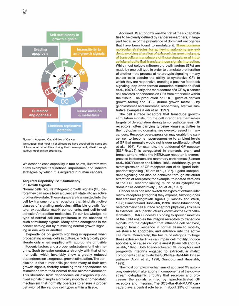
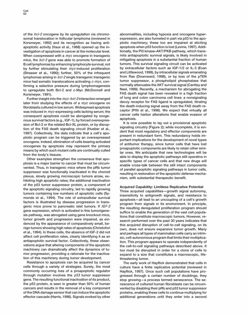
Figure 2. The Emergent Integrated Circuit of the Cell
Progress in dissecting signaling pathways has begun to lay out a circuitry that will likely mimic electronic integrated circuits in complexityand finesse, where transistors are replaced by proteins (e.g., kinases and phosphatases) and the electrons by phosphates and lipids, amongothers. In addition to the prototypical growth signaling circuit centered around Ras and coupled to a spectrum of extracellular cues, othercomponent circuits transmit antigrowth and differentiation signals or mediate commands to live or die by apoptosis. As for the geneticreprogramming of this integrated circuit in cancer cells, some of the genes known to be functionally altered are highlighted in red.
tumors, Ras proteins are present in structurally altered multiple cell biological effects. For example, the directinteraction of the Ras protein with the survival-promot-forms that enable them to release a flux of mitogenic
signals into cells, without ongoing stimulation by their ing PI3 kinase enables growth signals to concurrentlyevoke survival signals within the cell (Downward, 1998).normal upstream regulators (Medema and Bos, 1993).
We suspect that growth signaling pathways suffer While acquisition of growth signaling autonomy bycancer cells is conceptually satisfying, it is also tooderegulation in all human tumors. Although this point
is hard to prove rigorously at present, the clues are simplistic. We have traditionally explored tumor growthby focusing our experimental attentions on the geneti-abundant (Hunter, 1997). For example, in the best stud-
ied of tumors—human colon carcinomas—about half cally deranged cancer cells (Figure 3, left panel). It is,however, increasingly apparent that the growth deregu-of the tumors bear mutant ras oncogenes (Kinzler and
Vogelstein, 1996).Wesuggest that the remaining colonic lation within a tumor can only be explained once weunderstand the contributions of the ancillary cells pres-tumors carry defects in other components of the growth
signaling pathways that phenocopy ras oncogene acti- ent in a tumor—the apparently normal bystanders suchas fibroblasts and endothelial cells—which must playvation. The nature of these alternative, growth-stimulat-
ing mechanisms remains elusive. key roles in driving tumor cell proliferation (Figure 3,right panel). Within normal tissue, cells are largely in-Under intensive study for two decades, the wiring
diagram of the growth signaling circuitry of the mamma- structed to grow by their neighbors (paracrine signals)or via systemic (endocrine) signals. Cell-to-cell growthlian cell is coming into focus (Figure 2). Newdownstream
effector pathways that radiate from the central SOS- signaling is likely to operate in the vastmajority of humantumors as well; virtually all are composed of severalRas-Raf-MAP kinase mitogenic cascade are being dis-
covered with some regularity (Hunter, 1997; Rommel distinct cell types that appear to communicate via het-erotypic signaling.andHafen, 1998). This cascade is also linked via a variety
of cross-talking connections with other pathways; these Heterotypic signaling between the diverse cell typeswithin a tumor may ultimately prove to be as importantcross connections enable extracellular signals to elicit
Cell60
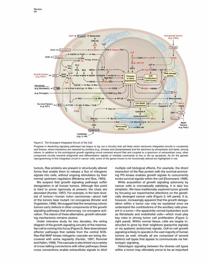
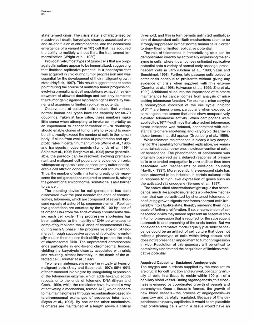
Figure 3. Tumors as Complex Tissues
The field of cancer research has largely beenguidedby a reductionist focus on cancer cellsand the genes within them (left panel)—a fo-cus that has produced an extraordinary bodyof knowledge. Looking forward in time, webelieve that important new inroads will comefrom regarding tumors as complex tissues inwhich mutant cancer cells have conscriptedand subverted normal cell types to serve asactive collaborators in their neoplastic agenda(right panel). The interactions between thegenetically altered malignant cells and thesesupporting coconspirators will prove criticalto understanding cancer pathogenesis and tothe development of novel, effective therapies.
in explaining tumor cell proliferation as the cancer cell- the components governing the transit of the cell throughthe G1 phase of its growth cycle. Cells monitor theirautonomous mechanisms enumerated above. For ex-
ample, we suspect that many of the growth signals driv- external environment during this period and, on the ba-sis of sensed signals, decide whether to proliferate, toing the proliferation of carcinoma cells originate from
the stromal cell components of the tumor mass. While be quiescent, or to enter into a postmitotic state. At themolecular level, many and perhaps all antiproliferativedifficult to validate at present, such thinking recasts the
logic of acquired GS autonomy: successful tumor cells signals are funneled through the retinoblastoma protein(pRb) and its two relatives, p107 and p130. When in aare those that have acquired the ability to co-opt their
normal neighbors by inducing them to release abundant hypophosphorylated state, pRb blocks proliferation bysequestering and altering the function of E2F transcrip-fluxes of growth-stimulating signals (Skobe and Fu-
senig, 1998). Indeed, in some tumors, these cooperating tion factors that control the expressionof banksof genesessential for progression from G1 into S phase (Wein-cells may eventually depart from normalcy, coevolving
with their malignant neighbors in order to sustain the berg, 1995).Disruption of the pRb pathway liberates E2Fs andgrowth of the latter (Kinzler and Vogelstein, 1998; Olumi
et al., 1999). Further, inflammatory cells attracted to sites thus allows cell proliferation, rendering cells insensitiveto antigrowth factors that normally operate along thisof neoplasia may promote (rather than eliminate) cancer
cells (Cordon-Cardo and Prives, 1999; Coussens et al., pathway to block advance through the G1 phase of thecell cycle. The effects of the soluble signaling molecule1999; Hudson et al., 1999), another example of normal
cells conscripted to enhance tumor growth potential, TGF� are the best documented, but we envision otherantigrowth factors will be found to signal through thisanother means to acquire necessary capabilities.pathway as well. TGF� acts in a number of ways, moststill elusive, to prevent the phosphorylation that inacti-Acquired Capability: Insensitivity
to Antigrowth Signals vates pRb; in this fashion, TGF� blocks advance throughG1. In some cell types, TGF� suppresses expressionWithin a normal tissue, multiple antiproliferative signals
operate to maintain cellular quiescence and tissue ho- of the c-myc gene, which regulates the G1 cell cyclemachinery in still unknown ways (Moses et al., 1990).meostasis; these signals include both soluble growth
inhibitors and immobilized inhibitors embedded in the More directly, TGF� causes synthesis of the p15INK4B andp21 proteins, which block the cyclin:CDK complexesextracellular matrix and on the surfaces of nearby cells.
These growth-inhibitory signals, like their positively act- responsible for pRb phosphorylation (Hannon andBeach, 1994; Datto et al., 1997).ing counterparts, are received by transmembrane cell
surface receptors coupled to intracellular signaling cir- The pRb signaling circuit, as governed by TGF� andother extrinsic factors, can be disrupted in a variety ofcuits.
Antigrowth signals can block proliferation by two dis- ways in different types of human tumors (Fynan andReiss, 1993). Some lose TGF� responsiveness throughtinct mechanisms. Cells may be forced out of the active
proliferative cycle into the quiescent (G0) state from downregulation of their TGF� receptors, while othersdisplay mutant, dysfunctional receptors (Fynan andwhich they may reemerge on some future occasion
when extracellular signals permit. Alternatively, cells Reiss, 1993; Markowitz et al., 1995). The cytoplasmicSmad4 protein, which transduces signals from ligand-maybe induced to permanently relinquish their prolifera-
tive potential by being induced to enter into postmitotic activated TGF� receptors to downstream targets, maybe eliminated through mutation of its encoding genestates, usually associated with acquisition of specific
differentiation-associated traits. (Schutte et al., 1996). The locus encoding p15INK4Bmay bedeleted (Chin et al., 1998). Alternatively, the immediateIncipient cancer cells must evade these antiprolifera-
tive signals if they are to prosper. Much of the circuitry downstream target of its actions, CDK4, may becomeunresponsive to the inhibitory actions of p15INK4B be-that enables normal cells to respond to antigrowth sig-
nals is associated with the cell cycle clock, specifically cause of mutations that create amino acid substitutions
Review61
in its INK4A/B-interacting domain; the resulting cyclin in virtually all cell types throughout the body. Once trig-gered by a variety of physiologic signals, this programD:CDK4 complexes are then given a free hand to inacti-
vate pRb by hyperphosphorylation (Zuo et al., 1996). unfolds in a precisely choreographed series of steps.Cellular membranes are disrupted, the cytoplasmic andFinally, functional pRb, the end target of this pathway,
may be lost through mutation of its gene. Alternatively, nuclear skeletons are broken down, the cytosol is ex-truded, the chromosomes are degraded, and the nu-in certain DNA virus-induced tumors, notably cervical
carcinomas, pRb function is eliminated through seques- cleus is fragmented, all in a span of 30–120 min. In theend, the shriveled cell corpse is engulfed by nearby cellstration by viral oncoproteins, such as the E7 oncoprotein
of human papillomavirus (Dyson et al., 1989). In addition, in a tissue and disappears, typically within 24 hr (Wyllieet al., 1980).cancer cells can also turn off expression of integrins and
other cell adhesion molecules that send antigrowth sig- The apoptotic machinery can be broadly divided intotwo classes of components—sensors and effectors. Thenals, favoring instead those that convey progrowth sig-
nals; these adherence-based antigrowth signals likely sensors are responsible for monitoring the extracellularand intracellular environment for conditions of normalityimpinge on the pRb circuit as well. The bottom line is
that the antigrowth circuit converging onto Rb and the or abnormality that influence whether a cell should liveor die. These signals regulate the second class of com-cell division cycle is, one way or another, disrupted in
a majority of human cancers, defining the concept and ponents, which function as effectors of apoptotic death.The sentinels include cell surface receptors that binda purpose of tumor suppressor loss in cancer.
Cell proliferation depends on more than an avoidance survival or death factors. Examples of these ligand/receptor pairs include survival signals conveyed by IGF-of cytostatic antigrowth signals. Our tissues also con-
strain cell multiplication by instructing cells to enter irre- 1/IGF-2 through their receptor, IGF-1R, and by IL-3 andits cognate receptor, IL-3R (Lotem and Sachs, 1996;versibly into postmitotic, differentiated states, using di-
verse mechanisms that are incompletely understood; it Butt et al., 1999). Death signals are conveyed by theFAS ligand binding the FAS receptor and by TNF� bind-is apparent that tumor cells use various strategies to
avoid this terminal differentiation. One strategy for ing TNF-R1 (Ashkenazi and Dixit, 1999). Intracellularsensors monitor the cell’s well-being and activate theavoiding differentiation directly involves the c-myc on-
cogene, which encodes a transcription factor. During death pathway in response to detecting abnormalities,including DNA damage, signaling imbalance provokednormal development, the growth-stimulating action of
Myc, in association with another factor, Max, can be by oncogene action, survival factor insufficiency, or hyp-oxia (Evan and Littlewood, 1998). Further, the life ofmostsupplanted by alternative complexes of Max with a
group of Mad transcription factors; the Mad–Max com- cells is in part maintained by cell–matrix and cell–celladherence-based survival signals whose abrogationplexes elicit differentiation-inducing signals (Foley and
Eisenman, 1999). However, overexpression of the c-Myc elicits apoptosis (Ishizaki et al., 1995; Giancotti and Ru-oslahti, 1999). Both soluble and immobilized apoptoticoncoprotein, as is seen in many tumors, can reverse this
process, shifting the balance back to favor Myc–Max regulatory signals likely reflect the needs of tissues tomaintain their constituent cells in appropriate architec-complexes, thereby impairing differentiation and pro-
moting growth. During human colon carcinogenesis, in- tural configurations.Many of the signals that elicit apoptosis convergeactivation of the APC/�-catenin pathway serves to block
the egress of enterocytes in the colonic crypts into a on the mitochondria, which respond to proapoptoticsignals by releasing cytochrome C, a potent catalyst ofdifferentiated, postmitotic state (Kinzler and Vogelstein,
1996). Analogously, during the generation of avian eryth- apoptosis (Green andReed, 1998).Members of theBcl-2family of proteins, whose members have either pro-roblastosis, the erbA oncogene acts to prevent irrevers-
ible erythrocyte differentiation (Kahn et al., 1986). apoptotic (Bax, Bak, Bid, Bim) or antiapoptotic (Bcl-2,Bcl-XL, Bcl-W) function, act in part by governing mito-While the components and interconnections between
the various antigrowth and differentiation-inducing sig- chondrial death signaling through cytochrome C re-lease. The p53 tumor suppressor protein can elicit apo-nals and the core cell cycle machinery are still being
delineated, the existence of an antigrowth signaling cir- ptosis by upregulating expression of proapoptotic Baxin response to sensing DNA damage; Bax in turn stimu-cuitry is clear (Figure 2), as is the necessity for its circum-
vention by developing cancers. lates mitochondria to release cytochrome C.The ultimate effectors of apoptosis include an array
of intracellular proteases termed caspases (ThornberryAcquired Capability: Evading Apoptosisand Lazebnik, 1998). Two “gatekeeper” caspases, �8The ability of tumor cell populations to expand in numberand �9, are activated by death receptors such as FASis determined not only by the rate of cell proliferationor by the cytochrome C released from mitochondria,but also by the rate of cell attrition. Programmed cellrespectively. These proximal caspases trigger the acti-death—apoptosis—represents a major source of thisvation of a dozen ormore effector caspases that executeattrition. The evidence is mounting, principally fromthe deathprogram, through selective destructionof sub-studies in mouse models and cultured cells, as well ascellular structures and organelles, and of the genome.from descriptive analyses of biopsied stages in humanThe possibility that apoptosis serves as a barrier tocarcinogenesis, that acquired resistance toward apo-
cancer was first raised in 1972, when Kerr, Wyllie, andptosis is a hallmark of most and perhaps all types ofCurrie describedmassive apoptosis in the cells populat-cancer.ing rapidly growing, hormone-dependent tumors follow-Observations accumulated over the past decade indi-
cate that the apoptotic program is present in latent form ing hormonewithdrawal (Kerr et al., 1972). The discovery
Cell62
of the bcl-2 oncogene by its upregulation via chromo- abnormalities, including hypoxia and oncogene hyper-expression, are also funneled in part via p53 to the apo-somal translocation in follicular lymphoma (reviewed inptotic machinery; these too are impaired at elicitingKorsmeyer, 1992) and its recognition as having anti-apoptosis when p53 function is lost (Levine, 1997). Addi-apoptotic activity (Vaux et al., 1988) opened up the in-tionally, the PI3 kinase–AKT/PKB pathway, which trans-vestigation of apoptosis in cancer at themolecular level.mits antiapoptotic survival signals, is likely involved inWhen coexpressed with a myc oncogene in transgenicmitigating apoptosis in a substantial fraction of humanmice, the bcl-2 gene was able to promote formation oftumors. This survival signaling circuit can be activatedBcell lymphomasby enhancing lymphocyte survival, notby extracellular factors such as IGF-1/2 or IL-3 (Evanby further stimulating their myc-induced proliferationand Littlewood, 1998), by intracellular signals emanating(Strasser et al., 1990); further, 50% of the infrequentfrom Ras (Downward, 1998), or by loss of the pTENlymphomas arising in bcl-2 single transgenic transgenictumor suppressor, a phospholipid phosphatase thatmice had somatic translocations activating c-myc, con-normally attenuates the AKT survival signal (Cantley andfirming a selective pressure during lymphomagenesisNeel, 1999). Recently, a mechanism for abrogating theto upregulate both Bcl-2 and c-Myc (McDonnell andFAS death signal has been revealed in a high fractionKorsmeyer, 1991).of lung and colon carcinoma cell lines: a nonsignalingFurther insight into the myc-bcl-2 interaction emergeddecoy receptor for FAS ligand is upregulated, titratinglater from studying the effects of a myc oncogene onthe death-inducing signal away from the FAS death re-fibroblasts cultured in lowserum.Widespreadapoptosisceptor (Pitti et al., 1998). We expect that virtually allwas induced in myc-expressing cells lacking serum; thecancer cells harbor alterations that enable evasion ofconsequent apoptosis could be abrogated by exoge-apoptosis.nous survival factors (e.g., IGF-1), by forced overexpres-It is now possible to lay out a provisional apoptoticsion of Bcl-2 or the related Bcl-XL protein, or by disrup-
signaling circuitry (Figure 2); while incomplete, it is evi-tion of the FAS death signaling circuit (Hueber et al.,dent that most regulatory and effector components are1997). Collectively, the data indicate that a cell’s apo-present in redundant form. This redundancy holds im-ptotic program can be triggered by an overexpressedportant implications for the development of novel typesoncogene. Indeed, elimination of cells bearing activatedof antitumor therapy, since tumor cells that have lostoncogenes by apoptosis may represent the primaryproapoptotic components are likely to retain other simi-
means by which suchmutant cells are continually culledlar ones. We anticipate that new technologies will be
from the body’s tissues.able to display the apoptotic pathways still operative in
Other examples strengthen the consensus that apo- specific types of cancer cells and that new drugs willptosis is a major barrier to cancer that must be circum- enable cross-talk between the still intact componentsvented. Thus, in transgenic mice where the pRb tumor of parallel apoptotic signaling pathways in tumor cells,suppressor was functionally inactivated in the choroid resulting in restoration of the apoptotic defense mecha-plexus, slowly growing microscopic tumors arose, ex- nism, with substantial therapeutic benefit.hibiting high apoptotic rates; the additional inactivationof the p53 tumor suppressor protein, a component of
Acquired Capability: Limitless Replicative Potentialthe apoptotic signaling circuitry, led to rapidly growing Three acquired capabilities—growth signal autonomy,tumors containing low numbers of apoptotic cells (Sy- insensitivity to antigrowth signals, and resistance tomonds et al., 1994). The role of extracellular survival apoptosis—all lead to an uncoupling of a cell’s growthfactors is illustrated by disease progression in trans- program from signals in its environment. In principle,genic mice prone to pancreatic islet tumors. If IG F-2 the resulting deregulated proliferation program shouldgene expression, which is activated in this tumorigene- suffice to enable the generation of the vast cell popula-sis pathway, was abrogated using gene knockout mice, tions that constitute macroscopic tumors. However, re-tumor growth and progression were impaired, as evi- search performed over the past 30 years indicates thatdenced by the appearance of comparatively small, be- this acquired disruption of cell-to-cell signaling, on itsnign tumors showing high rates of apoptosis (Christofori own, does not ensure expansive tumor growth. Manyet al., 1994). In these cells, the absence of IGF-2 did not and perhaps all types of mammalian cells carry an intrin-affect cell proliferation rates, clearly identifying it as an sic, cell-autonomousprogram that limits theirmultiplica-antiapoptotic survival factor. Collectively, these obser- tion. This program appears to operate independently ofvations argue that altering components of the apoptotic the cell-to-cell signaling pathways described above. Itmachinery can dramatically affect the dynamics of tu- too must be disrupted in order for a clone of cells tomor progression, providing a rationale for the inactiva- expand to a size that constitutes a macroscopic, life-tion of this machinery during tumor development. threatening tumor.Resistance to apoptosis can be acquired by cancer The early work of Hayflick demonstrated that cells in
cells through a variety of strategies. Surely, the most culture have a finite replicative potential (reviewed incommonly occurring loss of a proapoptotic regulator Hayflick, 1997). Once such cell populations have pro-through mutation involves the p53 tumor suppressor gressed through a certain number of doublings, theygene. The resulting functional inactivation of its product, stop growing—a process termed senescence. The se-the p53 protein, is seen in greater than 50% of human nescence of cultured human fibroblasts can be circum-cancers and results in the removal of a key component vented by disabling their pRb and p53 tumor suppressorof theDNAdamage sensor that can induce the apoptotic proteins, enabling these cells to continue multiplying for
additional generations until they enter into a secondeffector cascade (Harris, 1996). Signals evoked by other
Review63
state termed crisis. The crisis state is characterized by threshold, and this in turn permits unlimited multiplica-massive cell death, karyotypic disarray associated with tion of descendant cells. Both mechanisms seem to beend-to-end fusion of chromosomes, and the occasional strongly suppressed inmost normal human cells in orderemergence of a variant (1 in 107) cell that has acquired to deny them unlimited replicative potential.the ability to multiply without limit, the trait termed im- The role of telomerase in immortalizing cells can bemortalization (Wright et al., 1989). demonstrated directly by ectopically expressing the en-Provocatively, most types of tumor cells that are prop- zyme in cells, where it can convey unlimited replicative
agated in culture appear to be immortalized, suggesting potential onto a variety of normal early passage, prese-that limitless replicative potential is a phenotype that nescent cells in vitro (Bodnar et al., 1998; Vaziri andwas acquired in vivo during tumor progression and was Benchimol, 1998). Further, late passage cells poised toessential for the development of their malignant growth enter crisis continue to proliferate without giving anystate (Hayflick, 1997). This result suggests that at some evidence of crisis when supplied with this enzymepoint during the course of multistep tumor progression, (Counter et al., 1998; Halvorsen et al., 1999; Zhu et al.,evolving premalignant cell populations exhaust their en- 1999). Additional clues into the importance of telomeredowment of allowed doublings and can only complete maintenance for cancer comes from analysis of micetheir tumorigenic agendaby breaching themortality bar- lacking telomerase function. For example, mice carryingrier and acquiring unlimited replicative potential. a homozygous knockout of the cell cycle inhibitorObservations of cultured cells indicate that various p16INK4A are tumor prone, particularly when exposed to
normal human cell types have the capacity for 60–70 carcinogens; the tumors that arise show comparativelydoublings. Taken at face value, these numbers make elevated telomerase activity. When carcinogens werelittle sense when attempting to invoke cell mortality as applied to p16INK4A-null mice that also lacked telomerase,an impediment to cancer formation: 60–70 doublings tumor incidence was reduced, concomitant with sub-should enable clones of tumor cells to expand to num- stantial telomere shortening and karyotypic disarray inbers that vastly exceed the number of cells in the human those tumors that did appear (Greenberg et al., 1999).body. If clues from evaluation of proliferation and apo- While telomere maintenance is clearly a key compo-ptotic rates in certain human tumors (Wyllie et al., 1980) nent of the capability for unlimited replication, we remainand transgenic mouse models (Symonds et al., 1994; uncertain about another one, the circumvention of cellu-Shibata et al., 1996; Bergers et al., 1998) prove generaliz- lar senescence. The phenomenon of senescence wasable, the paradox can be resolved: evolving premalig- originally observed as a delayed response of primarynant and malignant cell populations evidence chronic, cells to extended propagation in vitro and has thus beenwidespread apoptosis and consequently suffer consid- associated with mechanisms of divisional countingerable cell attrition concomitant with cell accumulation. (Hayflick, 1997). More recently, the senescent state hasThus, the number of cells in a tumor greatly underrepre- been observed to be inducible in certain cultured cellssents the cell generations required to produce it, raising in response to high level expression of genes such asthe generational limit of normal somatic cells as a barrier the activated ras oncogene (Serrano et al., 1997).to cancer. The above-cited observationsmight argue that senes-The counting device for cell generations has been cence,much like apoptosis, reflects aprotectivemecha-
discovered over the past decade: the ends of chromo- nism that can be activated by shortened telomeres orsomes, telomeres, which are composed of several thou- conflicting growth signals that forces aberrant cells irre-sand repeats of a short 6 bp sequence element. Replica- versibly into aG0-like state, thereby rendering them inca-tive generations are counted by the 50–100 bp loss of pable of further proliferation. If so, circumvention of se-telomeric DNA from the ends of every chromosome dur- nescence in vivomay indeed represent an essential steping each cell cycle. This progressive shortening has in tumor progression that is required for the subsequentbeen attributed to the inability of DNA polymerases to approach to and breaching of the crisis barrier. But wecompletely replicate the 3� ends of chromosomal DNA
consider an alternative model equally plausible: senes-during each S phase. The progressive erosion of telo-
cence could be an artifact of cell culture that does notmeres through successive cycles of replication eventu-reflect a phenotype of cells within living tissues andally causes them to lose their ability to protect the endsdoes not represent an impediment to tumor progressionof chromosomal DNA. The unprotected chromosomalin vivo. Resolution of this quandary will be critical toends participate in end-to-end chromosomal fusions,completely understand the acquisition of limitless repli-yielding the karyotypic disarray associated with crisiscative potential.and resulting, almost inevitably, in the death of the af-
fected cell (Counter et al., 1992).Acquired Capability: Sustained AngiogenesisTelomere maintenance is evident in virtually all types ofThe oxygen and nutrients supplied by the vasculaturemalignant cells (Shay and Bacchetti, 1997); 85%–90%are crucial for cell function and survival, obligating virtu-of them succeed in doing so by upregulating expressionally all cells in a tissue to reside within 100 �m of aof the telomerase enzyme, which adds hexanucleotidecapillary blood vessel. During organogenesis, this close-repeats onto the ends of telomeric DNA (Bryan andness is ensured by coordinated growth of vessels andCech, 1999), while the remainder have invented a wayparenchyma. Once a tissue is formed, the growth ofof activating a mechanism, termed ALT, which appearsnew blood vessels—the process of angiogenesis—isto maintain telomeres through recombination-based in-transitory and carefully regulated. Because of this de-terchromosomal exchanges of sequence informationpendence on nearby capillaries, it would seem plausible(Bryan et al., 1995). By one or the other mechanism,
telomeres are maintained at a length above a critical that proliferating cells within a tissue would have an
Cell64
intrinsic ability to encourage blood vessel growth. But case angiogenesis was found to be activated in mid-stage lesions, prior to the appearance of full-blown tu-the evidence is otherwise. The cells within aberrant pro-
liferative lesions initially lack angiogenic ability, curtail- mors. Similarly, angiogenesis can be discerned in pre-malignant lesions of the human cervix, breast, and skining their capability for expansion. In order to progress
to a larger size, incipient neoplasiasmust develop angio- (melanocytes) (Hanahan and Folkman, 1996); we expectthat induction of angiogenesis will prove to be an earlygenic ability (Bouck et al., 1996; Hanahan and Folkman,
1996; Folkman, 1997). tomidstage event inmany human cancers. These obser-vations, taken together with the effects of angiogenesisCounterbalancing positive and negative signals en-
courage or block angiogenesis. One class of these sig- inhibitors, indicate that neovascularization is a prerequi-site to the rapid clonal expansion associated with thenals is conveyed by soluble factors and their receptors,
the latter displayed on the surface of endothelial cells; formation of macroscopic tumors.Tumors appear to activate the angiogenic switch byintegrins and adhesion molecules mediating cell–matrix
and cell–cell association also play critical roles. The changing the balance of angiogenesis inducers andcountervailing inhibitors (Hanahan and Folkman, 1996).angiogenesis-initiating signals are exemplified by vas-
cular endothelial growth factor (VEGF) and acidic and One common strategy for shifting the balance involvesaltered gene transcription. Many tumors evidence in-basic fibroblast growth factors (FGF1/2). Each binds to
transmembrane tyrosine kinase receptors displayed by creased expression of VEGF and/or FGFs compared totheir normal tissue counterparts. In others, expressionendothelial cells (Fedi et al., 1997; Veikkola and Alitalo,
1999). A prototypical angiogenesis inhibitor is throm- of endogenous inhibitors such as thrombospondin-1 or�-interferon is downregulated. Moreover, both transi-bospondin-1, which binds to CD36, a transmembrane
receptor onendothelial cells coupled to intracellular Src- tions may occur, and indeed be linked, in some tumors(Singh et al., 1995; Volpert et al., 1997).like tyrosine kinases (Bull et al., 1994). There are cur-
rently more than two dozen angiogenic inducer factors Themechanisms underlying shifts in the balances be-tween angiogenic regulators remain incompletely un-known and a similar number of endogenous inhibitor
proteins. derstood. In one well-documented example, the inhibi-tor thrombospondin-1 has been found to positivelyIntegrin signaling also contributes to this regulatory
balance. Quiescent vessels express one class of inte- regulated by the p53 tumor suppressor protein in somecell types. Consequently, loss of p53 function, whichgrins, whereas sprouting capillaries express another.
Interference with signaling from the latter class of inte- occurs inmost human tumors, can cause thrombospon-din-1 levels to fall, liberating endothelial cells from itsgrins can inhibit angiogenesis (Varner and Cheresh,
1996; Giancotti and Ruoslahti, 1999), underscoring the inhibitory effects (Dameron et al., 1994). The VE G F geneis also under complex transcriptional control. For exam-important contribution of cell adhesion to the angiogenic
program (Hynes andWagner, 1996). Extracellular prote- ple, activation of the ras oncogene or loss of the VHLtumor suppressor gene in certain cell types causesases are physically and functionally connectedwith pro-
angiogenic integrins, and both help dictate the invasive upregulation of VEGF expression (Rak et al., 1995; Max-well et al., 1999).capability of angiogenic endothelial cells (Stetler-Ste-
venson, 1999). Another dimension of regulation is emerging in theform of proteases, which can control the bioavailabilityExperimental evidence for the importance of inducing
and sustaining angiogenesis in tumors is both extensive of angiogenic activators and inhibitors. Thus, a varietyof proteases can release bFGF stored in the ECMand compelling (Bouck et al., 1996; Hanahan and Folk-
man, 1996; Folkman, 1997). The story begins almost 30 (Whitelock et al., 1996), whereas plasmin, a proangio-genic component of the clotting system, cancleave itselfyears ago with Folkman and colleagues, who used in
vivo bioassays to demonstrate the necessity of angio- into an angiogenesis inhibitor form called angiostatin(Gately et al., 1997). The coordinated expression of pro-genesis for explosive growth of tumor explants (re-
viewed in Folkman, 1997). Molecular proof of principle and antiangiogenic signaling molecules, and their mod-ulation by proteolysis, appear to reflect the complexcame, for example, when anti-VEGF antibodies proved
able to impair neovascularization and growth of subcu- homeostatic regulation of normal tissue angiogenesisand of vascular integrity.taneous tumors in mice (Kim et al., 1993), as did a domi-
nant-interfering version of the VEGF receptor 2 (flk-1) As is already apparent, tumor angiogenesis offers auniquely attractive therapeutic target, indeed one that(Millauer et al., 1994); both results have motivated the
development of specific VEGF/VEGF-R inhibitors now is shared in common by most and perhaps all types ofhuman tumors. The next decade will produce a catalogin late stage clinical trials.
The essential role of angiogenesis is further supported of the angiogenic regulatory molecules expressed bydifferent types of tumors, and in many cases, by theirby the ability of an increasing catalog of antiangiogenic
substances to impair the growth of tumor cells inocu- progenitor stages. Use of increasingly sophisticatedmouse models will make it possible to assign specificlated subcutaneously in mice (Folkman, 1997). Tumors
arising in cancer-prone transgenic mice are similarly roles to each of these regulators and to discern themolecular mechanisms that govern their production andsusceptible to angiogenic inhibitors (Bergers et al.,
1999). activity. Already available evidence indicates that differ-ent types of tumor cells use distinctmolecular strategiesThe ability to induce and sustain angiogenesis seems
to be acquired in a discrete step (or steps) during tumor to activate the angiogenic switch. This raises the ques-tion of whether a single antiangiogenic therapeutic willdevelopment, via an “angiogenic switch” from vascular
quiescence.When three transgenicmousemodels were suffice to treat all tumor types, or whether an ensembleof such therapeutics will need to be developed, eachanalyzed throughout multistep tumorigenesis, in each
Review65
responding to a distinct program of angiogenesis that Changes in expression of CAMs in the immunoglobu-lin superfamily also appear to play critical roles in thehas been developed by a specific class of humanprocesses of invasion and metastasis (Johnson, 1991).tumors.The clearest case involves N-CAM, which undergoes aswitch in expression from a highly adhesive isoform toAcquired Capability: Tissue Invasion and Metastasispoorly adhesive (or even repulsive) forms in Wilms’ tu-Sooner or later during the development of most typesmor, neuroblastoma, and small cell lung cancer (John-of human cancer, primary tumor masses spawn pioneerson, 1991; Kaiser et al., 1996) and reduction in overallcells that move out, invade adjacent tissues, and thenceexpression level in invasive pancreatic and colorectaltravel to distant sites where they may succeed in found-cancers (Fogar et al., 1997). Experiments in transgenicing new colonies. These distant settlements of tumormice support a functional role for the normal adhesivecells—metastases—are the causeof 90%of humancan-form of N-CAM in suppressing metastasis (Perl et al.,cer deaths (Sporn, 1996). The capability for invasion and1999).metastasis enables cancer cells to escape the primaryChanges in integrin expression are also evident intumor mass and colonize new terrain in the body where,
invasive and metastatic cells. Invading and metastasiz-at least initially, nutrients and space are not limiting. Theing cancer cells experience changing tissue microenvi-newly formed metastases arise as amalgams of cancerronments during their journeys, which can present novelcells and normal supporting cells conscripted from thematrix components. Accordingly, successful coloniza-host tissue. Like the formation of the primary tumortion of these new sites (both local and distant) demandsmass, successful invasion and metastasis depend uponadaptation, which is achieved through shifts in the spec-all of the other five acquired hallmark capabilities. Buttrum of integrin � or � subunits displayed by the migrat-what additional cellular changes enable the acquisitioning cells. These novel permutations result in differentof these final capabilities during tumorigenesis?integrin subtypes (of which there are greater than 22)Invasion and metastasis are exceedingly complexhaving distinct substrate preferences. Thus, carcinomaprocesses, and their genetic and biochemical determi-cells facilitate invasion by shifting their expression ofnants remain incompletely understood. At the mecha-integrins from those that favor the ECM present in nor-nistic level, they are closely allied processes,which justi-mal epithelium to other integrins (e.g., �3�1 and �V�3)fies their association with one another as one generalthat preferentially bind the degraded stromal compo-capability of cancer cells. Both utilize similar operationalnents produced by extracellular proteases (Varner andstrategies, involving changes in the physical couplingCheresh, 1996; Lukashev and Werb, 1998). Forced ex-of cells to their microenvironment and activation of ex-pression of integrin subunits in cultured cells can inducetracellular proteases.or inhibit invasive and metastatic behavior, consistentSeveral classes of proteins involved in the tetheringwith a role of these receptors in acting as central deter-of cells to their surroundings in a tissue are altered inminants of these processes (Varner and Cheresh, 1996).cells possessing invasive or metastatic capabilities. TheAttempts at explaining the cell biological effects ofaffected proteins include cell–cell adhesion molecules
integrins in terms of a small number of mechanistic rules(CAMs)—notably members of the immunoglobulin andhave been confounded by the large number of distinct
calcium-dependent cadherin families, both ofwhichme-integrin genes, by the even larger number of heterodi-
diate cell-to-cell interactions—and integrins, which link meric receptors resulting fromcombinatorial expressioncells to extracellular matrix substrates. Notably, all of of various � and � receptor subunits, and by the increas-these “adherence” interactions convey regulatory sig- ing evidence of complex signals emitted by the cyto-nals to the cell (Aplin et al., 1998). The most widely plasmic domains of these receptors (Aplin et al., 1998;observed alteration in cell-to-environment interactions Giancotti and Ruoslahti, 1999). Still, there is little doubtin cancer involves E-cadherin, a homotypic cell-to-cell that these receptors play central roles in the capabilityinteraction molecule ubiquitously expressed on epithe- for tissue invasion and metastasis.lial cells. Coupling between adjacent cells by E-cadherin The second general parameter of the invasive andbridges results in the transmission of antigrowth and metastatic capability involves extracellular proteasesother signals via cytoplasmic contacts with �-catenin (Coussens and Werb, 1996; Chambers and Matrisian,to intracellular signaling circuits that include the Lef/ 1997). Protease genes are upregulated, protease inhibi-Tcf transcription factor (Christofori and Semb, 1999). tor genes are downregulated, and inactive zymogenE-cadherin function is apparently lost in a majority of forms of proteases are converted into active enzymes.epithelial cancers, by mechanisms that include muta- Matrix-degrading proteases are characteristically asso-tional inactivation of the E-cadherin or �-catenin genes, ciated with the cell surface, by synthesis with a trans-transcriptional repression, or proteolysis of the extracel- membrane domain, binding to specific protease re-lular cadherin domain (Christofori and Semb, 1999). ceptors, or association with integrins (Werb, 1997;Forced expression of E-cadherin in cultured cancer cells Stetler-Stevenson, 1999). One imagines that docking ofand in a transgenic mouse model of carcinogenesis im- active proteases on the cell surface can facilitate inva-pairs invasive and metastatic phenotypes, whereas in- sion by cancer cells into nearby stroma, across bloodterference with E-cadherin function enhances both vessel walls, and through normal epithelial cell layers.capabilities (Christofori and Semb, 1999). Thus, E-cad- That notion notwithstanding, it is difficult to unambigu-herin serves as a widely acting suppressor of invasion ously ascribe the functions of particular proteases solelyand metastasis by epithelial cancers, and its functional to this capability, given their evident roles in otherelimination represents a key step in the acquisition of hallmark capabilities, including angiogenesis (Stetler-
Stevenson, 1999) and growth signaling (Werb, 1997;this capability.
Cell66
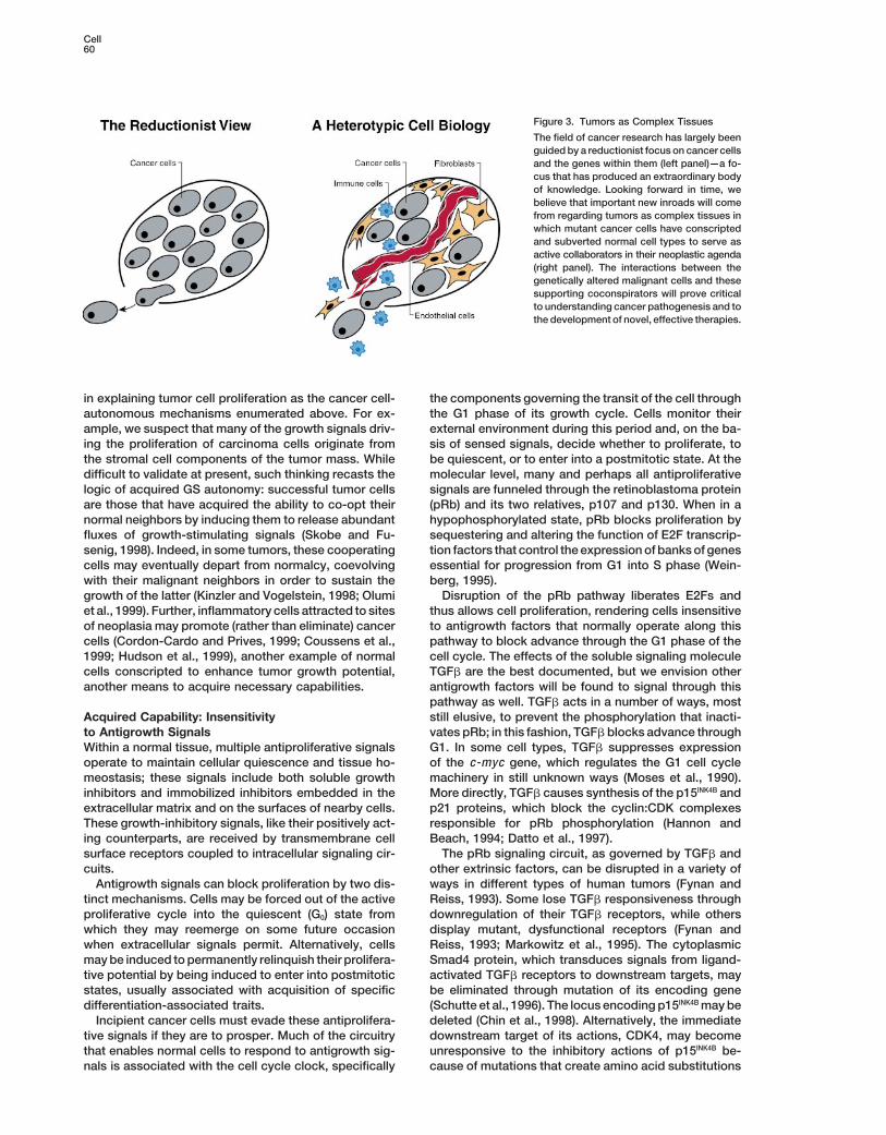
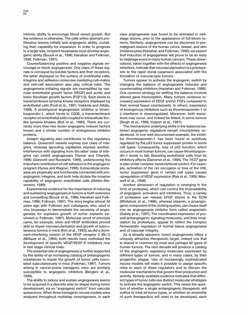
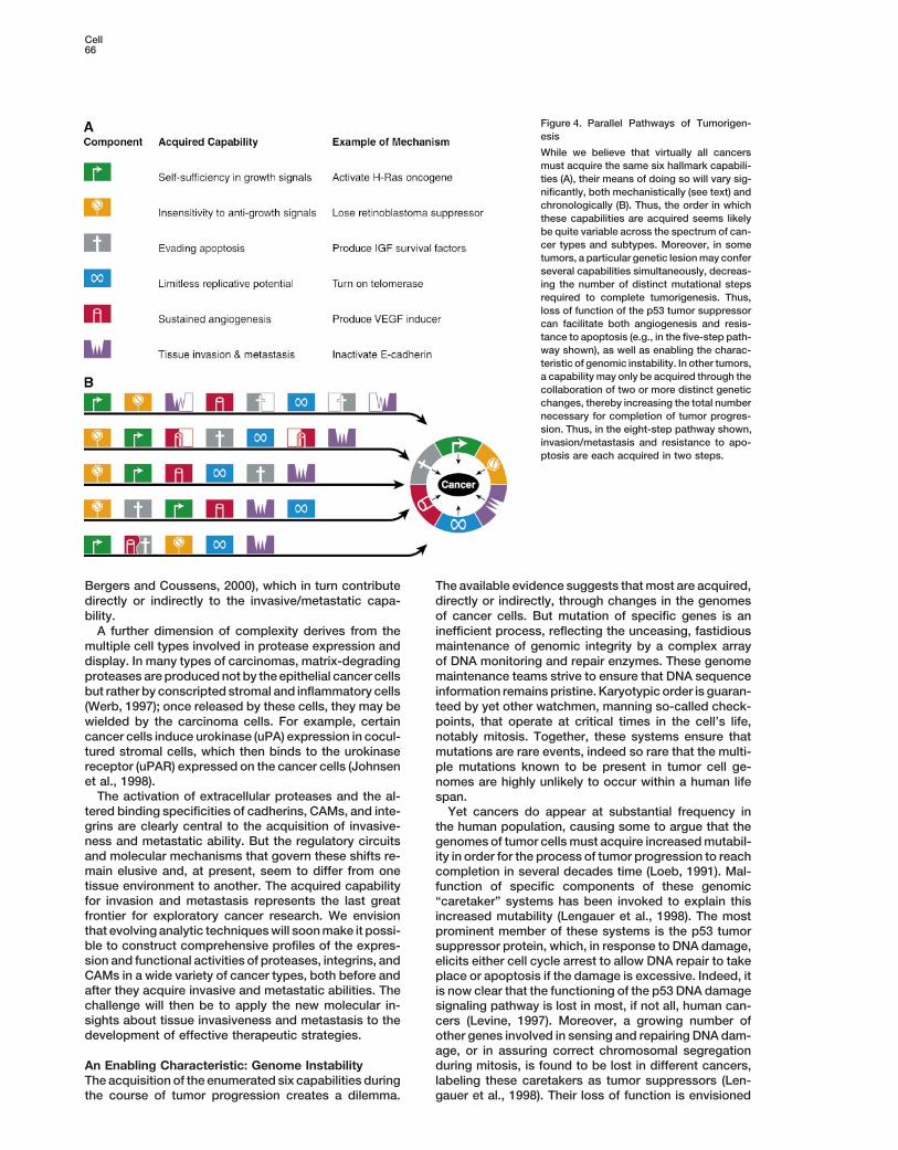
Figure 4. Parallel Pathways of Tumorigen-esis
While we believe that virtually all cancersmust acquire the same six hallmark capabili-ties (A), their means of doing so will vary sig-nificantly, both mechanistically (see text) andchronologically (B). Thus, the order in whichthese capabilities are acquired seems likelybe quite variable across the spectrum of can-cer types and subtypes. Moreover, in sometumors, a particular genetic lesionmay conferseveral capabilities simultaneously, decreas-ing the number of distinct mutational stepsrequired to complete tumorigenesis. Thus,loss of function of the p53 tumor suppressorcan facilitate both angiogenesis and resis-tance to apoptosis (e.g., in the five-step path-way shown), as well as enabling the charac-teristic of genomic instability. In other tumors,a capability may only be acquired through thecollaboration of two or more distinct geneticchanges, thereby increasing the total numbernecessary for completion of tumor progres-sion. Thus, in the eight-step pathway shown,invasion/metastasis and resistance to apo-ptosis are each acquired in two steps.
Bergers and Coussens, 2000), which in turn contribute The available evidence suggests thatmost are acquired,directly or indirectly, through changes in the genomesdirectly or indirectly to the invasive/metastatic capa-
bility. of cancer cells. But mutation of specific genes is aninefficient process, reflecting the unceasing, fastidiousA further dimension of complexity derives from the
multiple cell types involved in protease expression and maintenance of genomic integrity by a complex arrayof DNA monitoring and repair enzymes. These genomedisplay. In many types of carcinomas, matrix-degrading
proteases areproduced not by the epithelial cancer cells maintenance teams strive to ensure that DNA sequenceinformation remains pristine. Karyotypic order is guaran-but rather by conscripted stromal and inflammatory cells
(Werb, 1997); once released by these cells, they may be teed by yet other watchmen, manning so-called check-points, that operate at critical times in the cell’s life,wielded by the carcinoma cells. For example, certain
cancer cells induce urokinase (uPA) expression in cocul- notably mitosis. Together, these systems ensure thattured stromal cells, which then binds to the urokinase mutations are rare events, indeed so rare that the multi-receptor (uPAR) expressed on the cancer cells (Johnsen ple mutations known to be present in tumor cell ge-et al., 1998). nomes are highly unlikely to occur within a human lifeThe activation of extracellular proteases and the al- span.
tered binding specificities of cadherins, CAMs, and inte- Yet cancers do appear at substantial frequency ingrins are clearly central to the acquisition of invasive- the human population, causing some to argue that theness and metastatic ability. But the regulatory circuits genomes of tumor cells must acquire increasedmutabil-and molecular mechanisms that govern these shifts re- ity in order for the process of tumor progression to reachmain elusive and, at present, seem to differ from one completion in several decades time (Loeb, 1991). Mal-tissue environment to another. The acquired capability function of specific components of these genomicfor invasion and metastasis represents the last great “caretaker” systems has been invoked to explain thisfrontier for exploratory cancer research. We envision increased mutability (Lengauer et al., 1998). The mostthat evolving analytic techniqueswill soonmake it possi- prominent member of these systems is the p53 tumorble to construct comprehensive profiles of the expres- suppressor protein, which, in response to DNA damage,sion and functional activities of proteases, integrins, and elicits either cell cycle arrest to allow DNA repair to takeCAMs in a wide variety of cancer types, both before and place or apoptosis if the damage is excessive. Indeed, itafter they acquire invasive and metastatic abilities. The is now clear that the functioning of the p53 DNA damagechallenge will then be to apply the new molecular in- signaling pathway is lost in most, if not all, human can-sights about tissue invasiveness and metastasis to the cers (Levine, 1997). Moreover, a growing number ofdevelopment of effective therapeutic strategies. other genes involved in sensing and repairing DNA dam-
age, or in assuring correct chromosomal segregationduring mitosis, is found to be lost in different cancers,An Enabling Characteristic: Genome Instability
The acquisition of the enumerated six capabilities during labeling these caretakers as tumor suppressors (Len-gauer et al., 1998). Their loss of function is envisionedthe course of tumor progression creates a dilemma.
Review67
to allow genome instability and variability and the gener- the signals exchanged between the various cell typesexisting symbiotically within a tumor mass and knowingation of consequently mutant cells with selective advan-
tages. Interestingly, recent evidence suggests that apo- their effects on the integrated circuits of each of thosecell types.ptosis may also be a vehicle of genomic instability, in
that DNA within apoptotic cell bodies can be incorpo- Our ability to analyze individual human cancers atthe genetic and biochemical levels will also undergo arated into neighboring cells following phagoctytosis
(Holmgren et al., 1999), in principle genetically diversify- dramatic change. At present, description of a recentlydiagnosed tumor in terms of its underlying genetic le-ing any of the constituent cell types of a tumor. We place
this acquired characteristic of genomic instability apart sions remains a distant prospect. Nonetheless, we lookahead 10 or 20 years to the time when the diagnosis offrom the six acquired capabilities associated with tumor
cell phenotype and tumor physiology: it represents the all the somatically acquired lesions present in a tumorcell genome will become a routine procedure. By then,means that enables evolving populations of premalig-
nant cells to reach these six biological endpoints. genome-wide gene expression profiles of tumor cellswill also be routine. With all this information in hand, itwill become possible to test definitively our propositionAlternative Pathways to Cancerthat the development of all types of human tumor cellsThe paths that cells take on their way to becomingmalig-is governed by a common set of rules such as thosenant are highly variable. Within a given cancer type,implied by the six acquired capabilities enumeratedmutation of particular target genes such as ras or p53here.may be found in only a subset of otherwise histologicallyWe anticipate far deeper insight into the roles playedidentical tumors. Further, mutations in certain onco-
by inherited alleles in cancer susceptibility and patho-genes and tumor suppressor genes can occur early ingenesis. At present, our understanding of the interplaysome tumor progression pathways and late in others. Asat the cellular level between inherited cancer modifiera consequence, the acquisition of biological capabilitiesgeneswith oncogenes and tumor suppressor genes thatsuch as resistance to apoptosis, sustained angiogen-are altered somatically is rudimentary; modifiers can inesis, and unlimited replicative potential can appear atprinciple act in any of the constituent cell types of adifferent times during these various progressions. Ac-tumor, or elsewhere in the body, whereas the classicalcordingly, the particular sequence in which capabilitiescancer genes largely act in the cancer cells themselves.are acquired can vary widely, both among tumors of theThese gaps will be bridged in part by new informaticssame type and certainly between tumors of differenttechnologies, enabling us to process and interpret thetypes (Figure 4). Furthermore, in certain tumors, a spe-inundationof genetic information thatwill soon flow fromcific genetic event may, on its own, contribute only par-automated sequencing instruments. New technologiestially to the acquisition of a single capability, while inwill also aid us in rationalizing the complex constella-others, this event may aid in the simultaneous acquisi-tions of interacting alleles in terms of a systematics oftion of several distinct capabilities. Nonetheless, we be-cancer formation of the type that we propose here.lieve that independent of how the steps in these geneticThe metaphors used to conceptualize cancer cellpathways are arranged, the biological endpoints that
function will also shift dramatically. For decades now,are ultimately reached—the hallmark capabilities of can-we have been able to predict with precision the behaviorcer—will prove to be shared in common by all types ofof an electronic integrated circuit in terms of its constit-tumors.uent parts—its interconnecting components, each re-sponsible for acquiring, processing, andemitting signalsSynthesisaccording to a precisely defined set of rules. Two de-Cancer cells propagated in culture and dissected intocades from now, having fully charted the wiring dia-their molecular components have yielded much of thegrams of every cellular signaling pathway, it will be pos-wealth of information that we currently possess aboutsible to lay out the complete “integrated circuit of thethe molecular processes underlying cancer develop-cell” upon its current outline (Figure 2). We will then bement. Yet by simplifying the nature of cancer—por-able to apply the tools of mathematical modeling totraying it as a cell-autonomous process intrinsic to theexplain how specific genetic lesions serve to reprogramcancer cell—these experimental models have turnedthis integrated circuit in each of the constituent celltheir back on a central biological reality of tumor forma-types so as to manifest cancer.tion in vivo: cancer development depends upon changesWith holistic clarity of mechanism, cancer prognosisin the heterotypic interactions between incipient tumor
and treatment will become a rational science, unrecog-cells and their normal neighbors. Moreover, once formed,nizable by current practitioners. It will be possible tovirtually all types of human tumors, including their meta-understand with precision how and why treatment regi-static outgrowths, continue to harbor complex mixturesmens and specific antitumor drugs succeed or fail. Weof several cell types that collaborate to create malignantenvision anticancer drugs targeted to each of the hall-growth (Figure 3). This reconceptualization of cancermark capabilities of cancer; some, used in appropriatecell biology has begun to drive profound changes incombinations and in concert with sophisticated technol-how we study this disease experimentally. Continuingogies to detect and identify all stages of disease pro-elucidation of cancer pathogenesis will depend increas-gression, will be able to prevent incipient cancers fromingly upon heterotypic organ culture systems in vitrodeveloping, while others will cure preexisting cancers,and evermore refined mouse models in vivo. Lookingelusive goals at present. One day, we imagine that can-ahead into the future, these systems will help us chartcer biology and treatment—at present, a patchwork quiltcomprehensive maps of growth signaling networks in
cancer, an endeavor that will depend on defining all of of cell biology, genetics, histopathology, biochemistry,
Cell68
molecule E-cadherin as a tumour-suppressor gene. Trends Bio-immunology, and pharmacology—will become a sci-chem. Sci. 24, 73–76.ence with a conceptual structure and logical coherenceChristofori, G., Naik, P., and Hanahan, D. (1994). A second signalthat rivals that of chemistry or physics.supplied by insulin-like growth factor II in oncogene-induced tumori-genesis. Nature 369, 414–418.
Acknowledgments Cordon-Cardo, C., and Prives, C. (1999). At the crossroads of inflam-mation and tumorigenesis. J. Exp. Med. 190, 1367–1370.
We wish to thank Terry Schoop of Biomed Arts Associates, SanCounter, C.M., Avilion, A.A., LeFeuvre, C.E., Stewart, N.G., Greider,
Francisco, for preparation of the figures, Cori Bargmann and ZenaC.W., Harley, C.B., and Bacchetti, S. (1992). Telomere shortening
Werb for insightful comments on the manuscript, and Normita San- associated with chromosome instability is arrested in immortal cellstore for editorial assistance. In addition, we are indebted to Joe which express telomerase activity. EMBO J. 11, 1921–1929.Harford and Richard Klausner, who allowed us to adapt and expand
Counter, C.M., Hahn, W.C., Wei, W., Dickinson Caddle, S., Beijers-their depiction of the cell signaling network, and we appreciatebergen, R.L., Lansdorp, P.M., Sedivy, J.M., and Weinberg, R.A.suggestions on signaling pathways fromRandyWatnick, Brian Elen-(1998). Dissociation between telomerase activity, telomere mainte-bas, Bill Lundberg, Dave Morgan, and Henry Bourne. R. A. W. isnance and cellular immortalization. Proc. Natl. Acad. Sci. USA 95,
a Ludwig Foundation and American Cancer Society Professor of14723–14728.
Biology. His work has been supported by the Department of theCoussens, L.M., and Werb, Z. (1996). Matrix metalloproteinases andArmy and the National Institutes of Health. D. H. acknowledgesthe development of cancer. Chem. Biol. 3, 895–904.the support and encouragement of the National Cancer Institute.Coussens, L.M., Raymond, W.W., Bergers, G., Laig-Webster, M.,Editorial policy has rendered the citations illustrative but not com-Behrendtsen, O., Werb, Z., Caughey, G.H., and Hanahan, D. (1999).prehensive.Inflammatorymast cells up-regulate angiogenesis during squamousepithelial carcinogenesis. Genes Dev. 13, 1382–1397.
ReferencesDameron, K.M., Volpert, O.V., Tainsky, M.A., and Bouck, N. (1994).Control of angiogenesis in fibroblasts by p53 regulation of throm-Aplin, A.E., Howe, A., Alahari, S.K., and Juliano, R.L. (1998). Signalbospondin-1. Science 265, 1582–1584.
transduction and signal modulation by cell adhesion receptors: theDatto, M.B., Hu, P.P., Kowalik, T.F., Yingling, J., and Wang, X.F.role of integrins, cadherins, immunoglobulin-cell adhesion mole-(1997). The viral oncoprotein E1A blocks transforming growth factorcules, and selectins. Pharmacol. Rev. 50, 197–263.�-mediated induction of p21/WAF1/Cip1 and p15/INK4B Mol. Cell.
Ashkenazi, A., and Dixit, V.M. (1999). Apoptosis control by death Biol. 17, 2030–2037.and decoy receptors. Curr. Opin. Cell Biol. 11, 255–260.
DiFiore, P.P., Pierce, J.H., Kraus, M.H., Segatto, O., King, C.R., andBergers, G., and Coussens, L.M. (2000). Extrinsic regulators of epi- Aaronson, S.A. (1987). erbB-2 is a potent oncogene when overex-thelial tumor progression: metalloproteinases. Curr. Opin. Genet. pressed in NIH/3T3 cells. Science 237, 178–182.Dev., in press.
Downward, J. (1998). Mechanisms and consequences of activationBergers, G., Hanahan, D., and Coussens, L.M. (1998). Angiogenesis of protein kinase B/Akt. Curr. Opin. Cell Biol. 10, 262–267.and apoptosis are cellular parameters of neoplastic progression in Dyson, N., Howley, P.M., Munger, K., and Harlow, E. (1989). Thetransgenic mouse models of tumorigenesis. Int. J. Dev. Biol. 42, human papillomavirus-16 E7 oncoprotein is able to bind to the reti-995–1002. noblastoma gene product. Science 243, 934–937.Bergers, G., Javaherian, K., Lo, K.-M., Folkman, J., and Hanahan, Evan, G., and Littlewood, T. (1998). A matter of life and cell death.D. (1999). Effects of angiogenesis inhibitors on multistage carcino- Science 281, 1317–1322.genesis in mice. Science 284, 808–812.
Fedi, P., Tronick, S.R., and Aaronson, S.A. (1997). Growth factors.Bishop, J.M., and Weinberg, R.A., eds. (1996). Molecular Oncology In Cancer Medicine, J.F. Holland, R.C. Bast, D.L. Morton, E. Frei,(New York: Scientific American, Inc.). D.W. Kufe, and R.R. Weichselbaum, eds. (Baltimore, MD: Williams
and Wilkins), pp. 41–64.Bodnar, A.G., Ouellete, M., Frolkis, M., Holt, S.E., Chiu, C., Morin,G.B., Harley, C.B., Shay, J.W., Lichtsteiner, S., and Wright, W.E. Fogar, P., Basso, D., Pasquali, C., De Paoli, C., Sperti, C., Roveroni,(1998). Extension of life-span by introduction of telomerase into G., Pedrazzoli, G., and Plebani, M. (1997). Neural cell adhesionmole-normal human cells. Science 279, 349–352. cule (N-CAM) in gastrointestinal neoplasias. Anticancer Res. 17,
1227–1230.Bouck, N., Stellmach, V., and Hsu, S.C. (1996). How tumors becomeFoley, K.P., and Eisenman, R.N. (1999). Two MAD tails: what theangiogenic. Adv. Cancer Res. 69, 135–174.recent knockouts of Mad1 and Mx1 tell us about the MYC/MAX/Bryan, T.M., andCech, T.R. (1999). Telomerase and themaintenanceMAD network. Biochim. Biophys. Acta 1423, M37–47.of chromosome ends. Curr. Opin. Cell Biol. 11, 318–324.Folkman, J. (1997). Tumor angiogenesis. In Cancer Medicine, J.F.Bryan, T.M., Englezou, A., Gupta, J., Bacchetti, S., and Reddel,Holland, R.C. Bast, D.L. Morton, E. Frei, D.W. Kufe, and R.R. Weich-R.R. (1995). Telomere elongation in immortal human cells withoutselbaum, eds. (Baltimore, MD: Williams and Wilkins), pp. 181–204.detectable telomerase activity. EMBO J. 14, 4240–4248.Foulds, L. (1954). The Experimental Study of Tumor Progression.Bull, H.A., Brickell, P.M., and Dowd, P.M. (1994). Src-related proteinVolumes I–III (London: Academic Press).
tyrosine kinases are physically associated with the surface antigenFynan, T.M., and Reiss, M. (1993). Resistance to inhibition of cellCD36 in human dermal microvascular endothelial cells. FEBS Lett.growth by transforming growth factor-� and its role in oncogenesis.351, 41–44.Crit. Rev. Oncog. 4, 493–540.
Butt, A.J., Firth, S.M., and Baxter, R.C. (1999). The IGF axis andGately, S., Twardowski, P., Stack, M.S., Cundiff, D.L., Grella, D.,programmed cell death. Immunol. Cell Biol. 77, 256–262.Castellino, F.J., Enghild, J., Kwaan, H.C., Lee, F., Kramer, R.A., et
Cantley, L.C., and Neel, B.G. (1999). New insights into tumor sup- al. (1997). The mechanism of cancer-mediated conversion of plas-pression: PTEN suppresses tumor formation by restraining the minogen to the angiogenesis inhibitor angiostatin. Proc. Natl. Acad.phosphoinositide 3-kinase/AKT pathway. Proc. Natl. Acad. Sci. USA Sci. USA 94, 10868–10872.96, 4240–4245. Giancotti, F.G., and Ruoslahti, E. (1999). Integrin signaling. ScienceChambers, A.F., and Matrisian, L.M. (1997). Changing views of the 285, 1028–1032.role of matrix metalloproteinases in metastasis. J. Natl. Cancer Inst. Green, D.R., and Reed, J.C. (1998). Mitochondria and apoptosis.89, 1260–1270. Science 281, 1309–1312.Chin, L., Pomerantz, J., and DePinho, R.A. (1998). The INK4a/ARF Greenberg, R.A., Chin, L., Femino, A., Lee, K.H., Gottlieb, G.J.,tumor suppressor: one gene—two products—two pathways. Trends Singer, R.H., Greider, C.W., and DePinho, R.A. (1999). Short dysfunc-Biochem. Sci. 23, 291–296. tional telomeres impair tumorigenesis in the INK4a�2/3 cancer-prone
mouse. Cell 97, 515–525.Christofori, G., and Semb, H. (1999). The role of the cell-adhesion

Review69
Hahn, W.C., Counter, C.M., Lundberg, A.S., Beijersbgern, R.L., Markowitz, S., Wang, J., Meyeroff, L., Parsons, R., Sun, L., Lutter-baugh, J., Fan, R., Zborowska, E., Kinzler, K., Vogelstein, B., et al.Brooks, M.W., and Weinberg, R.A. (1999). Creation of human tumor
cells with defined genetic elements. Nature 400, 464–468. (1995). Inactivation of the type II TGF-� receptor in colon cancercells with microsatellite instability. Science 268, 1336–1338.Halvorsen, T.L., Leibowitz, G., and Levine, F. (1999). Telomerase
activity is sufficient to allow transformed cells to escape from crisis. Maxwell, P.H., Wiesener, M.S., Chang, G.-W., Clifford, S.C., Vaux,E.C., Cockman, M.E., Wykoff, C.C., Pugh, C.W., Maher, E.R., andMol. Cell. Biol. 19, 1864–1870.Ratcliffe, P.J. (1999). The tumour suppressor protein VHL targetsHanahan, D., and Folkman, J. (1996). Patterns and emerging mecha-hypoxia-inducible factors for oxygen-dependent proteolysis. Naturenisms of the angiogenic switch during tumorigenesis. Cell 86,399, 271–275.353–364.McDonnell, T.J., and Korsmeyer, S.J. (1991). Progression fromHannon, G.J., and Beach, D. (1994). P15INK4B is a potential effectorlymphoid hyperplasia to high-grade malignant lymphoma in miceof TGF-beta-induced cell cycle arrest. Nature 371, 257–261.transgenic for the t(14;18). Nature 349, 254–256.Harris, C.C. (1996). p53 tumor suppressor gene: from the basicMedema, R.H., and Bos, J.L. (1993). The role of p21-ras in receptorresearch laboratory to the clinic—an abridged historical perspec-tyrosine kinase signaling. Crit. Rev. Oncog. 4, 615–661.tive. Carcinogenesis 17, 1187–1198.Millauer, B., Shawver, L.K., Plate, K.H., Risau, W., and Ullrich, A.Hayflick, L. (1997). Mortality and immortality at the cellular level. A(1994). Glioblastomagrowth inhibited in vivo by a dominant-negativereview. Biochemistry 62, 1180–1190.Flk-1 mutant. Nature 367, 576–579.Holmgren, L., Szeles, A., Rajnavolgyi, E., Folkman, J., Klein, G.,Moses, H.L., Yang, E.Y., and Pietenpol, J.A. (1990). TGF-� stimula-Ernberg, I., and Falk, K.I. (1999). Horizontal transfer of DNA by thetion and inhibition of cell proliferation: new mechanistic insights.uptake of apoptotic bodies. Blood 93, 3956–3963.Cell 63, 245–247.Hudson, J.D., Shoaibi, M.A., Maestro, R., Carnero, A., Hannon, G.J.,Nowell, P.C. (1976). The clonal evolution of tumor cell populations.and Beach, D.H. (1999). A proinflammatory cytokine inhibits p53Science 194, 23–28.tumor suppressor activity. J. Exp. Med. 190, 1375–1382.Olumi, A.F., Grossfeld, G.D., Hayward, S.W., Carroll, P.R., Tlsty, T.D.,Hueber, A.O., Zornig, M., Lyon, D., Suda, T., Nagata, S., and Evan,and Cunha, G.R. (1999). Carcinoma-associated fibroblasts directG.I. (1997). Requirement for the CD95 receptor-ligand pathway intumor progression of initiated human prostatic epithelium. Cancerc-Myc-induced apoptosis. Science 278, 1305–1309.Res. 59, 5002–5011.Hunter, T. (1997). Oncoprotein networks. Cell 88, 333–346.Perl, A.-K., Dahl, U., Wilgenbus, P., Cremer, H., Semb, H., andHynes, R.O., and Wagner, D.D. (1996). Genetic manipulation of vas-Christofori, G. (1999). Reduced expresion of neural cell adhesioncular adhesion molecules in mice. J. Clin. Invest. 98, 2193–2195.molecule induces metastatic dissemination of pancreatic � tumor
Ishizaki, Y., Cheng, L., Mudge, A.W., and Raff, M.C. (1995). Pro- cells. Nat. Med. 5, 286–291.grammed cell death by default in embryonic cells, fibroblasts, and
Pitti, R.M., Marsters, S.A., Lawrence, D.A., Roy, M., Kischkel, F.C.,cancer cells. Mol. Biol. Cell 6, 1443–1458.Dowd, P., Huang, A., Donahue, C.J., Sherwood, S.W., Baldwin, D.T.,
Johnsen, M., Lund, L.R., Romer, J., Almholt, K., and Dano K. (1998). et al. (1998). Genomic amplification of a decoy receptor for FasCancer invasion and tissue remodeling: common themes in proteo- ligand in lung and colon cancer. Nature 396, 699–703.lytic matrix degradation. Curr. Opin. Cell Biol. 10, 667–671.
Rak, J., Filmus, J., Finkenzeller, G., Grugel, S., Marme, D., and Ker-Johnson, J.P. (1991). Cell adhesion molecules of the immunoglobu- bel, R.S. (1995). Oncogenes as inducers of tumor angiogenesis.lin supergene family and their role in malignant transformation and Cancer Metastasis Rev. 14, 263–277.progression to metastatic disease. Cancer Metastasis Rev. 10,
Renan,M.J. (1993). Howmanymutations are required for tumorigen-11–22.esis? Implications from human cancer data. Mol. Carcinogenesis 7,
Kahn, P., Frykberg, L., Brady, C., Stanley, I., Beug, H., Vennstrom, 139–146.B., and Graf, T. (1986). v-erbA cooperates with sarcoma oncogenes
Rommel, C., and Hafen, E. (1998). Ras—a versatile cellular switch.in leukemic cell transformation. Cell 45, 349–356.Curr. Opin. Genet. Dev. 8, 412–418.
Kaiser, U., Auerbach, B., and Oldenburg, M. (1996). The neural cellSchutte,M., Hruban, R., Hedrick, L., Cho, K., Nadasdy,G.,Weinstein,adhesion molecule NCAM in multiple myeloma. Leuk. LymphomaC., Bova, G., Isaacs, W., Cairns, P., Nawroz, H., et al. (1996). DP C420, 389–395.gene in various tumor types. Cancer Res. 56, 2527–2530.
Kerr, J.F., Wyllie, A.H., and Currie, A.R. (1972). Apoptosis: a basicSerrano, M., Lin, A.W., McCurrach, M.E., Beach, D., and Lowe, S.W.biological phenomenon with wide-ranging implications in tissue ki-(1997). Oncogeneic ras provokes premature cell senescence associ-netics. Br. J. Cancer 26, 239–257.ated with accumulation of p53 and p16INK4A. Cell 88, 593–602.
Kim, K.J., Li, B., Winer, J., Armanini, M., Gillett, N., Philipps, H.S., andShibata, M.A., Maroulakou, I.G., Jorcyk, C.L., Gold, L.G., Ward, J.M.,Ferrara, N. (1993). Inhibition of vascular endothelial growth factor-andGreen, J.E. (1996). p53-independent apoptosis duringmammaryinduced angiogenesis suppresses tumour growth in vivo. Naturetumor progression in C3(1)/SV40 large T antigen transgenic mice:362, 841–844.suppression of apoptosis during the transition from preneoplasia
Kinzler, K.W., and Vogelstein, B. (1996). Lessons from hereditary to carcinoma. Cancer Res. 56, 2998–3003.colorectal cancer. Cell 87, 159–170.
Shay, J.W., and Bacchetti, S. (1997). A survey of telomerase activityKinzler, K.W., and Vogelstein, B. (1998). Landscaping the cancer in human cancer. Eur. J. Cancer 33, 787–791.terrain. Science 280, 1036–1037.
Singh, R.K., Gutman, M., Bucana, C.D., Sanchez, R., Llansa, N.,Korsmeyer, S.J. (1992). Chromosomal translocations in lymphoid and Fidler, I.J. (1995). Interferons alpha and beta down-regulate themalignancies reveal novel proto-oncogenes. Annu. Rev. Immunol. expression of basic fibroblast growth factor in human carcinomas.10, 785–807. Proc. Natl. Acad. Sci. USA 92, 4562–4566.Lengauer, C., Kinzler, K.W., and Vogelstein, B. (1998). Genetic insta- Skobe, M., and Fusenig, N.E. (1998). Tumorigenic conversion ofbilities in human cancers. Nature 396, 643–649. immortal human keratinocytes through stromal cell activation. Proc.
Natl. Acad. Sci. USA 95, 1050–1055.Levine, A.J. (1997). p53, the cellular gatekeeper for growth and divi-sion. Cell 88, 323–331. Slamon, D.J., Clark, G.M., Wong, S.G., Levin, W.J., Ullrich, A., and
McGuire, W.L. (1987). Human breast cancer: correlation of relapseLoeb, L.A. (1991). Mutator phenotype may be required for multistepcarcinogenesis. Cancer Res. 51, 3073–3079. and survival with amplification of the HER-2/neu oncogene. Science
235, 177–182.Lotem, J., and Sachs, L. (1996). Control of apoptosis in hematopoie-sis and leukemia by cytokines, tumor suppressor and oncogenes. Sporn, M.B. (1996). The war on cancer. Lancet 347, 1377–1381.Leukemia 10, 925–931. Stetler-Stevenson, W.G. (1999). Matrix metalloproteinases in angio-
genesis: a moving target for therapeutic intervention. J. Clin. Invest.Lukashev, M.E., and Werb, Z. (1998). ECM signaling: orchestratingcell behaviour and misbehaviour. Trends Cell Biol. 8, 437–441. 103, 1237–1241.
Cell70
Strasser, A., Harris, A.W., Bath, M.L., and Cory, S. (1990). Novelprimitive lymphoid tumours induced in transgenic mice by coopera-tion between myc and bcl-2. Nature 348, 331–333.Symonds, H., Krall, L., Remington, L., Saenz-Robles, M., Lowe, S.,Jacks, T., and Van Dyke, T. (1994). p53-dependent apoptosis sup-presses tumor growth and progression in vivo. Cell 78, 703–711.Thornberry, N.A., and Lazebnik, Y. (1998). Caspases: enemieswithin.Science 281, 1312–1316.Varner, J.A., and Cheresh, D.A. (1996). Integrins and cancer. Curr.Opin. Cell Biol. 8, 724–730.Vaux, D.L., Cory, S., and Adams, T.M. (1988). B cl-2 promotes thesurvival of hematopoietic cells and cooperateswith c-myc to immor-talize pre-B cells. Nature 335, 440–442.Vaziri, H., and Benchimol, S. (1998). Reconstitution of telomeraseactivity in normal human cells leads to elongation of telomeres andextended replicative life span. Curr. Biol. 8, 279–282.Veikkola, T., and Alitalo, K. (1999). VEGFs, receptors and angiogen-esis. Semin. Cancer Biol. 9, 211–220.Volpert, O.V., Dameron, K.M., and Bouck, N. (1997). Sequential de-velopment of an angiogenic phenotype by human fibroblasts pro-gressing to tumorigenicity. Oncogene 14, 1495–1502.Weinberg, R.A. (1995). The retinoblastoma protein and cell cyclecontrol. Cell 81, 323–330.Werb, Z. (1997). ECM and cell surface proteolysis: regulating cellularecology. Cell 91, 439–442.Whitelock, J.M., Murdoch, A.D., Iozzo, R.V., and Underwood, P.A.(1996). The degradation of human endothelial cell-derived perlecanand release of bound basic fibroblast growth factor by stromelysin,collagenase, plasmin, and heparanases. J. Biol. Chem. 271, 10079–10086.
Wright, W.E., Pereira-Smith, O.M., and Shay, J.W. (1989). Reversiblecellular senescence: implications for immortalization of normal hu-man diploid fibroblasts. Mol. Cell. Biol. 9, 3088–3092.Wyllie, A.H., Kerr, J.F., and Currie, A.R. (1980). Cell death: the signifi-cance of apoptosis. Int. Rev. Cytol. 68, 251–306.Yarden, Y., and Ullrich, A. (1988). EGF and erbB2 receptor overex-pression in human tumors. Growth factor recepor tyrosine kinases.Annu. Rev. Biochem. 57, 443–478.Zhu, J., Wang, H., Bishop, J.M., and Blackburn, E.H. (1999). Telo-merase extends the lifespan of virus-transformed human cells with-out net telomere lengthening. Proc. Natl. Acad. Sci. USA 96, 3723–3728.
Zuo, L., Weger, J., Yang, Q., Goldstein, A.M., Tucker, M.A., Walker,G.J., Hayward, N., and Dracopoli, N.C. (1996). Germline mutationsin the p16INK4A binding domain of CDK4 in familial melanoma. Nat.Genet. 12, 97–99.


![Index [] · real world scenario, 70 restrictions in Windows 2000 domain controller, 55–57, 56 built-in accounts, securing, 58–59 IUSR_computername account, 292 disabling, 67 password,](https://img.pdfslide.us/doc/110x75/5eaaa01feb7f5c0a912bc79c/index-real-world-scenario-70-restrictions-in-windows-2000-domain-controller.jpg)