Embed Size (px)
Citation preview
1
Cell Signaling Exam 1 Spring 2007. To avoid problems (such as not knowing what question you are answering, or to avoid answering the questions out of order), please:
1. put your name on the first page 2. For each question, start a new piece of paper 3. Place the number of the question before you start writing, and remember to use
letters (a) or (b) for subparts of the question. 4. Keep your answers in correct order; Don’t hide question 5 after question 8 5. KEEP an eye out for the time – don’t spend too much time on one question and
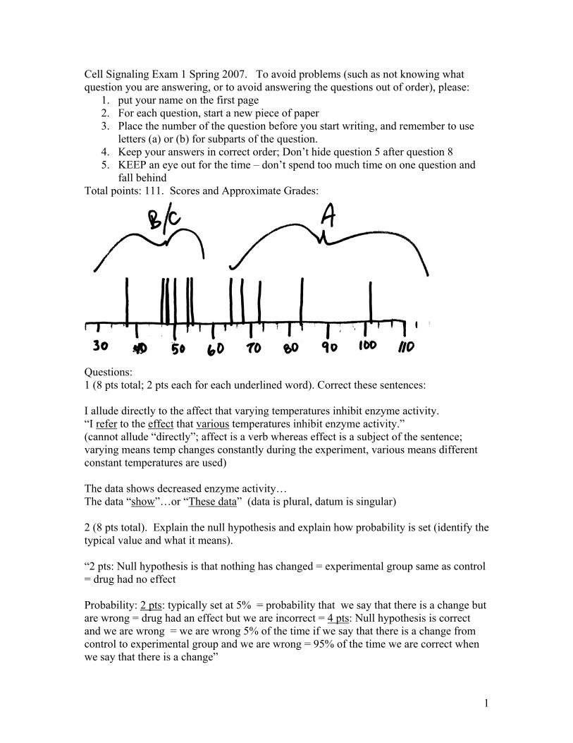
fall behind Total points: 111. Scores and Approximate Grades:
Questions: 1 (8 pts total; 2 pts each for each underlined word). Correct these sentences: I allude directly to the affect that varying temperatures inhibit enzyme activity. “I refer to the effect that various temperatures inhibit enzyme activity.” (cannot allude “directly”; affect is a verb whereas effect is a subject of the sentence; varying means temp changes constantly during the experiment, various means different constant temperatures are used) The data shows decreased enzyme activity… The data “show”…or “These data” (data is plural, datum is singular) 2 (8 pts total). Explain the null hypothesis and explain how probability is set (identify the typical value and what it means). “2 pts: Null hypothesis is that nothing has changed = experimental group same as control = drug had no effect Probability: 2 pts: typically set at 5% = probability that we say that there is a change but are wrong = drug had an effect but we are incorrect = 4 pts: Null hypothesis is correct and we are wrong = we are wrong 5% of the time if we say that there is a change from control to experimental group and we are wrong = 95% of the time we are correct when we say that there is a change”

2
3. (9 pts total) (a) Name and define the two types of t Tests, (b) tell me which is the best form to use and why, and (c) pick the one you would use based on the following experimental protocol: I measure enzyme activity in different plates of cells in culture by taking optical density readings with a spectrometer. I then alter the temperature to a non-lethal value and again measure the enzyme activity in the plates. “(a) 4 pts: the paired t test is where the same samples or subjects are used for both control and experimental groups, pooled t Test is where control group samples/subjects are different from those in the experimental group (b) 3 pts: Best test is the paired t test; variance is minimized to provide for the most powerful test. With a pooled t Test, you may say that there is no difference between control and experimental group yet there is. For example, an athlete with very low blood pressure is in your control group, this will increase the variance in the control group and obscure any difference in blood pressure between the control group and the experimental group if you use the pooled t Test. Use of the paired t test would not give you this problem as the athlete is in both control and experimental and you are looking only at the change (difference) between the athlete’s control and experimental value. The paired t Test examines the change/difference in each individual, whereas the pooled t test merely compares the average and standard deviation of the controls versus the experimental groups. That is, the paired t Test is better than the pooled t test because abnormal values –high or low- weaken the power of the pooled t Test- there might be a significant difference but you would not see it” (c) 2 pts: paired t Test (same sample used for both control and experimental group). 4. 11 pts total (a) Hormones typically do not regulate which steps noted below (write down the number(s) and the name)? In regulatory method number one below
(b) name the protein family that is directly involved, (c) how this protein is typically turned on by what kind of enzyme (discuss and
explain any conformational changes) (d) where the protein family member binds to DNA and why (e) how the protein family member binds to DNA (give examples)
3
“(a) 2pts: steps 2 and 3 are not typically regulated by hormones (b) 2 pts: transcription factors (c) 2 pts: kinases typically turn on transcription factors by putting a phosphate group onto an amino acid in the transcription factor; this new phosphate group makes new weak bonds (probably ionic) and distorts the transcription factor; this physical distortion activates the transcription factor (puts it in the correct shape to bind DNA) (d) 3 pts: the transcription factor binds to the (1pt) major groove typically at the enhancer region on DNA because (2 pts) there are more possible sites of interaction there than at the minor groove. Also possible: more room for protein in major groove. (e) 2 pts: (1pt) weak bonds (not strong bonds like the covalent bond) are used to bind the transcription factor to the DNA. Examples: hydrogen bond, hydrophobic interaction, weak ionic bond, …Or this example: 10-20 weak bonds involving 6-12 nucleotides” (1pt) The structure of the transcription factor is important: helix-turn-helix, leucine zipper, helix-loop-helix, zinc finger- is important as the protein wraps around the DNA (often like a close pin). 5. (10 pts total) Compare and contrast the affinity of endocrine and synaptic receptors for their ligand. Explain any differences in affinity. “(2pts)The endocrine hormone is dumped into the blood stream where it is diluted to as little as 10-10 Molar. (the blood stream rapidly moves the hormone through convection) This means that the target cells (2pts) must have a receptor that shows very high affinity to be able to bind this trace amount of hormone. However, the neurotransmitter is released right (2pts)into the area next to where the receptor is found (although the hormone has to move by diffusion not convection, the distance is small) and the concentration of the neurotransmitter is higher -for acetylcholine: 5 x 10⁻⁴ Molar. Thus, the (2pts) affinity of the receptor for the neurotransmitter is lower than that of endocrine hormone receptors.” 6. (9 pts total) Soluble Steroid receptors: (a) name the three domains found within them (b) explain how the receptors are activated and note the role of the three domains. (c) briefly note what the activated receptor does (do not get into the primary or secondary responses). (d) The receptor is what type of DNA binding protein? a) 3 pts: transcription activating domain, DNA binding domain, ligand binding domain b) see below; steroid binds, (1pt)ligand binding domain closes in over steroid and removes the inhibitory protein, (1pt) DNA binding domain binds to enhancer regions, (1pt) transcription activating domain binds coactivator. c) 1pt: activated receptor binds to enhancer regions to turn on a gene (increase mRNA production) d) 2pts: member of the zinc finger family of transcription factors

4
2525
for its movement, see next slide
FIG. 15-13 RECEPTOR ORIGINALLYBOUND TO INHIBITORNOTE THREE DOMAINSAND N, C TERMINI
NOTE HOW ACTIVATION OCCURS
2626
Note the extensiveAlpha helices
The blue alpha helix(ligand binding domain)Acts like a lid and slams closed (a 180 degree movement) when the Ligand is present
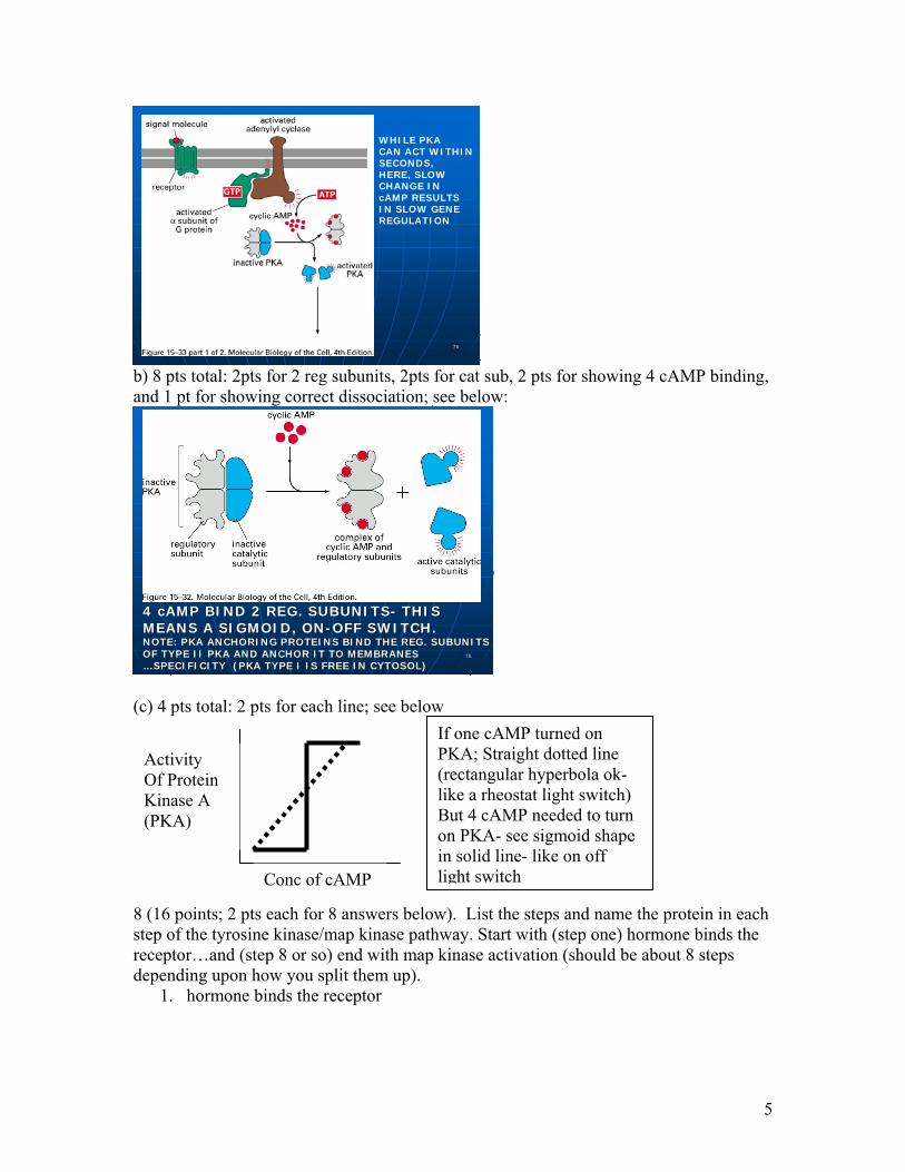
7. 16 pts total (a) Briefly, name the proteins in the cAMP system…start with “hormone binds receptor” and continue with a list of the proteins in the proper order through activation of protein kinase A. (b) Explain how cAMP activates protein kinase A (use a drawing). (c) for protein kinase A activation, show the kinetics when you graph kinase A activity on the Y axis, and the concentration of cAMP on the X axis. Use a solid line. Now draw a dotted line for the kinetics if only one cAMP would activate kinase A. “a) 4 pts total: hormone binds a serpentine/7 transmembrane receptor, (1pt) G protein turned on –Gs- as it binds GTP and dissociates GDP, (1pt) adenylate (or adenyl) cyclase turned on by Gs protein alpha subunit, (1pt) cAMP made from ATP, (1pt) cAMP binds and activates protein kinase A. See slide below.
5
7676
WHILE PKACAN ACT WITHINSECONDS,HERE, SLOWCHANGE IN cAMP RESULTSIN SLOW GENEREGULATION
b) 8 pts total: 2pts for 2 reg subunits, 2pts for cat sub, 2 pts for showing 4 cAMP binding, and 1 pt for showing correct dissociation; see below:
7575
4 4 cAMPcAMP BIND 2 REG. SUBUNITSBIND 2 REG. SUBUNITS-- THIS THIS MEANS A SIGMOID, ONMEANS A SIGMOID, ON--OFF SWITCH.OFF SWITCH.NOTE: PKA ANCHORING PROTEINS BIND THE REG. SUBUNITS NOTE: PKA ANCHORING PROTEINS BIND THE REG. SUBUNITS OF TYPE II PKA AND ANCHOR IT TO MEMBRANES OF TYPE II PKA AND ANCHOR IT TO MEMBRANES …SPECIFICITY (PKA TYPE I IS FREE IN CYTOSOL)…SPECIFICITY (PKA TYPE I IS FREE IN CYTOSOL)
(c) 4 pts total: 2 pts for each line; see below
8 (16 points; 2 pts each for 8 answers below). List the steps and name the protein in each step of the tyrosine kinase/map kinase pathway. Start with (step one) hormone binds the receptor…and (step 8 or so) end with map kinase activation (should be about 8 steps depending upon how you split them up).
1. hormone binds the receptor
If one cAMP turned on PKA; Straight dotted line (rectangular hyperbola ok- like a rheostat light switch) But 4 cAMP needed to turn on PKA- see sigmoid shape in solid line- like on off light switchConc of cAMP
Activity Of Protein Kinase A (PKA)

6
2. receptors dimerize (two come together) to activate the receptors, one receptor puts a tyrosine on the other receptor’s tyrosine (transphosphorylation)
3. grb2 binds to phosphorylated tyrosine and is activated 4. grb2 binds and activates Sos 5. Sos binds activates Ras 6. Ras binds and turns on Raf (map kinase kinase kinase) 7. Raf phosphorylates and activates MEK (map kinase kinase) 8. MEK phosphorylates and turns on ERK (map kinase)
9 (8 points; 2 pts each). Describe the last four events (in proper order; ignore any feedback loops) that result in cdk1 activation at the G2/M phase border. Name the proteins involved. Answer: 1.cyclin B binds cdk1, 2. Wee 1 kinase inhibits by putting two phosphates on cdk1, 3. CAK (kinase) puts on activating phosphate but wee1 phosphates still inhibits, 4. then cdc25 removes inhibitory phosphate and cdk1 is turned on.
10. (8 pts total) How does one signal result in activation of different signaling pathways that lead to cell division? It is known that addition of a hormone to T cells results in activation of the T cell through stimulation of the hormone receptor. In a paper in Nature Immunology, these data were found (I have added some supporting data): --If you knock out the gene for a soluble protein called Dlgh1, activation of T cells by agonist cannot rapidly turn on a relay protein called p38 kinase, and T cell activation is partially inhibited. Knocking out a gene “kills” the gene (protein it codes for is nonfunctional). --However, the knock out of Dlgh1 does not stop rapid activation of other kinases (Erk or Jnk) after hormone addition to T cells. --Neither NF-κB activation or calcium release are blocked by the knock out of Dlgh1. --If only the genes for 2 kinases - Zap70 and Lck - are knocked out, p38 does not get activated after agonist addition. However, in cells with these 2 kinase genes knocked out, Erk and Jnk are still rapidly activated after agonist addition to T cells. --If you precipitate (with antibodies; this is immunoprecipitation) Dlgh1, you find that p38, Zap70 and Lck are also precipitated. Erk and Jnk are not co-immunoprecipitated. --Dlgh1 does not have kinase activity and it is not a G protein.
7
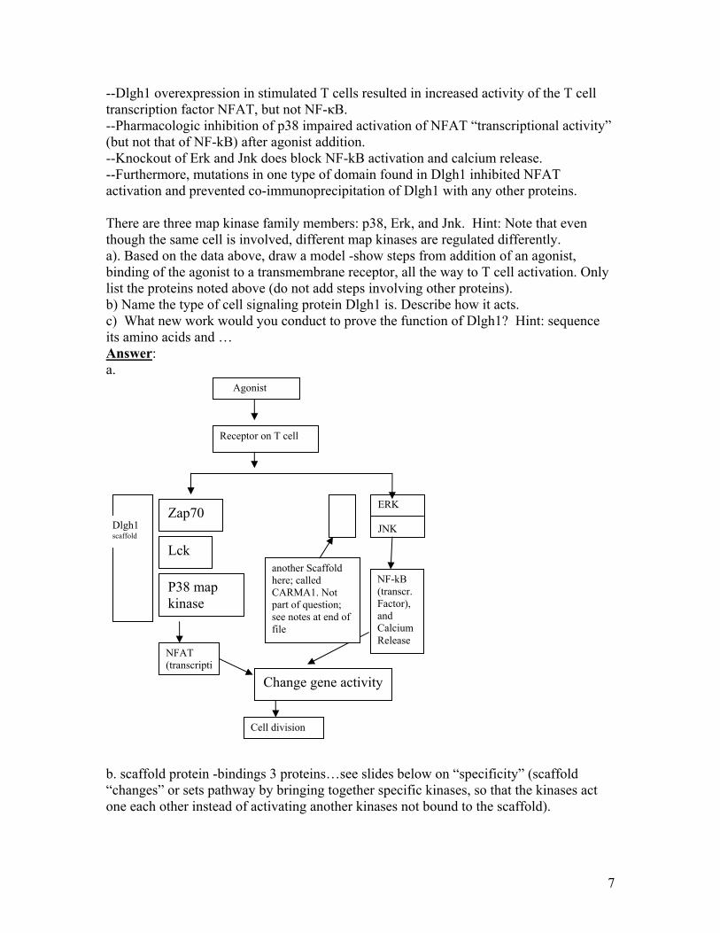
--Dlgh1 overexpression in stimulated T cells resulted in increased activity of the T cell transcription factor NFAT, but not NF-κB. --Pharmacologic inhibition of p38 impaired activation of NFAT “transcriptional activity” (but not that of NF-kB) after agonist addition. --Knockout of Erk and Jnk does block NF-kB activation and calcium release. --Furthermore, mutations in one type of domain found in Dlgh1 inhibited NFAT activation and prevented co-immunoprecipitation of Dlgh1 with any other proteins. There are three map kinase family members: p38, Erk, and Jnk. Hint: Note that even though the same cell is involved, different map kinases are regulated differently. a). Based on the data above, draw a model -show steps from addition of an agonist, binding of the agonist to a transmembrane receptor, all the way to T cell activation. Only list the proteins noted above (do not add steps involving other proteins). b) Name the type of cell signaling protein Dlgh1 is. Describe how it acts. c) What new work would you conduct to prove the function of Dlgh1? Hint: sequence its amino acids and … Answer: a.
b. scaffold protein -bindings 3 proteins…see slides below on “specificity” (scaffold “changes” or sets pathway by bringing together specific kinases, so that the kinases act one each other instead of activating another kinases not bound to the scaffold).
Agonist
Receptor on T cell
Dlgh1 scaffold
NFAT (transcripti
ERK JNK
NF-kB (transcr. Factor), and Calcium Release
Cell division
another Scaffold here; called CARMA1. Not part of question; see notes at end of file
Zap70
Lck
P38 map kinase
Change gene activity
8
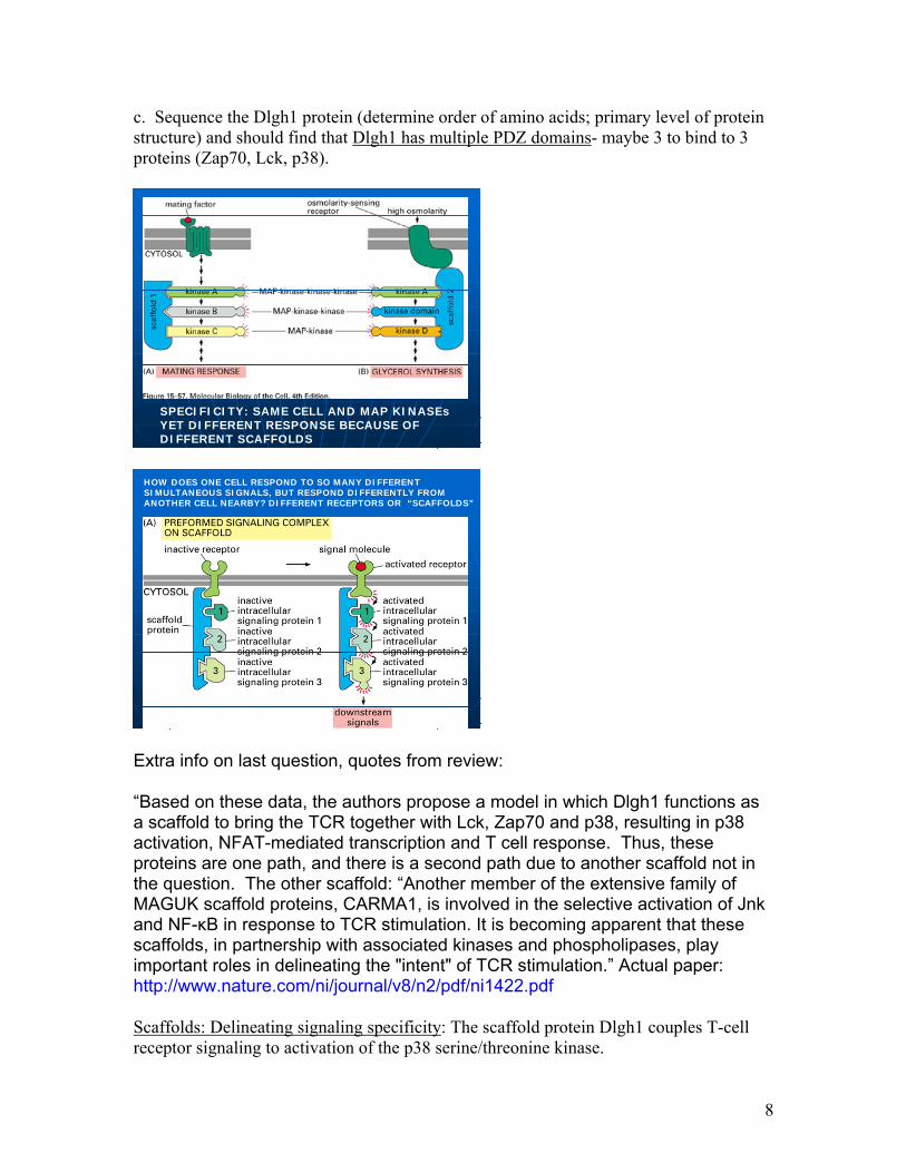
c. Sequence the Dlgh1 protein (determine order of amino acids; primary level of protein structure) and should find that Dlgh1 has multiple PDZ domains- maybe 3 to bind to 3 proteins (Zap70, Lck, p38).
SPECIFICITY: SAME CELL AND MAP KINASEsYET DIFFERENT RESPONSE BECAUSE OF DIFFERENT SCAFFOLDS
HOW DOES ONE CELL RESPOND TO SO MANY DIFFERENT SIMULTANEOUS SIGNALS, BUT RESPOND DIFFERENTLY FROM ANOTHER CELL NEARBY? DIFFERENT RECEPTORS OR “SCAFFOLDS”
Extra info on last question, quotes from review:
“Based on these data, the authors propose a model in which Dlgh1 functions as a scaffold to bring the TCR together with Lck, Zap70 and p38, resulting in p38 activation, NFAT-mediated transcription and T cell response. Thus, these proteins are one path, and there is a second path due to another scaffold not in the question. The other scaffold: “Another member of the extensive family of MAGUK scaffold proteins, CARMA1, is involved in the selective activation of Jnk and NF-κB in response to TCR stimulation. It is becoming apparent that these scaffolds, in partnership with associated kinases and phospholipases, play important roles in delineating the "intent" of TCR stimulation.” Actual paper: http://www.nature.com/ni/journal/v8/n2/pdf/ni1422.pdf
Scaffolds: Delineating signaling specificity: The scaffold protein Dlgh1 couples T-cell receptor signaling to activation of the p38 serine/threonine kinase.

9
Antigenic stimulation of T-cell receptors (TCRs) promotes the activation of a myriad of cytoplasmic signal transduction pathways to affect T cell-mediated adaptive immunity. How a common signal, such as TCR stimulation, activates several discrete signaling networks to promote distinct cellular responses is an area of intense research. In Nature Immunology, Miceli and colleagues report that TCR signaling causes the membrane-associated guanylate kinase (MAGUK) scaffold protein Dlgh1 to bind to and facilitate activation of the p38 serine/threonine kinase, which in turn specifically stimulates NFAT-mediated transcription.
TCR stimulation promotes the recruitment of adaptor and scaffold proteins (including Dlgh1), kinases (including Zap70 and Lck), phospholipases and small GTPases into complexes known as "signalosomes", which coordinate the T cell's response to antigens. p38 is a well known effector of TCR-mediated immune response, and previous work has shown that Lck and Zap70-mediated phosphorylation of p38 is involved in its activation following TCR stimulation. Miceli and colleagues found phosphorylated p38 in complex with Dlgh1 in stimulated T cells. siRNA-mediated depletion of Dlgh1 in stimulated T cells resulted in a decrease in p38 phosphorylation, but had no effect on phosphorylation of Erk or Jnk. NF-κB activity and calcium release were similarly unaffected by Dlgh1 depletion, suggesting that Dlgh1 specifically links TCR signaling to p38 activation. What, then, are the cellular consequences of this type of p38 activation? Dlgh1 overexpression in stimulated T cells resulted in increased activity of the T cell transcription factor NFAT, but not NF-κB. Pharmacologic inhibition of p38 impaired TCR-induced NFAT transcriptional activity. Furthermore, mutations introduced in the p38 binding domain of Dlgh1 abrogated NFAT phosphorylation and activity, supporting a role for Dlgh1 in coupling TCR signaling to p38 phosphorylation and subsequent NFAT activation. Based on these data, the authors propose a model in which Dlgh1 functions as a scaffold to bring the TCR together with Lck, Zap70 and p38, resulting in p38 activation, NFAT-mediated transcription and T cell response. Another member of the extensive family of MAGUK scaffold proteins, CARMA1, is involved in the selective activation of Jnk and NF-κB in response to TCR stimulation. It is becoming apparent that these scaffolds, in partnership with associated kinases and phospholipases, play important roles in delineating the "intent" of TCR stimulation. Emily J. Chenette Signaling Gateway Original References: June L. Round, Lisa A. Humphries, Tamar Tomassian, Paul Mittelstadt, Min Zhang & M. Carrie Miceli. Scaffold protein Dlgh1 coordinates alternative p38 kinase activation, directing T cell receptor signals toward NFAT but not NF-κB transcription factors Nature Immunology 8, 154 - 161 (2007) Full text | PDF | Subscribe to Nature Immunology Mercedes Rincón & Roger J. Davis. Choreography of MAGUKs during T cell activation Nature Immunology 8, 126 - 127 (2007) Full text | PDF | Subscribe to Nature Immunology See also: http://www.signaling-gateway.org/update/updates/200602/nri1799.html