Embed Size (px)
Citation preview

UNIVERSIDADE DE LISBOA
FACULDADE DE CIÊNCIAS
DEPARTAMENTO DE BIOLOGIA ANIMAL
Cell-extracellular matrix interactions during
early paraxial mesoderm development
Catarina Teixeira Lopes
Mestrado em Biologia Evolutiva e do Desenvolvimento
2007

2
UNIVERSIDADE DE LISBOA
FACULDADE DE CIÊNCIAS
DEPARTAMENTO DE BIOLOGIA ANIMAL
Cell-extracellular matrix interactions during
early paraxial mesoderm development
Catarina Teixeira Lopes
Mestrado em Biologia Evolutiva e do Desenvolvimento
Dissertação orientada por Prof. Doutora Maria Gabriela Rodrigues
2007

3
ACKNOWLEDGEMENTS
I would like to thank my tutors Gabriela Rodrigues and Sólveig Thorsteinsdóttir for their
always helpful guidance and advice throughout these past two years and for their suggestions to
make my work even better. I would like to thank Pedro Rifes for all the patience he had to teach
me almost everything he knows about Development Biology and basically how to work in a
laboratory. Besides, Pedro is the best co-worker one can aspire to work with. This thesis would
not be completed in such a short period of time if it wasn’t for Ana Gaspar and her expertise in
cryosectioning. I want also to thank Ana for being the only person I can really talk about art and
drawing. I would like to thank Gabriel Martins for being someone I can share my American
knowledge but especially for always being there to help us obtain the best microscopy images
ever. I could not forget to thank Luis Marques for being in these past months a thesis buddy but
especially for his sense of humour and his knowledge about movies! A special thank you to
Raquel Andrade and the Braga “girl power” group for sending those precious probes, which
were the starting point of my journey in Development Biology and also for Leonor Saúde and
Raquel Lourenço for teaching me how to reach that starting point. I would like to thank my
parents for being exactly that, my caring, patient and loving parents! I thank you, Tiago, for all
your love and patience, basically for being my comfort zone. Last, but not the least, a very
special thanks to all my friends for giving support to carry on and for not being mad for all the
times I couldn’t show up because of my work…Thank you Teresa, you are the special friend that
I want to treasure!

4
RESUMO
A gastrulação é uma das fases mais determinantes do desenvolvimento embrionário
animal, durante a qual a morfologia dos embriões é altamente reestruturada devido à elevada
taxa de remodelação e migração celular. No caso específico da galinha, inicialmente, o embrião
adquire uma forma discoidal e apresenta uma única camada de células, a blastoderme. Antes da
gastrulação iniciar, a blastoderme ainda vai dar origem a duas camadas celulares separadas por
uma cavidade: o epiblasto, dorsalmente, que vai sobretudo originar o embrião em si e o
hipoblasto que vai dar origem apenas a estruturas extra-embrionárias. A gastrulação inicia-se
com a formação de um sulco ao longo de todo o embrião, a linha primitiva, o qual estabelece o
eixo antero-posterior do embrião. Segue-se a formação do nó de Hensen, ou seja, um aglomerado
de células na extremidade anterior da linha primitiva que começa a regredir, percorrendo a linha
primitiva no sentido antero-posterior do embrião. Durante todo este processo, dá-se a migração
de células do epiblasto, através quer da linha primitiva, quer do nó de Hensen, em direcção à
cavidade (blastocélio) para originar a endoderme embrionária, que desloca o hipoblasto para os
lados, e o terceiro folheto embrionário, a mesoderme. Nesse sentido, as células do epiblasto que
migram através do nó vão dar origem à notocorda, ao mesênquima cefálico, e à parte dorsal do
tubo digestivo. Duas massas aproximadamente cilíndricas de células mesenquimatosas,
denominadas de mesoderme paraxial ou pré-somitica (MPS) individualizam-se de cada lado do
tubo neural emergente. Esta MPS acaba por sofrer um processo de segmentação, durante o qual
se separam periodicamente blocos esféricos de células, a partir da sua extremidade anterior,
designados por sómitos. Assim, à medida que a gastrulação prossegue através da adição de novas
células à MPS mais posterior, o embrião alonga-se e o processo de somitogénese continua a
epitelializar células da MPS mais anterior. Mais tarde, os sómitos dão origem ao esqueleto axial,
a todos os músculos esqueléticos, aos tendões e à derme do tronco e ainda definem o padrão
segmentado dos nervos axiais periféricos. Este processo de somitogénese é estritamente regulado
no tempo e espaço, sendo variável para cada espécie (no caso da galinha, cada sómito separa-se
da MPS a cada 90 minutos) (Christ and Ordahl, 1995; Gossler and Hrabe de Angelis, 1998), e é
essencial para o desenvolvimento normal do embrião nos vertebrados.

5
Todo o processo de gastrulação, nomeadamente a somitogénese, envolve rearranjos
celulares extensos, onde as células mesenquimatosas da MPS se convertem em sómitos
epiteliais, a qual consiste numa transição mesênquima-epitélio. Estas transições envolvem
rearranjos intensivos das interacções célula-célula e célula-matriz extracelular. A matriz
extracelular (MEC) consiste numa rede intrincada de diversas proteínas fibrosas e
glicosaminoglicanos, a qual é secretada localmente maioritariamente por células
mesenquimatosas (fibroblastos). Dada a sua variada composição, a MEC pode apresentar
diversas funções, entre as quais fornecer um substrato às células para aderirem e proliferarem,
modulando directamente a forma e funções celulares e regular a comunicação intercelular
durante a diferenciação (Roskelley et al., 1995). Um componente da MEC cuja importância na
somitogénese é amplamente reconhecida é a fibronectina. A fibronectina é uma glicoproteína
constituída por dois monómeros idênticos, ligados entre si por duas pontes bissulfito e que
contêm vários locais de ligação que podem interagir com uma grande variedade de moléculas,
nomeadamente com receptores proteicos da membrana celular chamados integrinas (Pankov and
Yamada, 2002; Yamada et al., 2003). As integrinas pertencem a uma família de proteínas
transmembranares diméricas que estabelecem a ligação física das moléculas da MEC ao
citosqueleto da célula, nomeadamente à rede intracelular de actina (Danen and Sonnenberg,
2003; Miranti and Brugge, 2002). Em relação à fibronectina, esta pode exercer diversas funções,
tais como promover a adesão, a migração e a proliferação celular, interferindo com a
morfogénese (Robinson et al., 2004). A fibronectina também desempenha um papel fundamental
durante a somitogénese uma vez que, em ratinhos mutantes nulos para fibronectina, não se
formam sómitos (George et al., 1993; Georges-Labouesse et al., 1996). Mais recentemente, num
artigo publicado pelo nosso laboratório, avaliou-se a contribuição relativa da ectoderme e de uma
matriz intacta de fibronectina na somitogénese. Neste trabalho, demonstrou-se que a matriz de
fibronectina é essencial para o processo morfológico da somitogénese e que esta matriz é
facilmente removida por enzimas como a dispase. Em (Rifes et al., 2007), foi também sugerido
que a principal função da ectoderme, durante a somitogénese, consiste na produção de
fibronectina, enquanto que a porção mais posterior da MPS expressa o seu principal receptor, a
integrina α5 (Itga5).
Tendo em conta este conhecimento prévio, o principal objectivo deste trabalho consistiu
em analisar o padrão de expressão da fibronectina e dois dos seus receptores mais comuns

6
(integrinas α4 e α5) no embrião de galinha ao longo das primeiras 72 horas de desenvolvimento.
Nesse sentido, recorreu-se à técnica de hibridação in situ para os três estádios de
desenvolvimento (HH4-5, HH12-14 e HH19-20). Como resultado verificou-se que o RNAm da
fibronectina é fortemente expresso na maioria dos epitélios, sobretudo no epiblasto e,
posteriormente, na ectoderme. Quanto aos receptores da fibronectina (Itga5 e Itga4), estes estão
expressos inicialmente na linha primitiva e nó de Hensen. No entanto, a partir do estadio HH12-
14, os RNAms destas integrinas encontram-se sobretudo na mesoderme e seus derivados,
nomeadamente nos sómitos. No entanto, verifica-se que a integrina Itga5 está também expressa
no coração e primórdios vasculares, enquanto que a integrina Itga4 está também expressa no
lado dorsal do tubo neural. No sentido de confrontar estes resultados com o padrão de expressão
da proteína da fibronectina, usou-se a técnica de imuno-histoquímica nos mesmos três estádios.
Como resultado, confirmou-se que a proteína da fibronectina constitui um dos componentes mais
abundantes da matriz extracelular, estando presente nas lâminas basais dos epitélios,
nomeadamente do epiblasto e, mais tarde, da ectoderme e nos espaços extracelulares do tecido
mesenquimatoso.
Adicionalmente, foi estabelecido um sistema in vitro de culturas de mesodermes pré-
somíticas isoladas, no sentido de discriminar o efeito de diferentes enzimas e substratos no
comportamento e morfologia celulares. Assim, recorreu-se novamente à técnica da imuno-
citoquímica para observar a expressão de diversas moléculas importantes para a somitogénese
em MPSs totalmente isoladas dos tecidos adjacentes. Nesse sentido, usaram-se duas enzimas
(dispase e colagenase) para obter os explantes de MPSs, os quais foram postos em cultura sobre
três substratos diferentes (lamelas cobertas com fibronectina plasmática, gelatina ou matrigel).
Numa primeira abordagem, verificou-se que as células das mesodermes paraxiais isoladas
conseguem refazer uma matriz de fibronectina à sua volta em qualquer um dos substratos,
mesmo após esta matriz ter sido destruída por acção da enzima dispase. Verificou-se também
que as células das MPSs, quando cultivadas sobre fibronectina, apresentam uma maior agregação
das células em torno do explante inicial, enquanto que as células cultivadas sobre matrigel
(substrato rico em laminina) apresentam uma maior migração celular a partir do explante. No
entanto, todas estas células apresentam baixos níveis de N-caderina e β-catenina em todas as
experiências, sugerindo que as MPSs em cultura permanecem mesenquimatosas e não fazem a
transição mesênquima-epitélio, característica da somitogénese. A N-caderina é uma proteína

7
transmembranar, dependente de cálcio, que medeia a adesão célula-célula ao formar junções
aderentes estáveis entre elas. No entanto, as caderinas também participam na sinalização
intracelular através da ligação dos seus domínios citoplasmáticos à β-catenina, a qual é também
um modelador da via canónica de sinalização Wnt (Derycke and Bracke, 2004; Lilien et al.,
2002). Por último, verificou-se se ocorreu alguma proliferação celular ou até apoptose (morte
celular programada). Através de uma marcação para a histona 3 fosforilada e caspase 3,
confirmou-se que tanta a proliferação como a morte celular não é significativa nas várias
condições experimentais, respectivamente.
Como conclusão, este trabalho demonstra como a presença da fibronectina e seus
principais receptores (integrinas α4 e α5) é significativa em vários tecidos diferentes, ao longo
das primeiras 72 horas de desenvolvimento do embrião de galinha (estádios HH4-20).
Futuramente, será interessante analisar os padrões de expressão de outros componentes
importantes da matriz extracelular, nomeadamente a laminina e os diversos proteoglicanos,
durante estas fases mais precoces do desenvolvimento embrionário. Outra questão a aprofundar
em futuros trabalhos será a análise do padrão de expressão da integrina Itga4, dado que o RNAm
deste receptor apresenta uma expressão variável em diferentes embriões do mesmo estádio
(HH12-14). Por fim, é também importante aperfeiçoar a técnica de cultura de mesodermes pré-
somíticas isoladas, dado que oferece uma maior acessibilidade e consequente manipulação
destas. Posteriormente, há que experimentar usar meio sem soro ou até adicionar factores
difusíveis, tais como o ácido retinóico, Wnts e Fgf8, de modo a mimetizar a ectoderme in vivo e
verificar se estes factores favorecem ou não a epitelização das células da MPS.
Palavras-chave: desenvolvimento precoce da galinha, mesoderme pré-somítica, matriz
extracelular, fibronectina, integrinas, cultura de células.

8
ABSTRACT
Fibronectin is an abundant glycoprotein of the extracellular matrix, which has been
shown to play a role in a variety of functions such as cell proliferation, survival, migration and
differentiation. Fibronectin is assembled into a fibrillar matrix through a cell-mediated process
involving their heterodimeric membrane receptors, integrins. Recently, the work of our group
has evidenced the importance of fibronectin in somitogenesis and how the ectoderm and
mesoderm collaborate in the construction of the fibronectin matrix of presomitic mesoderm
(Rifes et al., 2007). In the present work, we further characterised the expression patterns of
fibronectin (Fn1), integrin α5 (Itga5), the main sub-unit involved in fibronectin assembly, and
integrin α4 (Itga4), during chick embryonic development. Fn1 transcripts are strongly expressed
in most epithelia, mainly in the ectoderm in all stages studied. Itga5 is initially expressed in the
primitive streak and node and it is later found in the ingressing mesoderm and many of its
derivates, especially the heart and vascular primordia. Itga4 transcripts are also detected in the
node and mesoderm, but later it is expressed in the dorsal neural tube and somite derivates.
Finally, we also established an in vitro culture system of presomitic mesoderm cells by using
different enzymes and substrata. Immunofluorescence labelling of fibronectin shows that isolated
PSM cells in culture can assembly a fibronectin matrix, even when it was previously removed by
dispase. In the tested culture conditions, these cells remained in a non-proliferative
mesenchymal-like state, expressing very low levels of both N-cadherin and β-catenin. These data
suggest that fibronectin is not only an important component of the extracellular matrix in vivo
and in vitro, but also that its presence in various tissues during the early stages of chick
development is imperative.
Keywords: chick early development, presomitic mesoderm, extracellular matrix, fibronectin,
integrins, cell culture.

9
TABLE OF CONTENTS
ACKNOWLEDGEMENTS…………………………………………………………….................3
RESUMO ……………………………………………………………………………....................4
ABSTRACT……………………………………………………………………………….............8
INTRODUCTION
The early development of the chick embryo……………………………………………………..11
The extracellular molecules……………………………………………………………………...13
The importance of the ECM in the early development of the chick embryo…………………….14
MATERIALS AND METHODS
Eggs and embryos………………………………………………………………………………..17
Cryosection....……………………………………………………………………………………17
In situ hybridisation probes………………………………………………………………………17
Whole mount in situ hybridisation……………………………………………………………….18
PSM cell cultures………………………………………………………………………………...19
Antibodies and immunohistochemistry………………………………………………………….20
RESULTS
Expression pattern for chick fibronectin protein at stages HH4-HH20.........................................22
Expression pattern for chick Fn1 transcripts at stages HH4-HH20...............................................23
Expression pattern for chick Itga5 transcripts at stages HH4-HH20.............................................25
Expression pattern for chick Itga4 transcripts at stages HH4-HH20.............................................26
Assembly of fibronectin matrix and cell behaviour of PSM cells in vitro....................................29
Cell-extracellular matrix interactions in cell epithelialisation behaviour of PSM cells in vitro...31
Cell proliferation in PSM cells in vitro…………………………………………………………..33
Cell apoptosis in PSM cells in vitro……………………………………………………………...35

10
DISCUSSION
The presence of fibronectin is essential for normal gastrulation and somitogenesis.....................36
The integrin receptors for fibronectin in embryo development.....................................................37
Analysis of presomitic mesoderm cell behaviour growing on different extracellular matrix substrata in vitro.............................................................................................................................38
BIBLIOGRAPHY………………………………………………………………………………41

11
INTRODUCTION
The early development of the chick embryo
In the beginning of chick development, a flat monolayer disc of cells called blastoderm,
gives rise to two new layers: the upper layer of embryonic cells or epiblast and the underlying
hypoblast. When gastrulation begins, the primitive streak is formed, i.e., a furrow in the midline
of the embryonic disk that extends from the future posterior end of the embryo towards the
centre. Epiblast cells ingressing through the primitive streak into the blastocoel replace the
hypoblast cells and generate the endoderm and mesoderm layers. As gastrulation proceeds, a
thickening of cells in the anterior end of the primitive streak, the Hensen’s node, migrates
posteriorly, moving along the elongating embryo, and eventually reaching the region of tail bud.
As the Hensen’s Node migrates posteriorly, cells ingressing through the node will give rise to the
head mesenchyme, the dorsal endoderm and the notochord, anteriorly to Hensen’s node. As a
consequence, the neural tube forms anteriorly to Hensen’s node. Moreover, the mesodermal cells
will be placed in a medial-proximal position: axial (the notochord), and the paraxial,
intermediate and lateral mesoderm. The notochord, placed beneath the neural tube, is a transient
structure that defines the embryonic midline throughout organogenesis. The paraxial mesoderm,
placed on each side of the axis, will give rise to the skeletal, muscle and tendon precursors, as
well as the dorsal dermis. The urogenital system is derived from the intermediate mesoderm,
while the lateral mesoderm is the precursor of the vascular system and lining of the body
cavities.
The presomitic mesoderm (PSM) will originate the axial skeleton and all skeletal muscles
in the trunk and limbs. The segmentation of the rod-shaped mesenchymal PSM through
somitogenesis will underline the metamerism during vertebrate development. The periodicity of
segmentation is species-specific and, in the case of chick embryogenesis, a pair of somites buds
off from the anterior PSM every 90 minutes, reaching a total of 52 pairs of somites (Pourquié,
2004). The newly formed somites consist of epithelial balls of columnar cells enveloping
mesenchymal cells within a central cavity, the somitocoel. At the same time a new somite arises
from the anterior PSM new cells are added in the posterior end as gastrulation continues in the

12
region of the Hensen’s node, and later in the tail bud. Later, the somites develop from the
paraxial mesoderm through a process of segmentation and constitute the segmental pattern of the
body. However, prior to its morphological segmentation, the presomitic mesoderm undergoes a
rostral to caudal regionalization at the molecular level, which begins to define the prospective
somites (Saga and Takeda, 2001). Driven by changes in the expression of adhesion molecules,
somitomeres (loose masses of mesoderm) compact and give rise to successive pairs of somites
by a mesenchymal to epithelial transition (Duband et al., 1987; Kulesa and Fraser, 2002), first at
the cranial end of the paraxial mesoderm and proceeding caudally, while new mesenchyme cells
enter the posterior paraxial mesoderm as a consequence of continuous gastrulation (Christ and
Ordahl, 1995). These somites bud off of the paraxial or presomitic mesoderm in a pairwise
manner, bilateral to the axial notochord and neural tube (reviewed in (Gossler and Hrabe de
Angelis, 1998; Stockdale et al., 2000), The newly formed somites consist of epithelial balls of
columnar cells enveloping mesenchymal cells within a central cavity, the somitocoel. The
process of somitogenesis lasts until the number of somites characteristic of the species is
reached; in the chick embryo, it corresponds to the formation of 52 pair of somites during the
first five days of development (Aulehla and Herrmann, 2004; Freitas et al., 2005; Pourquié,
2004). Moreover, this process of gastrulation is followed by organogenesis, when individual
tissues and organs develop within the newly formed germ layers (ectoderm, endoderm, and
mesoderm). The anterior segment of the neural tube forms the three main parts of the brain: the
forebrain, midbrain, and the hindbrain, which are later divided into five sub-regions. The
subsequent maturation process of the somites along the anterior–posterior axis generates all
skeletal muscles of the body (originating from the dermomyotome), the axial skeleton
(sclerotome) and part of the dermis (originating from the dermomyotome) and imposes a
segmental pattern to peripheral nerves and vascular primordia (Christ and Ordahl, 1995; Gossler
and Hrabe de Angelis, 1998)

13
The extracellular molecules
During the process of gastrulation, embryonic structures are surrounded by extracellular
matrix (ECM) material connecting them to adjacent tissues. The ECM constitutes an intricate
network of macromolecules, composed by a variety of fibrous proteins and glycosaminoglycans
(GAGs) that are secreted via exocytosis mainly by resident cells of the mesodermal tissues
(fibroblasts) and assembled into an organized tri-dimensional structure. Due to its diverse nature
and composition, the ECM can serve many functions, such as providing support and anchorage
for cells to migrate and proliferate, segregating tissues from one another and regulating
intercellular communication during cell differentiation and morphogenesis (Hay, 2005; Larsen et
al., 2006; Zagris, 2001). Extracellular molecules are linked to one another by multiple binding
domains and form a stable, multifunctional matrix. The cell response to ECM is mediated
through plasma membrane receptors called Cell Adhesion Molecules (CAMs), which include
four protein families: the immunoglobulins, the selectins, the integrins and the cadherins (Hynes,
1992). This last class of transmembrane proteins play important roles in cell adhesion (via
adherens junctions) and cell migration. Their function is calcium-dependent and their
intracellular domain binds to β-catenin, which is also an important component in the Wnt
signalling pathway (Lilien et al., 2002).
Figure 1- Early stages of chick development (A) At stage HH4, the primitive streak is fully extended along the midline (lightly stained groove with darker staining in the ridges). (B) The Hensen's node (arrow) marks the anterior end of the streak, and begins to migrate posteriorly at stage HH5 (compare arrows in A and B). (C, D) Stage HH12-13 embryos have 16 to 22 pairs of somites and the tail bud continues to elongate the embryo posteriorly. (E, F) By stage HH18-20, the embryo elongation is nearly complete, while anteriorly, the cranial segmentation is advanced (vesicles numbered in F). Adapted from Hamburger and Hamilton (1951). Anterior is upwards; s=somites; tb=tail bud; h=heart; *=optic cup

14
In the ECM composition, there are also two functional types of fibrous proteins: the ones
that are mainly structural and used for support, such as collagen and elastin, and the ones that
confer primarily adhesive properties to the ECM, such as fibronectin and laminin. Fibronectin is
a dimer composed of two large subunits joined by a pair of disulfide bonds, which can be
secreted as unfolded, inactive plasma or cellular forms, although all forms of fibronectin are
encoded by a single large gene (Pankov and Yamada, 2002). Each monomer is composed by
three types of repeating units and can be a ligand for a great variety of molecules. Fibronectin
has a remarkable wide variety of functional activities besides binding to cell surfaces, such as
cell proliferation and tissue morphogenesis (Pankov and Yamada, 2002; Yamada et al., 2003).
The importance of the ECM in the early development of the chick embryo
Interactions between PSM cells and the ECM molecule fibronectin have been implicated
in somitogenesis (Duband et al., 1987; George et al., 1993; Lash et al., 1984; Lash and
Ostrovsky, 1986; Ostrovsky et al., 1983). Fibronectin (Fn1)-null mouse embryos initiate
gastrulation normally, however they incur into an embryonic lethal phenotype due to defects in
mesodermal cell migration and mesoderm formation devoid of somites (George et al., 1993;
Georges-Labouesse et al., 1996). Other studies also demonstrated that the fibronectin matrix is
important to cell compaction and tissue cohesion (Lash et al., 1984; Lash, 1985; Robinson et al.,
2003). The fibronectin matrix assembly is a complex cell-dependent process, which requires the
Figure 2 - Fibronectin fibrilogenesis Fibronectin is produced in a compact form (A) that requires attachment to integrins (orange doublets) to become extended. Integrins bind to the actin cytoskeleton (green strips) through associated proteins (grey circles), thus providing the necessary tension to unfold fibronectin (B). The exposed fibronectin-binding sites allow new molecules to be incorporated into fibrils (C).Adapted from (Mao and Schwarzbauer, 2005).

15
binding of fibronectin to specific cell surface integrins, usually the α5β1 integrin, and
fibrillogenesis involving fibronectin-fibronectin binding (Mao and Schwarzbauer, 2005;
Wierzbicka-Patynowski and Schwarzbauer, 2003). The integrins are a family of heterodimeric
transmembrane glycoproteins, composed of distinct α and β subunits that are non-covalently
bound together. These proteins are unique cell surface receptors that mediate the adhesion of
cells to ECM proteins and endothelial surfaces (Hynes, 1992; Ramos et al., 1996). The
attachment of fibronectin to the extracellular domain of the integrin receptors initiates
intracellular signalling pathways that regulate cell survival, proliferation, and migration, as well
as association with the cellular cytoskeleton, via a set of adaptor molecules such as actin. On the
other way, intracellular actin filaments also promote the assembly of secreted fibronectin
molecules into fibrils and influence fibril orientation during the assembly of the matrix of
fibronectin. Therefore, the binding to integrins, the organization of the actin cytoskeleton and the
activation of kinase cascades all contribute to the assembly process of the fibronectin matrix
(Wierzbicka-Patynowski and Schwarzbauer, 2003). Different integrins recognize distinct regions
of fibronectin, for example α4β1 binds to the CS1 site within the alternatively spliced V region
(Komoriya et al., 1991; Sechler et al., 2000) while α5β1 or αv integrins binds to the tripeptide
sequence Arg-Gly-Asp (RGD) in the type III9 module (Argraves et al., 1987; Pankov and
Yamada, 2002) along with the synergy sequence located in the adjacent type III10 module
(Danen and Sonnenberg, 2003). The α4, α5 and β1 integrin subunits have been shown to be
essential for embryonic development. The knockout of these genes in mouse embryos always
leads to embryonic lethality (Testaz et al., 1999; Yang et al., 1999). (Davis et al., 1997) have
shown that the cohesivity of the germ layers in amphibian gastrulae correlate perfectly with the
spatial positioning of these integrins. Fibronectin also plays a critical role in early development.
Mutation of integrin α5 in zebrafish prevents fibronectin accumulation, resulting in a defect in
somite boundaries and epithelialisation (Julich et al., 2005). Furthermore, embryos null for both
α5 and αv integrin subunits (Itga5 and Itgav) do not assemble a fibronectin matrix and fail to
form somites (Yang et al., 1999). More recently, the work of our group demonstrated that a
fibronectin matrix is essential for morphological somite formation and that a major, previously
unrecognised, role of ectoderm in somitogenesis is the synthesis of fibronectin, while all but the
anteriormost region of the presomitic mesoderm expresses the fibronectin assembly receptor
integrin α5 (Itga5) (Rifes et al., 2007). In fact, the α5β1 integrin is unique amongst the

16
fibronectin-binding integrins because it is the only integrin that naturally assembles fibronectin
into a matrix (Robinson et al., 2003). The importance of these interactions between the
fibronectin matrix and the PSM cells through integrin receptors in normal somitogenesis is
dramatically demonstrated by the fibronectin and its integrin receptors knockouts experiments
and expression patterns labelling.
Based on these facts, the main objective of the present work was to clarify a putative
connection between the fibronectin matrix and the process of somitogenesis. Thus, we addressed
an extensive evaluation of the expression patterns of fibronectin (Fn1) and its integrin receptors
(Itga5 and Itga4), during the first three days of chick embryo development (stages HH4-5 to
HH20-21). Whole mount in situ hybridizations and immunohistochemistry protocol were
performed to determine the exact location of the fibronectin mRNA and protein, respectively.
Finally, we also established an in vitro culture system of presomitic mesoderm cells, in order to
discriminate the effect of different substrates (gelatin, matrigel and fibronectin coated coverslips)
and enzymes (dispase and collagenase) on cell behaviour and morphology. Immunofluorescence
labelling was also performed for various molecules, namely the fibronectin from the ECM, actin
filaments from the cellular cytoskeleton, N-cadherin (epithelial tissue marker when concentrated
on the apical side of cells) and also β-catenin, which establishes the connection between the
cadherins and the actin cytoskeleton. Based on the results obtained, we gathered evidence that
fibronectin is not only an important component of the extracellular matrix in vivo and in vitro,
but also an essential player for a normal development of the chick embryo in many aspects, and
particularly during the process of somitogenesis.

17
MATERIALS AND METHODS
Eggs and embryos
Fertilised chicken (Gallus gallus) eggs were incubated in a humidified atmosphere at 37-
38ºC for 24, 48 and 72 hours. The ages of the embryos were determined according to Hamburger
and Hamilton stages (1951). The embryos between stages HH4-HH21 were removed from the
eggs, rinsed with Phosphate Buffered Saline solution (PBS) (77 mM Na2HPO4, 23 mM
NaH2PO4, 0,12 mM CaCl2) without Ca2+ and Mg 2+ and processed for different protocols
described below.
Cryosectioning
Embryos were fixed in 4% paraformaldehyde in phosphate buffer (77 mM Na2HPO4, 23
mM NaH2PO4, 0,12 mM CaCl2) with 4% sucrose overnight at 4ºC and processed for
cryoembedding according to (Bajanca et al., 2004), embryos are washed in phosphate buffer only
and later in phosphate buffer with 15% sucrose, both overnight at 4ºC. The embryos were
incubated for 1 hour at 37ºC in phosphate buffer with 15% sucrose and 7,5% gelatin followed by
freezing on chilled isopentane. 12 μm cryostat (Bright Clinicut 3020) sections were mounted in
Super-Frost slides (Menzel-Glaser) and processed for immunohistochemistry or, if embryos had
already been subjected to whole mount in situ hybridisation, 18 μm cryostat sections were
mounted in Aquatex (Merck) and photographed by an Olympus DP50 digital camera attached to
an Olympus BX60 microscope equipped with epifluorescence and Nomarski optics. ImageJ and
Adobe Photoshop were used for image analysis and processing.

18
In situ hybridisation probes
In situ hybridisation probes for Fn1, Itga5 and Itga4 were generated. The Fn1 probe was
designed against a region present in all splice variants of the chick Fn1 mRNA (Ffrench-
Constant and Hynes, 1988; Norton and Hynes, 1987). Reverse transcription and polymerase
chain reactions (RT-PCR) were used to isolate portions of chick Fn1, Itga5 and Itga4 using the
sense oligos 5’-CGTTCGTCTCACTGGCTACA-3’, 5’-AGGTGCTGAGGGGGCAA-3’, 5’-
CTTTGGGGCTTCAGTTTGTG-3’ and antisense oligos 5’-GGTCCTCTGGATGGGATTCT-
3’, 5’-CACGACGGTGAGCGAAG-3’, 5’-ATTCCATCTTCCCTGCCATT-3’, respectively. The
DNA fragments generated were cloned into the pCR®II-TOPO® vector (Invitrogen) and plasmid
DNA was isolated. The constructs were confirmed upon sequencing.
According to standard procedures adapted from (Sambrook et al., 1989), the digoxigenin-
labelled RNA probes were obtained by adding the adequate restriction enzyme (XhoI for Fn1
and BamHI for Itga5 and Itga4) and buffer to the initial DNA plasmids for 2 hours at 37ºC. A
phenol-chloroform extraction was performed on the linearised plasmids followed by the addition
of NaAc and 100% ethanol (1/10 and 2,5 of the final volume) overnight at -20ºC. In the second
day, the DNA was transferred gradually to DEPC water by a series of centrifugations at 4ºC
followed by the transcription reaction by adding transcription buffer 5x, DTT 0,1M, DIG RNA
labelling mix 10x (Roche), RNA polymerase (SP6 for Fn1 and T7 for Itga5 and Itga4) and
RNAsine for 4 hours at 37ºC. The addition of EDTA 0,5M; ph8, LiCl 4M and 100% ethanol for
2 hours at -20ºC was followed by a new transfer to DEPC water by a series of centrifugations at
4ºC to obtain the RNA probes.
Whole mount in situ hybridisation
According to Henrique et al. (1995), embryos were fixed overnight at 4ºC in 4%
formaldehyde with 2 mM ethylene glycol-bis (β-amino-ethyl ether) tetraacetic acid (EGTA) in
PBS w/Ca2+Mg2+, rinsed in PBT (PBS, 0,1% Tween 20) and treated with proteinase K (10μg/ml)
for 2, 4 or 45 minutes at room temperature, depending on the stage of the embryo (HH4-5,
HH12-14 and HH20-21, respectively). After new fixation in 4% formaldehyde in PBS, the
embryos were rinsed again in PBT and gradually transferred to a Hybmix solution (50%

19
formamide, 1,3x SSC, EDTA - Ethylenediamine tetraacetic acid at 5mM, 50 mg/ml yeast tRNA,
0,2% Tween 20, 0,05 g/l de CHAPS – 3- ((3-Cholamidopropyl) dimethylammonio)-1-
Propanesulfonic Acid and 100 mg/ml heparin) for 1 hour at 70ºC. Then, the embryos were
incubated in a pre-heated Hybmix solution with the mRNA probes overnight at 70ºC and
gradually transferred to MABT (maleic acid 0,01mM, NaCl 0,15mM, 1%Tween 20) followed by
a final incubation with MABT:blocking. At the end of the day, the embryos were incubated with
a digoxigenin-labelled polyclonal sheep antibody labelled with alkaline fosfatase (Roche),
overnight at room temperature. Finally, the embryos were washed several times with MABT
followed by a final wash with NTMT (0,1 M NaCl, 0,1 M Tris-HCl pH 9,5, 0,04 M MgCl2 and
1% Tween 20). BM Purple Substrate (Roche) was used as a staining solution and the time
necessary for revelation varied with each probe. Some embryos were cryosectioned as described
above and the cryostat sections were mounted in Aquatex (Mereck) and photographed by an
Olympus DP50 digital camera attached to an Olympus BX60 microscope equipped with
Nomarski optics. ImageJ and Adobe Photoshop were used for image analysis and processing.
PSM cells cultures
Briefly, 24-well plastic Petri dishes (Falcon, Becton-Dickinson Co.) were coated, during
approximately 1 hour, at room temperature with 100μl of 1% gelatin in distilled water or 100μl
of MatrigelTM Basement Membrane Matrix diluted in medium F12 (1:10). Coverslips precovered
with human plasma fibronectin (BD BiocoatTM Cellware) in 6-well plastic Petri dishes were also
used.
Embryos from day 2 (HH12-HH14) were pinned down on resin-coated Petri dishes with
phosphate-buffered saline with calcium and magnesium (PBS w/Ca2+Mg2+). PSM isolation,
including removal of ectoderm and/or endoderm was achieved using tungsten needles, with the
application of two enzymes: dispase (2,4 U/ml; Roche), which cleaves fibronectin and collagen
type IV (Stenn et al., 1989); and collagenase type II (125 U/ml; Sigma), which digests various
collagens. PSM isolation with dispase took 3-4 minutes, while collagenase isolation lasted for 8-
10 minutes. Dispase and collagenase were simply washed away with PBS w/Ca2+Mg2+.
According to the protocol described by (Palmeirim et al., 1997), isolated PSMs were added to

20
coated coverslips and incubated in culture medium (Medium 199 containing Earl’s Salts (Sigma)
with 10% chick serum (Sigma), 5% fetal bovine serum (Gibco, Invitrogen Cell Culture), 50 U/ml
of penicillin/streptomycin (Gibco, Invitrogen Cell Culture) and 2 mM L-glutamine (Sigma). The
incubation occurred in a humidified incubator at 37ºC in the presence of 5% of CO2 for
approximately 48 hours. The wells were observed with an Olympus inverted microscope to
confirm the adhesion of the explants and the migration of the cells.
Antibodies and immunohistochemistry
Whole embryos: after fixation overnight at 4ºC in 4% formaldehyde in PBS, embryos
were washed with PBS with agitation and permeabilised with 0,5% Triton X-100 for 2 hours
followed by incubation with Ab1 diluted in 1%Triton BSA in PBS at 4ºC overnight. Fibronectin
immunohistochemistry was performed using a polyclonal antibody (F3648, Sigma, 1:400).
Embryos were washed again with PBS with agitation and incubated with Ab2 diluted in
1%Triton BSA in PBS at 4ºC overnight. Secondary antibody was Alexa Fluor 488-conjugated
anti-rabbit IgG F(ab')2 fragments (Molecular Probes, 1:1000) and 4’,6-diamidine-2-phenylidole-
dihydrochloride (DAPI, 5 µg/ml in PBS 0,1% Triton X-100) was used for nuclear staining. After
new fixation with 0,2% PFA in PBS, embryos were mounted on microscope slides in
Vectashield (Vector Laboratories).
Cryosections: slides were treated with 0,2% Triton X-100 in PBS for 20 minutes, washed
in PBS and blocked for 30 minutes in 5% BSA in PBS. Incubation in primary antibody in
1%Triton BSA in PBS was overnight at 4ºC while secondary antibody was 1 hour, at room
temperature. The antibodies used were the same as used to label whole embryos. Sections were
finally fixed with 2% PFA in PBS and counterstained with 4’,6-diamidine-2-phenylidole-
dihydrochloride (DAPI, 5 µg/ml in PBS 0,1% Triton X-100).
PSM cell cultures: after incubation, the cells were washed twice briefly with PBS and
fixed with 4% paraformaldehyde in PBS for 10 minutes at room temperature. After new wash
with PBS, cells were permeabilised with 1% Triton X-100 in PBS for 5 minutes at room
temperature followed by blocking the cells with 1% BSA (Bovine Serum Albumen, Sigma) in
PBS also at room temperature. Cells were washed twice briefly with PBS again and transferred
onto parafilm with 30μl of Ab1 diluted in 1%Triton BSA in PBS at 4ºC overnight. Antibodies

21
used were a polyclonal fibronectin antibody (Sigma, 1:400), a monoclonal anti-N-cadherin
antibody (clone32, BD Transduction Laboratories, 1:100), a polyclonal β-catenin antibody
(Sigma, 1:100), a polyclonal phosphorilated histone 3 antibody (Upstate, 1:100) and a polyclonal
cleaved caspase-3 antibody (Cell Signaling 1:100). After transferring coverslips back to 6 or 24-
well plates and washed with PBS 3x5 minutes, the coverslips were put onto new parafilm with
30μl of Ab2 diluted in 1%Triton BSA in PBS at 4ºC overnight. Secondary antibodies were Alexa
Fluor 488, 568-conjugated anti-rabbit and anti-mouse IgG F(ab')2 fragments (Molecular Probes,
1:1000) For F-actin staining, Alexa Fluor 568-conjugated phalloidin was used (Molecular
Probes, 1:400) and 4’,6-diamidine-2-phenylidole-dihydrochloride (DAPI, 5 µg/ml in PBS 0,1%
Triton X-100) was used for nuclear staining. Finally, cells were fixed with 4% paraformaldehyde
in PBS for 30 minutes at room temperature and mounted on microscope slides in Vectashield
(Vector Laboratories).
All images were acquired with an Olympus DP50 digital camera attached to an Olympus
BX60 microscope equipped with epifluorescence. ImageJ and Adobe Photoshop were used for
image analysis and processing.
22
RESULTS
Expression pattern for chick fibronectin protein at stages HH4-HH20
At stage HH4, fibronectin immunoreactivity was mainly observed in the anterior borders
of area pellucida and along the primitive streak (Fig.3A,B). However, the transversal sections
reveal a strong labelling in the ventral surface of the epiblast and more sparse in the hypoblast
and primitive streak (Fig. 3C,D). Stage HH12 embryos showed a strong labelling along the
neural tube and between the somites (Fig. 3E,F). In the sections, fibronectin immunoreactivity is
intense surrounding the notochord (Fig. 3G-N) and in the heart and gut epithelia (Fig. 3G,H) but
faint among the PSM cells (Fig. 3M,N). Finally, the immunofluorescence on stage HH20
embryos showed a strong labelling in the heart and notochord (Fig. 4A,B). In anterior sections,
the fibronectin immunoreactivity is mainly present surrounding the neural tube and the optic
capsule (Fig. 4C,D). The posterior sections show an intense labelling in all vascular epithelia and
limb mesenchyme (Fig. 4E-J). The data confirmed that fibronectin is an abundant component of
the extracellular matrix by its presence in basement membranes of different epithelia and
mesenchymal tissue. In comparison with the Fn1 expression pattern, it seems that the fibronectin
is secreted by epithelial cells and then assembled in the underlying basal membrane and
neighbouring mesenchyme.
ntD
B
C
ap
ps
ps
G
K
I
H
NM
nt
h
nt* *
psm
g
FE
nt
Fn
F‐actin
Fn
F‐actin
L
*
A
C D
N
L
J
M
K
I
dmJ ntdm
Figure 3 - Immunofluorescence for fibronectin in the HH4 and HH12 embryos Fibronectin immunofluorescence labelling is particularly conspicuous in the heart and notochord (A,B) but it is also visible in the terminal portion of the pharyngeal arches. Cranial transverse sections (C,D) show immunoreactivity surrounding the neural tube, the optic cup and eye lens. Intense labelling is visible surrounding the notochord and all vascular epithelia: the descending aorta and the aortic arches within the pharyngeal arches(E,F) and also the endocardium (G,H). The lung bud and the limb bud are also visibly stained in the mesenchyme(G-J). Fn=fibronectin; pa=pharyngeal arches; h=heart; b=limb bud; n=notochord; e=eye; da=descending aorta;lb=lung bud. Scale bar: 100 μm (A-J)

23
Expression pattern for chick Fn1 transcripts at stages HH4-HH20
In situ hybridisation for Fn1 at stage HH4 revealed a strong expression throughout the
epiblast, including the primitive streak and the Hensen’s node (Fig. 5A). However, anteriorly to
the node, the head process is the only structure that doesn’t express Fn1 transcripts at stage HH4
(Fig. 5B). The section (Fig. 5C) also revealed a strong staining in the posterior hypoblast. Later
in development, at stage HH12, Fn1 mRNA is strongly expressed in the ectoderm and in the
epithelial somites (Fig. 5D,E). Furthermore, the endoderm underlying anterior PSM is also
strongly stained for Fn1 but only a faint signal was present in the caudal PSM (Fig. 5G,H).
Finally, at stage HH20, the transverse sections show that Fn1 mRNA is mainly present in
epithelia, such as the ectoderm, the gut endoderm and the heart endocardium (Fig. 6B,D,E,F,G).
Together these data show that the major site of Fn1 transcription is the ectoderm in all three
stages and that the PSM expresses only low levels of Fn1 mRNA.
B
A
E F
HGh
h
lb
e
papa
n
Fn
F‐actin
bda
da
pa
I J
DC
b
E
F
G
H
I
J
Figure 4 - Immunofluorescence for fibronectin in the HH20 embryos Fibronectin immunofluorescence labelling is particularly conspicuous in the heart and notochord (A,B) but it is also visible in the terminal portion of the pharyngeal arches. Cranial transverse sections (C,D) show immunoreactivity surrounding the neural tube, the optic cup and eye lens. Intense labelling is visible surrounding the notochord and all vascular epithelia: the descending aorta and the aortic arches within the pharyngeal arches (E,F) and also the endocardium (G,H). The lung bud and the limb bud are also visibly stained in the mesenchyme (G-J). Fn=fibronectin; pa=pharyngeal arches; h=heart; b=limb bud; n=notochord; e=eye; da=descending aorta; lb=lung bud. Scale bar: 100 μm (A-J)

24
C
B
D
H
G
F
E
H
G
F
EA
C
B
n
Figure 5 - Fn1 mRNA expression pattern in stage HH5 and HH12 embryos Stage HH5 embryos (A-C) show strong staining throughout the entire epiblast (A). The head process, anterior to the node, is lightly stained (B), while posteriorly to the node, Fn1 is strongly expressed in the primitive streak, epiblast and the underlying hypoblast (C). Whole mount visualization of stage HH12 embryos (D) show Fn1 expression throughout the embryo, but stronger in the somites. Stage HH12 embryos sections (E-H) evidence the strongly stained ectoderm (arrowheads), and anterior endoderm (arrows in E,F), while the PSM and posterior endoderm are barely stained (G,H). Anterior is upwards in A,D; n=Hensen’s node; *=epithelial somites; arrows=endoderm; arrowheads=ectoderm. Scale bar: 500 μm (A,D) and 100 μm (B,C,E,F,G,H),
D
hlb
C
e
B
papa
E
g
dm
C
* *
g
b
F G
cm
A
olocpa
h
tb
b
G
FE
D
BC
Figure 6 - Fn1 mRNA expression pattern in stage HH20 embryo Stage HH20 embryo (A) shows Fn1 transcripts throughout the ectoderm, with a more intense staining in the pharyngeal arches and heart. However, in the transverse sections (B-G), Fn1 appears to be present especially in epithelia, such as the ectoderm (B,D,E,F,G), the heart endocardium (D) and the gut endoderm (E,F). Staining for Fn1 transcripts is also visible lateral portion of the pharyngeal arches (B), in the eye lens (C) and in the lung bud (D). Note that the neural tube was open (visible in C) to avoid probe and substrate accumulation. op=otic capsule; oc=optic cup and lens; pa=pharyngeal arches; h=heart; tb=tail bud; e=eye; lb=lung bud; dm=dermomyotome; g=gut; b=limb bud; *=epithelial somites; c=cloaca; m=posterior mesoderm. Scale bar: 500 μm (A) and 100 μm (B-G)

25
Expression pattern for chick Itga5 transcripts at stages HH4-HH20
In HH4 stage embryos, the main fibronectin receptor Itga5 is mainly present in the
primitive streak, including the Hensen’s node and the ingressing mesoderm (Fig. 7A,B). At stage
HH6, Itga5 mRNA is downregulated anteriorly to the node (Fig. 7C). In situ hybridisation for
Itga5 on HH12 stage embryos showed a strong staining in the posterior two-thirds of the PSM
and epithelial somites (Fig. 7E). The sections revealed also a strong staining in the notochord
throughout the embryo (Fig. 7F-I). Later in development, at stage HH20, the Itga5 mRNA is
present in the eye, limb and tail buds (Fig. 8A). The transversal sections revealed a strong
staining in the limb mesenchyme and in the vascular extraembryonic membranes (Fig. 8E-G).
These data demonstrate that ectoderm is the major source of fibronectin while the PSM expresses
Itga5, the receptor necessary for its assembly into a fibrillar matrix.
A C
DB
BD I
H
G
F
I
HG
FE
Figure 7 - Itga5 mRNA expression pattern in stage HH4, HH6 and HH12 embryos In stage HH4 embryos (A), both the extended primitive streak and the Hensen’s node (arrow) are strongly stained for Itga5 transcripts. The transversal section (B) shows a stained ingressing mesoderm. By stage HH6 (C,D), Itga5 mRNA is restricted to the node (arrow) and the remaining posterior primitive streak. In stage HH12 embryos (E), somites and the posterior two-thirds of the PSM strongly express Itga5. Transverse sections (F-I) show the stained neural tube and notochord, and reveal the strongly stained intermediate mesoderm condensation. Anterior is upwards in A,C,E; arrow=Hensen’s node; *=epithelial somites; im=intermediate mesoderm; n=notochord, arrowhead=ectoderm. Scale bar: 500 μm (A,C,E) and 100 μm (B,D,F,G,H,I)

26
Expression pattern for chick Itga4 transcripts at stages HH4-HH20
In situ hybridisation for Itga4 on the early stages (HH4 and HH6) revealed an expression
pattern very similar to Itga5, i.e., a strong staining in the primitive streak and Hensen’s node and
later, the Itga4 mRNA is downregulated anteriorly to the node (Fig. 9A,C). The main difference
between the two integrins consists in a stronger staining of Itga4 throughout the epiblast and in
the head fold. In stage HH12 embryos, Itga4 transcripts are only detected along the neural tube
and posterior PSM (Fig. 9D-F). However, the definite pattern in the PSM remains unclear (Fig.
9D-F are only examples). The transversal sections showed a strong staining for Itga4 mRNA in
the dorsal neural tube and notochord (Fig. 9G,H). Stage HH20 embryos show Itga4 transcripts in
the cephalic mesenchyme, the eye lens and the tail bud (Fig. 10A). The sections show Itga4
mRNA mainly in the notochord, dorsal somites and dermomyotome (Fig. 10C-G). These results
Fdm
vvvb
E* *
gb
G
B
e
D
h
s
C
papa
pa
h
b
tbA
pa
oc
ol
G
F
ED
B
C
Figure 8 - Itga5 mRNA expression pattern in stage HH20 embryoWhole mount observation (A) shows strong Itga5 expression in the optic cup, tail bud, limb bud, pharyngeal arches and otic capsule. Transverse sections (B-G) show Itga5 mRNA present in the posterior somites (G) but almost absent in the anterior dermamyotomes (E,F). The sections also reveal a strong staining in the eye lens (B), vitelline blood vessels of the extraembryonic membranes (E,F) and in the limb mesenchyme (G). In contrast, the pharynx area, stomach and heart show a milder staining for Itga5 transcripts (C,D). oc=otic capsule; pa=pharyngeal arches; ol=optic cup and lens; h=heart; b=limb bud; tb=tail bud; e=eye; s=stomach; vb=vitelline blood vessels; vv=vitelline veins; dm=dermamyotome; *=epithelial somites; g=gut. Scale bar: 500 μm (A) and 100 μm (B-G)

27
show that the expression patterns of the two fibronectin receptors (Itga4 and Itga5) have some
similarities, especially in the early HH5 and HH6 stages. Therefore, Itga4 and Itga5 are both
expressed in the PSM, while the fibronectin is mainly being produced in the ectoderm above.
H im
n
dm
DC
B
B
*****
* * D ****
*
E FA
n
G
Figure 9 - Itga4 mRNA expression pattern in stage HH4, HH6 and HH12 embryosStage HH4 embryos (A) show Itga4 transcripts throughout the epiblast, with a more intense staining in the primitive streak and Hensen’s node. Transverse section evidences Itga4 expression in the ingressing mesoderm (B). In HH6 embryos (C), the epiblast is stained posteriorly to the Hensen’s node position, the latter showing a stronger staining. The head fold also shows an intense staining. HH12 embryos (D-F) show a strong Itga4 expression in the neural tube and in the posterior PSM. Nevertheless, the definite pattern in the PSM remains unclear (D-F are only examples). Transversal sections (G,H), not corresponding to the whole mount images, show a strong staining in the neural tube (stronger anteriorly, as in G), as well as the notochord, stained throughout the embryo (G,H). Anterior is upwards; *=epithelial somites; n=notochord; dm=dermomyotome; im=intermediate mesoderm. Scale bar: 500 μm (A,C,D,E,F) and 100 μm (B,G,H)

28
Aoc
pa
b
h
tb
ol
G
F
E
DC
B
B
e
h
vb
D
p
C
p
cm
G
a
**
b
Fdmb
g
E
n
Figure 10 - Itga4 mRNA expression pattern in stage HH20 embryo Stage HH20 embryo (A) shows a strong Itga4 expression in the cephalic mesenchyme, the eye lens, the otic capsule and tail bud. The sections (B-G) show Itga4 mRNA in the notochord (C-G), the amnion (E,F) and the posterior mesoderm (G). Itga4 transcripts are also present in the head mesenchyme, including the eye lens (B),dermomyotome (C-E), vitelline blood vessels (D), dorsal mesenchyme of the limb bud (E) and in the dorsal portion of the somites (F). Note that the neural tube was open (visible in B) to avoid probe and substrateaccumulation. ol=optic cup and lens; oc=otic capsule; pa=pharyngeal arches; h=heart; b=limb bud; tb=tail bud; e=eye; p=pharynx; vb=vitelline blood vessels; dm=dermomyotome; n=notochord; g=gut; *=epithelial somites;a=allantois; c=cloaca; m=posterior mesoderm. Scale bar: 500 μm (A) and 100 μm (B-G).

29
Assembly of fibronectin matrix and cell behaviour of PSM cells in vitro
As recently reported by our work group (Rifes et al., 2007), the use of dispase enzyme
destroys the PSM fibronectin matrix within the time period necessary for PSM isolation.
mRNA Stage HH4-5 Stage HH12-14 Stage HH19-20
Fn1
Epiblast
primitive streak
posterior hypoblast
ectoderm
anterior somites
anterior endoderm
ectoderm
heart, lung bud and gut epithelia
pharyngeal arches
lens of the eye
extraembryonic membranes
Itgα5
primitive streak
Hensen’s node
ingressing mesoderm
anterior somites
posterior 2/3 of PSM
notochord
intermediate mesoderm
posterior somites
limb and tail buds
lens of the eyes
extraembryonic membranes and veins
Itgα4
primitive streak
Hensen’s node
ingressing mesoderm
epiblast
dorsal neural tube
posterior PSM?
notochord
head mesenchyme
notochord
dermomyotomes
limb and tail buds
lens of the eyes
otic capsule
extraembryonic membranes and veins
Table 1 - Summary of the expression patterns for the Fn1, Itga4 and Itga5 transcripts at stages HH4-HH20

30
Therefore, cells grown on either fibronectin coverslips or gelatin showed a fibronectin matrix
assembly around the cell aggregation (Fig. 11C,G), while cells incubated on matrigel revealed a
milder immunofluorescence labelling for fibronectin (Fig. 11K). However, we noticed that some
PSM cells showed more cell aggregation around the intial explant, especially the cells cultured
on Fn coverslips (Fig. 11B) and that the cells cultured on matrigel presented a loose aggregation
with more cell migration (Fig. 11J).
In the same work, Rifes et al.(2007) also concluded that PSM isolation using collagenase
preserved an apparently intact fibrillar fibronectin matrix. Cells isolated with this enzyme and
plated on either fibronectin coverslips or gelatin showed that most of the fibronectin matrix
remained in the original explant, even though the cells on gelatin presented more cell migration
(Fig. 12B,C,F,G). On the other hand, cells growing on matrigel showed a significant longer
range cell migration combined with a fibronectin matrix assembly (Fig. 12J,K). These results
showed that both the enzyme used for PSM isolation, i.e., the presence or absence of a
fibronectin matrix around the recently isolated explant and the substrate on which PSM cells are
cultured, influence the assembly of the fibronectin matrix and the behaviour of the cells in
culture. We conclude that the presence of fibronectin around the explant restrains cell migration
from the initial PSM, unless these cells are cultured on a laminin-coated substratum (matrigel),
which will promote thus cell migration.
Fn coverslips
Gelatin
MatrigelTM
Merge FnPhalloidin DAPI
G
D
KJ L
H
A
E
I
F
B C
*
*
*H
Figure 11 - Assembly of fibronectin matrix and cell behaviour of PSM cells isolated by dispase (A-L) Isolated presomitic mesoderm cells were cultured on three different substrates and posteriorly immunolabelled with fibronectin (B,F,J) and phalloidin (C,G,K) antibodies. (A-D) Cells cultured on fibronectin coverslips show a more aggregate formation surrounding the initial explant (B) and produced amore reasonable amount of fibronectin (C). (E-H) Cells cultured on gelatin show a similar cell aggregation and fibronectin immunoreactivity as the ones cultured on fibronectin coverslips (F,G). (I-L) Cells incubated on matrigel present a looser aggregation and migrated further away from the original explant localization (J). Notice that the fibronectin labelling reveals a poor matrix assembly (K). (D,H,L) DAPI was used as a nuclear marker; *=original explant localization. Scale bar: 100 μm

31
Cell-extracellular matrix interactions in cell epithelialisation behaviour of PSM cells in
vitro
Our next question was whether the use of different enzymes to isolate PSMs and different
substrata to culture cells on would also interfere with cell epithelialisation.behaviour. For this,
we investigated the expression of N-cadherin and β-catenin, normaly used as a molecular
markers for cell epithelialisation. Additionally, β-catenin is also a component in the Wnt
signalling pathway necessary for the mesenchymal to epithelial transition during somitogenesis
(Lilien et al., 2002). Cells growing on fibronectin coverslips showed low levels of expression for
both N-cadherin and β-catenin in a few migrating cells (Fig. 13B,C,D), when using dispase to
isolate the initial PSM explants. However, cells cultured on gelatin also showed a very low
immunoreactivity for both N-cadherin and β-catenin (Fig. 13F,G), while cells grown on matrigel
presented longer range of cell migration (Fig.13L) and a slighty more intense expression of N-
cadherin and β-catenin (Fig.13J,K).
Figure 12 - Assembly of fibronectin matrix and cell behaviour of PSM cells isolated by collagenase (A-L) Isolated presomitic mesoderm cells were cultured on three different substrates and posteriorlyimmunolabelled with fibronectin (B,F,J) and phalloidin (C,G,K) antibodies. (A-D) Cells cultured on fibronectin coverslips show a higher cell aggregation around the explant with no significant cell migration (B)and almost all of the fibronectin matrix remains in the explant (C). (E-H) Cells cultured on gelatin show no cell aggregation (F) as well as a poor fibronectin assembly around them (G). (I-L) Cells cultured on matrigel present mainly cell migration (J) but the Fn labeling shows that a high amount of fibronectin was deposited around them (K). (D,H,L) DAPI was used as a nuclear marker; *=original explant localization. Scale bar: 100 μm
A B D
E GF
KJI
CBA
L
H
D
Fn coverslips
Gelatin
Merge Phalloidin Fn DAPI
*
*
*MatrigelTM

32
On the other hand, PSM cells isolated with collagenase and cultured on fibronectin
coverslips showed more N-cadherin and β-catenin immnunoreactivity (Fig. 14B,C) than the ones
cultured on gelatin (Fig. 14F,G). However, cells cultured on fibronectin or matrigel showed
some colocalisation of N-cadherin and β-catenin expressions (yellow labelling due to the two
different fluorescent labels overlapped in Fig. 14A,I). These data suggest that PSM cells in
culture express low levels of N-cadherin and β-catenin, and that these proteins are not expressed
in a polarizely way in the apical part of the cell. Both these proteins have a diffuse expression
pattern, showing that the PSM cells stay mesenchymal under the different culture conditions.
A
J K
FE
D
I
H
L
C
G
K
BFn
coverslips
Gelatin
Merge N‐cadherin β‐catenin DAPI
MatrigelTM
Figure 13 - Cell-extracellular matrix interactions in cell epithelialisation behaviour of PSM cells isolated by dispase (A-L) Isolated presomitic mesoderm cells were cultured on three different substrates and posteriorlyimmunolabelled with N-cadherin (B,F,J) and (C,G,K) antibodies. (A-D) Cells cultured on fibronectin coverslips show a weak labelling for N-cadherin (B) and also for β-catenin (C). (E-H) Cells cultured on gelatin also show a very low immunoreactivity for both N-cadherin and β-catenin (F,G). (I-L) Cells incubated on matrigel present a more intense expression for both proteins (J,K) and with the most cellmigration (L). (D,H,L) DAPI was used as a nuclear marker. Scale bar: 50 μm

33
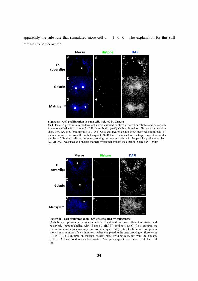
Cell proliferation in PSM cells in vitro
To assess if the different substrata and culture conditions used could affect PSM cell
proliferative properties, immunochemistry for phosphorilated histone 3 was used in all the
different conditions tested. Histone H3 is one of the five main histone proteins involved in the
structure of chromatin and, consequently, in cell division where its N-terminal tail suffers
phosphorilation during this cellular process. PSM cells isolated with dispase and cultured on
gelatin or matrigel presented more dividing cells (Fig. 15E,H) than the ones cultured on
fibronectin coverslips (Fig. 15B).
In PSM cells isolated with collagenase and cultured on matrigel, the number of mitotic
cells is slightly higher than the situation in the other two experimental conditions. (Fig. 16H).
Cells cultured on fibronectin coverslips or gelatin presented very few proliferating cells (Fig.
16B,C,E,F). Overall, our results suggest that the cell proliferating activity in these explants is
low, therefore, the size of the cell island around the initial explant localisation is manly due to
cell migration and can not be significantly attributed to cell proliferation. Matrigel was
Figure 14 - Cell-extracellular matrix interactions in cell epithelialisation behaviour of PSM cells isolated by collagenase (A-L) Isolated presomitic mesoderm cells were cultured on three different substrates and posteriorly immunolabelled with N-cadherin (B,F,J) and (C,G,K) antibodies. (A-D) Cells cultured on fibronectin coverslips show a strong labelling for both N-cadherin (B) and β-catenin (C) with some overlap of the two different fluorescent labels (yellow labeling in A), i.e., a colocalization of N-cadherin and β-catenin expressions. (E-H) Cells cultured on gelatin show a low immunoreactivity for both ptoteins (F,G). (I-L) Cells cultured on matrigel also show an overlap of the two different fluorescent labels (yellow labeling in I) but less intense than in (I). (D,H,L) DAPI was used as a nuclear marker. Scale bar: 50 μm
34
apparently the substrate that stimulated more cell d��1�0�0� The explanation for this still
remains to be uncovered.
Histone 3
DAPIMerge
Fn coverslips
Gelatin
MatrigelTM
A
D
B C
E F
G H I
*
**
* *
Figure 15 - Cell proliferation in PSM cells isolated by dispase(A-I) Isolated presomitic mesoderm cells were cultured on three different substrates and posteriorly immunolabelled with Histone 3 (B,E,H) antibody. (A-C) Cells cultured on fibronectin coverslips show very few proliferating cells (B). (D-F) Cells cultured on gelatin show more cells in mitosis (E), mainly in cells far from the initial explant. (G-I) Cells incubated on matrigel present a similar number of dividing cells as the ones growing on gelatin, mainly in the periphery of the explant. (C,F,I) DAPI was used as a nuclear marker; *=original explant localization. Scale bar: 100 μm
Merge Histone 3
DAPI
Fn coverslips
Gelatin
A B C
D E F
G H I
*
*
*MatrigelTM
Figure 16 - Cell proliferation in PSM cells isolated by collagenase(A-I) Isolated presomitic mesoderm cells were cultured on three different substrates and posteriorly immunolabelled with Histone 3 (B,E,H) antibody. (A-C) Cells cultured on fibronectin coverslips show very few proliferating cells (B). (D-F) Cells cultured on gelatin show similar number of cells in mitosis, when compared to the ones growing on fibronectin (E). (G-I) Cells cultured on matrigel present more dividing cells, far from the explant. (C,F,I) DAPI was used as a nuclear marker; *=original explant localization. Scale bar: 100 μm

35
Cell apoptosis in PSM cells in vitro
Finally, in order to verify that the results obtained by this in vitro system were not
compromised by cell apoptosis, immunochemistry for caspase 3 was used in all the different
conditions of PSM explant culture. Caspase 3 is included in the group of cysteine proteases
which carry out the degradation of the cell during the process of apoptosis, i.e., a form of
programmed cell death. Therefore, PSM cells, obtained by using two enzymatic treatments and
cultured on the three different substrates, all showed a non-relevant level of cell apoptosis (Fig.
17A-C as an example). The culture conditions used in this work allowed the PSM cells to
survive and remain attached to the substrate for at least for 48 hours. However, we have also to
consider that dying cells usually detach from the substrate and could be easily washed away
during the immunochemistry protocol. Nevertheless, we can assume that the process of apoptosis
did not interfere with the experiments performed nor did any of the culture conditions
particularly promote cell death in PSM explant cultures.
Gelatin
Merge Caspase 3
DAPIA B C
*
B
Figure 17 - Cell apoptosis in PSM cells isolated by collagenase(A-C) Isolated presomitic mesoderm cells were cultured on gelatin and immunolabelled with Caspase 3 (B) antibody. Cells cultured on gelatin did not show any cells in apoptosis (cellular death - B). (C) DAPI was used as a nuclear marker; *=original explant localization. Scale bar: 100 μm

36
DISCUSSION
The presence of fibronectin is essential for normal gastrulation and somitogenesis
Previous studies indicated that the adhesive properties of fibronectin could play an
influential role in chick somitogenesis (Lash et al., 1984; Lash et al., 1987). Moreover, mouse
embryos lacking the Fn1 gene (George et al., 1993; Georges-Labouesse et al., 1996) showed
severe defects in mesoderm, neural tube and vascular development, and represent one of the
most severe somitogenesis phenotypes in a single mouse gene (reviewed in Pourquié, 2001).
More recently, fibronectin was also described as a requirement for normal somitogenesis and
axial extension in Xenopus (Marsden and DeSimone, 2003) and for somite epithelialisation and
boundary maintenance in zebrafish (Julich et al., 2005; Koshida et al., 2005).
Immunofluorescence in stage HH4 embryos evidenced a fibronectin-rich network
underlying the epiblast and a sparser labelling in the extracellular spaces of the hypoblast similar
to previously described by (Harrisson et al., 1985). In later stages (HH12 and HH20), fibronectin
forms a complex matrix expressed throughout the ectoderm basement membrane, around the
neural tube and notochord and surrounding the somites. However, the PSM expresses low
fibronectin immunoreactivity in a sparse punctate pattern. Our results are in line with the ones
described by (Ostrovsky et al., 1983) and show that in HH10-HH24 chick embryos, the
fibronectin is mainly present in the ectoderm, the somites and neural tube. Using in situ
hybridisation of HH4 – HH20 stage embryos, the results confirmed that the epiblast and later the
ectoderm are major sources of Fn1 transcripts in all the observed developmental stages.
However, at stage HH12, the presomitic mesoderm expresses only low levels of Fn1 mRNA,
concomitantly with the expression pattern described by (Ffrench-constant and Hynes, 1988).
Surprisingly, the fibronectin immunofluorescence labelling in the PSM does not match with the
staining of the PSM for Fn1 transcripts. Moreover, these results contradict the fibronectin
expression in a sheet-like form along ventral and dorsal sides of the PSM, observed by (Lash et
al., 1987). The observations by Lash may be referring to the accumulation of fibronectin in the
ectoderm and endoderm basement membranes surrounding the PSM.

37
According to Suzuki et al. (1995), in the mouse embryo at the early primitive streak
stage, fibronectin mRNAs were detected throughout the mesoderm cells, except for the
mesoderm of the prechordal plate and the notochordal plate. As neurulation proceeds, during the
early somite stages, mesodermal Fn1 expression is largely down-regulated. Perkinson and
Norton (1997) showed, in mouse embryos between E8 and E12,5, that the fibronectin labelling is
specific for the neural tube, PSM and posterior somites but absent from the heart. Overall, in
early development, the chick embryo shows a strong Fn1 expression in the epiblast, different
from the mesodermal expression in the mouse embryo. However, as gastrulation proceeds, the
two models resemble more by revealing fibronectin expression in the same tissues, namely in the
neural tube, PSM and somites.
The integrin receptors for fibronectin in embryo development
Integrin α5 subunit-deficient mice die by day E10 due to severe vascular failure, showing
extensive mesodermal defects, forming only the anterior 10 somites and posterior trunk and
neural tube distortions (Yang et al., 1993; Goh et al., 1997). Furthermore, double knock-out
embryos for α5 and αv integrin do not assemble a fibronectin matrix and share the same
phenotype displaying an absence of somites with the Fn1-null mice (Yang et al., 1999).
More recently, (Takahashi et al., 2007) observed that mutating the fibronectin RGD motif
into a non-functional RGE, it could mimic the α5-null mice phenotype. By knocking-out the
RGD motif of fibronectin, essential for α5β1 or αv interactions and fibrils assembly, the mouse
embryos died at E10. This study showed that the mutated fibronectin can be assembled by αv
integrin, using the RGE motif, and this integrin maintains the normal embryo development until
the α5 integrin-fibronectin interaction is absolutely necessary for it. Therefore, the RGE mice
have a similar phenotype to the ones lacking the α5 subunit, although they have both integrins
and a fibronectin matrix apparently but without the RGD motif. Integrins also revealed to be
important for cell-cell adhesion during the 90º somite rotation in Xenopus and zebrafish
(Kragtorp, 2006).Work of our group has evidenced that mesodermal integrins collaborate with
the ectoderm (the major source of Fn1) to construct the fibronectin matrix necessary for
somitogenesis (Rifes et al., 2007).

38
Itga5 and Itga4 transcripts are initially expressed in the primitive streak, including the
node, but later in development, they are present in various derivates of the ingressed mesoderm,
especially in the presomitic mesoderm. Therefore, these two integrins are expressed in the PSM
and ready to interact with the fibronectin, which is being produced by the ectoderm cells above.
The α4 and α5 integrins may collaborate in binding a fibronectin matrix assembly around the
PSM, which participates in somitogenesis.
As described above, αv integrin is also an important fibronectin receptor and essential for
the process of fibrillogenesis in vivo (Yang et al, 1999). Furthermore, Yang et al. (1999) also
suggested possible roles for αv integrins in replacing the role of α5β1 in the fibronectin matrix
assembly in vitro. According to Takahashi et al. (2007), the αv, but not α5β1, integrins mediate
the assembly of fibronectin into matrix fibrils when the fibronectin RGD motif is inactive.
However, we used two different RNA probes to label Itgav transcripts in the early stages of
chick embryo but the results were inconclusive and further experiments will be done.
In summary, the fibronectin and its cell surface receptors (integrin α4 and α5) are
essential proteins for normal embryonic development, especially in the earliest stages. As future
prospects, it will be interesting to observe more carefully the expression pattern of the Itgα4
transcripts, especially due to the fact that its expression pattern is dynamic during stage HH12-14
embryos. However, analysing the expression patterns of other components of the extracellular
matrix, such as laminin and proteoglycans, in the same stages will be also an interesting issue to
explore.
Analysis of presomitic mesoderm cell behaviour growing on different extracellular matrix
substrata in vitro
In order to further understand the influence of the extracellular matrix, and especially
fibronectin, on the process of somitogenesis and in the epithelialisation of the PSM cells in
particular, we established an in vitro culture system using the presomitic mesoderm growing on
several substrates Mesenchymal cell behaviour is largely determined by its local environment
and, in vitro, by the properties of the available substrate, as demonstrated in the chick embryo by

39
(Solursh et al., 1979). Other studies revealed that the segmental plate cells, which are the
precursors of embryonic somites, normally show very little cell-cell or cell-substrate interaction
in culture (Lash et al., 1985). As previously described by Lash et al. (1987), the isolated PSMs
cultured on a fibronectin-coated substrate showed cell spreading. Indeed, fibronectin was shown
to be a structural component of the extracellular matrix used as a substrate for migration and
positioning of mesenchymal cells (Harrisson et al., 1985). In addition, cell attachment to
adhesive protein substrates other than fibronectin can also affect fibronectin fibrillogenesis itself
(Bae et al., 2004).
On the other hand, enzymes used for PSM isolation, such as dispase and collagenase, can
degrade differently the extracellular components (i.e. fibronectin), and ultimately influence PSM
cell behaviour and arrangement (Rifes et al 2007). Therefore, we also used this in vitro culture
system of mesenchymal cells to discriminate the effect of different substrates and enzymes, i.e.,
the presence or absence of a fibronectin matrix around the recently isolated explant, on cell
behaviour and morphology. We performed fibronectin immunostaining to assess if cells in
culture were capable of producing and assembling their own fibronectin matrix, when cultured
on fibronectin or laminin (gelatin as a control substrate). It has previously been demonstrated
that FN-null cells growing on FN or laminin substrates were able to assemble exogenously-
supplied FN into fibrils (reviewed in (Mao and Schwarzbauer, 2005b)). Our results showed that
the assembly of a fibronectin matrix in dispase-isolated PSMs is higher when cells are cultivated
on exogenous fibronectin and gelatin substrates, while PSM cells isolated with collagenase
assemble a fibronectin matrix preferentially when cultured on matrigel. Matrigel mimics
basement membranes, which is a specialized form of the extracellular matrix containing a set of
proteins that include, namely, laminin (Zagris, 2001). Laminins are generally known to be a
substrate that promotes cellular migration (reviewed by (Sanes, 1989)), while α5β1 integrin–
fibronectin interaction promotes strong intercellular cohesion in cellular aggregates (Robinson et
al., 2003). However, we have to consider that the PSM cells isolated with collagenase migrated
short distances from the explant and the fibronectin immunoreactivity was kept in the explants,
while the dispase-isolated PSM cells migrated and consequently the fibronectin labelling was
dispersed. On the other hand, our results showed that more cell migration is observed in the PSM
cells cultured on matrigel but less in cells growing on fibronectin. Considering the importance of
fibronectin in the somitogenesis, it is possible that the fibronectin is sending a signal for PSM

40
cells to compact instead of migrating from the explants. As for the cytoskeleton labelling, all the
cultured PSMs showed actin stress fibers, in sites of anchorage of PSM cells to the surrounding
extracellular matrix. The actin pattern displayed is characteristic of a mesenchymal cell
phenotype.
Furthermore, N-cadherin, a marker for epithelialisation and cell polarization and β-
catenin, a cytoskeleton regulator that binds to the intracellular domain of cadherins, were also
detected in our cultured PSM cells. The results showed that PSM cells in culture express low
levels of both N-cadherin and β-catenin, and, more importantly, that the distribution of these two
proteins is not polarized in the cells, suggesting that these cells stay mesenchymal under the
different conditions. Duband et al. (1987) also showed that cultured segmental plate cells were
organized as a loose mesenchyme with no apparent orientation of cells and therefore weakly
stained for N-cadherin. In epithelial branching morphogenesis in the mouse salivary glands,
fibronectin is required for cleft formation and induces local suppression of cadherin during the
conversion of cell–cell adhesions to cell–matrix adhesions (Sakai et al., 2003). We therefore
expect to explore further the influence of fibronectin on PSM cells in culture, namely, the
possible induction by fibronectin of an epithelial phenotype in PSM cells, with a polarized actin
cytoskeleton, and N-cadherin and β-catenin apical distribution.
Cells in vivo exist in complex environments, where they constantly interact with multiple
ECM molecules, diffusible substances and other cells. Nevertheless, the in vitro culture system
proposed in this work would allow a direct access to the morphology of PSM cells and their
behaviour under different and controlled experimental conditions. Indeed, in vitro studies show
that specific cell-ECM interactions affect the dynamics of other ECM components and the
physical state of the ECM itself (Larsen et al., 2006). Therefore, the present model could be used
for growing cells in serum-free medium and adding diffusable factors to the culture medium,
such as retinoic acid, Fgf8 and Wnts, which are important secreted molecules in vivo, and
analysing how the complex interaction of these factors altogether could affect the
epithelialisation of the PSM cells.

41
BIBLIOGRAPHY
Argraves,W.S., Suzuki,S., Arai,H., Thompson,K., Pierschbacher,M.D., and Ruoslahti,E. (1987). Amino acid sequence of the human fibronectin receptor. J. Cell Biol. 105, 1183-1190.
Aulehla,A. and Herrmann,B.G. (2004). Segmentation in vertebrates: clock and gradient finally joined. Genes Dev. 18, 2060-2067.
Bajanca,F., Luz,M., Duxson,M.J., and Thorsteinsdottir,S. (2004). Integrins in the mouse myotome: Developmental changes and differences between the epaxial and hypaxial lineage. Dev. Dyn. 231, 402-415.
Christ,B. and Ordahl,C.P. (1995). Early stages of chick somite development. Anat. Embryol. (Berl) 191, 381-396.
Danen,E.H. and Sonnenberg,A. (2003). Integrins in regulation of tissue development and function. J. Pathol. 201, 632-641.
Davis,G.S., Phillips,H.M., and Steinberg,M.S. (1997). Germ-layer surface tensions and "tissue affinities" in Rana pipiens gastrulae: quantitative measurements. Dev. Biol. 192, 630-644.
Derycke,L.D. and Bracke,M.E. (2004). N-cadherin in the spotlight of cell-cell adhesion, differentiation, embryogenesis, invasion and signalling. Int. J. Dev. Biol. 48, 463-476.
Duband,J.L., Dufour,S., Hatta,K., Takeichi,M., Edelman,G.M., and Thiery,J.P. (1987). Adhesion molecules during somitogenesis in the avian embryo. J. Cell Biol. 104, 1361-1374.
Ffrench-Constant,C. and Hynes,R.O. (1988). Patterns of fibronectin gene expression and splicing during cell migration in chicken embryos. Development 104, 369-382.
Freitas,C., Rodrigues,S., Saude,L., and Palmeirim,I. (2005). Running after the clock. Int. J. Dev. Biol. 49, 317-324.
George,E.L., Georges-Labouesse,E.N., Patel-King,R.S., Rayburn,H., and Hynes,R.O. (1993). Defects in mesoderm, neural tube and vascular development in mouse embryos lacking fibronectin. Development 119, 1079-1091.
Georges-Labouesse,E.N., George,E.L., Rayburn,H., and Hynes,R.O. (1996). Mesodermal development in mouse embryos mutant for fibronectin. Dev. Dyn. 207, 145-156.
Gossler,A. and Hrabe de Angelis,M. (1998). Somitogenesis. Curr. Top. Dev. Biol. 38, 225-287.

42
Hamburger,V. and Hamilton,H.L. (1992). A series of normal stages in the development of the chick embryo. 1951. Dev. Dyn. 195, 231-272.
Harrisson,F., Van Hoof,J., Vanroelen,C., and Vakaet,L. (1985). Transfer of extracellular matrix components between germ layers in chimaeric chicken-quail blastoderms. Cell Tissue Res. 239, 643-649.
Hay,E.D. (2005). The mesenchymal cell, its role in the embryo, and the remarkable signaling mechanisms that create it. Dev. Dyn. 233, 706-720.
Henrique,D., Adam,J., Myat,A., Chitnis,A., Lewis,J., and Ish-Horowicz,D. (1995). Expression of a Delta homologue in prospective neurons in the chick. Nature 375, 787-790.
Hynes,R.O. (1992). Integrins: versatility, modulation, and signaling in cell adhesion. Cell 69, 11-25.
Julich,D., Geisler,R., and Holley,S.A. (2005). Integrinalpha5 and delta/notch signaling have complementary spatiotemporal requirements during zebrafish somitogenesis. Dev. Cell 8, 575-586.
Komoriya,A., Green,L.J., Mervic,M., Yamada,S.S., Yamada,K.M., and Humphries,M.J. (1991). The minimal essential sequence for a major cell type-specific adhesion site (CS1) within the alternatively spliced type III connecting segment domain of fibronectin is leucine-aspartic acid-valine. J. Biol. Chem. 266, 15075-15079.
Koshida,S., Kishimoto,Y., Ustumi,H., Shimizu,T., Furutani-Seiki,M., Kondoh,H., and Takada,S. (2005). Integrinalpha5-dependent Fibronectin accumulation for maintenance of somite boundaries in zebrafish embryos. Dev. Cell 8, 587-598.
Kulesa,P.M. and Fraser,S.E. (2002). Cell dynamics during somite boundary formation revealed by time-lapse analysis. Science 298, 991-995.
Larsen,M., Artym,V.V., Green,J.A., and Yamada,K.M. (2006). The matrix reorganized: extracellular matrix remodeling and integrin signaling. Curr. Opin. Cell Biol. 18, 463-471.
Lash,J.W. (1985). Somitogenesis: investigations on the mechanism of compaction in the presomitic mass and a possible role for fibronectin. Prog. Clin. Biol. Res. 171, 45-60.
Lash,J.W., Linask,K.K., and Yamada,K.M. (1987). Synthetic peptides that mimic the adhesive recognition signal of fibronectin: differential effects on cell-cell and cell-substratum adhesion in embryonic chick cells. Dev. Biol. 123, 411-420.
Lash,J.W. and Ostrovsky,D. (1986). On the formation of somites. Dev. Biol. (N. Y. 1985. ) 2, 547-563.

43
Lash,J.W., Seitz,A.W., Cheney,C.M., and Ostrovsky,D. (1984). On the role of fibronectin during the compaction stage of somitogenesis in the chick embryo. J. Exp. Zool. 232, 197-206.
Lilien,J., Balsamo,J., Arregui,C., and Xu,G. (2002). Turn-off, drop-out: functional state switching of cadherins. Dev. Dyn. 224, 18-29.
Mao,Y. and Schwarzbauer,J.E. (2005a). Fibronectin fibrillogenesis, a cell-mediated matrix assembly process. Matrix Biol. 24, 389-399.
Mao,Y. and Schwarzbauer,J.E. (2005b). Stimulatory effects of a three-dimensional microenvironment on cell-mediated fibronectin fibrillogenesis. J. Cell Sci. 118, 4427-4436.
Miranti,C.K. and Brugge,J.S. (2002). Sensing the environment: a historical perspective on integrin signal transduction. Nat. Cell Biol. 4, E83-E90.
Norton,P.A. and Hynes,R.O. (1987). Alternative splicing of chicken fibronectin in embryos and in normal and transformed cells. Mol. Cell Biol. 7, 4297-4307.
Ostrovsky,D., Cheney,C.M., Seitz,A.W., and Lash,J.W. (1983). Fibronectin distribution during somitogenesis in the chick embryo. Cell Differ. 13, 217-223.
Palmeirim,I., Henrique,D., Ish-Horowicz,D., and Pourquie,O. (1997). Avian hairy gene expression identifies a molecular clock linked to vertebrate segmentation and somitogenesis. Cell 91, 639-648.
Pankov,R. and Yamada,K.M. (2002). Fibronectin at a glance. J. Cell Sci. 115, 3861-3863.
Perkinson,R.A. and Norton,P.A. (1997). Expression of the mouse fibronectin gene and fibronectin-lacZ transgenes during somitogenesis. Dev. Dyn. 208, 244-254.
Pourquié,O. (2004). The chick embryo: a leading model in somitogenesis studies. Mech. Dev. 121, 1069-1079.
Ramos,J.W., Whittaker,C.A., and DeSimone,D.W. (1996). Integrin-dependent adhesive activity is spatially controlled by inductive signals at gastrulation. Development 122, 2873-2883.
Rifes,P., Carvalho,L., Lopes,C., Andrade,R.P., Rodrigues,G., Palmeirim,I., and Thorsteinsdottir,S. (2007). Redefining the role of ectoderm in somitogenesis: a player in the formation of the fibronectin matrix of presomitic mesoderm. Development 134, 3155-3165.
Robinson,E.E., Foty,R.A., and Corbett,S.A. (2004). Fibronectin matrix assembly regulates alpha5beta1-mediated cell cohesion. Mol. Biol. Cell 15, 973-981.

44
Robinson,E.E., Zazzali,K.M., Corbett,S.A., and Foty,R.A. (2003). Alpha5beta1 integrin mediates strong tissue cohesion. J. Cell Sci. 116, 377-386.
Roskelley,C.D., Srebrow,A., and Bissell,M.J. (1995). A hierarchy of ECM-mediated signalling regulates tissue-specific gene expression. Curr. Opin. Cell Biol. 7, 736-747.
Saga,Y. and Takeda,H. (2001). The making of the somite: molecular events in vertebrate segmentation. Nat. Rev. Genet. 2, 835-845.
Sambrook,S.K., Fritsch,E.F., and Maniatis,T. (1989). Molecular cloning: a laboratory manual. USA: Cold Spring Harbor Laboratory Press.
Sechler,J.L., Cumiskey,A.M., Gazzola,D.M., and Schwarzbauer,J.E. (2000). A novel RGD-independent fibronectin assembly pathway initiated by alpha4beta1 integrin binding to the alternatively spliced V region. J. Cell Sci. 113 ( Pt 8), 1491-1498.
Solursh,M., Fisher,M., Meier,S., and Singley,C.T. (1979). The role of extracellular matrix in the formation of the sclerotome. J Embryol. Exp. Morphol. 54, 75-98.
Stenn,K.S., Link,R., Moellmann,G., Madri,J., and Kuklinska,E. (1989). Dispase, a neutral protease from Bacillus polymyxa, is a powerful fibronectinase and type IV collagenase. J. Invest Dermatol. 93, 287-290.
Stockdale,F.E., Nikovits,W., Jr., and Christ,B. (2000). Molecular and cellular biology of avian somite development. Dev. Dyn. 219, 304-321.
Suzuki HR, Solursh M, Baldwin HS. (1995). Relationship between fibronectin expression during gastrulation and heart formation in the rat embryo. Dev. Dyn. 204(3), 259-77.
Takahashi,S., Leiss,M., Moser,M., Ohashi,T., Kitao,T., Heckmann,D., Pfeifer,A., Kessler,H., Takagi,J., Erickson,H.P. et al. (2007). The RGD motif in fibronectin is essential for development but dispensable for fibril assembly. J. Cell Biol. 178, 167-178.
Testaz,S., Delannet,M., and Duband,J. (1999). Adhesion and migration of avian neural crest cells on fibronectin require the cooperating activities of multiple integrins of the (beta)1 and (beta)3 families. J. Cell Sci. 112 ( Pt 24), 4715-4728.
Wierzbicka-Patynowski,I. and Schwarzbauer,J.E. (2003). The ins and outs of fibronectin matrix assembly. J. Cell Sci. 116, 3269-3276.
Yamada,K.M., Pankov,R., and Cukierman,E. (2003). Dimensions and dynamics in integrin function. Braz. J. Med. Biol. Res. 36, 959-966.

45
Yang,J.T., Bader,B.L., Kreidberg,J.A., Ullman-Cullere,M., Trevithick,J.E., and Hynes,R.O. (1999). Overlapping and independent functions of fibronectin receptor integrins in early mesodermal development. Dev. Biol. 215, 264-277.
Zagris,N. (2001). Extracellular matrix in development of the early embryo. Micron. 32, 427-438.