Embed Size (px)
Citation preview
Cell Behavior and Signal Molecule Involvement in aCase Study of Malignant Histiocytosis: A Negative
Model of Morphine as an Immunoregulator
Gregory L. Fricchione, 1,2* Lawrence Cytryn, 3 Thomas V. Bilfinger, 2,4 andGeorge B. Stefano 1,2,4
1Department of Medicine, Brigham and Women’s Hospital, Harvard Medical School, Boston, Massachusetts2Neuroscience Research Institute, State University of New York at Old Westbury, Old Westbury, New York
3Department of Hematology, University Hospital, State University of New York at Stony Brook, Stony Brook, New York4Cardiac Research Program, Departments of Surgery and University Hospital, State University of New York at Stony Brook,
Stony Brook, New York
In a patient diagnosed with histiocytic medullary reticulosis (HM), we examined immu-nocytes for their responsiveness towards known signaling molecules. Both the granu-locytes and monocytes were found to exhibit a high level of spontaneous activation (96%compared to normal cells 7%; P < 0.001). These cells could not be downregulated whenexposed to morphine. Following patient treatment with doxorubicin and cyclophospha-mide, the immunocytes still exhibited a high spontaneous activation. They responded tomorphine exposure in vitro with a cell rounding and becoming immobile for only 20 minwhereas normal cells would remain round and immobile for up to 1–2 h. An examinationof the plasma from the HM patient revealed that monocyte colony stimulating factor(MCSF) levels were elevated (6.4 and 5.78 compared to a control range of 1–1.75 ng/ml).In the HM patient, the immunocytes did not express the opiate selective and opioidpeptide insensitive receptor, µ3, supporting the lack of opiate action. Given this finding,we incubated normal monocytes with MCSF and found that it significantly reduced the µ3Bmax. Given the role of intracellular calcium in the activation process of immunocytes,we examined the action of various calcium channel blockers for their ability to inhibit theactivated HM monocytes. The agents (nimodipine, cardiazem, and verapamil; 10 29 M)were able to inhibit the HM-associated chemokinesis. Taken together, the data indicatethat in the HM patient the immunocytes appear to be overactivated because stimulatorymolecules are high and have the ability to downregulate the normal ‘‘braking’’ process.Additionally, the data indicate that MCSF deregulation may be involved as an initiatingfactor for this disorder. Am. J. Hematol. 56:197–205, 1997. © 1997 Wiley-Liss, Inc.
Key words: medullary reticulosis; morphine; granulocytes; monocytes; mu3
INTRODUCTION
The term ‘‘histiocytic medullary reticulosis’’ (HM)was coined by Bodley and Smith in 1939 [1]. In theirdescription of these patients, a rapid fatal course charac-terized by fever, lymphadenopathy, hepatospleno-megally, anemia, and leukopenia was noted, yet it wasnot clear that the cause was a neoplastic one. The authorsmade a point of avoiding the term malignant for a con-dition described as ‘‘proliferation of erythrophagocytichistiocytes and their precursors.’’ Rappaport in 1966 in-troduced the term malignant histiocytosis in an attempt tofurther define this rare rapidly fatal disease [2]. The de-
bate about whether this disease is malignant from theonset or represents a malignant degeneration or a clini-cally fulminant but not neoplastic process of an initially
Contract grant sponsor: NIH; Contract grant numbers: MH/DA 17138,DA 09010, NIH Fogarty INT00045.
*Correspondence to: Dr. Gregory L. Fricchione, Division of Psychia-try, Brigham and Women’s Hospital, Harvard Medical School, Bos-ton, MA 02115. E-mail: [email protected]
Received for publication 22 November 1996; Accepted 12 February1997
American Journal of Hematology 56:197–205 (1997)
© 1997 Wiley-Liss, Inc.
benign condition is, even today, not resolved. Neither isit entirely clear how to categorize this disease despiteconsiderable progress in the classification of other he-mopoietic proliferative disorders [3].
In regard to HM, from a histological perspective, he-matophagocytosis can be carried out not only by macro-phages or tissue histiocytes, but also by hematopoieticand non-hematopoietic malignant tumor cells [4]. It iswell established that malignant lymphomas can recruitreactive cells [4]. In Hodgkin’s disease, for instance, re-active lymphocytes, macrophages, and leukocytes can bethe predominant cells [5]. This is far less typical formalignant histiocytosis. One possible explanation for thisrecruitment is the secretion of cytokines into the micro-environment by the malignant cells. In large cell lym-phoma, this may occur due to modifications of c-fms,colony stimulating factor (CSF-1), and/or monocytecolony stimulating factor (MCSF) and tumor necrosisfactor-alpha (TNA-a) with modulation of the chromo-somal 5q35 breakpoint region genes [6]. TNF levels havebeen found useful in predicting the severity and progno-sis of both malignant histiocytosis and virus-associatedhemophagocytic syndrome (VAHS). This informationcan be used successfully to monitor the progression ofthe disease and therapy [7].
Given this background, and based on the recent workregarding the role of neuropeptides, cytokines, and opiatealkaloids in immunoregulation and autoimmunoregula-tion [8–10], we examined immunocytes and serum ob-tained from a patient diagnosed with autopsy confirmedmalignant histiocytosis, to determine if deficits existed intheir immunocyte responsiveness to naturally occurringimmunocyte-suppressing substances [8–13]. We report,for the first time, that both granulocytes and monocytesobtained from this patient are dysfunctional, based ontheir level of spontaneous activation and lack of appro-priate responses to known immunosuppressory agents.Further, in vitro analysis of normal immunocytes ex-posed to MSCF mimics, in part, the cell behavior andbiochemical changes found in cells obtained from thispatient.
CASE REPORT
A sixty-five-year-old male was admitted with abdomi-nal pain of 5 days duration, and fever, and was found tohave a platelet count of 17,000 per microliter. He hadpreviously been in good health, except for a past historyof osteoarthritis treated with naproxen, and a history ofGuillain-Barre syndrome 12 years prior to admission.
Initial physical examination was significant for hepa-tosplenomegaly. The white blood cell count was 13.2 ×10³ per microliter, with 50% segmented neutrophils, 14%lymphocytes, and 16% monocytes. Hemoglobin was 16.1g per deciliter. The blood smear showed atypical mono-
cytes, large platelets, and schistocytes. Creatinine was2.2 mg per deciliter, BUN was 141 mg per deciliter, totalbilirubin was 4.4 mg per deciliter with 1.6 mg per deci-liter direct. AST was 28 IU per liter, ALT was 30 IU perliter, and LDH was 521 IU per liter. PT was 13.8 sec andPTT was 33.1 sec.
Bone marrow was aspirated with difficulty; aspirationand biopsy revealed extensive replacement of the normalhematopoietic elements by pleiomorphic, immature-appearing monocytoid cells, and numerous instances ofhemophagocytosis. The infiltrating population exhibitednonspecific esterase staining, with partial fluoride inhi-bition. Cytogenetics were normal, demonstrating akaryotype of 46, XY. Flow cytometric immunopheno-typing of the patient’s blood identified a population oflarge, mononuclear cells comprising 56% of circulatingleukocytes and co-expressing CD45, CD14, CD11b,CD11c, CD15, CD34, and CD33. A blood specimen sentto a research laboratory showed markedly elevated levelsof MCSF (patient 6.4 ng per ml, control 1.0 ng per ml),and G-CSF (patient 350.3 pg per ml, control 53.5 pg perml). Other measured cytokine levels included TNF (pa-tient 30.3 pg per ml, control <16 pg per ml), SCF (patient1.4 ng per ml, control 1.1 pg per ml), GMCSF (patientand both <8 pg/ml), and II-1beta (patient and controlboth <8 pg per ml). Southern hybridization analysis ofperipheral blood mononuclear cells showed no rear-rangement of immunoglobulin or T-cell receptor genes.The diagnosis of histiocytic medullary reticulosis wasmade.
Initial attempts to ameliorate severe and transfusion-refractory thrombocytopenia with corticosteroids and in-travenous gammaglobulin were unsuccessful. Combina-tion chemotherapy with cyclophosphamide, doxorubicin,vincristine, and prednisone (CHOP) was begun. Con-comitantly, the patient received 20 cGy splenic radio-therapy in eight fraction, with an approximately 50%reduction in spleen size; this was associated with a rise inplatelet count, and improvement in response to platelettransfusion. The first three cycles of chemotherapy wereadministered with 50–80% of standard doses of doxoru-bicin and cyclophosphamide, followed by full doses ofthe same regimen administered for a total of sevencycles.
Cytogenetics performed on a bone marrow aspirateobtained after cycle 1 indicated 46, XY, del(13),(q12q14). Flow cytometric immunophenotyping of mar-row cells at that time confirmed the phenotype originallyobtained on peripheral blood. Repeat cytogenetic studieson bone marrow after cycle 3 were normal, and Southernhybridization analysis of marrow mononuclear cellsagain showed no immunoglobulin or T-cell receptor generearrangements.
The patient experienced a progressive response,achieving normal white blood cell and platelet counts,
198 Fricchione et al.
and a rising hemoglobulin, at the time therapy wasstopped 6 months after presentation. At this time, mar-row examination showed hypercellularity, with scatteredatypical monocytoid cells, normal cytogenetics, andgerm line immunoglobulin and T-cell receptor genes bySouthern hybridization. A specimen of blood was storedat this time in the original research laboratory.
Hematologic improvement continued for 5 months af-ter discontinuation of chemotherapy, with persistent nor-mal white blood cell and platelet counts and normaliza-tion of hemoglobin. Fourteen months after the originalpresentation, the patient developed fever and thrombo-cytopenia. The white blood cell count was 8.4 × 10³ permicroliter, hemoglobin was 12.9 g per deciliter, andplatelets were 78,000 per microliter. Bone marrow aspi-ration and biopsy revealed near total replacement by im-mature monocytoid cells and mature hemophagocytichistiocytes. Cytogenetics performed on the bone marrowshowed 46, XY[9]/46, add/X(q22)Y, add(7)(p15), add(8)(p21), add (15)(q26)[cp11]. A blood specimen sent tothe research laboratory at this time showed an MCSFlevel of 5.78 ng per ml, with control of 1.75 ng per ml.The specimen sent at the time of completion of chemo-therapy 7 months earlier was also measured at this timeand showed an MCSF level of 1.25 ng per ml, with thesame control of 1.75 ng per ml.
The patient received broad spectrum antibiotics, in-cluding coverage forStaphylococcus aureus,which grewfrom an admission culture of an indwelling venous ac-cess device. The patient expired suddenly on the fourthhospital day, before receiving any antineoplastic therapy.Postmortem examination confirmed the bone marrowfindings and showed extensive systemic involvement,with tumor in the lungs, liver, spleen, lymph nodes, kid-neys, adrenals, and pituitary capsule. There was no evi-dence of myocardial infarction, pulmonary thromboem-bolism, or cerebrovascular accident.
MATERIALS AND METHODS
Human granulocytes and monocytes for cellular analy-ses were obtained from patient volunteers at UniversityHospital (State University of New York at Stony Brook)who had given their informed consent, as well as a singlepatient diagnosed as having histiocytic medullary reticu-losis (HM). Blood was obtained via central venous ac-cess in that patient. Cells were separated by the standardFicoll-Hypaque method as noted elsewhere in detail[9,10,14]; they were then washed three times in RPMImedium (RPMI, 25 m Hepes, Grand Island BiologicalCo., Grand Island, NY) and used for subsequent analysis.
Analysis of Cellular Activity
The analysis of chemotaxis was determined as notedextensively elsewhere [9,10,15]. Briefly, cells were
placed at the extreme right of a petroleum jelly ring insaline in 100-ml volumes on a slide, and then coveredwith a glass cover slip. At the right edge of the petroleumjelly ring, an opening was made and a 100-ml (in physi-ological saline) volume of either DAMA or f Met-Leu-Phe (fMLP) was added. The solution moves under theslide by capillary action [15]. Experimental measure-ments were initiated at this time.
Chemotaxis was differentiated from chemokinesis bydetermining the axis of cell alignment and subsequentmovement of the cell parallel to the chemical concentra-tion gradient, through the use of cell analysis software(American Innovision, Inc., San Diego, CA). About 20 to32 activated cells were observed for each 400-mm view-ing diameter, and four additional viewing diameters wereobserved per slide. The entire process was repeated threemore times and the resulting mean of these individualmeans (±SEM) was graphed. The variation for individualreadings was between 5 and 9%.
The mixture of human cells (separately) with mor-phine, an agent known to inhibit cell activation, chemo-taxis, chemokinesis, and spontaneous activation[9,10,12], was analyzed by phase-contrast microscopy,using a Zeiss (Mornwood, NY) Axiophot Microscope inconjunction with a JVC time-lapse video recording sys-tem, for conformational changes. Human granulocytesand monocytes each were analyzed at 30 min. Activationis defined as changes in cellular conformation rangingfrom inactive-rounded to active-amoeboid. This statewas determined by measurements of cellular area andperimeter and were mathematically expressed by use ofthe shape-factor formula of the American InnovisionAnalysis System or the Image Analytics (Hauppauge,NY) system as previously described [9,15–18]. The pro-portion of activated cells was determined as noted else-where [9,10,15,18]. Activated cells change their confor-mation in response to an inhibitory pharmacologicalstimulus; they become immobile and rounded [9–11,15,19].
Plasma was obtained from patient volunteers at Uni-versity Hospital who had given their informed consent,after the study was approved by the institutional reviewboard. Plasma was also obtained from the patient diag-nosed with HM. The plasma from these samples, as wellas the HM sample, were immediately frozen to −70°C. Inorder to determine if plasma obtained from control andthe HM patient contained signal molecules capable ofinfluencing immunocyte behavior it was exposed to na-ive control cells obtained from the Long Island BloodCenter (Melville, NY).
Opiate Binding Analysis
Human monocytes obtained from the Ficoll-Hypaquecentrifugation and subsequent washing were homog-enized −70°C in 50 volumes of 0.32 M sucrose, pH 7.4,
Cell Behavior in Malignant Histiocytosis 199
at 4°C, by the use of a Brinkmann (Westbury, NY) poly-tron (30 sec, setting no. 5) [10]. The crude homogenatewas centrifuged at 900g for 10 min at 4°C, and the su-pernatant was reserved on ice. The whitish crude pelletwas resuspended by homogenization (15 sec, setting no.5) in 30 volumes of 0.32 M sucrose/Tris-HCl buffer, pH7.4, and centrifuged at 900g for 10 min. The extractionprocedure was repeated one more time, and the combinedsupernatants were centrifuged at 900g for 10 min. Theresulting supernatants (S18) were used immediately.
Immediately prior to the binding experiment, the S18supernatant was centrifuged at 30,000g for 15 min andthe resulting pellet (P2) was washed once by centrifuga-tion in 50 volumes of the sucrose/Tris-HCl. The P2 pelletwas then resuspended with a Dounce hand-held homog-enizer (10 strokes) in 100 volumes of buffer. Bindinganalysis was then performed on the cell membrane sus-pensions.
Cytokine determinations were made by a commercialcompany (Osaka, Bethesda, MD) from numericallycoded blind samples.
Statistics
Analysis was performed by using the Student’st-testto compare controls with drug-exposed cells, as noted inthe text. Controls were run for each treatment so as toavoid individual variations due to spontaneous activationof the cells.
RESULTS
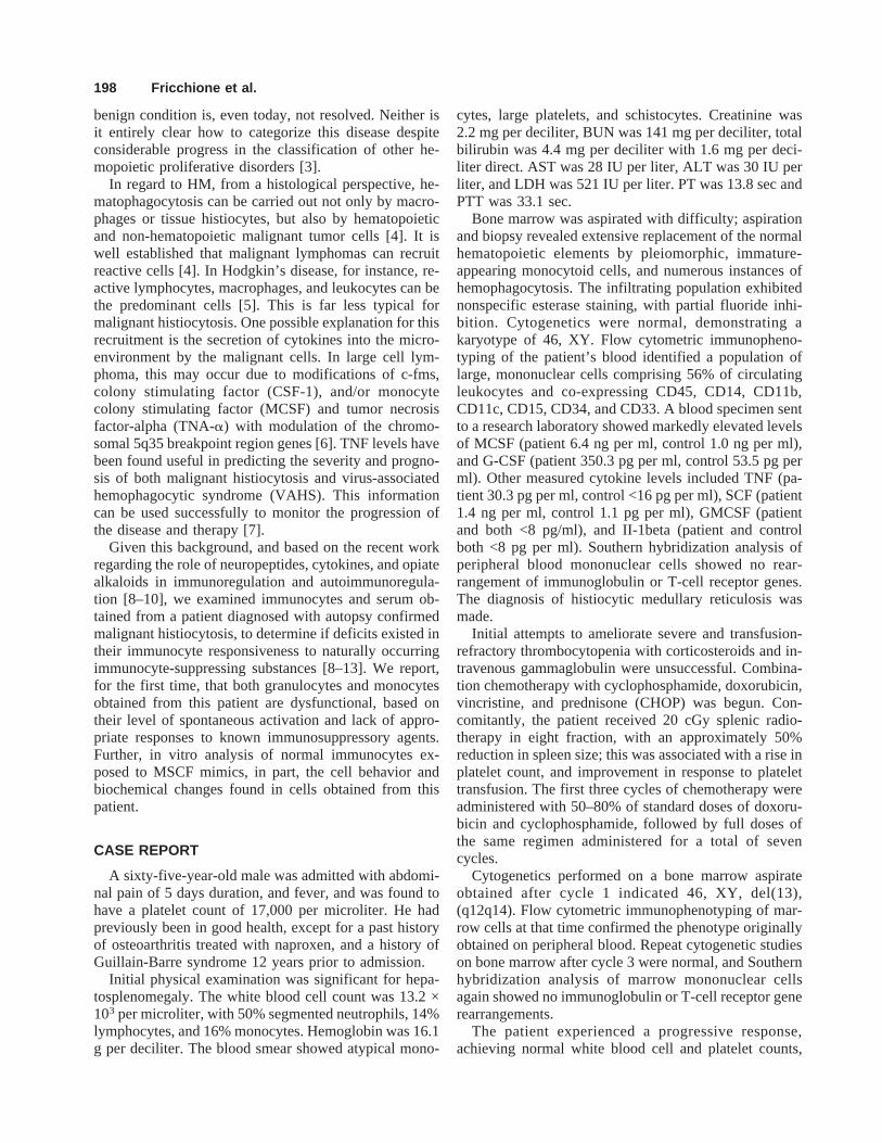
Control spontaneously active granulocytes and mono-cytes exhibit a non-directed migration path whereas cellsexposed to a concentration gradient of D-ala2, met en-kephalinamide (DAMA; 10-11 M) or FMLP (1.0 U/ml)[12], aligned their axis (i.e., elongated and became par-allel) [9,10,19] with the concentration gradient andmoved toward the higher concentration of the chemicalstimulus [15]. Both monocytes and granulocytes ob-tained from the patient diagnosed with MH did not ex-hibit chemotaxis when exposed to either agent; instead,they continued to move in a chemokinetic manner. Ad-ditionally, over 90% of both cell types from the MHpatient were spontaneously active compared to immuno-cytes obtained from controls (n4 42), which character-istically exhibit a spontaneous activation level of only6–9% (Fig. 1;P < 0.001) [9,10,14,15,19].
Given this high level of spontaneously activated im-munocytes, we attempted to determine if they could beinhibited by morphine, which has been previously shownto downregulate immunocyte activity [10]. Figure 1 dem-onstrates that the opiate alkaloid (10−6 M), regardless ofthe concentration, did not inhibit the HM-associated highlevel of immunocyte activation including chemokinesis.Interestingly, after 6 months of therapeutic treatment, a
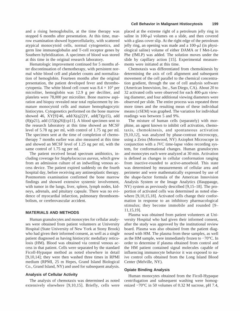
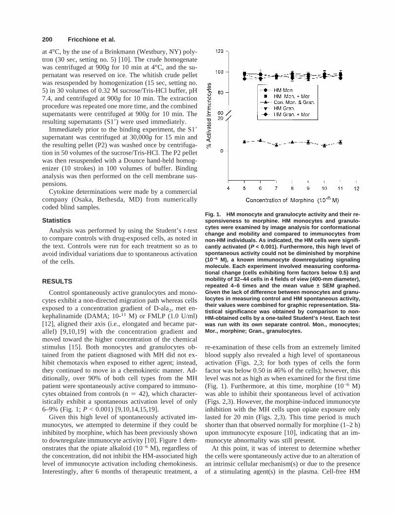
re-examination of these cells from an extremely limitedblood supply also revealed a high level of spontaneousactivation (Figs. 2,3; for both types of cells the formfactor was below 0.50 in 46% of the cells); however, thislevel was not as high as when examined for the first time(Fig. 1). Furthermore, at this time, morphine (10−6 M)was able to inhibit their spontaneous level of activation(Figs. 2,3). However, the morphine-induced immunocyteinhibition with the MH cells upon opiate exposure onlylasted for 20 min (Figs. 2,3). This time period is muchshorter than that observed normally for morphine (1–2 h)upon immunocyte exposure [10], indicating that an im-munocyte abnormality was still present.
At this point, it was of interest to determine whetherthe cells were spontaneously active due to an alteration ofan intrinsic cellular mechanism(s) or due to the presenceof a stimulating agent(s) in the plasma. Cell-free HM
Fig. 1. HM monocyte and granulocyte activity and their re-sponsiveness to morphine. HM monocytes and granulo-cytes were examined by image analysis for conformationalchange and mobility and compared to immunocytes fromnon-HM individuals. As indicated, the HM cells were signifi-cantly activated ( P < 0.001). Furthermore, this high level ofspontaneous activity could not be diminished by morphine(10−6 M), a known immunocyte downregulating signalingmolecule. Each experiment involved measuring conforma-tional change (cells exhibiting form factors below 0.5) andmobility of 32–44 cells in 4 fields of view (400-mm diameter),repeated 4–6 times and the mean value ± SEM graphed.Given the lack of difference between monocytes and granu-locytes in measuring control and HM spontaneous activity,their values were combined for graphic representation. Sta-tistical significance was obtained by comparison to non-HM-obtained cells by a one-tailed Student’s t-test. Each testwas run with its own separate control. Mon., monocytes;Mor., morphine; Gran., granulocytes.
200 Fricchione et al.
plasma (100ml) significantly stimulated monocytes(37% ± 5.7 [SEM]; n4 6 trials) obtained from non-HMindividuals whereas control plasma obtained from nor-mal non-stressed individuals did not induce this higherlevel of activity (11.4% + 2.6 [SEM]; n4 5 trials) dur-ing the observation period. Interestingly, not only werethe monocytes significantly activated (form factor40.50) over controls (P < 0.005) but they also exhibited ahigher percent of cells that were activated in comparisonto granulocytes (22.1 + 3.6 [SEM];P < 0.05). Addition-ally, the slow onset of MH-plasma activation (becomingmobile and ameboid) resembled kinetic curves obtainedin our laboratory with many cytokines, which occursover a 30-min period (data not shown) [15]. This datasuggest that the disorder may involve production of im-mune signal molecules resulting in stimulation of cellularactivation [12,13].
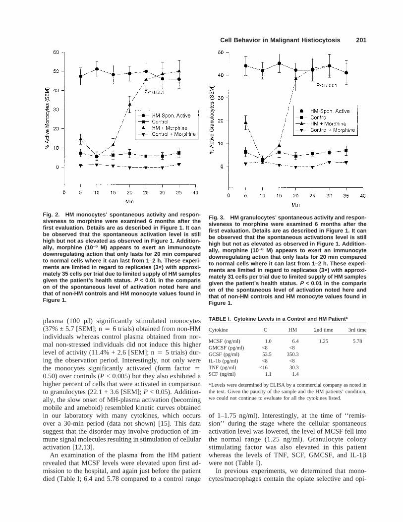
An examination of the plasma from the HM patientrevealed that MCSF levels were elevated upon first ad-mission to the hospital, and again just before the patientdied (Table I; 6.4 and 5.78 compared to a control range
of 1–1.75 ng/ml). Interestingly, at the time of ‘‘remis-sion’’ during the stage where the cellular spontaneousactivation level was lowered, the level of MCSF fell intothe normal range (1.25 ng/ml). Granulocyte colonystimulating factor was also elevated in this patientwhereas the levels of TNF, SCF, GMCSF, and IL-1bwere not (Table I).
In previous experiments, we determined that mono-cytes/macrophages contain the opiate selective and opi-
Fig. 2. HM monocytes’ spontaneous activity and respon-siveness to morphine were examined 6 months after thefirst evaluation. Details are as described in Figure 1. It canbe observed that the spontaneous activation level is stillhigh but not as elevated as observed in Figure 1. Addition-ally, morphine (10 −6 M) appears to exert an immunocytedownregulating action that only lasts for 20 min comparedto normal cells where it can last from 1–2 h. These experi-ments are limited in regard to replicates (3×) with approxi-mately 35 cells per trial due to limited supply of HM samplesgiven the patient’s health status. P < 0.01 in the comparison of the spontaneous level of activation noted here andthat of non-HM controls and HM monocyte values found inFigure 1.
Fig. 3. HM granulocytes’ spontaneous activity and respon-siveness to morphine were examined 6 months after thefirst evaluation. Details are as described in Figure 1. It canbe observed that the spontaneous activations level is stillhigh but not as elevated as observed in Figure 1. Addition-ally, morphine (10 −6 M) appears to exert an immunocytedownregulating action that only lasts for 20 min comparedto normal cells where it can last from 1–2 h. These experi-ments are limited in regard to replicates (3×) with approxi-mately 31 cells per trial due to limited supply of HM samplesgiven the patient’s health status. P < 0.01 in the comparison of the spontaneous level of activation noted here andthat of non-HM controls and HM monocyte values found inFigure 1.
TABLE I. Cytokine Levels in a Control and HM Patient*
Cytokine C HM 2nd time 3rd time
MCSF (ng/ml) 1.0 6.4 1.25 5.78GMCSF (pg/ml) <8 <8GCSF (pg/ml) 53.5 350.3IL-1b (pg/ml) <8 <8TNF (pg/ml) <16 30.3SCF (ng/ml) 1.1 1.4
*Levels were determined by ELISA by a commercial company as noted inthe text. Given the paucity of the sample and the HM patients’ condition,we could not continue to evaluate for all the cytokines listed.
Cell Behavior in Malignant Histiocytosis 201
oid peptide insensitive receptor,m3 [10], which down-regulates the cells’ activity and after exposure to activat-ing cytokines. Given the initial lack and later a partialresponse to morphine’s inhibitory action on the HM im-munocytes, we examined monocyte membrane homog-enates (limited supply for a one-point displacementanalysis) for the presence of this receptor. Compared tocontrol monocytes (5 nM3DHM bound to 149 fmol/mgprotein), we could not detect them3 receptor on the HMmonocytes, given the same level of protein per tube (0.12mg). It should be noted that even though our supply ofHM monocytes was quite small, in a comparable dilutionof control cells we were able to detect opiate binding.Thus, we believe this finding is valid despite the smallsample size due to the patient’s status.
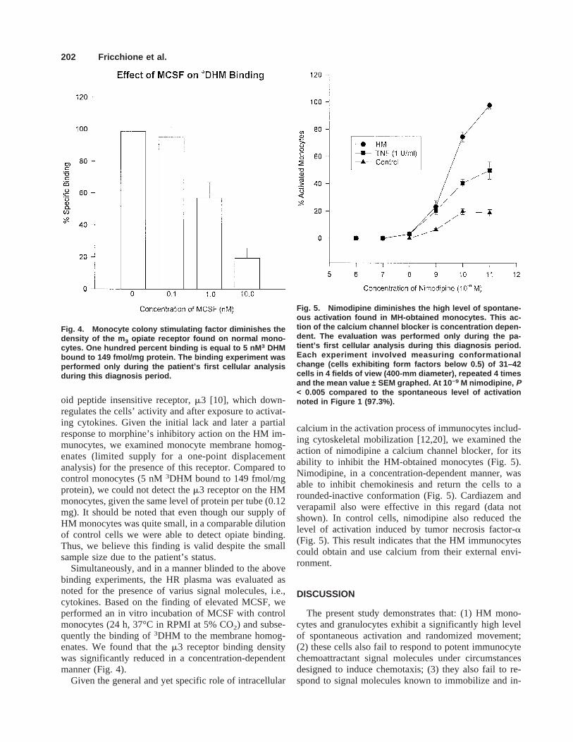
Simultaneously, and in a manner blinded to the abovebinding experiments, the HR plasma was evaluated asnoted for the presence of varius signal molecules, i.e.,cytokines. Based on the finding of elevated MCSF, weperformed an in vitro incubation of MCSF with controlmonocytes (24 h, 37°C in RPMI at 5% CO2) and subse-quently the binding of3DHM to the membrane homog-enates. We found that them3 receptor binding densitywas significantly reduced in a concentration-dependentmanner (Fig. 4).
Given the general and yet specific role of intracellular
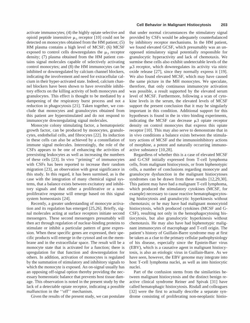
calcium in the activation process of immunocytes includ-ing cytoskeletal mobilization [12,20], we examined theaction of nimodipine a calcium channel blocker, for itsability to inhibit the HM-obtained monocytes (Fig. 5).Nimodipine, in a concentration-dependent manner, wasable to inhibit chemokinesis and return the cells to arounded-inactive conformation (Fig. 5). Cardiazem andverapamil also were effective in this regard (data notshown). In control cells, nimodipine also reduced thelevel of activation induced by tumor necrosis factor-a(Fig. 5). This result indicates that the HM immunocytescould obtain and use calcium from their external envi-ronment.
DISCUSSION
The present study demonstrates that: (1) HM mono-cytes and granulocytes exhibit a significantly high levelof spontaneous activation and randomized movement;(2) these cells also fail to respond to potent immunocytechemoattractant signal molecules under circumstancesdesigned to induce chemotaxis; (3) they also fail to re-spond to signal molecules known to immobilize and in-
Fig. 4. Monocyte colony stimulating factor diminishes thedensity of the m 3 opiate receptor found on normal mono-cytes. One hundred percent binding is equal to 5 nM 3 DHMbound to 149 fmol/mg protein. The binding experiment wasperformed only during the patient’s first cellular analysisduring this diagnosis period.
Fig. 5. Nimodipine diminishes the high level of spontane-ous activation found in MH-obtained monocytes. This ac-tion of the calcium channel blocker is concentration depen-dent. The evaluation was performed only during the pa-tient’s first cellular analysis during this diagnosis period.Each experiment involved measuring conformationalchange (cells exhibiting form factors below 0.5) of 31–42cells in 4 fields of view (400-mm diameter), repeated 4 timesand the mean value ± SEM graphed. At 10 −9 M nimodipine, P< 0.005 compared to the spontaneous level of activationnoted in Figure 1 (97.3%).
202 Fricchione et al.
activate immunocytes; (4) the highly opiate selective andopioid peptide insensitivem3 receptor [10] could not bedetected on monocytes obtained from the HM patient; (5)HM plasma contains a high level of MCSF; (6) MCSFexposed to control cells downregulates them3 receptordensity; (7) plasma obtained from the HM patient con-tains signal molecules capable of selectively activatingcontrol monocytes; and (8) the HM immunocytes can beinhibited or downregulated by calcium channel blockers,indicating the involvement and need for extracellular cal-cium in their hyper-activated state. Indeed, calcium chan-nel blockers have been shown to have reversible inhibi-tory effects on the killing activity of both monocytes andgranulocytes. This effect is thought to be mediated by adampening of the respiratory burst process and not areduction in phagocytosis [21]. Taken together, we con-clude that monocytes and granulocytes obtained fromthis patient are hyperstimulated and do not respond toimmunocyte downregulating signal molecules.
Monocyte colony stimulating factor, a hematopoieticgrowth factor, can be produced by monocytes, granulo-cytes, endothelial cells, and fibrocytes [22]. Its inductionin these cells can also be achieved by a constellation ofimmune signal molecules. Interestingly, the role of theCSFs appears to be one of enhancing the activities ofpreexisting leukocytes as well as increasing the numbersof these cells [23]. In vivo ‘‘priming’’ of immunocyteswith CSFs has been reported to increase their randommigration [23], an observation with great significance inthis study. In this regard, it has been surmised, as is thecase with the integration of many chemical signal sys-tems, that a balance exists between excitatory and inhibi-tory signals and that either a proliferative or a non-proliferative response will emerge based on this signalsystem homeostasis [24].
Recently, a greater understanding of monocyte activa-tion and its regulation has emerged [25,26]. Briefly, sig-nal molecules acting at surface receptors initiate secondmessengers. These second messengers presumably willthen act through regulation of nuclear-binding proteins tostimulate or inhibit a particular pattern of gene expres-sion. When these specific genes are expressed, their spe-cific products will emerge in the cytosol and on the mem-brane and in the extracellular space. The result will be amonocyte state that is activated for a function; there isupregulation for that function and downregulation forothers. In addition, activation of monocytes is regulatedby the summation of stimulatory and inhibitory signals towhich the monocyte is exposed. An on-signal usually hasan opposing off-signal option thereby providing the nec-essary homeostatic balance that prevents host tissue dam-age. This observation is noted in the present study by thelack of a detectable opiate receptor, indicating a possiblemalfunction in the ‘‘off’’ mechanism.
Given the results of the present study, we can postulate
that under normal circumstances the stimulatory signalprovided by CSFs would be adequately counterbalancedby inhibitory receptor mechanisms. In the HM patient,we found elevated GCSF, which presumably was an un-opposed stimulatory signal potentially responsible forgranulocytic hyperactivity and lack of chemotaxis. Wesurmise these cells also exhibit undetectable levels of them3 receptor, which downregulates its activity via nitricoxide release [27], since they normally express it [19].We also found elevated MCSF, which may have causedthe same picture in the MH monocytes. We speculate,therefore, that only continuous immunocyte activationwas possible, a result supported by the elevated serumlevel of MCSF. Furthermore, following a scan of cyto-kine levels in the serum, the elevated levels of MCSFsupport the present conclusion that it may be singularlyimportant in this condition. Additional support for thishypotheses is found in the in vitro binding experiments,indicating the MCSF can decreasem3 opiate receptordensity on control monocytes that express this opiatereceptor [10]. This may also serve to demonstrate that inin vivo conditions a balance exists between the stimula-tory actions of MCSF and the immunoinhibitory actionsof morphine, a potent and naturally occurring immuno-active substance [10,28].
Regardless of whether this is a case of elevated MCSFand G-CSF initially expressed from T-cell lymphomacells, from malignant histiocytosis, or from biphenotypiccells, a number of conclusions regarding monocyte andgranulocyte dysfunction in the malignant histiocytosessyndromes can be drawn from these results [12,29,30].This patient may have had a malignant T-cell lymphoma,which produced the stimulatory cytokines (MCSF, forexample) necessary to cause reactive hematophagocytos-ing histiocytosis and granulocytic hyperkinesis withoutchemotaxis; or he may have had malignant monocytoidhistiocytosis, which produced cytokines (MCSF and G-CSF), resulting not only in the hemophagocytosing his-tiocytosis, but also granulocytic hyperkinesis withoutchemotaxis. He may also have had biphenotypic malig-nant immunocytes of macrophage and T-cell origin. Thepatient’s history of Guillain-Barre syndrome may at firstbe taken as a clue to the primary cellular pathophysiologyof his disease, especially since the Epstein-Barr virus(EBV), which is a causative agent in malignant histiocy-tosis, is also an etiologic virus in Guillain-Barre. As wehave seen, however, the EBV genome may integrate intohost T-cell lymphoma nuclei, as well as into histiocyticnuclei.
Part of the confusion stems from the similarities be-tween malignant histiocytosis and the distinct benign re-active clinical syndrome Reiner and Spivak [31] havecalled hematophagic histiocytosis. Risdall and colleagues[32] were the first to actually describe a separate syn-drome consisting of proliferating non-neoplastic histio-
Cell Behavior in Malignant Histiocytosis 203
cytes that were strongly hematophagocytic. Initially fo-cused in response to viral infections, and thus termed bysome VAHS, hematophagic histiocytosis has since beendescribed as a reaction to almost any infectious pathogen,as well as to hematologic neoplasms and metastatic can-cer in the absence of infection [31]. It can occur even innon-immunosuppressed hosts. Reiner and Spivak believethat malignant histiocytosis is over-represented in theliterature, while hematophagic histiocytosis is actuallymore common than the literature would suggest. Thismay be understandable since the clinical and histologicaldifferences between malignant and hematophagic histio-cytosis are not always clear-cut. Fever, lymphadenopa-thy, hepatosplenomegaly, and pancytopenia are clinicalfeatures of both syndromes. They share the same demo-graphics (more common in middle age and in males) andwhile the prognosis in reactive hematophagic histiocyto-sis is more favorable, a rapid fatal outcome is still occa-sionally seen [33]. The presence of infection is not areliable marker because of its high incidence in patientswith hematopoietic malignancies. Malignant histiocyto-sis can complicate hematopoietic malignancies, but socan hematophagic histiocytosis.
Progress has been made in the last few years in usingclonal markers for tumor cell lines as a way to distinguishbetween malignant and benign forms of histiocytosis.Unfortunately, immunohistochemical techniques havenot helped characterize the origins of malignant histio-cytosis. Three very recent papers underscore this point.Oka et al. [29] reported on three cases of malignant his-tiocytosis that were diagnosed on the basis of clinicalsymptomatology, a rapidly fatal course, and the morphol-ogy and growth pattern of both blastoid and hemato-phagocytic cells. The blastoid and hematophagic cellsboth expressed the phenotype Mac-387+/KP1+/lysozyme+/polyclonal CD3+, with the Mac-387, KP1,and lysozyme markers evidencing monocytoid cell originand the CD3 marker suggesting T-lymphocyte origin.This result does not fully support the suggestion of Jaffeand colleagues that malignant histiocytosis may be ac-counted for by malignant lymphoma associated withlymphokine-stimulated non-neoplastic hematophagocy-tosing histiocytes [34]. It does suggest that, while manycases of so-called malignant histiocytosis have cells ex-pressing only T-lymphocyte antigens, there are othersthat have a biphenotypic character stemming from mac-rophage and T-cell populations. Su et al. [35] recentlypresented evidence showing clonotypic proliferation ofEBV genomes in the nuclei of large, atypical cells ex-pressing T-cell markers in four patients with clinicopath-ologic features of malignant histiocytosis. They surmisethat these are cases of T-cell lymphoma associated withcytokine-stimulated reactive histiocytosis. They also sug-gested that finding EBV genomes almost exclusively inthe nuclei of atypical T-cells can distinguish malignant
histiocytosis-like T-cell lymphoma from VAHS, inwhich EBV genomes are predominantly found in scat-tered B-cells, which then express the EBV antigens [36].However, even here there is variation, for Ohshima andassociates [37] recently reported a case of clinicopatho-logic malignant histiocytosis associated with chronicEBV infection. At necropsy, atypical hematophagocytos-ing histiocytes were noted to have extensively infiltratedbone marrow, liver, spleen, and lymph nodes. These cellswere positive for monocyte markers such as a-1-antitrypsin and MA935, but negative for T-cell or B-cellmarkers. In addition, in situ hybridization studies re-vealed many histiocytic nuclei with EBV genomes. Theysuggest that the EBV genome may integrate into histio-cytic host DNA, a situation akin to finding EBV genomein lymphoma nuclei, as noted above.
There is, therefore, evidence available for the malig-nant histiocytosis syndrome as secondary to (1) malig-nant T-cell lymphoma with reactive hematophagocytos-ing histiocytosis, (2) malignant monocytoid histiocytes,and (3) malignant biphenotypic immunocytes stemmingfrom both the macrophage and the T-cell. Reactive he-matophagocytic histiocytosis can be seen not only in re-sponse to T-cell lymphoma, but in reaction to other neo-plasms, as well as to immunodeficiency states, autoim-mune disorders, and to infectious diseases-as in VAHS[13]. It if of interest that the EBV has been associatedwith malignant histiocytosis of both the T-cell lymphomasubset, and true histiocytic varieties, as well as with re-active hematophagic histiocytosis.
In summary, MH may be regarded as a conditionwhereby the homeostatic balance controlling the activa-tion state of immunocytes is diminished or not present.Supporting this conclusion is our finding that MH mono-cytes lacked the ability to bind opiate alkaloids. Thus, thehigh level of immunocyte activation noted in this condi-tion could not be downregulated. In regard to the partialopiate alkaloid responsiveness of the monocyte duringthis patient’s treatment period, we can conclude that thechemotherapy given did not alleviate the condition.However, it appeared to provide for a partial recovery,perhaps due to a reduction in MCSF stimulation and aconsequent uncovering ofm3 opiate receptors. Further-more, the in vitro demonstration of MCSF action in di-minishing the opiate receptor levels points to that factthat in this condition the ability to regulate its synthesismay be damaged, leading to a lack of ‘‘braking’’ onimmunocytes. Indeed, this condition may itself be relatedto the presence of the viral condition, possibly leading todisinhibiting MCSF expression. In retrospect, these find-ings also may provide a rationale for combining a cal-cium blocker with traditional chemotherapy in this gen-erally fatal disease, given the in vitro inhibitory responseof the MH immunocytes to calcium channel blockers.
204 Fricchione et al.
ACKNOWLEDGMENTS
This work was in part supported by NIH grants MH/DA 17138, DA 09010, and NIH Fogarty INT00045(G.B.S.). The authors are indebted to Mr. FedericoCasares, Ms. Madeline Rodriguez, and Mr. RichardGlass at SUNY at Old Westbury (NIMH/NIDA CORFellow) for excellent technical assistance.
REFERENCES
1. Bodley SR, Smith R: Histiocytic medullary reticulosis. Lancet ii:194,1939.
2. Rappaport H: Tumors of the hemopoietic system. In Rappaport H (ed):‘‘Atlas of Tumor Pathology.’’ Washington, DC: Armed Forces Insti-tute of Pathology, 1966, pp 48–88.
3. Cline MJ: Histiocytes and histiocytosis. Blood 84:2840, 1994.4. Siegert W, Nell C, Engelhard M, Brittinger G, Tiemann M, Parware-
sch R, Heinz R, Huhn D: Peripheral T-cell non Hodgkin’s lymphomasof low malignancy: Prospective study of 25 patients with pleomorphicsmall cell lymphoma lymphoepithelial cell (Lehnert’s) lymphoma andT-zone lymphoma. The Kiel lymphoma study group. Br J Hematol87:529, 1994.
5. Editorial: Trends in lymphoma diagnosis. Lancet 1:249, 1989.6. Nezlof C: The 5q35bp chromosomal abnormality characterizes certain
CD30 positive anaplastic large cell lymphomas offering a new defi-nition of malignant histiocytosis in childhood. Nouv Rev Fr Hematol35:463, 1993.
7. Ishii E, Ohga S, Aoki T: Prognosis of children with virus-associatedhemophagocytic syndrome and malignant histiocytosis: Correlationwith levels of serum interleukin-1 and tumor necrosis factor. ActaHaematol 85:93, 1991.
8. Stefano GB: Role of opioid neuropeptides in immunoregulation. ProgNeurobiol 33:149, 1989.
9. Stefano GB, Melchiorri P, Negri L, Hughes TK, Scharrer B: (D-Ala2)-Deltorphin I binding and pharmacological evidence for a special sub-type of delta opioid receptor on human and invertebrate immune cells.Proc Natl Acad Sci USA 89:9316, 1992.
10. Stefano GB, Digenis A, Spector S, Leung MK, Bilfinger TV, MakmanMH, Scharrer B, Abumrad N: Opiatelike substances in an invertebrate,a novel opiate receptor on invertebrate and human immunocytes, anda role in immunosuppression. Proc Natl Acad Sci USA 90:11099,1993.
11. Smith EM, Hughes TKJ, Hashemi F, Stefano GB: Immunosuppressiveeffects of ACTH and MSH and their possible significance in humanimmunodeficiency virus infection. Proc Natl Acad Sci USA 89:782,1992.
12. Stefano GB, Bilfinger TV, Fricchione GL: The immune-neuro link andthe macrophage: Postcardiotomy delirium, HIV-associated dementiaand psychiatry. Prog Neurobiol 42:475, 1994.
13. Fricchione GL, Bilfinger TV, Stefano GB: The macrophage and neu-ropsychiatric disorders. Neuropsychiatry, neuropsychology and behav-ioral neurobiology. Neurobiology 9:16, 1996.
14. Shipp MA, Stefano GB, D’Adamio L, Switzer SN, Howard FD, Sin-isterra JI, Scharrer B, Reinkerz E: Downregulation of enkephalin-mediated inflammatory responses by CD10/neutral endopeptidase24.11. Nature 347:394, 1990.
15. Stefano GB, Bilfinger TV: Human neutrophil and macrophage che-mokinesis induced by cardiopulmonary bypass: Loss of DAME andIL-1 chemotaxis. J Neuroimmunol 47:189, 1993.
16. Stefano GB, Paemen LR, Hughes TKJ: Autoimmunoregulation: Dif-ferential modulation of CD10/neutral endopeptidase 24.11 by tumornecrosis factor and neuropeptides. J Neuroimmunol 41:9, 1992.
17. Stefano GB, Kushnerik V, Rodriquez M, Bilfinger TV: Inhibitoryeffect of morphine on granulocyte stimulation by tumor necrosis factorand substance p. Int J Immunopharmacol 16:329, 1994.
18. Stefano GB, Rodriquez M, Glass R, Casares F, Hughes TKJ, BilfingerTV: Hyperstimulation of leukocytes by plasma obtained from cardio-pulmonary bypass patients is diminished by morphine and IL-10 pre-treatment. J Cardiovasc Surg 36:25, 1995.
19. Makman MH, Bilfinger TV, Stefano GB: Human granulocytes containan opiate receptor mediating inhibition of cytokine-induced activationand chemotaxis. J Immunol 154:1323, 1995.
20. Stefano GB, Smith EM, Cadet P, Hughes TKJ: HIV GP120 alterationof DAMA and IL-1a induced chemotaxis responses in human andinvertebrate immunocytes. J Neuroimmunol 43:177, 1993.
21. Levy R, Dana R, Gold B, Alkan M, Schlaeffer F: Influence of calciumchannel blockers on polymorphonuclear and monocyte bactericidaland fungicidal activity. Israel J Med Sci 27:301, 1991.
22. Herrmann F, Mertelsmann R: Regulatory effects of cytokines on my-elopoiesis. In Coffey RR (ed): ‘‘Granulocyte Responses to Cyto-kines.’’ New York: Marcel Dekker, Inc., 1992, pp 339–349.
23. Donahue RE, Clark SC: Granulocyte colony stimulating factors astherapeutic agents. In Coffey RR (ed): ‘‘Granulocyte Responses toCytokines.’’ New York: Marcell Dekker, Inc., 1992, pp 637–649.
24. Axelrad AA: Some hemopoietic negative regulators. Exp Hematol18:143, 1990.
25. Adams DO, Hamilton TA: Molecular basis of macrophage activation:Diversity and its origins. In Lewis C (ed): The Natural Immune Sys-tem.’’ Oxford: University of Oxford Press, 1992, pp 1020–1026.
26. Adams DO: Macrophage activation. In Roitt IM, Delves PJ, (eds):‘‘Encyclopedia of Immunology,’’ Philadelphia: W.B. Saunders, 1992,pp 1020–1026.
27. Magazine HI, Liu Y, Bilfinger TV, Fricchione GL, Stefano GB: Mor-phine-induced conformational changes in human monocytes, granulo-cytes, and endothelial cells and in invertebrate immunocytes and mi-croglia are mediated by nitric oxide. J Immunol 156:4845, 1996.
28. Stefano GB, Scharrer B: Endogenous morphine and related opiates, anew class of chemical messengers. Adv Neuroimmunol 4:57, 1994.
29. Oka K, Mori N, Yatabe Y, Kojima M: Malignant hisitocytosis. Areport of 3 cases. Arch Pathol Lab Med 116:1228, 1992.
30. Nathan CF: Secretory products of macrophages. J Clin Invest 79:319,1987.
31. Reiner AP, Spivak JL: Hematophagic histiocytosis. A report of 23 newpatients and a review of the literature. Medicine 67:369, 1988.
32. Risdall RJ, McKenna RW, Nesbit ME, Drivit W, Balfrow HHJ, Sim-mons RL, Bunning RD: Virus associated hemophagocytic syndrome:A benign histiocytic proliferation distinct from malignant histiocyto-sis. Cancer 44:993, 1979.
33. Wong KF, Chan JK: Reactive hemophagocytic syndrome. A clinico-pathological study of 40 patients in an Oriental population. Am J Med93:177, 1992.
34. Jaffe ES, Costa J, Fauci AS, Cossman J, Isokos M: Malignant lym-phoma and erythrophagocytosis simulating malignant histiocytosis.Am J Med 304:648, 1981.
35. Su IJ, Hsu YH, Lin MT, Cheng AL, Wang CH, Weiss LM: Epstein-Barr virus containing T-cell lymphoma presents with hemophagocyticsyndrome mimicking malignant histiocytosis. Cancer 72:2019, 1993.
36. Muller-Hermelink HK: Epstein-Barr virus,b-cells, T-cells, and a lym-phoproliferative disorder. Lancet 343:922, 1994.
37. Ohshima K, Fujisaki T, Nagafudii S, Niho Y, Koloari S, Kikudii M:Malignant histiocytosis derived from a common histiocyte clone in apatient with chronic Epstein Barr virus infection. Leuk Lymph 17:355,1995.
Cell Behavior in Malignant Histiocytosis 205