Embed Size (px)
Citation preview
1
Crest® + Oral-B® at dentalcare.com
Course Author(s): Maria L. Geisinger, DDS, MSCE Credits: 2 hoursIntended Audience: Dentists, Dental Hygienists, Dental Assistants, Dental Students, Dental Hygiene Students, Dental Assistant StudentsDate Course Online: 04/27/2020Last Revision Date: N/ACourse Expiration Date: 04/26/2023Cost: FreeMethod: Self-instructionalAGD Subject Code(s): 148, 750
Online Course: www.dentalcare.com/en-us/professional-education/ce-courses/ce619
Disclaimer: Participants must always be aware of the hazards of using limited knowledge in integrating new techniques or procedures into their practice. Only sound evidence-based dentistry should be used in patient therapy.
Please Note: This course may not satisfy individual state requirements on CDC/Infection Control. Please check with your State Board to verify.
Conflict of Interest Disclosure Statement• The author reports no conflicts of interest associated with this course.
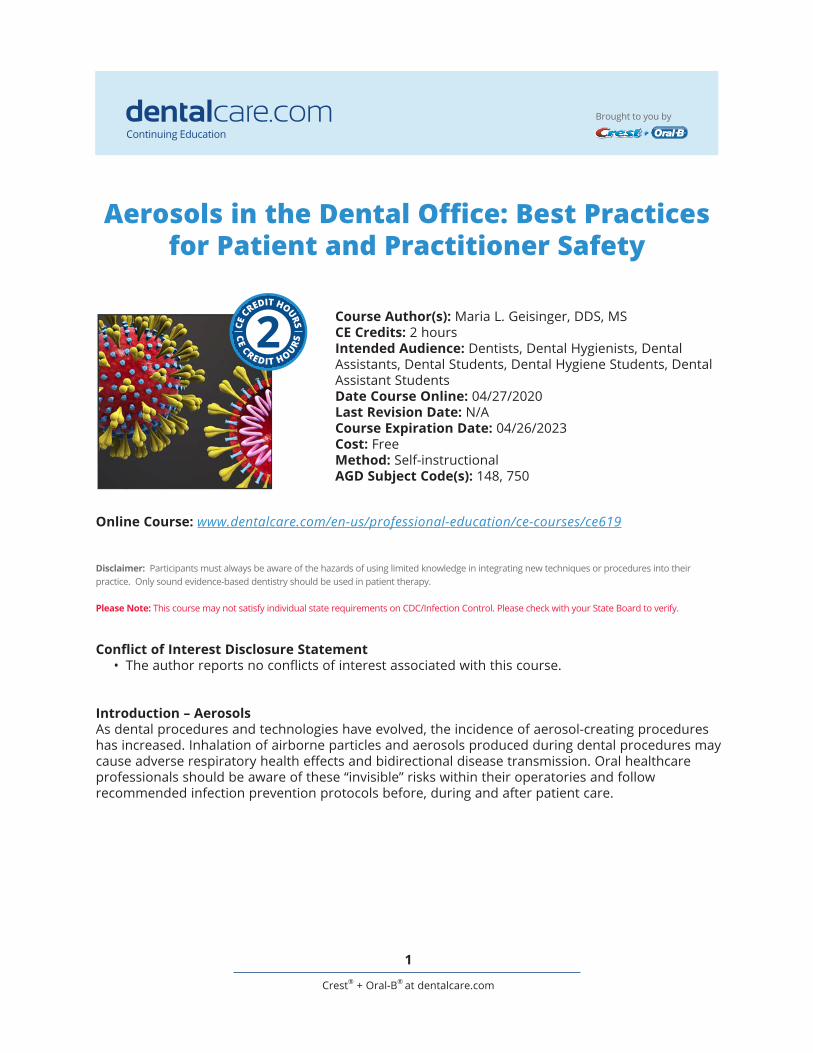
Introduction – AerosolsAs dental procedures and technologies have evolved, the incidence of aerosol-creating procedures has increased. Inhalation of airborne particles and aerosols produced during dental procedures may cause adverse respiratory health effects and bidirectional disease transmission. Oral healthcare professionals should be aware of these “invisible” risks within their operatories and follow recommended infection prevention protocols before, during and after patient care.
Aerosols in the Dental Office: Best Practices for Patient and Practitioner Safety
Continuing Education
Brought to you by

2
Crest® + Oral-B® at dentalcare.com
Course Contents• Overview• Learning Objectives• Introduction• Aerosols in the Dental Office: How, When,
and Where?• Airborne Droplets: Aerosols vs. Splatter?• Dental Procedures Associated with
Aerosols• Infectious Diseases Associated with
Aerosols• SARS-CoV-2 (Viral Cause of COVID-19) and
Aerosol Transmission• Characteristics of SARS-CoV-2• Potential Modes of Transmission for SARS-
CoV-2 in the Dental Office• Standard and Transmission-based
Precautions: Best Practices for Dental Professionals• Prevention Airborne Disease Transmission
Dental Office• Special Precautions for High-aerosol
Procedures (Known or Suspected COVID-19 Cases)
• Emerging Infection Prevention Practices for Use in Dental Practice after SARS-CoV-2
• Summary• Course Test• References/Additional Resources• About the Author
OverviewWith the rapid pandemic of COVID-19 and the spread of its underlying viral agent, SARS-CoV-2, there has been a renewed interest in aerosol-generating dental procedures in the dental office. Airborne disease transmission for SARS-CoV-2 has been established as a method of community spread. What is less well-defined is the role of aerosols generated during dental care in disease transmission. While previous research has demonstrated that microorganisms originating in the oral cavity and/or present in dental unit water lines (DUWL) are capable of causing respiratory infections, the current pandemic has highlighted the potential risks of dental aerosols and potential mitigation strategies. In addition to the close contact that routinely occurs during the delivery of dental care, the use of dental handpieces, powered scalers, air/water syringes, and other dental instruments
are known to create aerosols and spatter. As dental procedures and technologies have evolved, the incidence of aerosol-generating dental procedures has increased and a focus on methodologies to reduce aerosol bioload, to mitigate aerosol spread throughout the dental office, and to decrease aerosol generation is warranted.
Learning ObjectivesUpon completion of this course, the dental professional should be able to:• Explain the risk factors and basic properties
of aerosols generated during routine dental procedures.
• Describe what types of dental procedures result in significant dental aerosol production.
• Understand the types of pathogens and resultant illnesses are associated with such aerosols.
• Differentiate between standard and transmission-based precautions and their utility in the dental office for safe delivery of care.
• List infection control and aerosol mitigation techniques that may reduce the risk of cross-contamination to patients and providers.
IntroductionA novel β-coronavirus (SARS-CoV-2) causing severe and potentially fatal pneumonia (COVID-19) purportedly originating in a seafood market in Wuhan city, Hubei province, China has undergone pandemic spread throughout the globe.1-3 The typical clinical symptoms of SARS-CoV-2 infection are fever, dry cough, myalgia, fatigue, and pneumonia with abnormal chest CT. Less commonly observed symptoms include sputum production, headache, hemoptysis, and diarrhea.4-6 A zoonotic origin for SARS-CoV-2 is presumed. SARS-CoV-2 demonstrates 96.2% of whole-genome identity to the horseshoe bat (Rhinolophus affinis) virus RaTG13 and this novel virus demonstrates over 99% genetic similarity to β-CoV samples found in pangolins, (scaly anteaters).7,8 While approximately 70% of viruses become pathogenic in humans after moving from animals to humans, the exact species-level source of this virus has not yet been well-established. The person-to-person transmission of SARS-CoV-2 include direct
3
Crest® + Oral-B® at dentalcare.com
transmission, such as cough, sneeze, saliva and other droplet inhalation transmission, and contact transmission, such as the contact with oral, nasal, and eye mucous membranes and contaminated environmental surfaces.9-12
Dental health care personnel (DHCP) and their patients are at risks associated with aerosols in the dental office due to the frequency of close, personal face-to-face communication and exposure to saliva, blood, and other body fluids, and—indirectly—by the handling of sharp instruments and touching contaminated dental surfaces.13-16 Previous studies have shown that microorganisms in the mouth and respiratory tract can be transported in aerosols, splash and spatter generated during dental procedures and can contaminate the skin and mucous membranes of the mouth, respiratory passages, and eyes of DHCP as well as environmental surfaces and materials exposed to such aerosols and droplets. As such, DHCP play an important role in preventing disease transmission within the dental practice.17-24 This course seeks to assess the risks posed by aerosols in the dental office and assess infection control measures that can be implemented during dental practice to block the person-to-person transmission routes through standard and transmission-based precautions.25
Aerosols in the Dental Office: How, When, and Where?Airborne transmission of various pathogens has been demonstrated in both healthcare and community settings. Airborne transmission of tuberculosis and measles has been documented on commercial aircraft and in the clinic waiting area, where increased proximity to infected persons conveyed increased transmission risk.26,27 Viral transmission after airborne droplets/particles have settled on surfaces in these environments has also been demonstrated.26,27 Furthermore, isolation of several different pathogens capable of aerosol transmission have been noted in the dental offices.28,29
Airborne Droplets: Aerosols vs. Splatter?Aerosols are defined as liquid or solid particles less than 50 micrometers in diameter.20,21,30,31
Particles of this size are small enough to stay airborne for an extended period before they settle on environmental surfaces or enter the respiratory tract after inhalation.20,21 Smaller particles of in aerosols (droplets and droplet nuclei 0.5 to 10 μm in diameter) have the potential to enter the lungs and settle within the bronchial passages, reaching as far as the pulmonary alveoli.20,21 These droplets are thought to convey a high level of risk infection transmission in the dental office.30,31
Splash and splatter a mixture of air, water, and/or solid substances larger than 50 μm in diameter are visible to the naked eye20,21 and behave in a ballistic or projectile manner.20 These mixtures are ejected forcibly from their origin in an arc and travel along a bullet-like trajectory until they contact a surface or fall to the ground under the influence of gravitational forces.20 Unlike aerosols, splash and splatter are airborne only briefly.20,21 Because of this, they demonstrate limited penetration into the respiratory system.20,21
Within the dental office, airborne droplets and droplet nuclei present unique risks to DHCP and patients.30,31 They can remain in the air for a long time, may be transported with air flows for long distances, and can contaminate wide areas within the dental operatory.30-32 Splash and splatter, on the other hand, are generally deposited on surfaces closer to their origin, an estimated 15-120 cm from the source.31,32 These particles are a risk due to their contact with mucous membrane and close surfaces, including DHCP.31,32 Furthermore, there is evidence that some microorganisms may survive within splash and spatter and when the contaminated surfaces dry organisms may even become airborne as dust particles.31,32
Dental Procedures Associated with AerosolsAirborne contamination during dental procedures may come from a variety of sources. Foremost among these are: dental instrumentation, salivary, and respiratory sources.33 Dental handpieces, ultrasonic scalers, and the air-water syringes used in common dental practice are capable of producing aerosols, which are usually a mix of air and water derived from these devices

4
Crest® + Oral-B® at dentalcare.com
potential for disease transmission, even after the infected person has left the vicinity.34,38-40 SARS-CoV-2 can survive on such environmental surfaces for prolonged periods of time.34,41 Table 1 describes the risk of aerosols associated with dental procedures.
Infectious Diseases Associated with AerosolsAside from the common cold (rhinoviruses and other viruses), several types of bacteria and viruses have demonstrated airborne person-to-person transmission (Table 2).30,42-45
For many of these microorganisms, the overall microbial load within aerosols, splash, and splatter vary greatly predicated on disease status and the particular microorganism.11-13,25 For example, the reproduction number (R0) differs significantly between microorganisms.46
The R0 is the number of cases, on average, an infected patient will cause during their infectious period. This number, in practical application, is also influenced by the overall susceptibility within the population (e.g., vaccination rates, previous infection rates, cross-immunity from similar diseases, the novelty of a pathogen).46 Lastly, the likelihood of transmission is also influenced by the susceptibility of the host and related
and the patient’s saliva.34 Dental instruments, surfaces within the dental operatory, and dental equipment, when improperly cleaned, sterilized, and stored, or disinfected can also serve as fomites and contribute to cross-infection.
The oral environment is naturally wet and contains a high number of microorganisms. Dental plaque is a major source of such organisms, containing more than 700 known pathogens,35 but the mouth also harbors bacteria from the respiratory tract, including the nasopharynx and the lower pulmonary system.30 Gingival crevicular fluid, debris from tooth preparation, and dental materials may also be aerosolized during dental procedures and contribute to disease transmission.36,37
The most intense aerosol and splash and splatter has been shown to occur during use of ultrasonic scalers and high speed handpieces without a rubber dam;30,32,36 however, aerosols in the dental setting have also been associated with the use of low-speed handpieces, air/water syringes, patient coughing, and intraoral radiography.34 Because of the ability of aerosols to remain suspended in the air and travel further than splash and splatter, and distant contamination may occur, and there is
Table 1. Dental Devices and Procedures Known to Produce Airborne Contamination.30

5
Crest® + Oral-B® at dentalcare.com
converting enzyme 2 receptor (ACE2).57-60 Affinity for the ACE2 receptor is postulated to explain increased viral loads seen in older individuals, since ACE2 expression increases with age.61
It is well-established that RNA viruses have higher rates of mutation than DNA-viruses.62 On a per-site level, DNA viruses typically have mutation rates on the order of 10−8 to 10−6 substitutions per nucleotide site per cell infection (s/n/c). RNA viruses have mutation rates that range between 10−6 and 10−4 s/n/c. Given this rapid mutation rate, RNA viruses with high rates of pathogenicity are able generally to develop and propagate more frequently in the environment.62 However, current data suggests that SARS-CoV-2 may have a lower mutation rate than influenza, which may be advantageous during the development of vaccines and target medications.63 Additionally, the reproduction number (R0) of SARS-CoV-2 has been estimated to be between 1.5-6.49 with a median value of 2.78.64 This R0 is greater than that of SARS-CoV-1 (median R0=1.3)65 and H1N1 influenza (median R0=1.46),66 but less than that of measles (median R0=16.1).67
SARS-CoV-2 shares numerous similarities with SARS-CoV-1 [the causative agent in the severe acute respiratory syndrome (SARS) outbreak in 2002-2003], but there are some significant differences. Both originated in China and both
factors such as, overall health status, genetic influences, immunocompetence, vaccination/infection history, and previous exposure to similar diseases.47,48
SARS-CoV-2 (Viral Cause of COVID-19) and Aerosol Transmission
Characteristics of SARS-CoV-2Coronaviruses (Coronaviridae, of the order Nidovirales) are large, single stranded RNA viruses.49,50 Currently, there are four known genera of coronaviruses: α-CoV, β-CoV, γ-CoV, and δ-CoV.51,52 Coronaviruses have been identified as the causative agents of diseases in humans and other vertebrates. SARS-CoV-2 belongs to the β-COV family, which along with α-CoV viruses, are known to infect mammals and humans.49,53,54 SARS-CoV-2 possesses an ultrastructure typical of other coronaviruses, namely a membrane envelope with multiple “spike glycoprotein” (S-protein) extensions (Figure 1).55 The viral capsule has also been found to express other polyproteins, nucleoproteins, and membrane proteins, including specifically RNA polymerase, 3-chymotrypsin-like protease, papain-like protease, helicase, glycoprotein, and accessory proteins.7,55,56 The S-proteins from coronaviruses binds to receptors on host cells to facilitate viral entry into the target cells. For SARS-CoV-2 the target receptor is the human angiotensin-
Table 2. Diseases Known to be Spread by Droplets or Aerosols.30

6
Crest® + Oral-B® at dentalcare.com
demonstrate stability in the environment. In an in vitro study, SARS-CoV-2 was detectable in aerosols for up to three hours, up to four hours on copper, up to 24 hours on cardboard and up to two to three days on plastic and stainless steel.68 However, while SARS-CoV-1 was eradicated by intensive contact tracing and case isolation measures and no cases have been detected since 2004.69 SARS-CoV-2 has proven to be significantly more difficult to eradicate. Emerging evidence suggests that asymptomatic people infected with SARS-CoV-2 may be transmitting the virus prior to the onset of symptoms.70 The occurrence of asymptomatic transmission decreases the effectiveness of disease control measures that were effective against SARS-CoV-1.70 In contrast to SARS-CoV-1, most secondary cases of virus transmission of SARS-CoV-2 appear to be occurring in community settings.70
Both of viruses demonstrate binding affinity to the ACE2 receptor to enter host cells. However, the S-proteins from SARS-CoV-2 are less stable than those of SARS-CoV-1 and polyclonal anti-SARS S1 antibodies that inhibit entry of SARS-CoV-1, are not effective against SARS-CoV-2 pseudovirions.71 Further studies using recovered SARS and COVID-19 patients’ sera
show limited cross-neutralization, suggesting that recovery from one infection might not protect against the other.71
Potential Modes of Transmission for SARS-CoV-2 in the Dental OfficeEvidence suggests that SARS-CoV-2 can be transmitted both directly from person-to-person by respiratory droplets and via indirect fomite-mediated transmission.4,5 Viral shedding by asymptomatic individuals has been reported during an asymptomatic prodromal period of up to 14 days with the potential for viral shedding up to 24 days.72 Live SARS-CoV-2 viruses have been isolated from saliva of infected individuals and the concentration of virus in saliva has been shown in some cases to be significantly higher than that on nasopharyngeal testing swabs.13,73 Not surprisingly, ACE2+ cells are abundant throughout the respiratory tract and salivary gland duct epithelium.25,65 In the SARS epidemic in the early 2000s caused by SARS-CoV-1, epithelial cells of the salivary gland ducts were early targets for viral infection.74
Transmission of SARS-CoV-2 is increased in the dental setting due to the close interpersonal contact between individuals involved and by
Figure 1. Diagram of the ultrastructure of the SARS-CoV-2 virus.91

7
Crest® + Oral-B® at dentalcare.com
other bloodborne pathogens such as hepatitis B virus (HBV) and hepatitis C virus (HCV), which has been expanded in the intervening years to include other potentially infectious material (OPIM). Today infection prevention is predicated on Standard and Transmission based Precautions.81-86 There are three categories of Transmission-based Precautions: contact precaution, droplet precautions, and airborne precautions associated with droplet nuclei.84-87
Airborne precautions include administrative controls, environmental controls, and respiratory-protection controls. While typical outpatient dental facilities must incorporate administrative controls into their infection prevention protocol, they are not expected to be in full compliance with environmental and respiratory-prevention controls.
Prevention of Airborne Disease Transmission in the Dental OfficeInfection control standards were initially developed for dentistry in response to the HIV epidemic and included Standard and Transmission-based Precautions. Based upon emerging evidence regarding SARS-CoV-2
nature of the procedures performed during the delivery of dental care.75-77 Both DHCPs and patients are at risk due to droplets containing microorganisms or direct contact with conjunctival, nasal, or oral mucosal tissues.17-20,30,75-78 Furthermore, SARS-CoV-2 may survive between 4 to 72 hours on hard surfaces, which can lead to indirect exposure after touching such surfaces.70 The likelihood of such transmissions may be dependent upon the viral load of the infected individual and the susceptibility of the host individual.79 Potential pathways of SARS-CoV-2 transmission in the dental office are outlined in Figure 2.
Standard and Transmission-based Precautions: Best Practices for Dental ProfessionalsIn 1985, the Centers for Disease Control [now the Centers for Disease Control and Prevention (CDC)] introduced the concept that all blood and body fluids that might be contaminated with blood should be treated as infectious.80 Initial infection control measures were introduced largely because of the human immunodeficiency virus (HIV) epidemic; it was expanded to Universal Precautions to include
Figure 2. Potential transmission pathways for SARS-CoV-2 in the dental office.25

8
Crest® + Oral-B® at dentalcare.com
• If N95 or higher respirator masks are not available due to supply-chain or other issues, use of a surgical mask and full-face shield should be worn and patients should be referred for COVID-19 testing after the procedure.
• Practice how to properly don, use, and doff PPE in a manner to prevent self-contamination.
• Perform hand hygiene with alcohol-based hand rub before and after all patient contact, contact with potentially infectious material, and before putting on and upon removal of PPE, including gloves. Use soap and water if hands are visibly soiled.
• Clean and disinfect clinical surfaces with approved disinfection protocols and utilizing disinfectants from EPA-approved emerging viral pathogens claims (List N).90
• Screen all DHCP at the beginning of their shift for fever and respiratory symptoms. Document shortness of breath, new or change in cough, and sore throat. If they are ill, have them put on a facemask and leave the workplace.
Special Precautions for High-aerosol Procedures (Known or Suspected COVID-19 Cases)If emergency dental care is medically necessary for a patient who has, or is suspected of having COVID-19, Airborne Precautions including the following:88,89
• HCP in the room should wear an N95 or higher-level respirator, eye protection, gloves, and a gown.
• The number of healthcare providers present during the procedure should be limited to only those essential for patient care and procedure support. All other individuals should avoid contact within six feet of the patient.
• Procedures should ideally take place in an airborne infection isolation room (AIIR).
• Clean and disinfect procedure room surfaces promptly using approved protocols and disinfectants identified by the Environmental Protection Agency as effective against SARS-CoV-2.90
Dental treatment should be provided in a hospital or other facility that can treat the patient using the appropriate precautions.
and previous investigations studying other coronaviruses, spread is thought to occur mostly from person-to-person via respiratory droplets among close contacts.88 Close contact can occur while delivering patient care and is currently defined by the CDC as: 1) being within approximately 6 feet (2 meters) of a patient with COVID-19 for a prolonged period of time (≥30 minutes) or 2) having direct contact with infectious secretions from a patient with COVID-19. Infectious secretions may include sputum, saliva, serum, blood, and respiratory droplets.88 No reliable data currently exist to assess the risk of SARS-CoV-2 during routine dental practice or to determine whether the routine PPE and Standard Precautions used by DHCP is adequate to protect them.
CDC and ADA interim recommendations updated on April 7, 2020 for infection prevention and control include:88,89
• Postponement of elective and non-essential dental procedures until deemed safe by public health and governmental authorities.
• DHCP and patients should stay at home if experiencing COVID-19 symptoms and seek medical care as recommended based upon symptoms and healthcare provider assessment.
• Assess and triage patients using teledentistry prior to an in-person dental visit. Assess the emergent nature of the patient’s dental needs and any symptoms related to COVID-19. Patients with confirmed or suspected COVID-19 should be referred to contact the emergency department to determine the optimal patient care options, even for dental emergencies.
• If urgent and/or emergent dental treatment must be delivered for an asymptomatic patient, DHCP should then assess the likelihood of aerosol production during care.
• If the needed procedure is unlikely to produce aerosols, DHCP can use routine PPE and the procedure is considered low-risk.
• If aerosols are likely to be produced, the following PPE standards are recommended:• N95 (KN95) or higher-level respirator masks• Full face shield or goggles• Gloves• Disposable gown• Head coverings

9
Crest® + Oral-B® at dentalcare.com
coronavirus (SARS-CoV-2) has been implicated in the COVID-19 global pandemic. Similar to a previous coronavirus (SARS-CoV-1), SARS-CoV-2 enters host cells through human cell receptor ACE2 but appears to demonstrate higher binding affinity and SARS-CoV-2 has been shown to potentially have a higher reproduction number, indicating a higher level of transmissibility. Given these emerging data and what is known about the transmission of other coronaviruses, airborne transmission via droplets and possibly droplet nuclei are the likely main mode of person-to-person transmission.
Dental healthcare providers are urged to closely monitor advisory statements from governmental agencies and professional organizations regarding best practices.
Emerging Infection Prevention Practices for Use in Dental Practice after SARS-CoV-2As science and our understanding of the SARS-CoV-2 virus evolves and the pandemic subsides, it is likely that we may find ourselves in an endemic situation where lower levels of this virus exist in the population demonstrating periodic or even seasonal spikes in infection. In this scenario, it may become imperative that new strategies be developed to interrupt the chain of infection. Until such time Standard and Transmission-Based Precaution, including rigorous enforcement of Point-of-Care implementation of Respiratory Hygiene, and adherence to ADA and CDC Guidelines represent the Standard of Care.
SummarySince its first identification in Wuhan, China in November-December 2019, the novel

10
Crest® + Oral-B® at dentalcare.com
Course Test PreviewTo receive Continuing Education credit for this course, you must complete the online test. Please go to: www.dentalcare.com/en-us/professional-education/ce-courses/ce619/test
1. COVID-19 is a disease-causing severe pneumonia in patients infected by _______________.A. Yersinia pestisB. SARS-CoV-1C. SARS-CoV-2D. BatCoV RaTG13
2. The person-to-person transmission routes for COVID-19 of concern in dental practice include all of the following EXCEPT:A. Direct transmission through airborne particles produced by a cough or sneezeB. Direct transmission through airborne particles produced by dental proceduresC. contact transmission after touching contaminated surfaces and then touching oral, nasal, and
eye mucous membranesD. Fecal transmission
3. Aerosols are defined as liquid or solid particles less than 50 micrometers in diameter. Particles of this size are small enough to stay airborne for an extended period but can only travel limited distances (less than 120 cm).A. Both statements are true.B. The first statement is true, the second statement is false.C. The first statement is false, the second statement is true.D. Both statements are false.
4. All of the following are true about splatter droplets, EXCEPT:A. Splatter particles are usually a mixture of air, water, and/or solid substance and are larger
than 50 μm in diameterB. They may become suspended in air for long periods of timeC. Splatter particles follow a ballistic pattern and travel in an arc after they are emitted until they
contact a surface or fall to the groundD. They may be visible to the naked eye
5. The highest levels of aerosol and splatter emission has been shown to occur with the use of _______________.A. ultrasonic scalersB. intraoral radiograph captureC. high speed handpiece used with a rubber damD. low speed handpiece
6. The reproduction number (R0) describes _______________.A. the number of cases, on average, who will become infected annuallyB. the percentage of cases, on average, who will be infected, but asymptomatic during the
course of a disease outbreakC. the number of cases, on average, an infectious patient will cause during their infectious
periodD. the number of times a virus will replicate prior to one meaningful genetic mutation

11
Crest® + Oral-B® at dentalcare.com
7. Entry into host cells of coronaviruses is facilitated by the S-protein. In the case of both SARS-CoV-1 and SARS-CoV-2, this entry is through binding to the _______________.A. CD4 receptorB. Major histocompatibility complex (MHC)C. Angiotensin II receptor (ARB)D. angiotensin-converting enzyme 2 receptor (ACE2)
8. Viral shedding by asymptomatic individuals infected by SARS-COV-2 has been reported during a prodromal period of up to _______________.A. 5 daysB. 7 daysC. 14 daysD. 24 days
9. SARS-CoV-2 may survive on hard surfaces up to _______________.A. 6 hoursB. 12 hoursC. 24 hoursD. 72 hours
10. Transmission-based Precautions include all of the following categories EXCEPT:A. AirborneB. DropletC. ContactD. Distance
11. The United States Centers for Disease Control and Prevention (CDC) states that close contact with a patient infected with SARS-CoV-2 conveys significant risk for development of COVID-19. The CDC defines “close contact” as: 1) being within approximately 6 feet (2 meters) of a patient with COVID-19 for a prolonged period of time (≥30 minutes) or 2) having direct contact with infectious secretions from a patient with COVID-19.A. Both statements are true.B. The first statement is true, the second statement is false.C. The first statement is false, the second statement is true.D. Both statements are false.
12. The CDC recommends that healthcare workers take precautions to avoid direct contact with infectious secretions from patients who are known or possible cases of COVID-19. All of the following are considered infectious secretions, EXCEPT:A. SputumB. SalivaC. BloodD. Sweat
12
Crest® + Oral-B® at dentalcare.com
13. Current CDC guidance suggests that procedures that produce high levels of aerosols, in particular those that demonstrate exposure to infectious secretions or are likely to induce coughing should be avoided, if possible. If such procedures must be performed, the CDC suggests use of an N95 or greater respirator and other PPE, limiting the number of healthcare providers to those that are essential for the procedure, and performance of aerosol-generating procedures in an airborne infection isolation room.A. Both statements are true.B. The first statement is true, the second statement is false.C. The first statement is false, the second statement is true.D. Both statements are false.
13
Crest® + Oral-B® at dentalcare.com
References1. Zhu N, Zhang D, Wang W, et al. A Novel Coronavirus from Patients with Pneumonia in China,
2019. N Engl J Med. 2020;382(8):727–733. doi:10.1056/NEJMoa2001017.2. Wang C, Horby PW, Hayden FG, Gao GF. A novel coronavirus outbreak of global health concern
[published correction appears in Lancet. 2020 Jan 29;:]. Lancet. 2020;395(10223):470–473. doi:10.1016/S0140-6736(20)30185-9.
3. Liu T, Hu J, et al. Transmission Dynamics of 2019 Novel Coronavirus (2019-nCoV). The Lancet. 2020 Feb 05. Available at SSRN: doi:10.2139/ssrn.3526307. Accessed April 22, 2020.
4. Huang C, Wang Y, Li X, et al. Clinical features of patients infected with 2019 novel coronavirus in Wuhan, China [published correction appears in Lancet. 2020 Jan 30;:]. Lancet. 2020;395(10223):497–506. doi:10.1016/S0140-6736(20)30183-5.
5. Guan WJ, Ni ZY, Hu Y, et al. Clinical Characteristics of Coronavirus Disease 2019 in China [published online ahead of print, 2020 Feb 28]. N Engl J Med. 2020;NEJMoa2002032. doi:10.1056/NEJMoa2002032.
6. Wang D, Hu B, Hu C, et al. Clinical Characteristics of 138 Hospitalized Patients With 2019 Novel Coronavirus-Infected Pneumonia in Wuhan, China [published online ahead of print, 2020 Feb 7]. JAMA. 2020;e201585. doi:10.1001/jama.2020.1585.
7. Zhou P, Yang XL, Wang XG, et al. A pneumonia outbreak associated with a new coronavirus of probable bat origin. Nature. 2020;579(7798):270–273. doi:10.1038/s41586-020-2012-7.
8. Wahba L, Jain N, Fire AZ, et al. Identification of a pangolin niche for a 2019-nCoV-like coronavirus through an extensive meta-metagenomic search. bioRxiv 2020 Feb 14. doi:10.1101/2020.02.08.939660. Accessed April 22, 2020.
9. Lu CW, Liu XF, Jia ZF. 2019-nCoV transmission through the ocular surface must not be ignored. Lancet. 2020;395(10224):e39. doi:10.1016/S0140-6736(20)30313-5.
10. Belser JA, Rota PA, Tumpey TM. Ocular tropism of respiratory viruses. Microbiol Mol Biol Rev. 2013;77(1):144–156. doi:10.1128/MMBR.00058-12.
11. Rothe C, Schunk M, Sothmann P, et al. Transmission of 2019-nCoV Infection from an Asymptomatic Contact in Germany. N Engl J Med. 2020;382(10):970–971. doi:10.1056/NEJMc2001468.
12. Wax RS, Christian MD. Practical recommendations for critical care and anesthesiology teams caring for novel coronavirus (2019-nCoV) patients. Directives concrètes à l’intention des équipes de soins intensifs et d’anesthésiologie prenant soin de patients atteints du coronavirus 2019-nCoV. Can J Anaesth. 2020;67(5):568–576. doi:10.1007/s12630-020-01591-x.
13. To KK, Tsang OT, Chik-Yan Yip C, et al. Consistent detection of 2019 novel coronavirus in saliva [published online ahead of print, 2020 Feb 12]. Clin Infect Dis. 2020;ciaa149. doi:10.1093/cid/ciaa149.
14. Rodríguez-Morales AJ, MacGregor K, Kanagarajah S, Patel D, Schlagenhauf P. Going global - Travel and the 2019 novel coronavirus. Travel Med Infect Dis. 2020;33:101578. doi:10.1016/j.tmaid.2020.101578.
15. Faecher RS, Thomas JE, Bender BS. Tuberculosis: a growing concern for dentistry?. J Am Dent Assoc. 1993;124(1):94–104. doi:10.14219/jada.archive.1993.0003.
16. Nash KD. How infection control procedures are affecting dental practice today. J Am Dent Assoc. 1992;123(3):67–73. doi:10.14219/jada.archive.1992.0076.
17. Earnest R, Loesche W. Measuring harmful levels of bacteria in dental aerosols. J Am Dent Assoc. 1991;122(12):55–57. doi:10.14219/jada.archive.1991.0187.
18. Travaglini EA, Larato DC, Martin A. Dissemination of organisms bearing droplets by high-speed dental drills. J Prosthet Dent 1966;16:132-9. doi:10.1016/0022-3913(66)90120-X. Accessed April 22, 2020.
19. Miller RL. Generation of airborne infection...by high speed dental equipment. J Am Soc Prev Dent. 1976;6(3):14–17.
20. Micik RE, Miller RL, Mazzarella MA, Ryge G. Studies on dental aerobiology. I. Bacterial aerosols generated during dental procedures. J Dent Res. 1969;48(1):49–56. doi:10.1177/00220345690480012401.

14
Crest® + Oral-B® at dentalcare.com
21. Miller RL, Micik RE, Abel C, Ryge G. Studies on dental aerobiology. II. Microbial splatter discharged from the oral cavity of dental patients. J Dent Res. 1971;50(3):621–625. doi:10.1177/00220345710500031701.
22. Holbrook WP, Muir KF, Macphee IT, Ross PW. Bacteriological investigation of the aerosol from ultrasonic scalers. Br Dent J. 1978;144(8):245–247. doi:10.1038/sj.bdj.4804072.
23. Williams GH 3rd, Pollok NL 3rd, Shay DE, Barr CE. Laminar air purge of microorganisms in dental aerosols: prophylactic procedures with the ultrasonic scaler. J Dent Res. 1970;49(6):1498+. doi:10.1177/00220345700490065701.
24. Bentley CD, Burkhart NW, Crawford JJ. Evaluating spatter and aerosol contamination during dental procedures. J Am Dent Assoc. 1994;125(5):579–584. doi:10.14219/jada.archive.1994.0093.
25. Peng X, Xu X, Li Y, Cheng L, Zhou X, Ren B. Transmission routes of 2019-nCoV and controls in dental practice. Int J Oral Sci. 2020;12(1):9. Published 2020 Mar 3. doi:10.1038/s41368-020- 0075-9.
26. Kenyon TA, Valway SE, Ihle WW, Onorato IM, Castro KG. Transmission of multidrug-resistant Mycobacterium tuberculosis during a long airplane flight. N Engl J Med. 1996;334(15):933–938. doi:10.1056/NEJM199604113341501.
27. Bloch AB, Orenstein WA, Ewing WM, et al. Measles outbreak in a pediatric practice: airborne transmission in an office setting. Pediatrics. 1985;75(4):676–683.
28. Prospero E, Savini S, Annino I. Microbial aerosol contamination of dental healthcare workers’ faces and other surfaces in dental practice. Infect Control Hosp Epidemiol. 2003;24(2):139–141. doi:10.1086/502172.
29. Araujo MW, Andreana S. Risk and prevention of transmission of infectious diseases in dentistry. Quintessence Int. 2002;33(5):376–382.
30. Harrel SK, Molinari J. Aerosols and splatter in dentistry: a brief review of the literature and infection control implications. J Am Dent Assoc. 2004;135(4):429–437. doi:10.14219/jada.archive.2004.0207.
31. Szymańska J. Dental bioaerosol as an occupational hazard in a dentist’s workplace. Ann Agric Environ Med. 2007;14(2):203–207.
32. Leggat PA, Kedjarune U. Bacterial aerosols in the dental clinic: a review. Int Dent J. 2001;51(1):39–44. doi:10.1002/j.1875-595x.2001.tb00816.x.
33. Murdoch-Kinch CA, Andrews NL, Atwan S, Jude R, Gleason MJ, Molinari JA. Comparison of dental water quality management procedures. J Am Dent Assoc. 1997;128(9):1235–1243. doi:10.14219/jada.archive.1997.0400.
34. ADA Center for Professional Success. Summary of ADA Guidance During the SARS-CoV-2 Crisis. ADA. Accessed April 22, 2020.
35. Kilian M, Chapple IL, Hannig M, et al. The oral microbiome - an update for oral healthcare professionals. Br Dent J. 2016;221(10):657–666. doi:10.1038/sj.bdj.2016.865.
36. King TB, Muzzin KB, Berry CW, Anders LM. The effectiveness of an aerosol reduction device for ultrasonic scalers. J Periodontol. 1997;68(1):45–49. doi:10.1902/jop.1997.68.1.45.
37. Bennett AM, Fulford MR, Walker JT, Bradshaw DJ, Martin MV, Marsh PD. Microbial aerosols in general dental practice. Br Dent J. 2000;189(12):664–667. doi:10.1038/sj.bdj.4800859.
38. Al Maghlouth A, Al Yousef Y, Al Bagieh N. Qualitative and quantitative analysis of bacterial aerosols. J Contemp Dent Pract. 2004;5(4):91–100. Published 2004 Nov 15.
39. Grenier D. Quantitative analysis of bacterial aerosols in two different dental clinic environments. Appl Environ Microbiol. 1995;61(8):3165–3168.
40. Legnani P, Checchi L, Pelliccioni GA, D’Achille C. Atmospheric contamination during dental procedures. Quintessence Int. 1994;25(6):435–439.
41. Harrel SK, Barnes JB, Rivera-Hidalgo F. Aerosol and splatter contamination from the operative site during ultrasonic scaling. J Am Dent Assoc. 1998;129(9):1241–1249. doi:10.14219/jada.archive.1998.0421.
42. Veena HR, Mahantesha S, Joseph PA, Patil SR, Patil SH. Dissemination of aerosol and splatter during ultrasonic scaling: a pilot study. J Infect Public Health. 2015;8(3):260–265. doi:10.1016/j.jiph.2014.11.004.

15
Crest® + Oral-B® at dentalcare.com
43. Watanabe A, Tamaki N, Yokota K, Matsuyama M, Kokeguchi S. Use of ATP bioluminescence to survey the spread of aerosol and splatter during dental treatments. J Hosp Infect. 2018;99(3):303–305. doi:10.1016/j.jhin.2018.03.002.
44. Zemouri C, de Soet H, Crielaard W, Laheij A. A scoping review on bio-aerosols in healthcare and the dental environment. PLoS One. 2017;12(5):e0178007. Published 2017 May 22. doi:10.1371/journal.pone.0178007.
45. Volgenant CMC, de Soet JJ. Cross-transmission in the Dental Office: Does This Make You Ill?. Curr Oral Health Rep. 2018;5(4):221–228. doi:10.1007/s40496-018-0201-3.
46. Delamater PL, Street EJ, Leslie TF, Yang YT, Jacobsen KH. Complexity of the Basic Reproduction Number (R0). Emerg Infect Dis. 2019;25(1):1–4. doi:10.3201/eid2501.171901.
47. Chen Y, Li L. SARS-CoV-2: virus dynamics and host response [published online ahead of print, 2020 Mar 23]. Lancet Infect Dis. 2020;S1473-3099(20)30235-8. doi:10.1016/S1473-3099(20)30235-8.
48. Zhou F, Yu T, Du R, et al. Clinical course and risk factors for mortality of adult inpatients with COVID-19 in Wuhan, China: a retrospective cohort study [published correction appears in Lancet. 2020 Mar 28;395(10229):1038] [published correction appears in Lancet. 2020 Mar 28;395(10229):1038]. Lancet. 2020;395(10229):1054–1062. doi:10.1016/S0140-6736(20)30566-3.
49. Fehr AR, Perlman S. Coronaviruses: an overview of their replication and pathogenesis. Methods Mol Biol. 2015;1282:1–23. doi:10.1007/978-1-4939-2438-7_1.
50. Gorbalenya AE, Enjuanes L, Ziebuhr J, Snijder EJ. Nidovirales: evolving the largest RNA virus genome. Virus Res. 2006;117(1):17–37. doi:10.1016/j.virusres.2006.01.017.
51. Nakagawa K, Lokugamage KG, Makino S. Viral and Cellular mRNA Translation in Coronavirus-Infected Cells. Adv Virus Res. 2016;96:165–192. doi:10.1016/bs.aivir.2016.08.001.
52. Fan Y, Zhao K, Shi ZL, Zhou P. Bat Coronaviruses in China. Viruses. 2019;11(3):210. Published 2019 Mar 2. doi:10.3390/v11030210.
53. Weiss SR, Leibowitz JL. Coronavirus pathogenesis. Adv Virus Res. 2011;81:85–164. doi:10.1016/B978-0-12-385885-6.00009-2.
54. Yin Y, Wunderink RG. MERS, SARS and other coronaviruses as causes of pneumonia. Respirology. 2018;23(2):130–137. doi:10.1111/resp.13196.
55. Li F. Structure, Function, and Evolution of Coronavirus Spike Proteins. Annu Rev Virol. 2016;3(1):237–261. doi:10.1146/annurev-virology-110615-042301.
56. Wu F, Zhao S, Yu B, et al. Author Correction: A new coronavirus associated with human respiratory disease in China. Nature. 2020;580(7803):E7. doi:10.1038/s41586-020-2202-3.
57. Hantak MP, Qing E, Earnest JT, Gallagher T. Tetraspanins: Architects of Viral Entry and Exit Platforms. J Virol. 2019;93(6):e01429-17. Published 2019 Mar 5. doi:10.1128/JVI.01429-17.
58. Belouzard S, Millet JK, Licitra BN, Whittaker GR. Mechanisms of coronavirus cell entry mediated by the viral spike protein. Viruses. 2012;4(6):1011–1033. doi:10.3390/v4061011.
59. Wan Y, Shang J, Graham R, Baric RS, Li F. Receptor Recognition by the Novel Coronavirus from Wuhan: an Analysis Based on Decade-Long Structural Studies of SARS Coronavirus. J Virol. 2020;94(7):e00127-20. Published 2020 Mar 17. doi:10.1128/JVI.00127-20.
60. Chai X, Hu L, Zhang Y, et al. Specific ACE2 Expression in Cholangiocytes May Cause Liver Damage After 2019-nCoV Infection. bioRxiv 2020 Feb 04. doi:10.1101/2020.02.03.931766. Accessed April 22, 2020.
61. Polymorphisms for ACE2, the Cell-Entry Receptor of SARS-CoV-2. Preprints 2020, 2020020258. doi:10.20944/preprints202002.0258.v2. Accessed April 22, 2020.
62. Peck KM, Lauring AS. Complexities of Viral Mutation Rates. J Virol. 2018;92(14):e01031-17. Published 2018 Jun 29. doi:10.1128/JVI.01031-17.
63. Schleunes A. Relatively Stable SARS-CoV-2 Genome Is Good News for a Vaccine. The Scientist. 2020 Mar 25. Accessed April 22, 2020.
64. Liu Y, Gayle AA, Wilder-Smith A, Rocklöv J. The reproductive number of COVID-19 is higher compared to SARS coronavirus. J Travel Med. 2020;27(2):taaa021. doi:10.1093/jtm/taaa021.

16
Crest® + Oral-B® at dentalcare.com
65. Chowell G, Castillo-Chavez C, Fenimore PW, Kribs-Zaleta CM, Arriola L, Hyman JM. Model parameters and outbreak control for SARS. Emerg Infect Dis. 2004;10(7):1258–1263. doi:10.3201/eid1007.030647.
66. Biggerstaff M, Cauchemez S, Reed C, Gambhir M, Finelli L. Estimates of the reproduction number for seasonal, pandemic, and zoonotic influenza: a systematic review of the literature. BMC Infect Dis. 2014;14:480. Published 2014 Sep 4. doi:10.1186/1471-2334-14-480.
67. Guerra FM, Bolotin S, Lim G, et al. The basic reproduction number (R0) of measles: a systematic review. Lancet Infect Dis. 2017;17(12):e420–e428. doi:10.1016/S1473-3099(17)30307-9.
68. National Institues of Health. News Releases. New coronavirus stable for hours on surfaces. 2020 Mar 17. Accessed April 22, 2020.
69. Centers for Disease Control (CDC). Recommendations for preventing transmission of infection with human T-lymphotropic virus type III/lymphadenopathy-associated virus in the workplace. MMWR Morb Mortal Wkly Rep. 1985;34(45):681–695..
70. Liu Y, Yan LM, Wan L, et al. Viral dynamics in mild and severe cases of COVID-19 [published online ahead of print, 2020 Mar 19]. Lancet Infect Dis. 2020;S1473-3099(20)30232-2. doi:10.1016/S1473-3099(20)30232-2.
71. van Doremalen N, Bushmaker T, Morris DH, et al. Aerosol and Surface Stability of SARS-CoV-2 as Compared with SARS-CoV-1. N Engl J Med. 2020;382(16):1564–1567. doi:10.1056/NEJMc2004973.
72. Backer JA, Klinkenberg D, Wallinga J. Incubation period of 2019 novel coronavirus (2019-nCoV) infections among travellers from Wuhan, China, 20-28 January 2020. Euro Surveill. 2020;25(5):2000062. doi:10.2807/1560-7917.ES.2020.25.5.2000062.
73. Cheng VCC, Wong SC, Chen JHK, et al. Escalating infection control response to the rapidly evolving epidemiology of the coronavirus disease 2019 (COVID-19) due to SARS-CoV-2 in Hong Kong [published online ahead of print, 2020 Mar 5]. Infect Control Hosp Epidemiol. 2020;1–6. doi:10.1017/ice.2020.58.
74. Liu L, Wei Q, Alvarez X, et al. Epithelial cells lining salivary gland ducts are early target cells of severe acute respiratory syndrome coronavirus infection in the upper respiratory tracts of rhesus macaques. J Virol. 2011;85(8):4025–4030. doi:10.1128/JVI.02292-10.
75. Kampf G, Todt D, Pfaender S, Steinmann E. Persistence of coronaviruses on inanimate surfaces and their inactivation with biocidal agents. J Hosp Infect. 2020;104(3):246–251. doi:10.1016/j.jhin.2020.01.022.
76. Chen J. Pathogenicity and transmissibility of 2019-nCoV-A quick overview and comparison with other emerging viruses. Microbes Infect. 2020;22(2):69–71. doi:10.1016/j.micinf.2020.01.004
77. Cleveland JL, Gray SK, Harte JA, Robison VA, Moorman AC, Gooch BF. Transmission of blood-borne pathogens in US dental health care settings: 2016 update. J Am Dent Assoc. 2016;147(9):729–738. doi:10.1016/j.adaj.2016.03.020.
78. van Doremalen N, Bushmaker T, Morris DH, et al. Aerosol and Surface Stability of SARS-CoV-2 as Compared with SARS-CoV-1. N Engl J Med. 2020;382(16):1564–1567. doi:10.1056/NEJMc2004973.
79. Centers for Disease Control (CDC). Recommendations for preventing transmission of infection with human T-lymphotropic virus type III/lymphadenopathy-associated virus in the workplace. MMWR Morb Mortal Wkly Rep. 1985;34(45):681–695.
80. Centers for Disease Control (CDC). Recommendations for prevention of HIV transmission in health-care settings. MMWR Suppl. 1987;36(2):1S–18S.
81. Centers for Disease Control (CDC). Update: universal precautions for prevention of transmission of human immunodeficiency virus, hepatitis B virus, and other bloodborne pathogens in health-care settings. MMWR Morb Mortal Wkly Rep. 1988;37(24):377–388.
82. U.S. Department of Labor, Occupational Safety and Health Administration. 29 CFR Part 1910.1030: Occupational exposure to bloodborne pathogens—OSHA, final rule. Fed Regist. 1991;56:64004–64182. Accessed April 22, 2020.

17
Crest® + Oral-B® at dentalcare.com
83. Garner JS. Guideline for isolation precautions in hospitals. The Hospital Infection Control Practices Advisory Committee [published correction appears in Infect Control Hosp Epidemiol 1996 Apr;17(4):214]. Infect Control Hosp Epidemiol. 1996;17(1):53–80. doi:10.1086/647190.
84. Siegel JD, Rhinehart E, Jackson M, Chiarello L; Health Care Infection Control Practices Advisory Committee. 2007 Guideline for Isolation Precautions: Preventing Transmission of Infectious Agents in Health Care Settings. Am J Infect Control. 2007;35(10 Suppl 2):S65–S164. doi:10.1016/j.ajic.2007.10.007.
85. Harte JA. Standard and transmission-based precautions: an update for dentistry. J Am Dent Assoc. 2010;141(5):572–581. doi:10.14219/jada.archive.2010.0232.
86. CDC. What healthcare personnel should know about caring for patients with confirmed or possible coronavirus disease 2019 (COVID-19). Accessed April 22, 2020.
87. CDC. oral Health. Infection Prevention & Control in Dental Settings. Summary of Infection Prevention Practices in Dental SettingsStandard Precautions. 2018 Jun 18. Accessed April 22, 2020.
88. CDC. Coronavirus Disease 2019 (COVID-19). Healthcare Professionals. Infection Control. Dental Settings. 2020 Apr 08. Accessed April 22, 2020.
89. ADA. ADA Interim Guidancefor Management of Emergency and Urgent Dental Care. 2020 Apr 01. Accessed April 22, 2020.
90. EPA. Pesticide Registration. List N: Disinfectants for Use Against SARS-CoV-2. 2020 Apr 21. Accessed April 22, 2020.
91. Wikimedia Commons. 3D medical animation corona virus. Accessed April 22, 2020.
Additional Resources• JADA. ADA Coronavirus Resource Center for Dentists. Accessed April 22, 2020.• CDC. Coronavirus (COVID-19). Accessed April 22, 2020.• EPA. Pesticide Registration. List N: Disinfectants for Use Against SARS-CoV-2. Accessed April 22, 2020.• ADA. ADA Coronavirus (COVID-19) Center for Dentists. Accessed April 22, 2020.
About the Author
Maria L. Geisinger, DDS, MSDr. Geisinger is a Professor and Director of Advanced Education in Periodontology in the Department of Periodontology in the University of Alabama at Birmingham (UAB) School of Dentistry. She received her BS in Biology from Duke University, her DDS from Columbia University School of Dental Medicine, and her MS and Certificate in Periodontology and Implantology from the University of Texas Health Science Center at San Antonio. Dr. Geisinger is a Diplomate in the American Board of Periodontology. She has served as the President of the American Academy of Periodontology Foundation and Chair of
the American Academy of Periodontology’s (AAP) Women in Periodontics Task Force. She currently serves as Chair of the ADA’s Council on Scientific Affairs and a member of the AAP’s Board of Trustees. She has authored over 40 peer-reviewed publications and her research interests include periodontal and systemic disease interaction, implant dentistry in the periodontally compromised dentition, and novel treatment strategies for oral soft and hard tissue growth. She lectures nationally and internationally on topics in periodontology and oral healthcare.
Email: [email protected]