Embed Size (px)
Citation preview
CD49d-Specific Single Domain Antibodies for the Treatment of Multiple
Sclerosis
By Jawaher Alsughayyir
Thesis submitted to the
Department of Biochemistry, Microbiology and Immunology
in partial fulfillment of the requirements
for the degree of Master of Science
Department of Biochemistry, Microbiology and Immunology (BMI)
Faculty of Medicine
University of Ottawa
© Jawaher Alsughayyir, Ottawa, Canada, 2012
ii
ABSTRACT
Multiple sclerosis is a neurodegenerative disorder affecting the central nervous
system (CNS). Currently, the disease is incurable and immunomodulating drugs are the only
option to control the disease. CD49d is an adhesion receptor expressed on most immune
cells. Antibodies that bind to CD49d and block immune cells from trafficking toward the
CNS are being pursued as one class of therapeutics. In this work, by combining recombinant
antibody and phage display technologies we isolated 10 anti-CD49d single domain
antibodies from a synthetic antibody light chain variable domain (VL) phage display library.
Isolated VLs (~ 12 kDa) were expressed in Escherichia coli, purified and analysed for
biophysical characteristics. The majority were expressed in good yields and were non-
aggregating. All 10 VLs bound recombinant CD49d by ELISA, and 7 bound to CD49d-
expressing cells in flow cytometry experiments. To empower the VLs for better therapeutic
efficacy (thru increasing avidity and half-life), three of the lead VLs were re-engineered as
fusions to fragment crystallisable (Fc) of human immunoglobulin gamma (IgG). The
engineered hFc-VL fragments (~ 70 – 90 kDa) retained their specificity for CD49d by flow
cytometry. With (i) being less immunogenic due to their human nature, (ii) their efficient
access to cryptic epitopes (iii) having half-lives comparable to IgGs’ and (iv) being more
cost effective compared to IgGs, these novel antibody fragments (monovalent VLs and
bivalent hFc-VLs) provide a promising therapeutic platform against multiple sclerosis.

iii
ACKNOWLEDGEMENTS
Foremost, I would like to express my sincere gratitude to my Supervisor Dr. Jamshid Tanha
for his continuous support, motivation, patience, and understanding. His wide knowledge,
logical thinking, detailed and constructive comments have provided a good basis for the
present thesis. His thought-provoking guidance and good advice have been valuable on both
an academic and a personal level, for which I am extremely grateful.
Besides my supervisor, I would like to thank Dr. Sad Subash and Dr. Alain Stintzi for their
time to be in my advisory committee. Your insightful comments and criticisms along the way
have been most helpful.
My sincere thanks also go to Dr. Yanal Murad, and Dr. Toya Baral for their guidance and
assistance in cell culture, subcloning, and transfection techniques.
My special gratitude is due to Dr. Dae Young Kim and Henk van Fassen for their great effort
in explaining things clearly and simply, and for their encouragement and good company. I
would have been lost without them.
I thank my fellow lab mates in the Antibody and Protein Engineering group at the National
Research Council of Canada. It has been a pleasure to work and study with you. I would like
to offer special thanks to Hiba Kandalaft for taking the time to help revise the first chapters
of this thesis, and to Yonghong Guan, who by her continuous inspiration make the laboratory
a much brighter place to work.
I would like to thank my dearest friends for helping me get through the difficult times, the
care they provided, and entertainment. I owe my loving thanks to my family, without their
encouragement and support it would have been impossible for me to finish this work.
iv
TABLE OF CONTENTS
ABSTRACT ............................................................................................................................. ii
ACKNOWLEDGEMENTS .................................................................................................... iii
TABLE OF CONTENTS ........................................................................................................ iv
LIST OF ABBREVIATIONS ................................................................................................ vii
LIST OF FIGURES .................................................................................................................. x
LIST OF TABLES .................................................................................................................. xi
1. Introduction .......................................................................................................................... 1
1.2 Project objectives ....................................................................................................... 3
2.Literature review………...…………………..……………….……………………………..3
2.1 Multiple sclerosis ....................................................................................................... 3
2.2 Contributing factors to MS development .................................................................. 4
2.3 MS pathogenesis and development ........................................................................... 5
2.3.1 Events occurring outside the CNS ..................................................................... 6
2.3.2 Events occurring within the BBB ...................................................................... 7
2.3.3 Events occurring inside the CNS ....................................................................... 9
2.4 Current therapies for MS ........................................................................................... 9
2.4.1 β-interferon therapies: ........................................................................................ 9
2.4.2 Glatiramer acetate (CopaxoneTM
) .................................................................... 10
2.4.3 Mitoxantrone (NovantroneTM
) ......................................................................... 11
2.4.4 Fingolimod (GilenyaTM
) ................................................................................... 11
2.4.5 Natalizumab (TysabriTM
) ................................................................................. 11
2.5 Very late antigen-4 (VLA-4) ................................................................................... 12
2.5.1 Functional roles of VLA-4 under physiological and pathological conditions . 17
2.5.2 Possible consequences of blocking VLA-4 by antibodies in vivo ................... 17
2.6 Immunoglobulins ......................................................................................................... 20
2.6.1 Basic structure and function of conventional IgG ................................................. 20
2.6.2 Heavy chain antibodies (HCAbs) and IgNARs .................................................... 24
2.6.3 Recombinant antibody fragments ......................................................................... 24
2.6.4 Types of single domain antibodies ........................................................................ 27
2.6.5 Sources of “monoclonal antibodies” ..................................................................... 33
2.7 Conclusion .................................................................................................................... 41
3. Materials and Methods ....................................................................................................... 42
v
3.1 Materials ........................................................................................................................... 42
3.1.1 VL phage display library, mammalian cell lines, and bacterial strains .................. 42
3.1.2 Antibodies and integrin ......................................................................................... 43
3.1.3 Primers, subcloning reagents, and expression vectors........................................... 43
3.1.4 Solutions, media, and consumables ....................................................................... 46
3.2 Methods ............................................................................................................................ 47
3.2.1 VL phage display library panning .......................................................................... 47
3.2.2 Clonal analysis by DNA sequencing ..................................................................... 51
3.2.3 Subcloning, expression, and purification of VLs ................................................... 51
3.2.3.1 VL subcloning ................................................................................................. 51
3.2.3.2 VL expression and extraction ......................................................................... 52
3.2.3.3 VL purification by immobilized-metal affinity chromatography ................... 53
3.2.4 Subcloning, expression and purification of hFc-VLs ............................................. 54
3.2.4.1 hFc-VL subcloning .............................................................................................. 54
3.2.4.2 Transient expression and purification of hFc-VL antibodies.............................. 58
3.2.5 Size exclusion chromatography (SEC) ..................................................................... 59
3.2.6 Binding assays ........................................................................................................... 60
3.2.6.1 ELISA ................................................................................................................ 60
3.2.6.2 Surface plasmon resonance (SPR) ..................................................................... 60
3.2.6.3 Flow cytometry .................................................................................................. 61
4. Results ................................................................................................................................ 64
4.1 Selection of CD49d-specific VLs by panning .............................................................. 64
4.1.1 Sequence analysis of isolated anti-CD49d VLs ..................................................... 68
4.2 VLs expression and purification ................................................................................... 69
4.3 Binding analysis of VLs ................................................................................................ 75
4.3.1 VL binding to recombinant human CD49d by ELISA .......................................... 75
4.3.2 VL binding to recombinant human CD49d by surface plasmon resonance .......... 77
4.3.3 VL binding to Jurkat cells by flow cytometry ....................................................... 80
4.3.4 VL screening for CD49d specificity using K-562 cell lines .................................. 81
4.4 hFc-VLs expression and purification ............................................................................ 89
4.5 Engineered hFc-VLs retained specificity to CD49d on K-562 cells ............................ 94
5. Discussion ........................................................................................................................ 100
5.1 Selection efficiency of anti-CD49d VLs ..................................................................... 101
5.2 Expression and SEC analysis of anti-CD49d VLs ................................................. 104
vi
5.3 Binding analyses of anti-CD49d VLs .................................................................... 105
5.4 Engineering homodimeric anti-CD49d hFc-VLs ........................................................ 107
5.5 Anti-CD49d hFc-VLs binding to CD49d-expressing cells ......................................... 111
5.6 Recommendations and future directions .................................................................... 114
5.6.1 On the anti-CD49d VL isolation .......................................................................... 114
5.6.2 On the efficacy enhancement of VLs and hFc-VLs ............................................. 115
5.6.3 On the functional assays ...................................................................................... 116
6. Conclusion ........................................................................................................................ 116
References ........................................................................................................................... 117
CONTRIBUTION OF COLLABORATORS ...................................................................... 138
Appendix 1 ........................................................................................................................... 139
Appendix 2 ........................................................................................................................... 140
Appendix 3 ........................................................................................................................... 141
vii
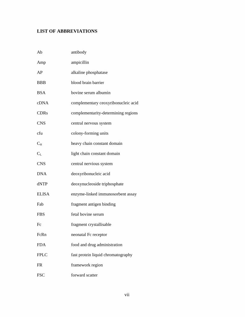
LIST OF ABBREVIATIONS
Ab
Amp
AP
BBB
BSA
cDNA
CDRs
CNS
cfu
CH
CL
CNS
DNA
dNTP
ELISA
Fab
FBS
Fc
FcRn
FDA
FPLC
FR
FSC
antibody
ampicillin
alkaline phosphatase
blood brain barrier
bovine serum albumin
complementary ceoxyribonucleic acid
complementarity-determining regions
central nervous system
colony-forming units
heavy chain constant domain
light chain constant domain
central nervious system
deoxyribonucleic acid
deoxynucleoside triphosphate
enzyme-linked immunosorbent assay
fragment antigen binding
fetal bovine serum
fragment crystallisable
neonatal Fc receptor
food and drug administration
fast protein liquid chromatography
framework region
forward scatter
viii
HRP
Ig
IMAC
IPTG
KD
kDa
mAb
MFI
MFIR
MRI
MS
MW
OD
PAGE
PBS
PCR
PE
PEG
PEI
PML
PPMS
pSJF2H
pTT5-hFc
Rmax
RPMI-1640
horseradish peroxidase
immunoglobulin
Immobilized metal-ion affinity chromatography
isopropyl-β-D-thiogalactopyranoside
affinity constant
kiloDaltons
monoclonal antibody
mean fluorescence intensity
mean fluoresence intensity ratio
magnetic resonance imaging
multiple sclerosis
molecular weight
optical density
polyacrylamide gel electrophoresis
phosphate-buffered saline
polymerase chain reaction
phycoerythrin
polyethylene glycol
polyethyleneimine
progressive multifocal leukoencephalopathy
primary progressive multiple sclerosis
VL expression vector
hFc-VL expression vector
maximum response defined as saturation of surface plasmon resonance
Roswell Park Memorial Institute medium-1640
ix
RRMS
RU
scFv
sdAb
SDS
SEC
SOC
SOE
SPMS
SPR
SSC
Tet
VCAM-1
Ve
VH
VHH
VL
VLA-4
relapsing-remitting phase of multiple sclerosis
resonance unit
single chain variable fragment
single domain antibody
sodium dodecyl sulfate
size exclusion chromatography
super optimized culture medium
Splice overlap extension
secondary progressive phase of multiple sclerosis
surface plasmon resonance
side scatter
tetracycline
vascular cell adhesion molecule-1
elution volume
heavy chain variable domain
heavy-chain antibody variable domain
light chain variable domain
very late antigen-4

x
LIST OF FIGURES
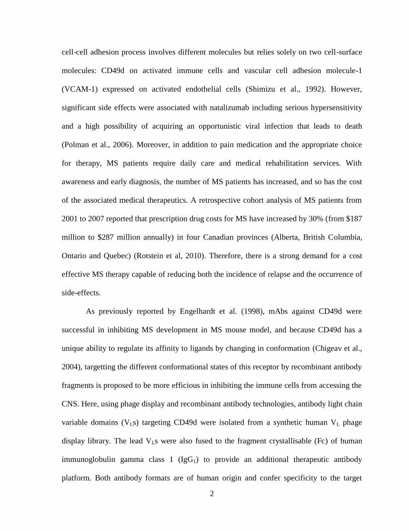
Figure 1. Predominant molecules involved in cell adhesion cascade.
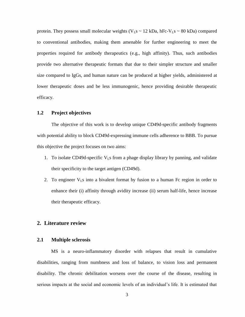
Figure 2. Schematic presentation of the extracellular structure of an integrin.
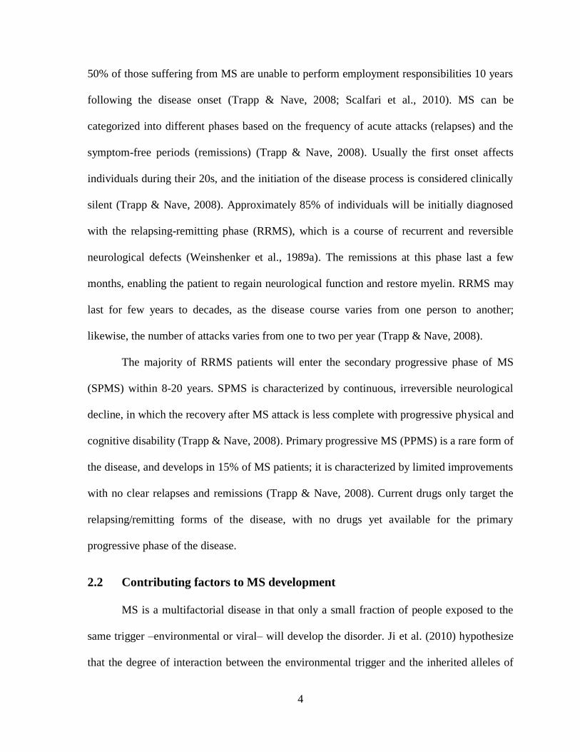
Figure 3. Conformational change of integrin (αIIbβ3) from bent to extended form.
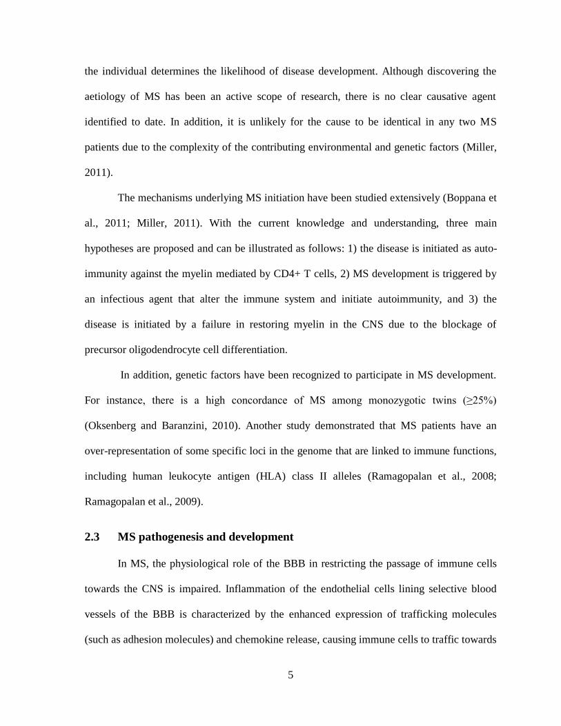
Figure 4. Possible consequences of VLA-4 blocking by antibodies.
Figure 5. Schematic representation of IgG antibody structure.
Figure 6. Representation of antibody fragments resulting from enzymatic digestion.
Figure 7. Shcematic representation of naturally occurring antibodies and engineered antibody
fragments.
Figure 8. Schematic representation of filamentous phage displaying sdAbs.
Figure 9. Schematic representation of the generation and selection of recombinant antibody
phage display libraries.
Figure 10. Panning scheme. Synthetic VL phage display library was added to Jurakt cells
expressing CD49d.
Figure 11. Schematic representation of SOE-PCR and final assembly of the recombinant
hFc-VL antibody fragment.
Figure 12. Schematic drawing of CDR randomizations on synthetic human VL phage display
library.
Figure 13. Monitoring library enrichment for binders by phage titration.
Figure 14. Representative purification profile of clone VL-2 by IMAC.
Figure 15. Anti-CD49d VL purification.
Figure 16. Screening VLs by ELISA for binding to recombinant CD49d.
Figure 17. SPR sensorgrams.
Figure 18. Determining the specificity of VLs towards CD49d by flow cytometry.
Figure 19. VLs show preferential binding to K562-CD49d cells in flow cytometry
experiments.
Figure 20. Determination of apparent molecular weight and aggregation status of engineered
hFc-VLs.
Figure 21. Binding of hFc-VLs to K-562 cell line variants measured by flow cytometry.
Figure 22. Concentration dependent binding of hFc-VL-6 measured by flow cytometry.
xi
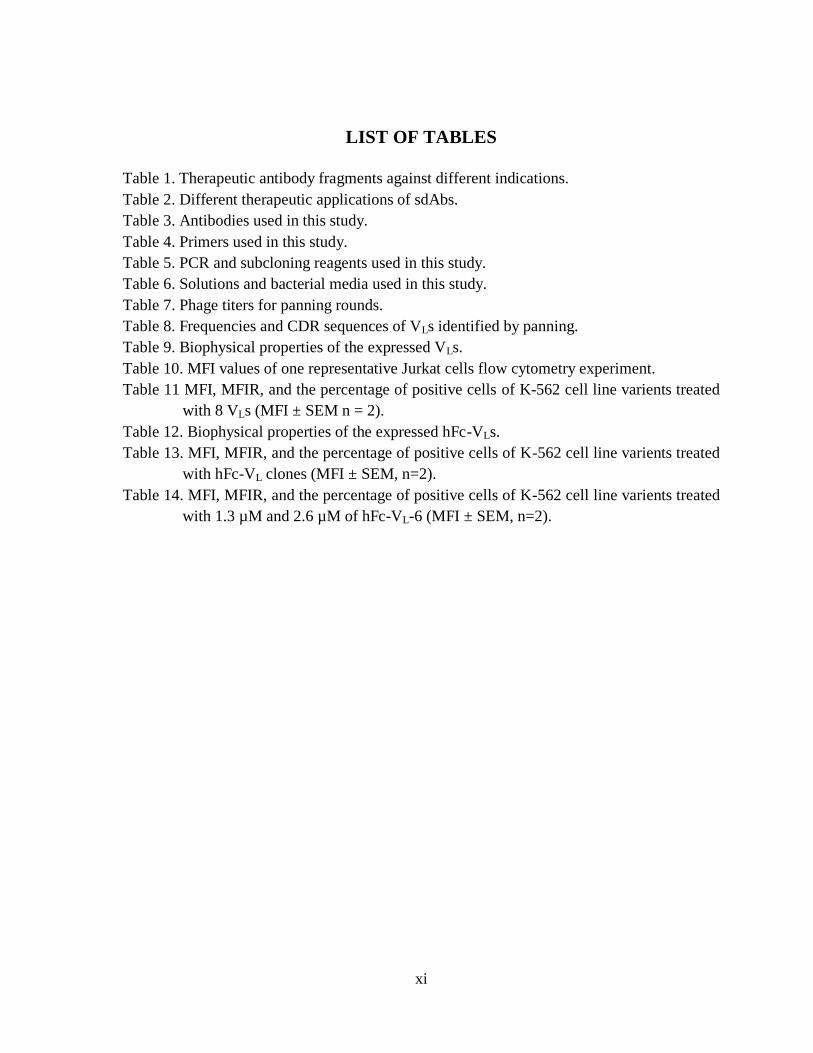
LIST OF TABLES
Table 1. Therapeutic antibody fragments against different indications.
Table 2. Different therapeutic applications of sdAbs.
Table 3. Antibodies used in this study.
Table 4. Primers used in this study.
Table 5. PCR and subcloning reagents used in this study.
Table 6. Solutions and bacterial media used in this study.
Table 7. Phage titers for panning rounds.
Table 8. Frequencies and CDR sequences of VLs identified by panning.
Table 9. Biophysical properties of the expressed VLs.
Table 10. MFI values of one representative Jurkat cells flow cytometry experiment.
Table 11 MFI, MFIR, and the percentage of positive cells of K-562 cell line varients treated
with 8 VLs (MFI ± SEM n = 2).
Table 12. Biophysical properties of the expressed hFc-VLs.
Table 13. MFI, MFIR, and the percentage of positive cells of K-562 cell line varients treated
with hFc-VL clones (MFI ± SEM, n=2).
Table 14. MFI, MFIR, and the percentage of positive cells of K-562 cell line varients treated
with 1.3 µM and 2.6 µM of hFc-VL-6 (MFI ± SEM, n=2).
1
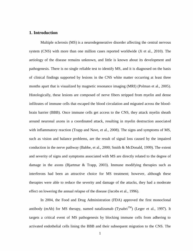
1. Introduction
Multiple sclerosis (MS) is a neurodegenerative disorder affecting the central nervous
system (CNS) with more than one million cases reported worldwide (Ji et al., 2010). The
aetiology of the disease remains unknown, and little is known about its development and
pathogenesis. There is no single reliable test to identify MS, and it is diagnosed on the basis
of clinical findings supported by lesions in the CNS white matter occurring at least three
months apart that is visualized by magnetic resonance imaging (MRI) (Polman et al., 2005).
Histologically, these lesions are composed of nerve fibers stripped from myelin and dense
infiltrates of immune cells that escaped the blood circulation and migrated across the blood-
brain barrier (BBB). Once immune cells get access to the CNS, they attack myelin sheath
around neuronal axons in a coordinated attack, resulting in myelin destruction associated
with inflammatory reaction (Trapp and Nave, et al., 2008). The signs and symptoms of MS,
such as vision and balance problems, are the result of signal loss caused by the impaired
conduction in the nerve pathway (Babbe, et al., 2000; Smith & McDonald, 1999). The extent
and severity of signs and symptoms associated with MS are directly related to the degree of
damage in the axons (Bjartmar & Trapp, 2003). Immune modifying therapies such as
interferons had been an attractive choice for MS treatment; however, although these
therapies were able to reduce the severity and damage of the attacks, they had a moderate
effect on lowering the annual relapse of the disease (Jacobs et al., 1996).
In 2004, the Food and Drug Administration (FDA) approved the first monoclonal
antibody (mAb) for MS therapy, named natalizumab (TysabriTM
) (Leger et al., 1997). It
targets a critical event of MS pathogenesis by blocking immune cells from adhering to
activated endothelial cells lining the BBB and their subsequent migration to the CNS. The
2
cell-cell adhesion process involves different molecules but relies solely on two cell-surface
molecules: CD49d on activated immune cells and vascular cell adhesion molecule-1
(VCAM-1) expressed on activated endothelial cells (Shimizu et al., 1992). However,
significant side effects were associated with natalizumab including serious hypersensitivity
and a high possibility of acquiring an opportunistic viral infection that leads to death
(Polman et al., 2006). Moreover, in addition to pain medication and the appropriate choice
for therapy, MS patients require daily care and medical rehabilitation services. With
awareness and early diagnosis, the number of MS patients has increased, and so has the cost
of the associated medical therapeutics. A retrospective cohort analysis of MS patients from
2001 to 2007 reported that prescription drug costs for MS have increased by 30% (from $187
million to $287 million annually) in four Canadian provinces (Alberta, British Columbia,
Ontario and Quebec) (Rotstein et al, 2010). Therefore, there is a strong demand for a cost
effective MS therapy capable of reducing both the incidence of relapse and the occurrence of
side-effects.
As previously reported by Engelhardt et al. (1998), mAbs against CD49d were
successful in inhibiting MS development in MS mouse model, and because CD49d has a
unique ability to regulate its affinity to ligands by changing in conformation (Chigeav et al.,
2004), targetting the different conformational states of this receptor by recombinant antibody
fragments is proposed to be more efficious in inhibiting the immune cells from accessing the
CNS. Here, using phage display and recombinant antibody technologies, antibody light chain
variable domains (VLs) targeting CD49d were isolated from a synthetic human VL phage
display library. The lead VLs were also fused to the fragment crystallisable (Fc) of human
immunoglobulin gamma class 1 (IgG1) to provide an additional therapeutic antibody
platform. Both antibody formats are of human origin and confer specificity to the target
3
protein. They possess small molecular weights (VLs ~ 12 kDa, hFc-VLs ~ 80 kDa) compared
to conventional antibodies, making them amenable for further engineering to meet the
properties required for antibody therapeutics (e.g., high affinity). Thus, such antibodies
provide two alternative therapeutic formats that due to their simpler structure and smaller
size compared to IgGs, and human nature can be produced at higher yields, administered at
lower therapeutic doses and be less immunogenic, hence providing desirable therapeutic
efficacy.
1.2 Project objectives
The objective of this work is to develop unique CD49d-specific antibody fragments
with potential ability to block CD49d-expressing immune cells adherence to BBB. To pursue
this objective the project focuses on two aims:
1. To isolate CD49d-specific VLs from a phage display library by panning, and validate
their specificity to the target antigen (CD49d).
2. To engineer VLs into a bivalent format by fusion to a human Fc region in order to
enhance their (i) affinity through avidity increase (ii) serum half-life, hence increase
their therapeutic efficacy.
2. Literature review
2.1 Multiple sclerosis
MS is a neuro-inflammatory disorder with relapses that result in cumulative
disabilities, ranging from numbness and loss of balance, to vision loss and permanent
disability. The chronic debilitation worsens over the course of the disease, resulting in
serious impacts at the social and economic levels of an individual’s life. It is estimated that
4
50% of those suffering from MS are unable to perform employment responsibilities 10 years
following the disease onset (Trapp & Nave, 2008; Scalfari et al., 2010). MS can be
categorized into different phases based on the frequency of acute attacks (relapses) and the
symptom-free periods (remissions) (Trapp & Nave, 2008). Usually the first onset affects
individuals during their 20s, and the initiation of the disease process is considered clinically
silent (Trapp & Nave, 2008). Approximately 85% of individuals will be initially diagnosed
with the relapsing-remitting phase (RRMS), which is a course of recurrent and reversible
neurological defects (Weinshenker et al., 1989a). The remissions at this phase last a few
months, enabling the patient to regain neurological function and restore myelin. RRMS may
last for few years to decades, as the disease course varies from one person to another;
likewise, the number of attacks varies from one to two per year (Trapp & Nave, 2008).
The majority of RRMS patients will enter the secondary progressive phase of MS
(SPMS) within 8-20 years. SPMS is characterized by continuous, irreversible neurological
decline, in which the recovery after MS attack is less complete with progressive physical and
cognitive disability (Trapp & Nave, 2008). Primary progressive MS (PPMS) is a rare form of
the disease, and develops in 15% of MS patients; it is characterized by limited improvements
with no clear relapses and remissions (Trapp & Nave, 2008). Current drugs only target the
relapsing/remitting forms of the disease, with no drugs yet available for the primary
progressive phase of the disease.
2.2 Contributing factors to MS development
MS is a multifactorial disease in that only a small fraction of people exposed to the
same trigger –environmental or viral– will develop the disorder. Ji et al. (2010) hypothesize
that the degree of interaction between the environmental trigger and the inherited alleles of
5
the individual determines the likelihood of disease development. Although discovering the
aetiology of MS has been an active scope of research, there is no clear causative agent
identified to date. In addition, it is unlikely for the cause to be identical in any two MS
patients due to the complexity of the contributing environmental and genetic factors (Miller,
2011).
The mechanisms underlying MS initiation have been studied extensively (Boppana et
al., 2011; Miller, 2011). With the current knowledge and understanding, three main
hypotheses are proposed and can be illustrated as follows: 1) the disease is initiated as auto-
immunity against the myelin mediated by CD4+ T cells, 2) MS development is triggered by
an infectious agent that alter the immune system and initiate autoimmunity, and 3) the
disease is initiated by a failure in restoring myelin in the CNS due to the blockage of
precursor oligodendrocyte cell differentiation.
In addition, genetic factors have been recognized to participate in MS development.
For instance, there is a high concordance of MS among monozygotic twins (≥25%)
(Oksenberg and Baranzini, 2010). Another study demonstrated that MS patients have an
over-representation of some specific loci in the genome that are linked to immune functions,
including human leukocyte antigen (HLA) class II alleles (Ramagopalan et al., 2008;
Ramagopalan et al., 2009).
2.3 MS pathogenesis and development
In MS, the physiological role of the BBB in restricting the passage of immune cells
towards the CNS is impaired. Inflammation of the endothelial cells lining selective blood
vessels of the BBB is characterized by the enhanced expression of trafficking molecules
(such as adhesion molecules) and chemokine release, causing immune cells to traffic towards
6
the site of inflammation and reach the CNS to further amplify the inflammatory response
(Engelhardt et al., 1998; Engelhardt, 2006). In the CNS, the axonal demyelination is thought
to be mediated either by the direct attack of immune cells or by the failure of proliferation
and differentiation of oligodendrocyte precursor cells, the source of new myelin-forming
cells (Wolswijk, 2002). The failure of myelin regeneration can be attributed to different
factors and mechanisms (reviewed in Franklin and ffrench-Constant, 2008), that
subsequently lead to the progressive impairments in the neural signals conduction pathways,
and to neurological dysfunction. There are three different events contributing to the
development of MS; these are: 1) events occurring outside the CNS, 2) events occurring at
the BBB level, and 3) events occurring inside the CNS.
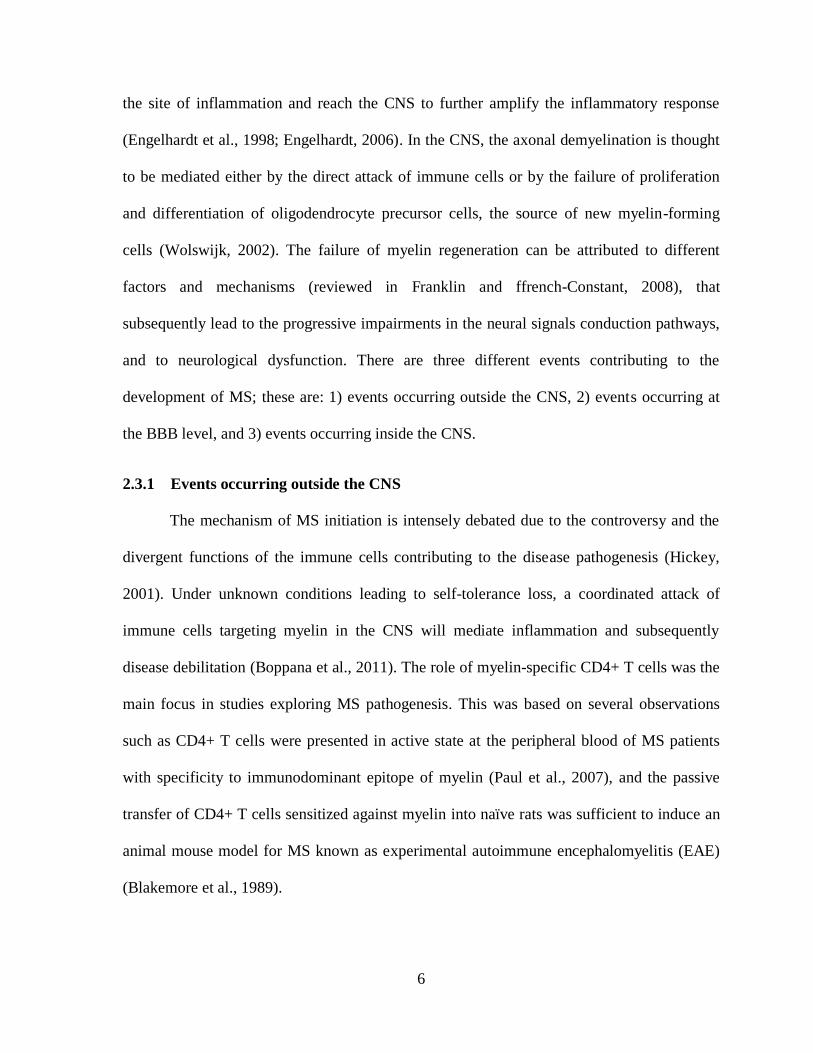
2.3.1 Events occurring outside the CNS
The mechanism of MS initiation is intensely debated due to the controversy and the
divergent functions of the immune cells contributing to the disease pathogenesis (Hickey,
2001). Under unknown conditions leading to self-tolerance loss, a coordinated attack of
immune cells targeting myelin in the CNS will mediate inflammation and subsequently
disease debilitation (Boppana et al., 2011). The role of myelin-specific CD4+ T cells was the
main focus in studies exploring MS pathogenesis. This was based on several observations
such as CD4+ T cells were presented in active state at the peripheral blood of MS patients
with specificity to immunodominant epitope of myelin (Paul et al., 2007), and the passive
transfer of CD4+ T cells sensitized against myelin into naïve rats was sufficient to induce an
animal mouse model for MS known as experimental autoimmune encephalomyelitis (EAE)
(Blakemore et al., 1989).
7
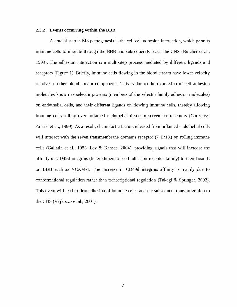
2.3.2 Events occurring within the BBB
A crucial step in MS pathogenesis is the cell-cell adhesion interaction, which permits
immune cells to migrate through the BBB and subsequently reach the CNS (Butcher et al.,
1999). The adhesion interaction is a multi-step process mediated by different ligands and
receptors (Figure 1). Briefly, immune cells flowing in the blood stream have lower velocity
relative to other blood-stream components. This is due to the expression of cell adhesion
molecules known as selectin proteins (members of the selectin family adhesion molecules)
on endothelial cells, and their different ligands on flowing immune cells, thereby allowing
immune cells rolling over inflamed endothelial tissue to screen for receptors (Gonzalez-
Amaro et al., 1999). As a result, chemotactic factors released from inflamed endothelial cells
will interact with the seven transmembrane domains receptor (7 TMR) on rolling immune
cells (Gallatin et al., 1983; Ley & Kansas, 2004), providing signals that will increase the
affinity of CD49d integrins (heterodimers of cell adhesion receptor family) to their ligands
on BBB such as VCAM-1. The increase in CD49d integrins affinity is mainly due to
conformational regulation rather than transcriptional regulation (Takagi & Springer, 2002).
This event will lead to firm adhesion of immune cells, and the subsequent trans-migration to
the CNS (Vajkoczy et al., 2001).

8
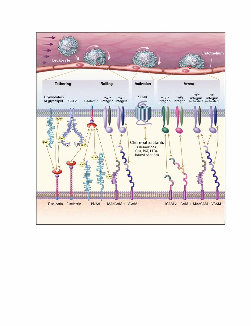
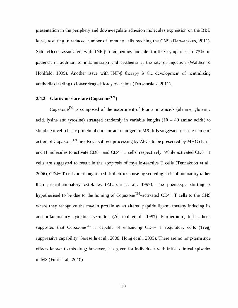
Figure 1. Predominant molecules involved in cell adhesion cascade. Trafficking of
immune cells (e.g., leukocytes) from bloodstream to sites of inflammation is mediated by
number of molecules and receptors. L-selectin, P-selectin glycoprotein ligand-1 (PSGL-1),
and CD49d interactions with corresponding receptors are involved in the initial tethering and
rolling of leukocytes. Rolling leukocytes express a receptors with seven Transmembrane
Domains (7 TMR), mediating the transmission of intracellular signals for leukocyte
activation through binding to chemokine molecules, resulting in a rapid activation to CD49d
(also known as integrin α-4) and will change in conformation. The active form of integrin α-
4 (represented by the slightly opened confirmation in the arrest section of the figure) will
have an increase affinity to its receptor VCAM-1 resulting in cell arrest and its subsequent
migration to the CNS. Reproduced with permission from (von Andrian & Mackay, 2000).
©Massachusetts Medical Society.
9
2.3.3 Events occurring inside the CNS
When T cells gain access to the CNS, they encounter the processed self-myelin in the
context of major histocompatibility complex (MHC) class II on the surface of antigen
presenting cells (APCs). This interaction results in the continuous release of cytokines, such
as interferon-gamma (INF-γ), thereby contributing to the increase in the BBB permeability
and further enhancing the local immune response against myelin. B cells will produce
antibodies that directly attack the myelin sheath, while macrophages destroy myelin
(Boppana et al., 2011).
2.4 Current therapies for MS
Currently, 8 therapies are approved by the FDA offering different options for MS
treatment (Derwenskus, 2011). All offered medications are disease-modifying and not curing
therapies. In other words, they are prescribed to prolong the time between MS attacks of the
relapsing remitting phase of the disease, allowing better myelin recovery and reduce changes
in MRI; however, individuals under therapy will still have active disease (Trapp & Nave,
2008; Derwenskus, 2011).
Therapeutics includes the four β-interferons (INFs)-based drugs AvonexTM
,
BetaseronTM
, ExtaviaTM
, and RebifTM
and four non-INF-based drugs which includes
fingolimod (GilenyaTM
), glatiramer acetate (CopaxoneTM
), mitoxantrone (NovantroneTM
),
and natalizumab (TysabriTM
) (Derwenskus, 2011).
2.4.1 β-interferon therapies:
INF-β therapeutics can alter the immune system in various ways with poorly
understood mode of action (Yong et al., 1998). In general, INF-β will inhibit antigen
10
presentation in the periphery and down-regulate adhesion molecules expression on the BBB
level, resulting in reduced number of immune cells reaching the CNS (Derwenskus, 2011).
Side effects associated with INF-β therapeutics include flu-like symptoms in 75% of
patients, in addition to inflammation and erythema at the site of injection (Walther &
Hohlfeld, 1999). Another issue with INF-β therapy is the development of neutralizing
antibodies leading to lower drug efficacy over time (Derwenskus, 2011).
2.4.2 Glatiramer acetate (CopaxoneTM
)
CopaxoneTM
is composed of the assortment of four amino acids (alanine, glutamic
acid, lysine and tyrosine) arranged randomly in variable lengths (10 – 40 amino acids) to
simulate myelin basic protein, the major auto-antigen in MS. It is suggested that the mode of
action of CopaxoneTM
involves its direct processing by APCs to be presented by MHC class I
and II molecules to activate CD8+ and CD4+ T cells, respectively. While activated CD8+ T
cells are suggested to result in the apoptosis of myelin-reactive T cells (Tennakoon et al.,
2006), CD4+ T cells are thought to shift their response by secreting anti-inflammatory rather
than pro-inflammatory cytokines (Aharoni et al., 1997). The phenotype shifting is
hypothesised to be due to the homing of CopaxoneTM
–activated CD4+ T cells to the CNS
where they recognize the myelin protein as an altered peptide ligand, thereby inducing its
anti-inflammatory cytokines secretion (Aharoni et al., 1997). Furthermore, it has been
suggested that CopaxoneTM
is capable of enhancing CD4+ T regulatory cells (Treg)
suppressive capability (Saresella et al., 2008; Hong et al., 2005). There are no long-term side
effects known to this drug; however, it is given for individuals with initial clinical episodes
of MS (Ford et al., 2010).
11
2.4.3 Mitoxantrone (NovantroneTM
)
NovantroneTM
is a cancer therapeutic that was first approved in 1987 for acute
myeloid leukemia. Later in 1996, it was approved for hormone refractory prostate cancer
(Fox, 2004) and in 2000 for MS therapy. It possesses a small molecular weight that can pass
through the BBB and act by inhibiting DNA replication, resulting in the apoptosis of
proliferating cells in the CNS. NovantroneTM
prescription is limited for worsening relapsing
remitting cases of MS due to safety issues including urinary tract infections and amenorrhea
(i.e., absence of period) (Fox, 2004), as well as early cardiac toxicity reported after 1-3 doses
(Avasarala et al., 2003; Paul et al., 2007).
2.4.4 Fingolimod (GilenyaTM
)
GilenyaTM
is a sphingosine-1-phosphate receptor modulator (i.e., a critical regulator
of lymphocyte trafficking) that acts by trapping lymphocytes in lymph nodes. The trapping
will result in lowering circulating lymphocytes number and reducing their chance to reach
the CNS (Horga et al., 2010). Although GilenyaTM
showed higher efficacy in respect to MRI
follow up and relapse rates compared to INF-β therapy, it is associated with safety issues
including fatal infections by disseminated primary varicella zoster and herpes simplex
encephalitis and other nonfatal infections, hypertension and elevated liver-enzyme levels
(Cohen et al., 2010).
2.4.5 Natalizumab (TysabriTM
)
TysabriTM
is a humanized mAb targeting CD49d and blocks its binding to VCAM-
1, resulting in lower immune cells migration to the CNS (Ransohoff, 2007). A two year
placebo-controlled study carried on patients with relapsing MS, reported that individuals
with MS receiving TysabriTM
had 17% probability of disease progression relative to 29% in
12
the placebo group. Moreover, TysabriTM
reduced the chance of clinical relapse by 68%
annually and reduced the possibility of new lesion formation as detected by MRI (Polman et
al., 2006). The main side effect associated with TysabriTM
is the risk of acquiring progressive
multifocal leukoencephalopathy (PML) caused by human polyomavirus when administered
with other immunosuppressive agents. Therefore, TysabriTM
is only prescribed as a
monotherapy through the TYSABRI Outreach: unified commitment to health (TOUCH)
program. Other side effects include headache and allergic hypersensitivity. In addition, 9%
of individuals receiving TysabriTM
developed neutralizing antibodies to the therapy that
lowers the therapeutic efficacy and requires frequent therapeutic dose adjustment (Calabresi
et al., 2007).
All of the above approved therapies demonstrated beneficial results for relapsing
remitting MS. However, none have any effectiveness in treating the more aggressive forms
of the disease. The cause of this bias is not clear, but it is likely that relapsing remitting form
is more of inflammatory process while progressive phases of MS are more of degenerative
process, indicating that the disease mechanism is different and partially explains why such
therapeutics can control MS by their anti-inflammatory roles (Derwenskus, 2011).
2.5 Very late antigen-4 (VLA-4)
Integrins are members of glycoprotein family and are composed of non-covalently
linked heterodimers of α and β subunits, each spanning the plasma membrane once (Arnaout
et al., 2005). There are 18 different α subunits and 8 β subunits identified in vertebrates and
their different pairing results in 24 integrins in mammals. According to electron microscopy
studies (Weisel et al., 1992), α and β heterodimer adopt a globular structure containing the
site for ligand binding and is connected to the cell membrane by two stalks and terminate in a
13
short cytoplasmic domain. VLA-4 is a member of the integrin family proteins. It lacks the α-
A domain found in most integrins and is composed of α-4 subunit (also known as CD49d)
and a β-1 chain (CD29) (Sanchez-Madrid et al., 1986; Hemler et al., 1987). VLA-4 is
involved in mediating extracellular matrix-cell, cell-cell and cell-pathogen interactions
(Hynes, 2002; Hogg et al., 2003). It has been reported that CD49d subunit is the responsible
compartment in mediating CD49d-expressing immune cells adhesion to VCAM-1 (Elices et
al., 1990). VLA-4 is expressed on a wide range of cells including T cells, B cells, natural
killer cells and dendritic cells.
Because integrins adopts multiple conformational states regulated by inside-out
signalling as well as ligand binding (Campbell and Humphries, 2011), it is difficult to obtain
a three-dimensional structure for such proteins, and a detailed quaternary structure for VLA-
4 is lacking.
In 2001, the first crystal structure of an integrin (αVβ3) extracellular domain was solved
(Xiong et al., 2001). This breakthrough has provided a better understanding of the overall
structure of integrins. A general schematic model of an integrin displaying different modules
of the protein is represented in (Figure 2). Each subunit of VLA-4 heterodimer is comprised
of number of domains which have complex interconnections with overlapping functions
detailed in different reviews (Arnaout et al., 2005; Campbell and Humphries, 2011). In VLA-
4, the α-subunit (or CD49d) is composed of four main extracellular domains: seven-bladed β-
propeller, a thigh, and two calf domains. The thigh and calf domains possess similar
immunoglobulin-like β-sandwich folds. The β-subunit is composed of eight domains: β-A
domain, hybrid domain, plexin-semaphorin-integrin (PSI) domain, four epidermal growth
factor (EGF)-like domains and a β-tail domain.
14
Like any other member of the integrin family, VLA-4 is highly dynamic and adopts
different conformations, each exhibiting different affinity towards its ligands, VCAM-1 and
fibronectin (Arnaout et al., 2005). There are two distinguished conformations for VLA-4: the
extended (or active) and the bent (or resting) conformations (Figure 3). The shift from the
bent to the extended conformation is rapid – occurring in sub-seconds – whereas its reversal
to the bent form occurs in less than a minute. The mechanism underlying the conformational
switch is represented by different models, including the “switchblade” and the “deadbolt”
models detailed elsewhere (Luo et al. 2007, Arnaout et al. 2005); however, controversy
remains especially concerning the microenvironmental requirements and the physiological
relevance of the conformational shift and affinity regulation.
The most important aspect of integrin structure in regard to this study is the two
regions within the CD49d subunit mediating conformation switching: the linker between the
β-propeller and the thigh, and the genu – also known as the knee – at the bend between the
thigh and calf-1 domain (Campbell and Humphries, 2011). This knowledge suggests that
stabilizing the resting state of CD49d by antibody fragments to prevent the conformational
change or to mask an active conformation will possibly reduce the likelihood of the integrin
activation or binding to its ligand.
15
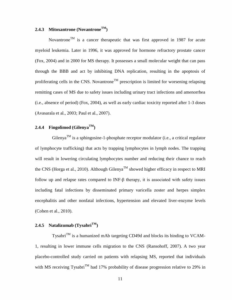
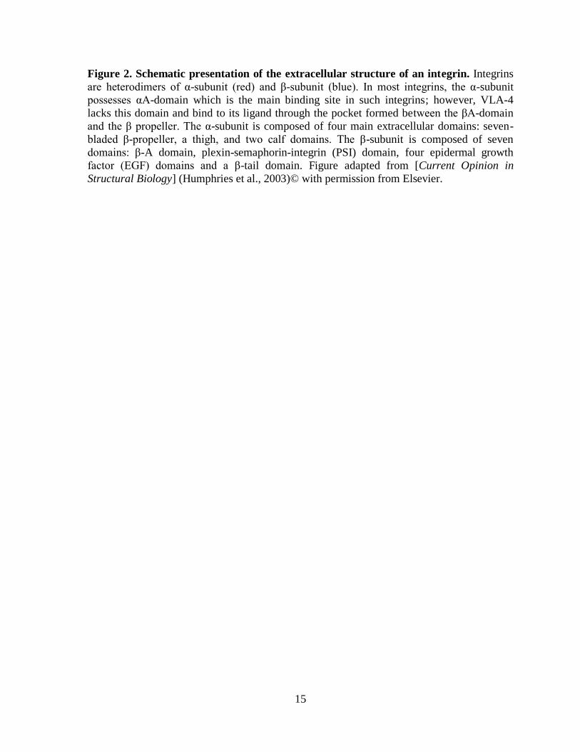
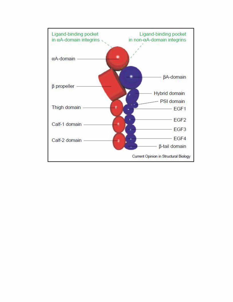
Figure 2. Schematic presentation of the extracellular structure of an integrin. Integrins
are heterodimers of α-subunit (red) and β-subunit (blue). In most integrins, the α-subunit
possesses αA-domain which is the main binding site in such integrins; however, VLA-4
lacks this domain and bind to its ligand through the pocket formed between the βA-domain
and the β propeller. The α-subunit is composed of four main extracellular domains: seven-
bladed β-propeller, a thigh, and two calf domains. The β-subunit is composed of seven
domains: β-A domain, plexin-semaphorin-integrin (PSI) domain, four epidermal growth
factor (EGF) domains and a β-tail domain. Figure adapted from [Current Opinion in
Structural Biology] (Humphries et al., 2003)© with permission from Elsevier.

16
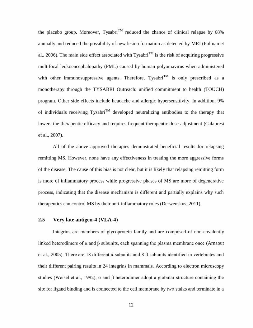
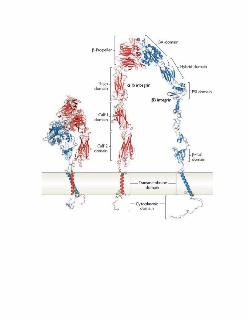
Figure 3. Conformational change of integrin (αIIbβ3) from bent to extended form. In
similarity with VLA-4, αIIbβ3 integrin lacks the αA-domain. The exact mechanism
underlying conformational switch is still under investigation. The switching from the bent
form (left) to the extended form (right) will expose hidden epitopes of the protein. The
change in conformation is suggested to have a direct role in the affinity status of the integrin.
Figure adapted by permission from Macmillan Publishers Ltd: [Nature Reviews Molecular
Cell Biology] (Shattil et al., 2010)©.
17
2.5.1 Functional roles of VLA-4 under physiological and pathological conditions
Under physiological conditions, VLA-4 has an essential role during embryo
development and immune response (Takada et al., 1987). It is also required for B- and T- cell
precursor development in the bone marrow (Miyake et al., 1991; Arroyo et al., 1999;
Arroyo,et al., 1996). Additionally, it is expressed on thymic epithelial cells and involved in
thymocyte development (Nieto et al., 1996). VLA-4 participates in the differentiation of Th1
lymphocytes (Mittelbrunn et al., 2004), and during inflammation VLA-4 has a clear role in
trafficking leukocytes toward the inflamed region (Lobb & Hemler, 1994; Ibbotson et al.,
2001).
2.5.2 Possible consequences of blocking VLA-4 by antibodies in vivo
Immune response generation and T cell differentiation may be affected by long-term
administration of antibodies targeting CD49d subunit of VLA-4. For instance, during
immune synapse formation between CD4+ T cells and APCs, antibodies against CD49d may
act as agonists or antagoniss (Gonzalez-Amaro et al., 2005; Hogg et al., 2003). Agonist anti-
CD49d antibodies are capable of stimulating the ligand of VLA-4 in an immune synapse that
is expressed on APCs, thus promoting Th-1 cells differentiation (Mittelbrunn et al., 2004).
On the contrary, antagonist antibodies will interfere with the immune synapse formation
leading to a deviation toward Th-2 lymphocytes differentiation. Because MS is thought to be
mediated by Th-1 immune response, antagonist antibodies may be beneficial in suppressing
MS attacks by their immunomodulatory role (Hemler et al., 1987; Elices et al., 1990 and
Wayner et al., 1989). The overall consequences of blocking CD49d subunit of VLA-4 are
illustrated in Figure 4.
18
In conclusion, CD49d is an adhesion receptor that mediates different biological
functions in healthy individuals. Antibodies targeting this receptor will affect the
differentiation of Th-1/Th-2 lymphocytes and immune cells extravasation to inflammation
sites among other undiscovered effects. There is no doubt that CD49d-antibodies have a
beneficial effect in reducing the annual attacks of MS. This therapeutic effect is believed to
be a consequence of blocking immune cells binding to their ligand on inflamed BBB. The
use of antibody fragments such as VLs or hFc-VLs with the potential to block CD49d binding
to VCAM-1 are proposed to be more advantageous therapeutic strategy. They are suggested
to have lower side effects associated with long-term administration due to their human
nature. In addition, the small molecular weight antibody fragments are more efficacious in
targeting multiple epitopes of CD49d. This critical feature will increase the chance of
“locking” CD49d conformation in the resting state from switching to the active state, thus,
inhibiting its binding to VCAM-1 on inflamed BBB.
19
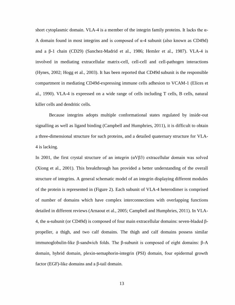
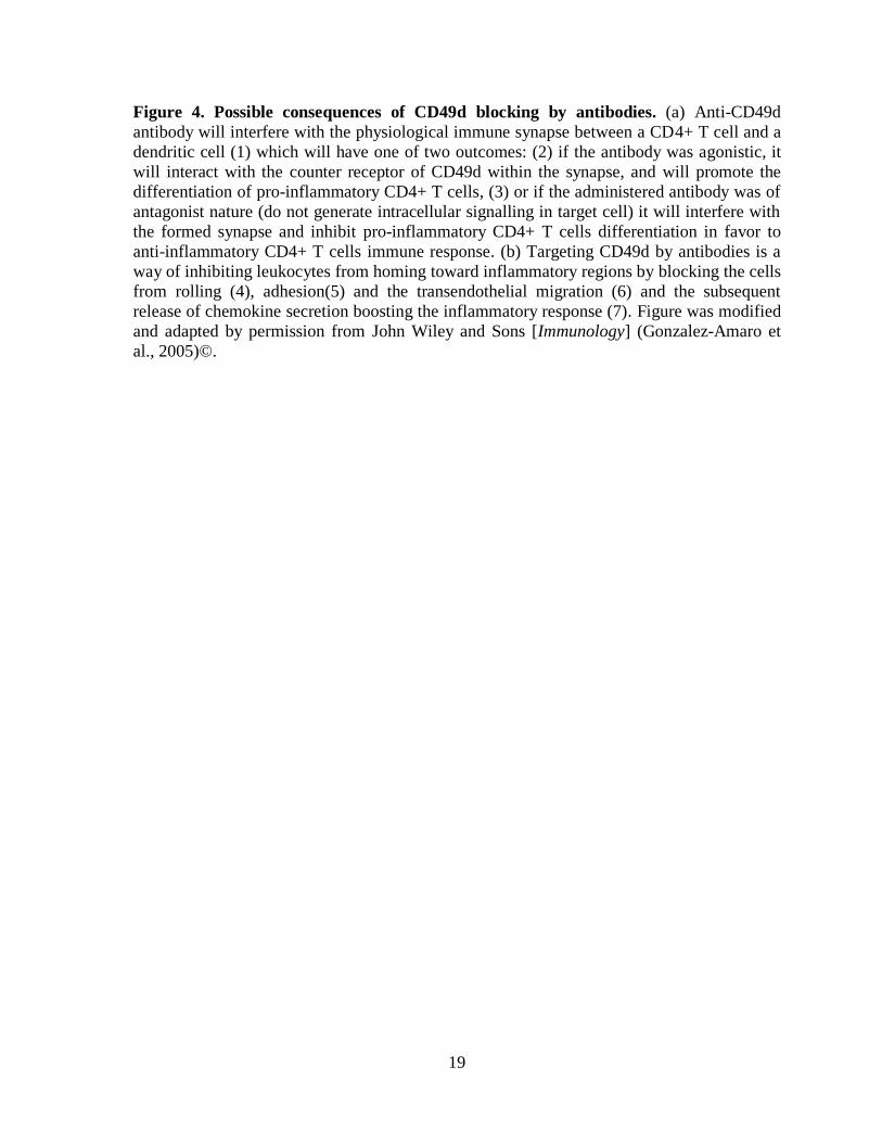
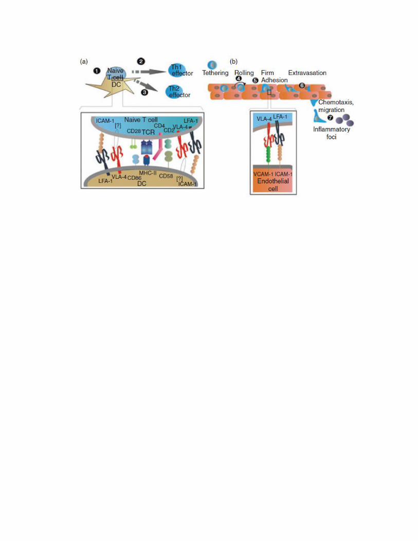
Figure 4. Possible consequences of CD49d blocking by antibodies. (a) Anti-CD49d
antibody will interfere with the physiological immune synapse between a CD4+ T cell and a
dendritic cell (1) which will have one of two outcomes: (2) if the antibody was agonistic, it
will interact with the counter receptor of CD49d within the synapse, and will promote the
differentiation of pro-inflammatory CD4+ T cells, (3) or if the administered antibody was of
antagonist nature (do not generate intracellular signalling in target cell) it will interfere with
the formed synapse and inhibit pro-inflammatory CD4+ T cells differentiation in favor to
anti-inflammatory CD4+ T cells immune response. (b) Targeting CD49d by antibodies is a
way of inhibiting leukocytes from homing toward inflammatory regions by blocking the cells
from rolling (4), adhesion(5) and the transendothelial migration (6) and the subsequent
release of chemokine secretion boosting the inflammatory response (7). Figure was modified
and adapted by permission from John Wiley and Sons [Immunology] (Gonzalez-Amaro et
al., 2005)©.
20
2.6 Immunoglobulins
Antibodies (Abs) are glycoproteins released from plasma cells in response to an
immunogen (i.e., substance that provoke immune response), and are also termed
immunoglobulins (Ig). Igs are grouped into 5 classes based on differences in their amino
acid sequences; these are: IgG, IgA, IgM, IgD and IgE, arranged in order of decreasing
concentration found in human plasma (reviewed in Amzel & Poljak, 1979).
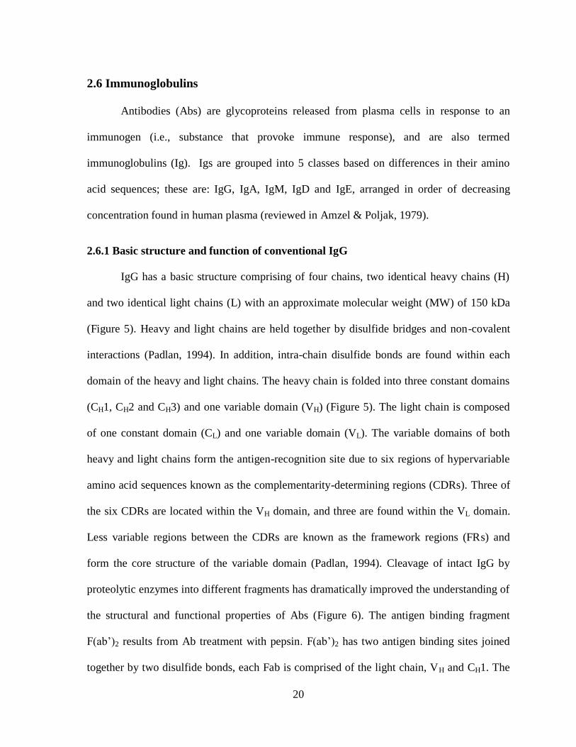
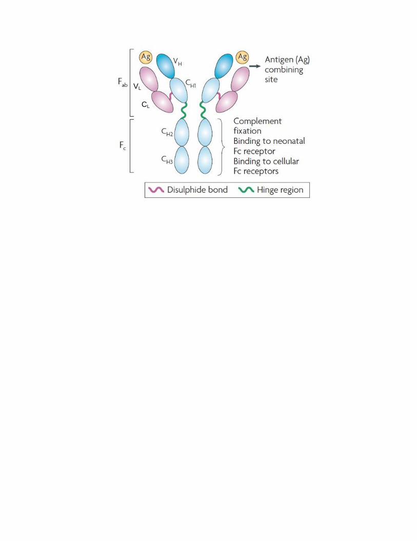
2.6.1 Basic structure and function of conventional IgG
IgG has a basic structure comprising of four chains, two identical heavy chains (H)
and two identical light chains (L) with an approximate molecular weight (MW) of 150 kDa
(Figure 5). Heavy and light chains are held together by disulfide bridges and non-covalent
interactions (Padlan, 1994). In addition, intra-chain disulfide bonds are found within each
domain of the heavy and light chains. The heavy chain is folded into three constant domains
(CH1, CH2 and CH3) and one variable domain (VH) (Figure 5). The light chain is composed
of one constant domain (CL) and one variable domain (VL). The variable domains of both
heavy and light chains form the antigen-recognition site due to six regions of hypervariable
amino acid sequences known as the complementarity-determining regions (CDRs). Three of
the six CDRs are located within the VH domain, and three are found within the VL domain.
Less variable regions between the CDRs are known as the framework regions (FRs) and
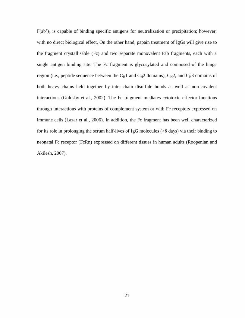
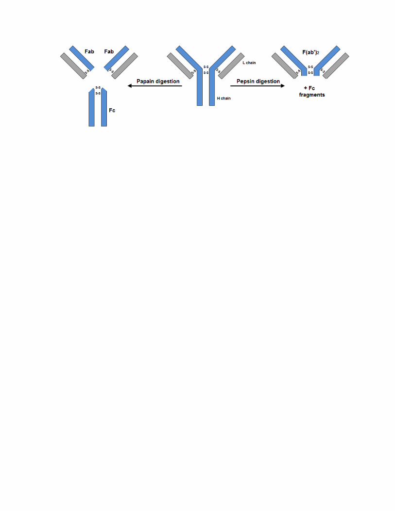
form the core structure of the variable domain (Padlan, 1994). Cleavage of intact IgG by
proteolytic enzymes into different fragments has dramatically improved the understanding of
the structural and functional properties of Abs (Figure 6). The antigen binding fragment
F(ab’)2 results from Ab treatment with pepsin. F(ab’)2 has two antigen binding sites joined
together by two disulfide bonds, each Fab is comprised of the light chain, VH and CH1. The
21
F(ab’)2 is capable of binding specific antigens for neutralization or precipitation; however,
with no direct biological effect. On the other hand, papain treatment of IgGs will give rise to
the fragment crystallisable (Fc) and two separate monovalent Fab fragments, each with a
single antigen binding site. The Fc fragment is glycosylated and composed of the hinge
region (i.e., peptide sequence between the CH1 and CH2 domains), CH2, and CH3 domains of
both heavy chains held together by inter-chain disulfide bonds as well as non-covalent
interactions (Goldsby et al., 2002). The Fc fragment mediates cytotoxic effector functions
through interactions with proteins of complement system or with Fc receptors expressed on
immune cells (Lazar et al., 2006). In addition, the Fc fragment has been well characterized
for its role in prolonging the serum half-lives of IgG molecules (˃8 days) via their binding to
neonatal Fc receptor (FcRn) expressed on different tissues in human adults (Roopenian and
Akilesh, 2007).

22
Figure 5. Schematic representation of IgG antibody structure. VH: variable domain of
heavy chain; VL: variable domain of light chain; CH; constant domain of heavy chain; CL:
constant domain of light chain; Fab: antigen binding fragment; Fc: fragment crystallisable.
Adapted by permission from Macmillan Publishers Ltd: [Nature Reviews Cancer]© (Weiner,
2007).
CL
VL

23
Figure 6. Representation of antibody fragments resulting from enzymatic digestion.
When an antibody is treated with papain, 2 Fab fragments and one Fc fragment can be
generated, while treatment with pepsin will generate a bivalent fragment F(ab’)2 and the rest
of Fc fragment will be degraded as the enzyme cleaves the antibody at a site below the
disulfide bonds that holds the two heavy chains together. L chain: light chain of IgG. H
chain: heavy chain of IgG Fab and F(ab’)2: antigen binding IgG fragments; Fc: fragment
crystallisable; S-S: disulfide bond.
24
2.6.2 Heavy chain antibodies (HCAbs) and IgNARs
In Camelidae (camels, llamas and alpacas) and cartilaginous sharks, antibodies devoid
of light chains are found to co-exist naturally with conventional antibodies (Hamers-
Casterman et al., 1993; Greenberg et al., 1995). These are referred to as HCAbs in
Camelidae and IgNARs in sharks. These antibodies confer antigen specificity via a single
variable domain of the heavy chain designated VHH for camelids HCAbs and VNAR for
sharks IgNARs (Figure 7). In camels, HCAbs comprise 75% of total serum Igs (van der
Linden et al., 2000). Camelids’ HCAbs are homodimers with MW of ~ 80 – 90 kDa. Each
monomer is composed of a variable domain linked to the CH2 and CH3 constant domains by
a hinge region (Hamers-Casterman et al., 1993). The unique ability of VHHs and VNARs to
function via single unpaired antigen-recognition site makes them suitable candidates for the
generation of minimal size immunoreagents (see below for details).
2.6.3 Recombinant antibody fragments
Recombinant antibody fragments have been increasingly used for potential diagnostic
and therapeutic applications. They demonstrate target specificity and affinity similar to
whole mAbs and have been engineered to provide various formats of basic antigen binding
units such as Fab, single chain variable fragments (scFvs, composed of VH and VL domains
linked by a linker peptide) and single domain antibodies (Figure 7) (Holliger and Hudson,
2005; Weisser and Hall, 2009). Due to their small size and simple structure, they can be
expressed efficiently in various expression systems including bacteria, yeast, plants and
mammalian cells (Weisser and Hall, 2009). They are also amenable to be re-built into
different formats (e.g., generating bivalent and bispecific antibodies targeting two epitopes
found in close proximity to one another) to be more efficacious agents (reviewed in Schaefer
25
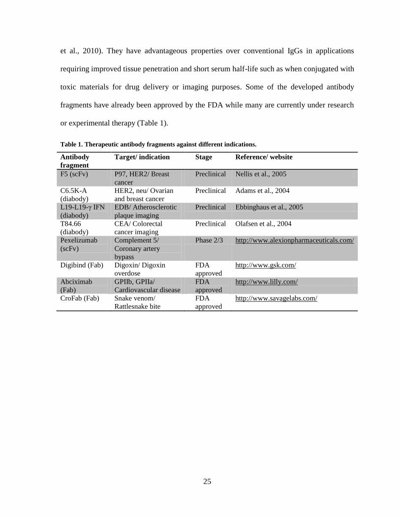
et al., 2010). They have advantageous properties over conventional IgGs in applications
requiring improved tissue penetration and short serum half-life such as when conjugated with
toxic materials for drug delivery or imaging purposes. Some of the developed antibody
fragments have already been approved by the FDA while many are currently under research
or experimental therapy (Table 1).
Table 1. Therapeutic antibody fragments against different indications.
Antibody
fragment
Target/ indication Stage Reference/ website
F5 (scFv) P97, HER2/ Breast
cancer
Preclinical Nellis et al., 2005
C6.5K-A
(diabody)
HER2, neu/ Ovarian
and breast cancer
Preclinical Adams et al., 2004
L19-L19-γ IFN
(diabody)
EDB/ Atherosclerotic
plaque imaging
Preclinical Ebbinghaus et al., 2005
T84.66
(diabody)
CEA/ Colorectal
cancer imaging
Preclinical Olafsen et al., 2004
Pexelizumab
(scFv)
Complement 5/
Coronary artery
bypass
Phase 2/3 http://www.alexionpharmaceuticals.com/
Digibind (Fab) Digoxin/ Digoxin
overdose
FDA
approved
http://www.gsk.com/
Abciximab
(Fab)
GPIIb, GPIIa/
Cardiovascular disease
FDA
approved
http://www.lilly.com/
CroFab (Fab) Snake venom/
Rattlesnake bite
FDA
approved
http://www.savagelabs.com/

26
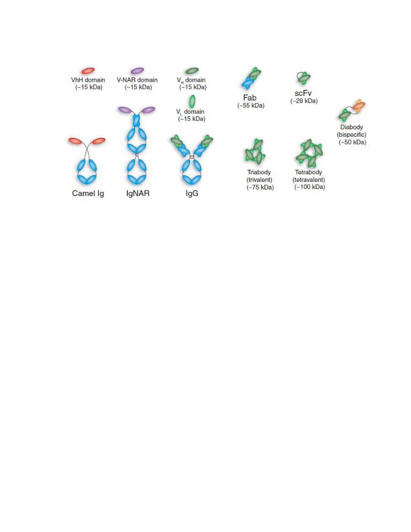
Figure 7. Schematic representation of naturally occurring antibodies and engineered
antibody fragments. Conventaional human IgG and heavy chain antiboies (camels’ Ig and
IgNAR) are found naturally in humans, camels and sharks respectivly. Both camel and shark
Ig lacks the light chain. Different formats of recombinant antigen binding fragments derived
from naturally occuring antibodies are represented. VHH: variable domain of camel Ig heavy
chain; VNAR: variable domain of shark Ig heavy chain; VH domain: variable domain of
human Ig heavy chain; VL domain: variable domain of human Ig light chain; Fab: antigen
binding fragment; scFv: single chain variable fragment; Diabody: a bivalent scFv, can be
engineered to genarate a bispecific heterodimer molecule. Adapted and modified by
permission from Macmillan Publishers Ltd: [Nature Biotechnology] (Holliger and Hudson,
2005)©.
27
2.6.4 Types of single domain antibodies
A single domain antibody (sdAb) is the simplest variant among other antibody
fragments with antigen-binding activity (Figure 7). It is derived from the variable domain of
whole Igs such as: VHs or VLs of human Igs (Reiter et al., 1999; van den Beucken et al.,
2001), VHHs of Camelidae HCAbs (Arbabi-Ghahroudi et al., 1997), or VNARs of shark
IgNARs (Lauwereys et al., 1998). In contrast to conventional antibodies, VHHs retains
efficient refolding and complete biological activity following thermal or chemical denaturing
conditions (van der Linden et al., 1999; Dumoulin et al., 2002). In addition, they are well-
expressed in bacterial and yeast systems with resistance to aggregation (Dumoulin et al.,
2002). According to amino acid sequence analyses, the apparent stability of VHHs is
attributed to conserved hydrophilic amino acids at positions 37, 44, 45 and 47 (Kabat
numbering system (Kabat et al., 1991)) located at the VL interface of VH domains in
conventional human IgGs (Muyldermans et al., 1994). This finding has led to the rational
design of “camelized” human sdAb libraries (i.e., substituting hydrophobic residues in
human sdAbs with hydrophilic residues to mimic those of VHHs) (Davies and Riechmann,
1996). Camelized sdAbs have provided minimal size agents capable of antigen binding at
high affinities with enhanced non-aggregating expression (Holt et al., 2003). This knowledge
was also employed to “humanize” VHHs with attractive biophysical properties to generate
less immunogenic sdAbs for potential therapeutic applications (Vincke et al., 2009).
Although VHs and VHHs have been extensively studied and characterized for research and
therapeutic potentials, VLs have also gained considerable interest. The limited success of
initial attempts in isolating VL domains with binding activities from synthetic (Soderlind et
al., 1995) or immune (Bonnert, 1993) repertoires in mice was probably due to their lower
28
sequence diversity when compared to VH repertoires (Tonegawa, 1983; Berek and Milstein,
1988). However, advances in library design strategies permitted the construction of VL
libraries with diversities up to 1010
clones (Paz et al., 2005). In consistence with the
theoretical argument suggesting library size correlation with antibody affinity (Perelson et
al., 1979), a number of studies have reported the isolation of antigen-specific VL domains
with high affinities from highly diverse libraries (Beucken et al., 2001; Hussack et al., 2012).
Furthermore, some VL domains have been isolated based on their major contribution in
antigen binding over VH domains. For instance, Colby et al. (2004) have isolated scFv
against huntingtin protein and reported that the paratope (i.e., antibody binding site) of the
isolated scFv was exclusively mapped to the VL domain. The huntingtin-specific VL was re-
cloned independently from the parental scFv and retained full binding activity to the target
antigen with five-fold increase in expression level (Colby et al., 2004). In addition,
hyperstable VL domains have been generated with complete reversible unfolding property
even when the disulfide bridge – which is critical for the domain’s folding stability – was
reduced (Ohage and Steipe, 1999). This unique property has opened the opportunity of
expressing functional VLs in reducing environments (i.e., cellular cytoplasm) as intrabody
domains that can serve as excellent tools for intracellular targeting applications.
2.6.4.1 Advantages and limitations of sdAbs
There are several properties that make sdAbs unique from other antibody formats.
They are produced efficiently in timely and cost-effective manner. The small size provided
by the single domain nature and extruding CDR3 loops results in the enhanced stability and
accessibility to hidden epitopes (Nuttall et al., 2000). They penetrate tissues efficiently and
are amenable to be engineered in multivalent formats or conjugated to other compounds to
29
enhance their utility in desired diagnostic or therapeutic applications (Hudson, 1998; Els
Conrath et al., 2001). They possess high chemical and thermal stability making them suitable
for selection under denaturing condition to isolate highly stable clones (Dumoulin et al.,
2003; Graef et al., 2011). In addition, they are advantageous over other antibody fragments
in terms of simplicity of construction and further engineering. For instance, they only require
a small set of primers for amplification and library construction purposes, and do not require
assembly reactions necessary for other antibody fragments generation (e.g., scFv) (Arbabi-
Ghahroudi et al., 1997).
The main disadvantage associated with sdAbs is their tendency to aggregate in
solution, a problem that is most frequent with sdAbs generated from human frameworks.
This issue can be solved by “camelization” which introduces key solubilizing residues from
camelid sdAb into human sdAb (Davies and Riechmann, 1996; Tanha et al., 2001). Another
way to isolate non-aggregating sdAbs is by the selection of aggregation-resistant clones by
heat denaturation method (Jespers et al., 2004). The short serum half-life of sdAbs may be
disadvantageous for applications requiring persistence serum half-life (e.g., toxin
neutralization). This issue can be solved by increasing the size of sdAbs above the
glomerular filtration threshold (~ 60 kDa) (Trejtnar & Laznicek, 2002). For instance, the
attachment of polyethylene glycol (PEG) to antibody fragments (PEGylation) is one efficient
technique proven to increase antibody fragments’ serum half-lives (Chapman, 2002).
Another successful technique is to physically link naturally existing proteins known for their
extended serum half-lives such as serum albumin (Smith et al., 2001), or Fc region of IgG
(Zhang et al., 2009) to the antibody fragment.
30
2.6.4.2 Engineering sdAbs for improved pharmaceutical properties
The desired pharmaceutical properties for sdAbs depend largely on their potential
application. In general, sdAbs’ aggregation status, target specificity and affinity are key
parameters for determining the quality of sdAbs (Lie et al., 2008). Molecular manipulation
via site-directed mutagenesis can be employed to significantly improve the therapeutic value
of sdAbs. For example, two single mutations at specific framework residues have given rise
to sdAb libraries with enhanced stability, thus, reducing the chance of developing
immunogenicity due to aggregation (Tanha et al., 2006; Hermeling et al., 2004). In addition,
introducing highly precise diversities at the CDRs to generate “biased libraries” is an
effective approach for the isolation of specific-antibody fragments with high-affinities
(Fellouse et al., 2005; Persson et al., 2006). The generation of innovative sdAbs capable of
mediating effector functions is beneficial for some therapeutic applications as well. This
highly desirable trait can be obtained by fusing the Fc fragment of IgG to recombinant sdAb
by cloning techniques. For instance, antibody-dependent and complement-dependent
cytotoxicity (ADCC and CDC, respectively) are two mechanisms mediated by the Fc
fragment and are known to enhance tumour eradication (Lazar et al., 2006). Zhang et al.
(2009) have isolated six VHHs with different specificities and fused them to the Fc fragment
of human IgG. All six fusion proteins retained specificity of their parental VHHs, with
improved tissue penetration and the property of ADCC and CDC induction. Furthermore,
introducing mutations at residues essential for the Fc fragment/FcRn binding is one strategy
to develop antibodies with variable levels of serum half-lives (Kenanova et al., 2005;
Kenanova et al., 2007). This pharmacokinetic property becomes crucial for antibodies
administered via injectable routs (e.g., intravenous) to maintain efficient antibody
31
distribution, persistence and protection from catalysis or intracellular degradation (Kenanova
et al., 2010; Roopenian and Akilesh, 2007).
2.6.4.3 Therapeutic and research applications of sdAbs
Over the past several years, sdAbs have been isolated for different therapeutic
applications such as toxin neutralization, enzyme inhibition and immunomodulation (Table
2). Besides the traditional therapeutic applications, sdAbs can be used as intrabodies (i.e.,
intracellular antibodies) with the ability of recognizing and abrogating intracellular signalling
molecules (Paz et al., 2005; Tanaka et al., 2003). Furthermore, sdAb libraries can by
employed as rapid discovery platforms for research applications. For example, Zhang et al.
(2004b) have described a method for identifying novel cancer-related antigen (CEA6) based
on sdAbs selection against tumor cells and pentamerization techniques (Zhang et al., 2004a).
The pentamerized CEA6-specific sdAb (named ES1) was further characterized and showed
excellent biophysical properties with enhanced antigen binding avidity conferred by
multivalency (Zhang et al., 2004a). In addition, ES1 was applicable as an
immunohistochemical reagent for CEA6 identification with high sensitivity (Mai et al.,
2006). Isolation of sdAbs on the basis of their capability of migrating across BBB model in
vitro is another example of sdAbs’ research application (Muruganandam et al., 2002; Tanha
et al, 2003). This technique allows for isolating sdAbs specific to certain receptors that
undergo transcytosis, thereby serving as potential vehicles for therapeutic delivery
towards/across target tissues. For instance, Abulrob et al. (2005) have engineered a
previously isolated sdAb with the capability of crossing the BBB in vitro and in vivo to be
conjugated to a large molecule model (~ 190 kDa). The sdAb-conjugate was successfully

32
shuttled into the brain across the BBB in vivo, implying the ability of sdAbs to deliver
molecules that are up to 10 times larger than their MW (Iqbal et al., 2010).
33
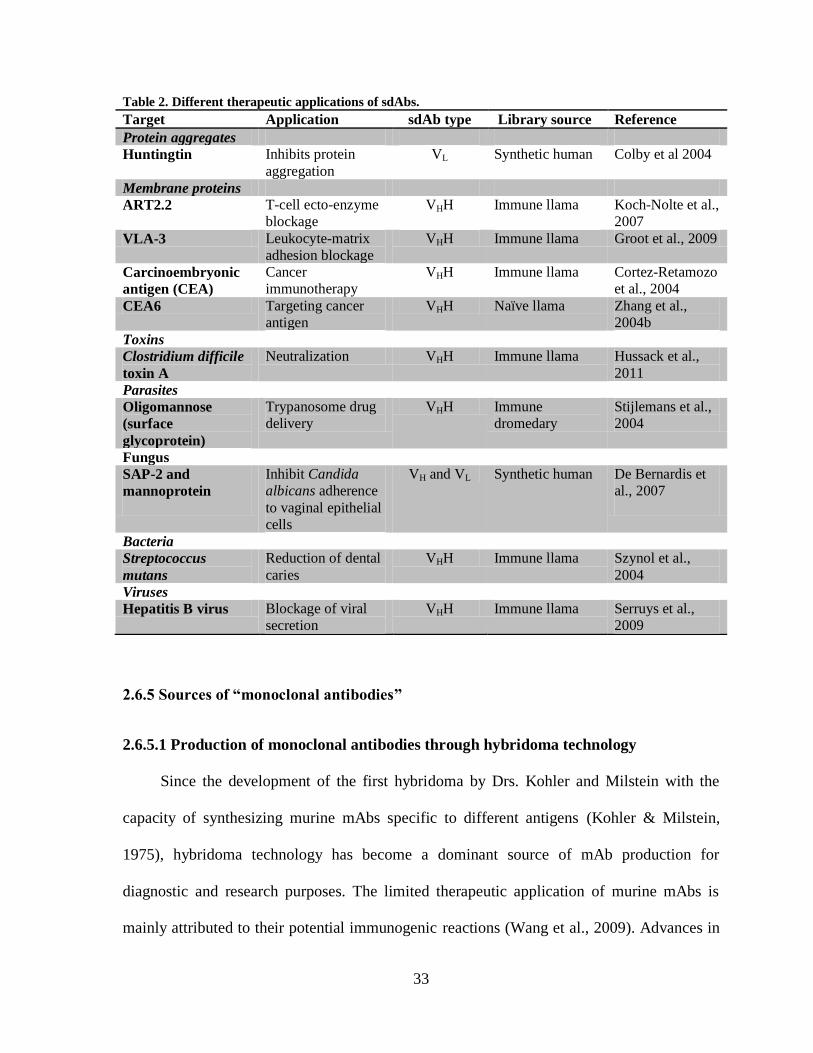
Table 2. Different therapeutic applications of sdAbs.
Target Application sdAb type Library source Reference
Protein aggregates
Huntingtin Inhibits protein
aggregation
VL Synthetic human Colby et al 2004
Membrane proteins
ART2.2 T-cell ecto-enzyme
blockage
VHH Immune llama Koch-Nolte et al.,
2007
VLA-3 Leukocyte-matrix
adhesion blockage
VHH Immune llama Groot et al., 2009
Carcinoembryonic
antigen (CEA)
Cancer
immunotherapy
VHH Immune llama Cortez-Retamozo
et al., 2004
CEA6 Targeting cancer
antigen
VHH Naïve llama Zhang et al.,
2004b
Toxins
Clostridium difficile
toxin A
Neutralization VHH Immune llama Hussack et al.,
2011
Parasites
Oligomannose
(surface
glycoprotein)
Trypanosome drug
delivery
VHH Immune
dromedary
Stijlemans et al.,
2004
Fungus
SAP-2 and
mannoprotein
Inhibit Candida
albicans adherence
to vaginal epithelial
cells
VH and VL Synthetic human De Bernardis et
al., 2007
Bacteria
Streptococcus
mutans
Reduction of dental
caries
VHH Immune llama Szynol et al.,
2004
Viruses
Hepatitis B virus Blockage of viral
secretion
VHH Immune llama Serruys et al.,
2009
2.6.5 Sources of “monoclonal antibodies”
2.6.5.1 Production of monoclonal antibodies through hybridoma technology
Since the development of the first hybridoma by Drs. Kohler and Milstein with the
capacity of synthesizing murine mAbs specific to different antigens (Kohler & Milstein,
1975), hybridoma technology has become a dominant source of mAb production for
diagnostic and research purposes. The limited therapeutic application of murine mAbs is
mainly attributed to their potential immunogenic reactions (Wang et al., 2009). Advances in
34
genetic engineering and molecular biology techniques have offered different approaches to
enhance murine mAb therapeutic use. One approach is to reduce mAbs immunogenicity in
humans. This can be achieved by modifying conventional mAbs to chimeric (murine Ab
with human CH1-CH2-CH3 domains) or humanized mAbs (human Ab with murine CDRs)
(Neuberger et al., 1985; Jones et al., 1986). While chimeric and humanized antibodies have
been able to reduce the human anti-mouse Ab (HAMA) response, reports have also
demonstrated their association with human anti-chimeric (HACA) or human anti-humanized
(HAHA) antibody immune responses. Both responses have been noted to reduce drug
efficacy over time by neutralizing antibodies which often requires frequent dose adjustment
(Hwang & Foote, 2005).
2.6.5.2 Production of monoclonal antibody fragments through phage display technology
Antibodies with desired specificity or property can be isolated from pools containing
billions of highly diverse antibodies in a complete in vitro process. Different antibody
display platforms have been generated including phage-display, microbial-display, and
ribosomal-display (reviewed in Hoogenboom, 2005). While all three display platforms are
currently in use for antibody screening and selection, phage display is the oldest and
dominant platform (Sidhu and Fellouse, 2006). It is robust, amendable to automation, can be
used in versatile applications and allow the display of variable formats of Ab fragments (e.g.,
Fab or sdAb) with diversities up to 1010
– 1011
clones (reviewed in Hoogenboom, 2005).
Unlike the hybridoma technology, it enables the selection of functional antibody fragments
against potentially unlimited array of targets including self-antigens (Griffiths et al., 1993),
toxic antigens (Hmila et al., 2008) or those with poor immunogenicity (Rahman et al., 2003).
Phage display technology is based on engineering filamentous bacteriophage (e.g., M13 or
35
Fd-tet) coat-protein to express functional antibody fragments (Figure 8) (reviewed in Winter
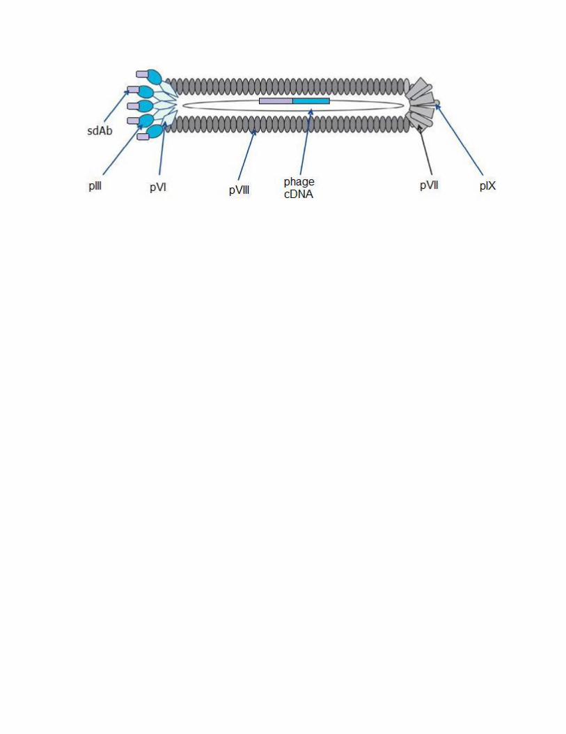
et al., 1994). The genome of filamentous bacteriophage is composed of a single stranded
DNA, encapsulated by a proteinaceous coat composed of five coat-proteins. Thousands of
copies of the major coat protein pVIII are arrayed on the surface, while the minor proteins
are arranged at the tips of the phage particle in two pairs of proteins (pVII-pIX and pIII-pVI)
(Rakonjac et al., 2011). Genes encoding for antibody fragments are usually subcloned
upstream of the pIII gene as it is well tolerated and does not affect phage infectivity (Barbass
III et al, 2004). Subsequently, the antibody library undergoes rounds of selection against
specific target antigen in a process called panning (Arbabi-Ghahroudi et al., 2009). In each
round, the entire phage library displaying antibodies is exposed to the antigen, and non-
specific binding phage is removed by several washing steps. Specifically bound phage can be
eluted by different methods, including treatment with extreme pH (MacKenzie and To, 1998;
Pincus et al., 1988), protease cleavage (Goletz et al., 2002) or competitive elution
(Meulemans et al., 1994). Specific phage is then amplified and enriched in the appropriate
bacterial host for propagation. Rounds of selection can be repeated three to five times and
genes encoding the isolated antibodies are subcloned into an E. coli for protein production
and subsequent characterization. Depending on the desired application of the isolated
antibody fragments, they can be engineered to be expressed in different formats including
their cloning and expression as fully assembled human antibodies. In fact, the first human
mAb (adalimumab, a tumor necrosis factor inhibitor) was generated from Fab phage display
library and was re-cloned in mouse myeloma cell line to be expressed as a complete IgG
molecule (Mahler et al., 1997).
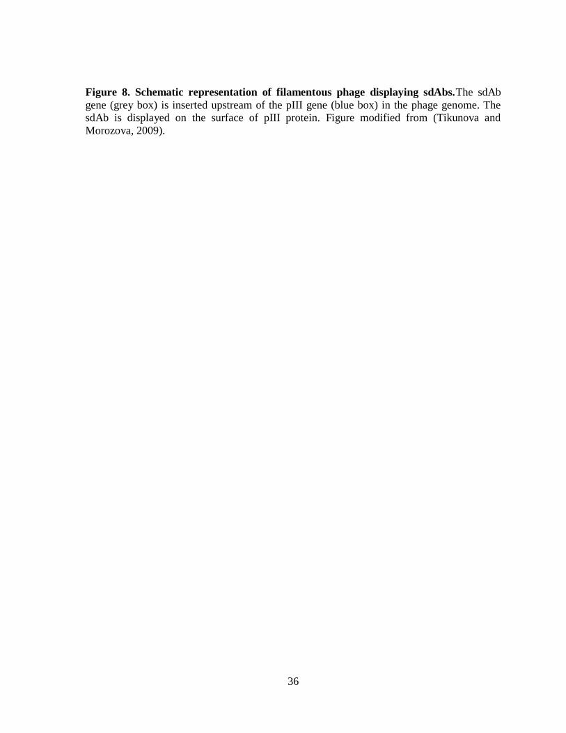
36
Figure 8. Schematic representation of filamentous phage displaying sdAbs.The sdAb
gene (grey box) is inserted upstream of the pIII gene (blue box) in the phage genome. The
sdAb is displayed on the surface of pIII protein. Figure modified from (Tikunova and
Morozova, 2009).
37
2.6.5.3 Types of antibody phage display formats
An important aspect of phage display technology is the direct linkage of displayed
antibody fragment (phenotype) to its encoding gene (genotype) by a phenotype-genotype
linker, thereby allowing antibody retrieval following cycles of selection (Hoogenboom,
2005). This feature can be achieved by phage or phagemid based display systems. In a phage
display system, the gene encoding Ab fragment is directly fused upstream of the phage coat-
protein gene (Smith, 1985). On the other hand, a phagemid display system requires two
components: a phagemid encoding the phage coat-protein (to which the Ab fragment gene
will be fused) and a helper phage to provide genes essential for phage assembly and
replication (Barbas III et al., 1991). The main difference between the two systems is in the
antibody display level. While a phage vector displays 3 – 5 copies of the antibody
(multivalent display), a phagemid vector usually expresses one copy of the Ab fragment on
the phage particle surface (monovalent display) (Ponsel et al., 2011). Therefore, phage
display system usually provides a greater number of Ab fragments following the initial round
of selection due to avidity effects. However, the affinities of isolated Ab fragments are
usually low to medium. There are several methods to overcome the low affinities of Ab
fragments by molecular biology techniques (O’Connell et al., 2002). In addition, increasing
the selective pressure in following rounds of selection is another way of isolating antibodies
with high affinities (Hoogenboom, 2005). In contrast, phagemid based Ab fragment display
libraries enables the retrieval of low number of unique antibodies at the initial selection;
however, with higher affinities due to the monovalent display nature (O’Connell et al.,
2002).
38
2.6.5.4 Types of antibody phage display libraries
Different types of recombinant antibody phage display libraries have been
constructed including immune, naïve, synthetic and semi-synthetic libraries (Figure 9)
(reviewed in: (Holliger and Hudson, 2005; Hoogenboom, 2005; Ponsel et al., 2011). Immune
libraries can be generated by amplifying antibody genes derived from B cells of immunized
donors by molecular biology techniques. The amplified genes are then incorporated by
subcloning into an appropriate phage vector and transformed into E. coli cells to create the
library. Naïve libraries are constructed in a similar manner; however the B cells are obtained
from a non-immunized donor. Naïve libraries are typically more diverse than immune
libraries, and can theoretically provide antibodies against any desired antigen (Shayne,
2007). In addition, antibodies isolated from naïve libraries often have lower affinities for
antigens in contrast to those derived from immunized libraries. This can be attributed to the
fact that immune libraries are already enriched for high affinity antibodies through in vivo
affinity maturation in the host.
Synthetic libraries are generated in vitro by polymerase chain reaction (PCR) using
randomized synthetic oligonucleotides that have been designed on the basis of previous
structural and functional analyses of antibodies (Sidhu and Fellouse, 2006). These
oligonucleotides introduce randomization into select CDR codons, thus generating diversity.
The synthetic CDR codons repertoire will be incorporated into defined framework genes in
phage display vectors. A semi-synthetic library combines the advantages of both naïve and
synthetic libraries. They can be generated by applying synthetic diversity into pre-
determined CDR regions while other CDRs had already undergone diversity naturally (Hoet,
et al, 2005). Antibodies derived from synthetic libraries have some advantages over those
derived from natural libraries. They provide a better understanding of both the structural and

39
functional properties of constructed libraries such as stability and non-aggregation (Sidhu
and Fellouse, 2006). This knowledge can subsequently improve design strategies and lead to
the generation of future synthetic antibody libraries with properties surpassing those
previously generated.
40
Figure 9. Schematic representation of the generation and selection of recombinant
antibody phage display libraries. Antibody libraries are generated from synthetic genes of
antibody’s variable domain or genes cloned from B cells of donors. The antibody phenotype
(represented by green, blue and orange boxes) is coupled to its genotype (wavy line) that is
packaged within a phage particle by phenotype-genotype coupling. Each phage particle will
display a unique antibody on its proteinaceous coat. Next, the library will undergo a selective
pressure on the phenotype to isolate potentially-specific antibodies followed by an
amplification step to amplify selected phage. The selection process will be repeated in
number of cycles, and lead antibodies will be tested for binding to the antigen of interest in
different binding assays. Adapted by permission from Macmillan Publishers Ltd: [Nature
Biotechnology]© (Hoogenboom, 2005).
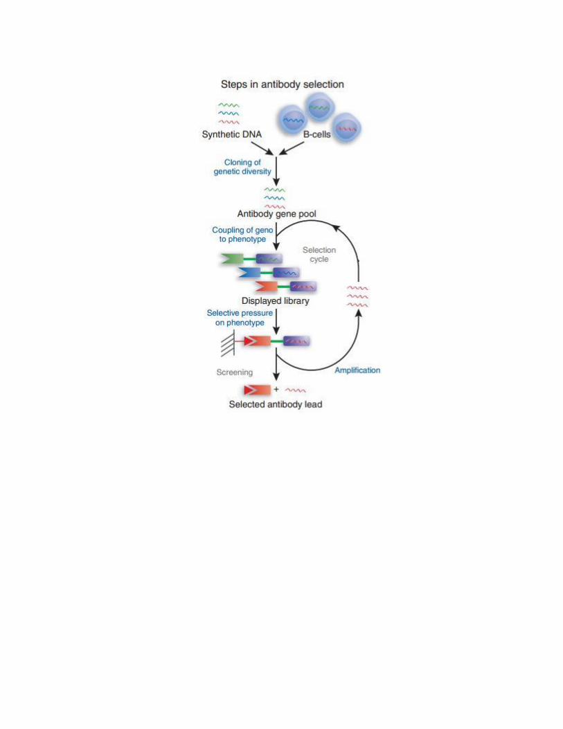
41
2.7 Conclusion
Antibodies and disease modifying agents have been developed to treat different
disorders. The complexity of MS pathogenesis has resulted in a number of different targets to
be explored as potential therapeutics. Despite the association of natalizumab with fatal viral
infection, its desirable outcome and superior efficacy in reducing the annual relapse of MS
compared to other therapeutics has encouraged the FDA to re-approve this therapy in 2006
following its withdrawal from the market in 2005 (Stuve et al., 2007). In this work, phage
display and molecular biology techniques have been applied to develop antibody fragments
(VLs and hFc-VLs) against CD49d. The antibody fragments are aimed at blocking the
interaction between CD49d-expressing immune cells and VCAM-1 expressed on the BBB in
order to limit subsequent transmigration of active immune cells toward the CNS. The
developed fragments are of low MW (VLs ~ 12 kDa and VL-Fc ~ 80 kDa), thereby allowing
them to induce a potent effect similar to that of whole IgG molecule while minimizing the
required therapeutic dose. Furthermore, both antibody types are based on human Fc and
human light chain framework sequences, and the hFc-VL fragments were expressed in a
human cell-line to minimize their immunogenicity, a key consideration for human
immunotherapeutics. While both antibody fragments may display low affinities towards the
target, their affinities can be improved by in vitro affinity maturation. In fact, it may be more
desirable to have therapeutics of lower affinity as they may reduce MS attacks with far fewer
adverse effects that would otherwise be caused by higher affinity antibodies and prolonged
CD49d blockage. The monovalent format (VL) or bivalent format (hFc-VL) can be used
alone or in combination to saturate CD49d surface in an efficient manner by accessing
42
cryptic epitopes that could not be reached by conventional IgGs, thus providing a promising
method for targeting physiologically relevant protein receptors.
3. Materials and Methods
3.1 Materials
3.1.1 VL phage display library, mammalian cell lines, and bacterial strains
The synthetic VL phage display library used in this study is described elsewhere
(Hussack et al., 2012). Jurkat cell line – immortalized leukemic T cells – was cultured in
Roswell Park Memorial Institute medium (RPMI-1640) (GIBCO, Grand Island, NY)
supplemented with 10% (v/v) fetal bovine serum (FBS) (GIBCO), and 1% (v/v) antibiotic-
antimycotic cocktail (GIBCO). K562-mock and K562-CD49d, engineered erythroleukemia
cell lines transfected with either the expression vector pFneo or with pFneo harboring
human cDNA of integrin CD49d, respectively, were kindly provided by Dr. Y. Takada
(Dana Farber Cancer Center, Boston, MA). K562-mock cell line was maintained in RPMI-
1640 supplemented with 10% (v/v) bovine serum albumin (BSA). K562-CD49d cell line
was maintained in the same culture medium containing 1 mg/mL of geneticin (GIBCO) for
selection. Human embryonic kidney cell line stably expressing Epstein-Barr virus nuclear
antigen-1 (HEK 293-EBNA-1) was kindly provided by Dr. Y. Murad (NRC-Institute for
Biological Sciences (IBS), Ottawa, ON, Canada) and cultured in serum-free F-17 medium
(GIBCO) supplemented with 1% (v/v) antibiotic-antimycotic cocktail. All cells were
incubated at 37°C, in humidified 5% CO2 incubator. Escherichia coli cells strain TG1 were
used as host for phage replication and sdAb expression. Electrocompetent TG1 cells were
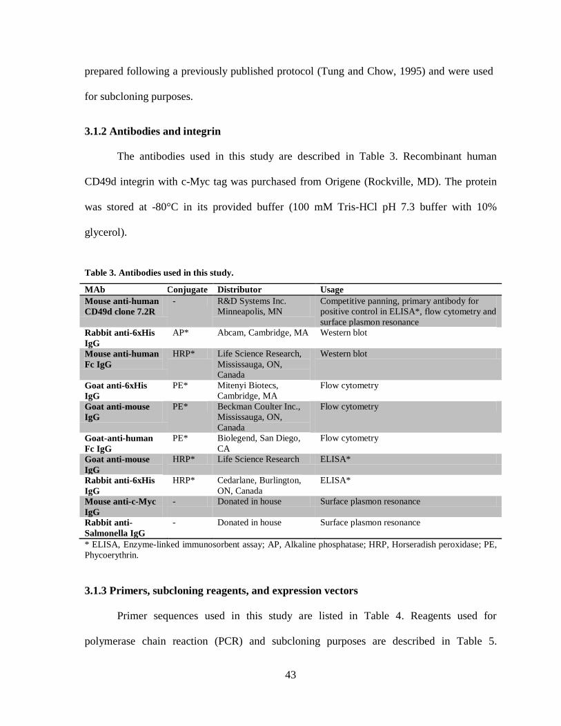
43
prepared following a previously published protocol (Tung and Chow, 1995) and were used
for subcloning purposes.
3.1.2 Antibodies and integrin
The antibodies used in this study are described in Table 3. Recombinant human
CD49d integrin with c-Myc tag was purchased from Origene (Rockville, MD). The protein
was stored at -80°C in its provided buffer (100 mM Tris-HCl pH 7.3 buffer with 10%
glycerol).
Table 3. Antibodies used in this study.
MAb Conjugate Distributor Usage
Mouse anti-human
CD49d clone 7.2R
- R&D Systems Inc.
Minneapolis, MN
Competitive panning, primary antibody for
positive control in ELISA*, flow cytometry and
surface plasmon resonance
Rabbit anti-6xHis
IgG
AP* Abcam, Cambridge, MA Western blot
Mouse anti-human
Fc IgG
HRP* Life Science Research,
Mississauga, ON,
Canada
Western blot
Goat anti-6xHis
IgG
PE* Mitenyi Biotecs,
Cambridge, MA
Flow cytometry
Goat anti-mouse
IgG
PE* Beckman Coulter Inc.,
Mississauga, ON,
Canada
Flow cytometry
Goat-anti-human
Fc IgG
PE* Biolegend, San Diego,
CA
Flow cytometry
Goat anti-mouse
IgG
HRP* Life Science Research ELISA*
Rabbit anti-6xHis
IgG
HRP* Cedarlane, Burlington,
ON, Canada
ELISA*
Mouse anti-c-Myc
IgG
- Donated in house Surface plasmon resonance
Rabbit anti-
Salmonella IgG
- Donated in house Surface plasmon resonance
* ELISA, Enzyme-linked immunosorbent assay; AP, Alkaline phosphatase; HRP, Horseradish peroxidase; PE,
Phycoerythrin.
3.1.3 Primers, subcloning reagents, and expression vectors
Primer sequences used in this study are listed in Table 4. Reagents used for
polymerase chain reaction (PCR) and subcloning purposes are described in Table 5.

44
Expression vectors pSJF2H (Arbabi-Gharoudi et al., 2010) and pTT5-hFc (Zhang et al.,
2009) were used for VLs and hFc-VLs expression, respectively.
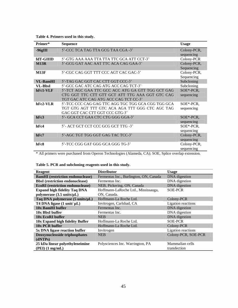
45
Table 4. Primers used in this study.
Primer* Sequence Usage
-96gIII 5’-CCC TCA TAG TTA GCG TAA CGA -3’ Colony-PCR,
sequencing
fdT-GIIID 5’-GTG AAA AAA TTA TTA TTC GCA ATT CCT-3’ Colony-PCR
M13R 5’-GCG GAT AAC AAT TTC ACA CAG GAA-3’ Colony-PCR,
Sequencing
M13F 5’-CGC CAG GGT TTT CCC AGT CAC GAC-3’ Colony-PCR,
Sequencing
VL-BamHI 5’-TAG GAC GGT CAC CTT GGT CCC-3’ Subcloning
VL-BbsI 5’-GCC GAC ATC CAG ATG ACC CAG TCT-3’ Subcloning
hFc1-VLF 5’-TCT AGC GAA TTC GCC ACC ATG GA GTT TGG GCT GAG
CTG GGT TTC CTT GTT GCT ATT TTG AAA GGT GTC CAG
TGT GAC ATC CAG ATG ACC CAG TCT CC-3’
SOE*-PCR,
sequencing
hFc2-VLR 5’-TCC CCC CAG GAG TTC AGG TGC TGG GCA CGG TGG GCA
TGT GTG AGT TTT GTC ACA AGA TTT GGG CTC AGC TAG
GAC GGT CAC CTT GGT CCC GTG-3’
SOE*-PCR,
sequencing
hFc3 5’- GCA CCT GAA CTC CTG GGG GGA-3’ SOE*-PCR,
sequencing
hFc4 5’- ACT GCT CCT CCC GCG GCT TTG -3’ SOE*-PCR,
sequencing
hFc7 5’-AGC TGT TGG GGT GAG TAC TCC-3’ Colony-PCR,
sequencing
hFc8 5’-TCC CGG GAT GGG GCA GGG TG-3’ Colony-PCR,
sequencing
* All primers were purchased from Operon Technologies (Alameda, CA); SOE, Splice overlap extension.
Table 5. PCR and subcloning reagents used in this study.
Reagent Distributor Usage
BamHI (restriction endonuclease) Fermentas Inc., Burlington, ON, Canada DNA digestion
BbsI (restriction endonuclease) Fermentas Inc. DNA digestion
EcoRI (restriction endonuclease) NEB, Pickering, ON, Canada DNA digestion
Expand high fidelity Taq DNA
polymerase (3.5 units/µL)
Hoffmann-LaRoche Ltd., Mississauga,
ON, Canada.
SOE-PCR
Taq DNA polymerase (5 units/µL) Hoffmann-La Roche Ltd. Colony-PCR
T4 DNA ligase (1 unit/ µL) Invitrogen, Carlsbad, CA Ligation reactions
10x BamHI buffer Fermentas Inc. DNA digestion
10x BbsI buffer Fermentas Inc. DNA digestion
10x EcoRI buffer NEB DNA digestion
10x Expand high fidelity Buffer Hoffmann-La Roche Ltd. SOE-PCR
10x PCR buffer Hoffmann-La Roche Ltd. Colony-PCR
5x DNA ligase reaction buffer Invitrogen Ligation reactions
Deoxynucleoside triphosphates
(dNTPs)
NEB Colony-PCR, SOE-PCR
25 kDa linear polyethyleneimine
(PEI) (1 mg/mL)
Polysciences Inc. Warrington, PA Mammalian cells
transfection
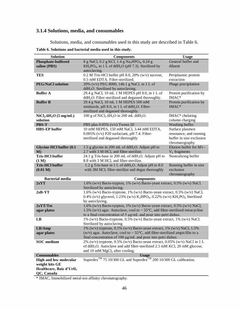
46
3.1.4 Solutions, media, and consumables
Solutions, media, and consumables used in this study are described in Table 6.
Table 6. Solutions and bacterial media used in this study.
Solution Components Usage
Phosphate-buffered
saline (PBS)
8 g NaCl, 0.2 g KCl, 1.4 g Na2HPO4, 0.24 g
KH2PO4, in 1 L of ddH2O (pH 7.3). Sterilized by
autoclaving.
General buffer and
diluent
TES 0.2 M Tris-HCl buffer pH 8.0, 20% (w/v) sucrose,
0.5 mM EDTA. Filter-sterilized.
Periplasmic protein
extraction
PEG/NaCl solution 20% (v/v) PEG 8000, 146.1 g NaCl, in 1 L of
ddH2O. Sterilized by autoclaving.
Phage precipitation
Buffer A 29.4 g NaCl, 10 mL 1 M HEPES pH 8.0, in 1 L of
ddH2O. Filter-sterilized and degassed thoroughly.
Protein purification by
IMAC*
Buffer B 29.4 g NaCl, 10 mL 1 M HEPES 500 mM
imidazole, pH 8.0, in 1 L of ddH2O. Filter-
sterilized and degassed thoroughly.
Protein purification by
IMAC*
NiCl2.6H2O (5 mg/mL)
solution
100 g of NiCl2.6H2O in 200 mL ddH2O. IMAC* chelating
column charging
PBS-T PBS plus 0.05% (v/v) Tween 20 Washing buffer
HBS-EP buffer 10 mM HEPES, 150 mM NaCl, 3.44 mM EDTA,
0.005% (v/v) P20 surfactant, pH 7.4. Filter-
sterilized and degassed thoroughly
Surface plasmon
resonance, and running
buffer in size exclusion
chromatography
Glycine-HCl buffer (0.1
M)
1.5 g glycine in 200 mL of ddH2O. Adjust pH to
2.7 with 3 M HCl, and filter-sterilize.
Elution buffer for hFc-
VL fragments
Tris-HCl buffer
(1 M)
24.1 g Tris-base in 200 mL of ddH2O. Adjust pH to
8.8 with 3 M HCl, and filter-sterilize.
Neutralizing buffer
Tris-HCl buffer
(0.01 M)
1.2 g Tris-base in 1 L of ddH2O. Adjust pH to 8.0
with 3M HCl, filter-sterilize and degas thoroughly
Running buffer in size
exclusion
chromatography
Bacterial media Components
2xYT 1.6% (w/v) Bacto-trypton, 1% (w/v) Bacto-yeast extract, 0.5% (w/v) NaCl.
Sterilized by autoclaving.
2xB-YT 1.6% (w/v) Bacto-tryptone, 1% (w/v) Bacto-yeast extract, 0.5% (w/v) NaCl,
0.4% (v/v) glycerol, 1.23% (w/v) K2HPO4, 0.22% (w/v) KH2PO4. Sterilized
by autoclaving.
2xYT/Tet
agar plates
1.6% (w/v) Bacto-tyrpton, 1% (w/v) Bacto-yeast extract, 0.5% (w/v) NaCl,
1.5% (w/v) agar. Autoclave, cool to ~ 55°C, add filter-sterilized tetracycline
to a final concentration of 5 μg/mL and pour into petri dishes.
LB 1% (w/v) Bacto-tryptone, 0.5% (w/v) Bacto-yeast extract, 1% (w/v) NaCl.
Sterilized by autoclaving
LB/Amp
agar plates
1% (w/v) tryptone, 0.5% (w/v) Bacto-yeast extract, 1% (w/v) NaCl, 1.5%
(w/v) agar. Autoclave, cool to ~ 55°C, add filter-sterilized ampicillin to a
final concentration of 100 μg/mL and pour into petri dishes.
SOC medium 2% (w/v) tryptone, 0.5% (w/v) Bacto-yeast extract, 0.05% (w/v) NaCl in 1 L
of ddH2O. Autoclave and add filter-sterilized 2.5 mM KCl, 20 mM glucose,
and 10 mM MgCl2 after cooling.
Consumables Usage
High and low molecular
weight kits GE
Healthcare, Baie d’Urfé,
QC, Canada
SuperdexTM
75 10/300 GL and SuperdexTM
200 10/300 GL calibration.
* IMAC, Immobilized metal-ion affinity chromatography.
47
3.2 Methods
3.2.1 VL phage display library panning
Jurkat cells were used as bait to select CD49d-specific VLs from a VL phage display
library by panning (Figure 10). To start the panning, thirty mL of exponentially growing
TG1 E. coli cells grown in 2xYT medium (Table 6) were prepared as previously described
(Arbabi-Ghahroudi et al., 2009) and incubated in a rotary shaker (37°C, 220 rpm) until the
OD600 reached 0.5 (3 – 4 h). At that time, ≅ 2x10¹²
phage in 100 μL of PBS was blocked in 2
mL of RPMI-1640 containing 2.5% (v/v) BSA, and incubated for 1 h at room temperature
with gentle agitation. One mL of Jurkat cells (≅ 4 x 10⁶ viable cells, counted by trypan blue
exclusion method (Strober, 2001)) was transferred to a 15-mL sterile tube and centrifuged at
2,000 rpm for 1 min at 4°C (Sorvall Legend RT Easyset bench-top centrifuge, Thermo
Scientific, Austin, TX). The extra medium was discarded, and the cells were washed twice
by re-suspending the pellet in 1 mL of fresh RPMI-1640 containing no BSA, mixed gently
by pipetting, and re-centrifuged as above. After the second wash, the cell pellet was pooled
with the 1 mL blocked phage, and incubated for 1 h at room temperature with gentle
agitation to allow phage binding to cells. Unbound phage was removed by a total of four
washes. In each washing step, the cell-phage suspension was centrifuged at 2,000 rpm for 1
min at 4ºC (Sorvall Legend RT Easyset bench-top centrifuge). The supernatant was
discarded and the pellet was re-suspended by gentle pipetting in 1 mL of washing buffer
(PBS-T for the first two washes and PBS for the latter two washes (Table 6)), transferred to a
fresh tube to reduce the amount of contaminating phage, and re-centrifuged as above. To
elute bound phage, the cell-phage pellet from the last washing step was re-suspended in 50
μL of mouse anti-human CD49d-specific mAb (Table 3) at a final concentration of 2 mg/mL
48
(~ 13 µM), transferred to a 1.5-mL sterile microtube, and incubated for 1.5 h at room
temperature with gentle agitation. Next, the cell suspension was centrifuged at 5,000 rpm for
2 min at 4ºC (Sorvall Legend Micro 17 Microcentrifuge, Thermo Scientific, Austin, TX).
The supernatant (containing eluted phage) was transferred to a sterile 1.5-mL microtube and
stored on ice temporarily. A small aliquot (100 µL) of the exponentially growing TG1 cells
was spread on 2xYT/Tet agar plate (Table 6) and incubated overnight at 37°C to ensure cells
were not pre-infected with phage. The remaining 30 mL of TG1 cells were infected with the
50 µL eluted phage by incubating the cell/phage mixture in a water bath at 37°C for 30 min.
A small aliquot of the infected TG1 cells was used to determine the phage output (eluted
phage titer) as follows: 100-fold dilutions (10-2
– 10-8
) of infected TG1 cells were prepared in
2xYT medium in a total volume of 500 µL. Next, 100 μL of each dilution were spread on
2xYT/Tet agar plate and incubated overnight at 37°C. The remaining 30 mL of infected TG1
cells were centrifuged at 3,000 rpm for 10 min at 4°C (Sorvall Legend RT Easyset bench-top
centrifuge). The supernatant was discarded and the cell pellet was re-suspended in 1 mL of
fresh 2xYT medium, spread on 4 large 2xYT/Tet agar Petri dishes (250 µL of infected cells
per plate), and incubated overnight at 37°C to amplify the eluted phage.

49
Figure 10. Panning scheme. (A) Synthetic VL phage display library was added to Jurakt
cells expressing CD49d. (B) Non-binders were washed out. (C) Binding phages were eluted
by the addition of monoclonal anti-human CD49d Ab at a high concentration (~ 13 µM)
which competed with the bound phages and released them to supernatant. (D) The eluted
phage was used to infect E.coli cells for phage amplification. Amplified phages were used as
input for the next round of panning.
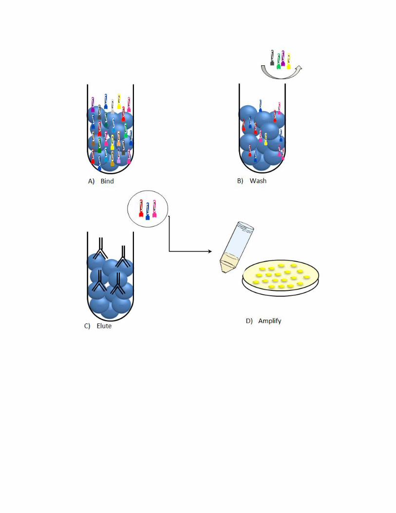
50
On the following day, colony-forming units (cfu) grown on the titer plates were
counted and the phage output was determined using the following formula:
Output (cfu) = number of colonies x dilution factor x culture volume (30,000
μL)/ plating volume (100 μL))
On the same day, phage particles were purified from the 4 large amplification culture
plates as follows: infected bacterial colonies were scraped by adding 2 x 5 mL of 2xYT
medium per plate, the cells were combined, transferred to a 50-mL sterile tube and
centrifuged at 3,000 rpm for 10 min at 4°C (Sorvall Legend RT bench-top centrifuge). The
supernatant containing phage particles was transferred to a new 50-mL sterile tube, and the
cell pellet was re-suspended in 2xYT/15% (v/v) glycerol, aliquoted in 1 mL volumes, and
stored at -80°C as a backup. The supernatant was centrifuged at 10,000 rpm for 10 min at
4°C (Beckman J2-21M/E centrifuge, Beckman Coulter Inc., Brea, CA; J-17 rotor), and
filtered through a Millipore-Stericup® filter unit (Millipore, Billerica, MA). Next, phage
particles were precipitated by adding 6 mL of PEG/NaCl solution (Table 6) to the filtrate,
incubated on ice for 1 h, and centrifuged at 4,100 rpm for 30 min at 4°C (Sorvall Legend RT
bench-top centrifuge). The supernatant was discarded and the pelleted phage was re-
suspended in 5 mL of PBS and transferred to a 15-mL sterile tube containing 1 mL of
PEG/NaCl solution, mixed gently by pipetting, and incubated on ice for 20 min. Next, the
suspension was centrifuged at 4,100 rpm for 30 min at 4°C (Sorvall Legend RT bench-top
centrifuge), the supernatant was discarded and the precipitated phage was re-suspended in 1
mL of sterile PBS. The purified phage titer was determined as follows: serially diluted phage
(10-6
– 10-12
) were prepared in PBS, and 10 μL of each dilution were used to infect 100 μL
of exponentially growing TG1 cells prepared as previously described (Arbabi-Ghahroudi et
51
al., 2009), and incubated for 15 min at 37°C without shaking. Next, the infected cells were
spread on 2xYT/Tet agar plates and incubated overnight at 37°C. On the following day, the
colonies were counted to determine phage input titer for the next round of panning using the
following formula:
Input (cfu) = number of colonies x dilution factor/ 10 µL (infecting volume) x 1000
The panning process was repeated for 3 additional rounds and the phage output and
input were tracked in all subsequent rounds. For each round of panning, 100 µL of the
purified phage from the previous round was used as the input phage for the following round
of selection.
3.2.2 Clonal analysis by DNA sequencing
After the fourth round of panning, 50 colonies were randomly selected from the
output phage titer plate for clonal analysis. Primers -96gIII and fdT-GIIID (Table 4) were
used to amplify the VL inserts by standard colony-PCR (Arbabi-Ghahroudi, 2009), and the
PCR products were sequenced as previously described (Strachan and Read, 1999). The
sequences were translated and aligned using DNASTAR Lasergene® softwares: SeqBuilder
and Megalign (DNASTAR Inc., Madison, WI).
3.2.3 Subcloning, expression, and purification of VLs
3.2.3.1 VL subcloning
Primers VL-BamHI and VL-BbsI (Table 3) were used to introduce BamHI and BbsI
restriction sites at both ends of the VL genes by standard PCR, and the PCR products were
purified with QIAquick PCR purificationTM
kit (Qiagen, Streetsville, ON, Canada). The
purified VL DNA and the expression vector pSJF2H were digested by restriction
52
endonucleases BbsI and BamHI (Table 4) and ligated together according to a previously
published protocol (Arbabi-Ghahroudi et al., 2010) to produce 6xHis tagged recombinant
VLs at the protein level. Next, 50 µL of electrocompetent TG1 cells were transformed with 3
µL of the ligated product as previously described (Arbabi-Ghahroudi et al., 2010). The
transformed cells were recovered from electroporation cuvettes by adding 1 mL of SOC
medium (Table 6) and incubated for 30 minutes at 37°C in rotary incubator adjusted to 220
rpm. Next, 100 μL of the transformed TG1 cells were spread on LB/Amp agar plate (Table
6) and incubated at 37°C overnight. The following day, positive colonies (i.e., colonies
harbouring corresponding VL genes) were screened by colony-PCR and sequencing using
M13R and M13F primers (Table 4). Positive colonies were selected for large scale protein
expression as described below.
3.2.3.2 VL expression and extraction
Multiple touches of a single positive colony were used to inoculate 10 mL of 2xB-YT
medium (Table 6) supplemented with 100 μg/mL ampicillin (2xB-YT/AMP). The culture
was incubated overnight at 27°C in a rotary shaker incubator adjusted to 220 rpm. On the
following day, the culture was used to seed a 1 L of 2xB-YT/AMP medium contained in a 4-
L baffled flask and incubated for 3 – 5 hours (37°C, 220 rpm). When the culture OD600
reached 0.3 – 0.5, the culture was induced with a final concentration of 0.1 mM isopropyl-β-
D-thiogalactopyranoside (IPTG) and incubated overnight (37°C, 220 rpm). The next day,
VLs were extracted from the bacterial periplasm by osmotic shock method (Arbabi-
Ghahroudi et al., 2010). In breif, cell cultures were harvested by centrifugation at 4,000 rpm
for 20 min at 4°C (Beckman J2-21M/E centrifuge, J-10 rotor) and the supernatant was
discarded. The cell pellet was re-suspended in 20 mL of ice-cold TES buffer (Table 6) and
53
incubated for 30 min on ice with occasional shaking. Thirty mL of ice-cold 0.125X TES
buffer were added to the cell suspension, mixed and left on ice for additional 30 min with
occasional shaking. The cells were then centrifuged at 11,000 rpm for 30 min at 4°C
(Beckman J2-21M/E centrifuge, J-17 rotor). A small aliquot of the supernatant was retained
and verified for protein expression by a standard Western blot against the 6xHis tag using
rabbit anti-6xHis IgG-AP conjugate (Table 3) as the detection agent. Next, the protein extract
was dialyzed overnight against 6 L of buffer A (Table 6) at 4°C.
3.2.3.3 VL purification by immobilized-metal affinity chromatography
All purification steps were performed following a previously described method
(Arbabi-Ghahroudi et al., 2009) under the control of ÄKTATM
FPLC system (GE
Healthcare) system. Briefly, dialyzed VLs were loaded onto 5 mL HiTrapTM
chelating HP
column (GE Healthcare) previously charged with Ni2+
by loading 30 mL of NiCl2.6H2O
solution (Table 6). To elute, Buffer A (Table 6) was applied at a flow rate of 1 mL/min with
decreased concentration from 100% to 0% over a period of 50 min with concurrent flow of
Buffer B (Table 6) but with increased concentration from 0% to 100%. The fractions
corresponding to the eluted peaks were further examined by sodium dodecyl sulfate-
polyacrylamide gel electrophoresis (SDS-PAGE) and Western blot (detected by rabbit anti-
6xHis IgG conjugated to AP (Table 3)) to confirm VLs expression. The fractions containing
purified VLs were pooled together and dialyzed overnight at 4°C against 6 L of buffer A to
remove imidazole, and VLs concentrations were determined by absorbance measurement
(A280) using their MWs and molar extinction coefficients (Pace et al., 1995).

54
3.2.4 Subcloning, expression and purification of hFc-VLs
3.2.4.1 hFc-VL subcloning
The DNA coding for CD49d-binding VL was amplified by SOE-PCR (Figure 11) and
subcloned into pTT5-hFc expression vector (Appendix 1). The SOE-PCR protocol was
designed to ensure that the extra DNA residue found within the human IgG1 hinge region of
the vector (labeled as extra TCPPCP in Appendix 1) would be deleted during the consecutive
PCRs. To start, the CD49d-binding VL DNA was amplified by PCR (PCR-1) using the
primers hFc1-VLF and hFc2-VLR (Table 4). This was done to flank the VL DNA with
EcoRI restriction site, Kozak sequence, and the signal sequence of human IgG1 heavy chain
at the 5’ end, and with the DNA coding for the hinge region and the first seven residues of
the Fc of human IgG1 Fc at the 3’ end. The reaction mixture was prepared according to the
following protocol:
dNTPs (2.5 mM each) 1 µL
10x Expand High Fidelity Buffer 5 µL
hFc1-VLF (10 pmol/µL) 1 µL
hFc2-VLR (10 pmol/µL) 1 µL
CD49d-specific VL (5 ng/µL) 1 µL
Expand High Fidelity Enzyme mix (3.5
units/µL)
0.5 µL
Sterile H2O 40.5 µL
On the second PCR (PCR-2), hFc3 and hFc4 primer pair (Table 4) was used to
amplify the first 65 amino acid residues of the human Fc region using pTT5-hFc vector as a
template according to the same reaction mixture above. The reaction tubes of PCR-1 and
PCR-2 were placed in a thermocycler to amplify the two DNA fragments with a program
consisting of a preheating step at 94°C for 5 min, 30 cycles of 94°C for 30 s, 55°C for 30 s,
and 72°C for 30 s. Next, the PCR products were loaded into a 1% agarose gel and
55
electrophoresed for 20 min at 120 V. The products of PCR-1 (~ 468 bp) and PCR-2 (~ 196
bp) were extracted from the 1% agarose gel and purified using the QIAquick Gel
ExtractionTM
kit (Qiagen) and DNA concentrations were determined based on OD260
measurements (Sambrook et al., 1989).
The third PCR (PCR-3) was carried on a template comprising of a mixture of PCR-1
and PCR-2 purified products. The primers hFc1-VLF and hFc4 (Table 4) were used to fuse
the two DNA fragments together through their overlapping complementary sequences (in a
context not containing the extra DNA residue) according to the following protocol:
dNTPs (2.5 mM each) 1 µL
10x Expand High Fidelity Buffer 5 µL
Primers hFc1-VLF and hFc4 (10
pmol/µL)
1 µL each
Gel-purified PCR-1 (~ 50ng/µL)
Gel-purified PCR-2 (~ 20ng/µL)
1 µL
1 µL
Expand High Fidelity Enzyme mix (3.5
units/µL)
0.5 µL
Sterile H2O 39.5 µL
The reaction tubes were placed in a thermal cycler with a program consisting of a
preheating step at 94°C for 5 min, 30 cycles of 94°C for 30 s, 55°C for 30 s, and 72°C for 90
s. The product of PCR-3 (~ 643 bp) was loaded into 1% agarose gel and the corresponding
band was extracted and purified with QIAquick Gel ExtractionTM
kit (Qiagen). The PCR-3
purified product and the expression vector pTT5-hFc were both digested with the restriction
endonucleases EcoRI and SacII (Table 5) in two separate digestion reactions as described
previously (Zhang et al., 2009). Subsequently, the digestion products were loaded into 1%
agarose gel and corresponding bands were purified with QIAquick Gel ExtractionTM
kit
(Qiagen). The DNA concentrations of both digestion products were determined by OD260
56
measurements and were ligated together according to a previously published protocol (Zhang
et al., 2009). The ligated products were transformed into TG1 cells by electroporation as
described above for VLs subcloning. Positive colonies were screened by colony-PCR and
sequencing using hFc7 and hFc8 primers (Table 4). Colonies harbouring sequences
corresponding to hFc-VL clones of interest were grown in 50 mL of LB medium
supplemented with 100 μg/mL ampicillin and incubated at 37°C and 220 rpm. When the
OD600 reached 0.4 – 0.6, the cells were harvested by centrifugation at 4,000 rpm for 10 min
at 25°C (Sorvall Legend RT bench-top centrifuge). The extra medium was discarded and the
plasmid was purified from the bacterial pellet by OMEGA fast filter plasmid MAXI prep
columns® (Omega, Norcross, GA) following the procedure provided by the manufacturer.
The LB bacterial culture preparation was repeated when the total yield of the extracted
plasmid was lower than 300 μg.
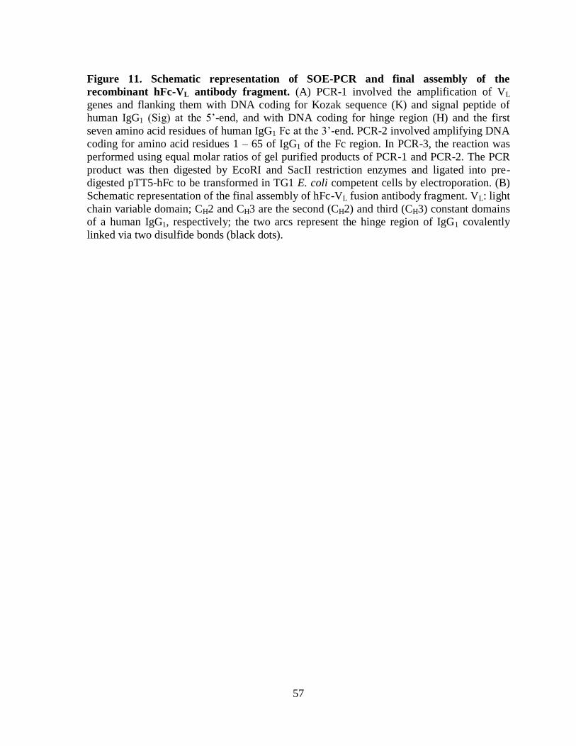
57
Figure 11. Schematic representation of SOE-PCR and final assembly of the
recombinant hFc-VL antibody fragment. (A) PCR-1 involved the amplification of VL
genes and flanking them with DNA coding for Kozak sequence (K) and signal peptide of
human IgG1 (Sig) at the 5’-end, and with DNA coding for hinge region (H) and the first
seven amino acid residues of human IgG1 Fc at the 3’-end. PCR-2 involved amplifying DNA
coding for amino acid residues 1 – 65 of IgG1 of the Fc region. In PCR-3, the reaction was
performed using equal molar ratios of gel purified products of PCR-1 and PCR-2. The PCR
product was then digested by EcoRI and SacII restriction enzymes and ligated into pre-
digested pTT5-hFc to be transformed in TG1 E. coli competent cells by electroporation. (B)
Schematic representation of the final assembly of hFc-VL fusion antibody fragment. VL: light
chain variable domain; CH2 and CH3 are the second (CH2) and third (CH3) constant domains
of a human IgG1, respectively; the two arcs represent the hinge region of IgG1 covalently
linked via two disulfide bonds (black dots).
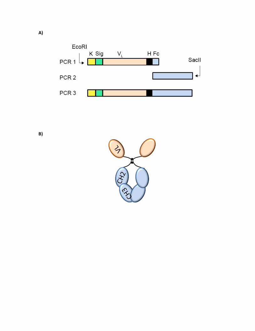
A)
B)
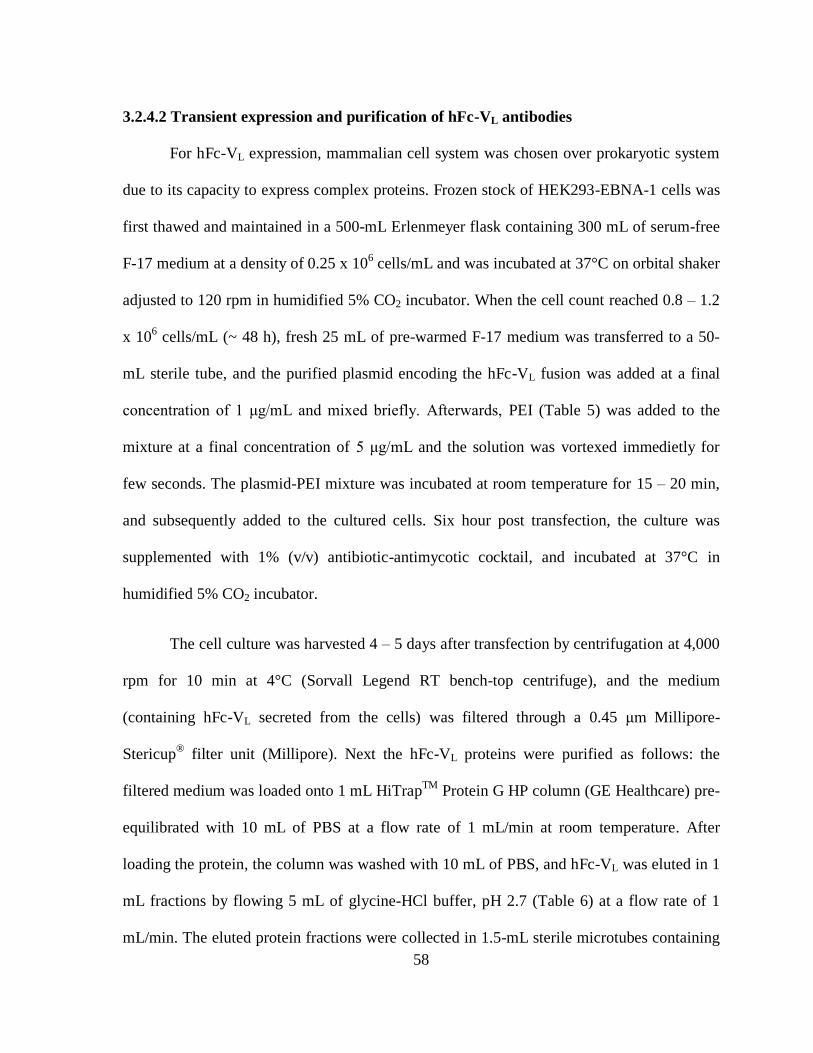
58
3.2.4.2 Transient expression and purification of hFc-VL antibodies
For hFc-VL expression, mammalian cell system was chosen over prokaryotic system
due to its capacity to express complex proteins. Frozen stock of HEK293-EBNA-1 cells was
first thawed and maintained in a 500-mL Erlenmeyer flask containing 300 mL of serum-free
F-17 medium at a density of 0.25 x 10⁶ cells/mL and was incubated at 37°C on orbital shaker
adjusted to 120 rpm in humidified 5% CO2 incubator. When the cell count reached 0.8 – 1.2
x 106 cells/mL (~ 48 h), fresh 25 mL of pre-warmed F-17 medium was transferred to a 50-
mL sterile tube, and the purified plasmid encoding the hFc-VL fusion was added at a final
concentration of 1 μg/mL and mixed briefly. Afterwards, PEI (Table 5) was added to the
mixture at a final concentration of 5 μg/mL and the solution was vortexed immedietly for
few seconds. The plasmid-PEI mixture was incubated at room temperature for 15 – 20 min,
and subsequently added to the cultured cells. Six hour post transfection, the culture was
supplemented with 1% (v/v) antibiotic-antimycotic cocktail, and incubated at 37°C in
humidified 5% CO2 incubator.
The cell culture was harvested 4 – 5 days after transfection by centrifugation at 4,000
rpm for 10 min at 4°C (Sorvall Legend RT bench-top centrifuge), and the medium
(containing hFc-VL secreted from the cells) was filtered through a 0.45 μm Millipore-
Stericup® filter unit (Millipore). Next the hFc-VL proteins were purified as follows: the
filtered medium was loaded onto 1 mL HiTrapTM
Protein G HP column (GE Healthcare) pre-
equilibrated with 10 mL of PBS at a flow rate of 1 mL/min at room temperature. After
loading the protein, the column was washed with 10 mL of PBS, and hFc-VL was eluted in 1
mL fractions by flowing 5 mL of glycine-HCl buffer, pH 2.7 (Table 6) at a flow rate of 1
mL/min. The eluted protein fractions were collected in 1.5-mL sterile microtubes containing
59
100 µL Tris-HCl buffer, pH 8.8 (Table 6) for immediate neutralization. Next, the eluted
fractions were verified for protein expression by a standard Western blot using mouse anti-
human Fc IgG-HRP conjugate (Table 3) as a detection agent. Fractions containing purified
protein were pooled and dialyzed overnight against 2 L of PBS at 4°C. On the following day,
200 μL aliquots of the dialyzed protein were prepared and stored at -20°C.
3.2.5 Size exclusion chromatography (SEC)
SuperdexTM
75 10/300 GL gel filtration column (optimal resolution of 3,000 – 70,000
Da) and SuperdexTM
200 10/300 GL gel filtration column (optimal resolution of 10,000 –
600,000 Da) (GE Healthcare) were used to determine the structural integrity, purity and
aggregation status of VL and hFc-VL antibody fragments, respectively. SEC was performed
on ÄKTATM
FPLC purification system (GE Healthcare) using HBS-EP (Table 6) for
SuperdexTM
75 and PBS for SuperdexTM
200 according to a previously described protocol
(Arbabi-Ghahroudi et al., 2010). The experiment was performed at room temperature and all
used running buffers (HBS-EP for VLs and PBS for hFc-VLs) were filtered through a 0.22
μm Millipore-Stericup® filter unit (Millipore) and degassed extensively under vacuum. The
column was pre-equilibrated with two column volumes (50 mL) of the filtered running buffer
at a pump speed of 0.5 mL/min. After the column was equilibrated, 200 μL of the purified
antibody fragments were loaded (one clone at a time) and allowed to flow through the
column. Normalized profiles were grafted and fractions corresponding to the monomeric
eluted protein peak were collected and stored at 4°C.
60
3.2.6 Binding assays
3.2.6.1 ELISA
ELISA microwell titer plate was coated overnight at 4°C with 100 μL of human
recombinant CD49d (Origene) at a final concentration of 5 μg/mL (45 nM) in PBS. On the
following day, coating solution was discarded and wells were blocked with 200 µL of 2%
(w/v) casein in PBS for 2 h at 37°C. The blocking solution was discarded and 100 µL of 50
μg/mL (~ 4 µM) of SEC purified VLs were added, and the plate was sealed and incubated for
1.5 h at 37°C. Next, the ELISA plate was washed 4 times with PBS-T, blotted against a clean
paper towel, and 100 µL of rabbit anti-6xHis IgG-HRP (Table 3) diluted 1:2,500 in PBS
were added per well, and the plate was incubated for 1 h at room temperature. Next, the
ELISA plate was washed 4 times with PBS-T, blotted against a paper towel, and bound
antibodies were detected colorimetrically by adding 100 µL of TMB substrate (KPL,
Gaithersburg, MD) and incubated for 15 – 20 min at room temperature. The reaction was
stopped by adding 100 µL of 1 M phosphoric acid, and the optical density was determined at
A450 nm using Dynatech MR5000 ELISA plate reader (Pegasus Scientific Inc., Rockville,
MD). Positive control wells were treated with 100 µL of 5 µg/mL (33 nM) mouse anti-
human CD49d mAb (Table 3) and detected with 100 µL of goat anti-mouse IgG-HRP (Table
3) diluted 1:5000 in PBS. Negative controls, which included those (i) having non-relevant VL
(50 µg/mL (~ 4 µM)), (ii) lacking VL, and (iii) lacking CD49d coating, received the same
treatment as test and positive control samples.
3.2.6.2 Surface plasmon resonance (SPR)
Binding kinetics for the interaction between VLs and the recombinant human CD49d
protein (Origene) were determined by SPR under the control of BIACORE 3000TM
optical
61
sensor platform (GE Healthcare). All SPR experiments were carried out at room temperature
in the running buffer HBS-EP (Table 6) at a flow rate of 40 μL/min. The surfaces were
regenerated by extensive washing with the running buffer.
In this experiment, CD49d sample was dialyzed in PBS to remove glycerol (10%)
which could interfere with SPR experiments. After dialysis, the protein was immobilized on
CM5 sensorchip (GE Healthcare) at 3,503 resonance units (RUs) by standard amine coupling
kit. SEC purified VLs (200 nM) were injected over the immobilized CD49d, and data was
analysed by BIAevaluation 4.1 software (GE Healthcare). The initial attempt for
immobilizing CD49d directly on the sensorchip was unsuccessful and resulted in reduced
activity of the protein as noted when injecting the mouse anti-CD49d mAb control (Table 3)
(explained in Results section). Therefore, CD49d (187 RUs) was indirectly captured via its c-
Myc tag after coupling the sensorchip with 15,122 RUs of anti-c-Myc IgG (Table 3). Surface
activity of indirectly captured CD49d was investigated by injecting 100 nM of anti-human
CD49d mAb control (Table 3). Controls for SPR experiment included CD49d (50 nM)
injected over a reference channel with no ligand immobilized on the sensorchip channel, or
injected over immobilized non-relevant IgG (Table 3). The following formula was used for
calculating the maximum binding capacity of the immobilized ligand (i.e., Rmax): (MWanalyte /
MWligand) x immobilized ligand amount (RU) x stoichiometric ratio) of the control IgG)
3.2.6.3 Flow cytometry
In all flow cytometry experiments, at least 15,000 cells were acquired on BD
FACScantoTM
flow cytometer (BD Biosciences, San Jose, CA). Jurkat cells were harvested
from suspension cultures by centrifugation at 1,600 rpm for 8 min at 4°C (Sorvall Legend
RT bench-top centrifuge). Afterwards, media was discarded and cells were re-suspended in 2
62
mL filtered 1% (w/v) BSA in PBS. The number of viable cells was determined by trypan
blue exclusion method (Stober, 2001), and the cells were adjusted to 2 x 10⁶ cells/mL.
Aliquots of 100 μL of the cell suspension were transferred to round-bottom FalconTM
tubes
(Fisher scientific, Toronto, ON, Canada), and 125 µg/mL (~ 10 µM final concentration) of
the SEC purified VLs were added to the cells in a total volume of 200 μL, mixed gently, and
incubated for 30 min at 4ºC. Non-binding VLs were washed by adding 3 mL of cold PBS and
cells were centrifuged as above. After discarding the washing buffer, the cells were re-
suspended in 100 μL of 1% (w/v) BSA in PBS, and 10 μL of undiluted anti-6xHis-PE (Table
3) were added. The cells were incubated for 10 min at 4ºC in the dark. Excess secondary
antibody was washed by adding 3 mL of cold PBS, and the cells were centrifuged again at
1,600 rpm for 8 min at 4°C (Sorvall Legend RT bench-top centrifuge). The washing buffer
was discarded and cells were re-suspended in 0.5 mL of 1% (v/v) formaldehyde in PBS.
Samples were acquired on BD FACScantoTM
flow cytometer (BD Biosciences), and data
were exported and analyzed with FlowJo®
7.6.5 software (TreeStar, San Carlos, CA). For the
positive control, cells were incubated with 10 μL of 25 μg/mL (~ 166 nM) of mouse anti-
human CD49d mAb in a total volume of 200 μL (Table 3) for 30 min at 4ºC, washed and re-
suspended in 100 μL of 1% (w/v) BSA as above, and detected by adding 1 μL of undiluted
0.5 mg/mL (~ 3 nM) goat anti-mouse IgG-PE secondary antibody (Table 3). The negative
controls included Jurkat cells incubated with anti-6xHis-PE (no VL added), and un-stained
Jurkat cells to detect the level of auto-fluorescence.
In order to confirm VLs’ specificity to CD49d, flow cytometry experiments were
done using K562-mock cells (not expressing CD49d) which represents the control cell line,
and K562-CD49d cells (stably expressing human CD49d) which represents the test cell line.
63
Each tested antibody fragment was added to both cell line at a final concentration of (125
µg/mL (~ 10 µM) for tested VLs or at a final concentration of 100 µg/mL (~ 1.3 µM) of hFc-
VLs according to the same protocol described above for Jurkat cells. Binding hFc-VL
antibody fragments were detected with 10 µL of undiluted anti-human Fc IgG-PE conjugate
(Table 3). The negative controls included K562-mock cells treated with: (1) mouse anti-
human CD49d IgG (~ 16 nM final concentration) detected with goat anti-mouse IgG-PE
conjugate (~ 15 nM); (2) non-relevant VL detected with goatanti-6xHis-IgG PE conjugate;
(3) the secondary antibody goat anti-mouse IgG-PE conjugate (~ 15 nM); and (4) the
secondary antibody goat anti-6xHis-IgG PE conjugate (10 µL of undiluted stock). The same
treatment conditions were applied on K562-CD49d cells to determine CD49d expression,
and background binding levels. Controls used for determining hFc-VLs binding specificity to
CD49d included: 1) non-stained cells, 2) K-562 cell line variants treated with non-relevant
hFc-VHH added at a final concentration of 100 µg/mL (~ 1.3 µM), and 3) K-562 cell line
variants treated with10 µL of the undiluted secondary antibody goat anti-human Fc IgG-PE
conjugate. In addition, the specific binding of VL or hFc-VL antibody fragments to CD49d-
expressing cells was determined by: 1) mean fluorescence intensity (MFI), 2), and the
percentage of positive cells. For flow cytometry concentration dependent experiments, two
concentrations (1.3 μM and 2.6 μM) of SEC purified hFc-VL were added to the test and
control cell lines in a total volume of 200 μL, and the rest of the flow cytometry experiment
was performed as previously described.
64
4. Results
4.1 Selection of CD49d-specific VLs by panning
A synthetic VL phage display library was generated on the basis of a pre-identified
human VL scaffold (HVLP324, hereafter referred to as VL-24) that possessed desired
properties such as high yield expression, non-aggregation, and reversible thermal unfolding
(Hussack et al., 2012). The library was constructed through in vitro mutagenesis at 18 amino
acid positions distributed at selected locations of CDR1, CDR2, and CDR3 (Figure 12A).
Library diversity was further enhanced by randomizing CDR3 using three mutagenic
oligonucleotides to give rise to CDR3 of variable lengths, namely 9, 10, and 11 amino acid
residues (Figure 12B). The library was subjected to four rounds of panning against CD49d-
expressing Jurkat T-cell line, and CD49d-binding phage was selectively eluted with a high
concentration (~ 13 µM) of a commercially available anti-CD49d mAb as a competitor as
previously described in the Methods section. Eluted phage was then amplified in bacterial
cells, purified, and used for subsequent round of selection. Four rounds of panning were
performed to enrich the library for phage clones displaying VLs that selectively bind to the
extracellular domain of human CD49d. Phage recovery efficiency was monitored in each
round of panning by determining the ratio of output to input phage titers (O/I ratio); where
the phage titers were calculated using the formulas described in the Methods section. It is a
common practise to obtain the O/I ratio from one trial of titration per round of panning in
order to check for enrichment trend that is indicated by the increase in the enrichment factor.
As expected, the phage recovery efficiency at the initial rounds of panning was lower than
that of subsequent rounds (Table 7; Figure 13). For example, by the end of round 3, the O/I
ratio had increased by over 1,000 fold compared to round 1, indicating enrichment for
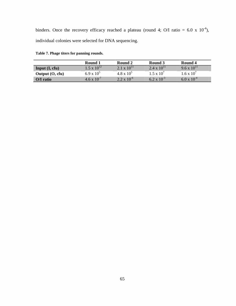
65
binders. Once the recovery efficacy reached a plateau (round 4; O/I ratio = 6.0 x 10⁻⁴
),
individual colonies were selected for DNA sequencing.
Table 7. Phage titers for panning rounds.
Round 1 Round 2 Round 3 Round 4
Input (I, cfu) 1.5 x 10¹²
2.1 x 10¹¹
2.4 x 10¹¹
9.6 x 10¹¹
Output (O, cfu) 6.9 x 10⁵ 4.8 x 10
5 1.5 x 10
7 1.6 x 10
7
O/I ratio 4.6 x 10⁻⁷
2.2 x 10⁻⁶
6.2 x 10⁻³
6.0 x 10⁻⁴
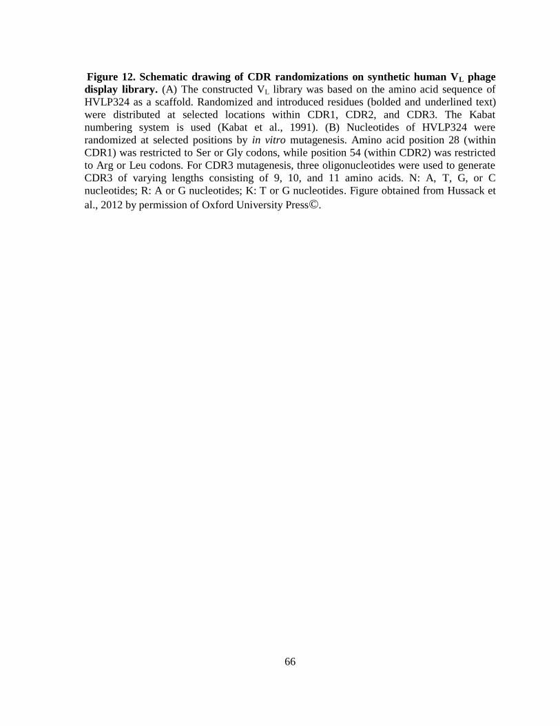
66
Figure 12. Schematic drawing of CDR randomizations on synthetic human VL phage
display library. (A) The constructed VL library was based on the amino acid sequence of
HVLP324 as a scaffold. Randomized and introduced residues (bolded and underlined text)
were distributed at selected locations within CDR1, CDR2, and CDR3. The Kabat
numbering system is used (Kabat et al., 1991). (B) Nucleotides of HVLP324 were
randomized at selected positions by in vitro mutagenesis. Amino acid position 28 (within
CDR1) was restricted to Ser or Gly codons, while position 54 (within CDR2) was restricted
to Arg or Leu codons. For CDR3 mutagenesis, three oligonucleotides were used to generate
CDR3 of varying lengths consisting of 9, 10, and 11 amino acids. N: A, T, G, or C
nucleotides; R: A or G nucleotides; K: T or G nucleotides. Figure obtained from Hussack et
al., 2012 by permission of Oxford University Press©.
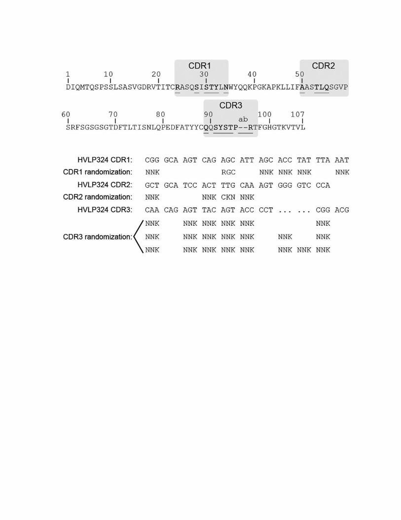

67
Figure 13. Monitoring library enrichment for binders by phage titration. The ratio of
output to input phage titers is shown as a function of panning round.
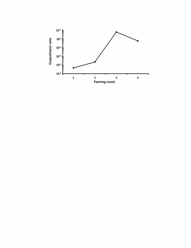
68
4.1.1 Sequence analysis of isolated anti-CD49d VLs
After the fourth round of panning, a total of 50 colonies were picked at random from
output titer plates and subjected to colony-PCR. Sequence analysis revealed 12 unique
sequences (Table 8; Appendix 2) with varying frequencies of occurrence. Clones VL-1, VL-2,
and VL-3 were the most enriched sequences with frequencies of 30%, 16%, and 14%,
respectively, while the rest of the clones had a frequency of occurrence of 4% or 2%. Out of
the 12 identified VLs, 3 VLs (VL-2, VL-11, and VL-17) were randomized in all 3 CDRs and
displayed totally unique sequences, while the remaining VLs had one or two wild-type (i.e.,
VL-24) CDRs (Table 8). Thus, 6 VLs (VL-3, VL-5, VL-6, VL-31, VL-33, and VL-35) were
wild-type for CDR1, 2 VLs (VL-21, VL-48) were wild-type for CDR1 and CDR2, and one VL
(VL-1) was wild-type for CDR3. Furthermore, an over-presentation of wild-type amino acid
residues (S28, Y32 and N34 of CDR1 and A50 of CDR2, Kabat numbering system (1991))
was noted in all sequenced VLs with the exception of clone VL-1 (which possessed G28 and
R34), VL-2 (G50), and VL-35 (R50). In addition, an over-presentation of a unique CDR2
amino acid sequence (A50, H53, L54, and N55) was shared by the clones VL-3, VL-6, VL-11,
VL-17, VL-31, and VL-33. The randomised residues of CDR3 (residues 89, 91 – 94, and 95a
– 96) of all sequenced VL clones displayed mutations with respect to the wild-type VL-24
(except for VL-1 which possessed wild-type CDR3, VL-2 and VL-33 which possessed wild-
type R96, and VL-5 and VL-17 which possessed wild-type S93). The sequence analysis also
revealed an over-presentation of R91 in all sequenced VL clones except for VL-1, VL-35, and
VL-48. The clones VL-2 and VL-3 shared similar CDR3 amino acid sequences and differed
only in three amino acid residues located at positions 93, 95a, and 96. Similarly, the clones
VL-2 and VL-33 shared similar CDR3 amino acid sequences and differed in 3 amino acid
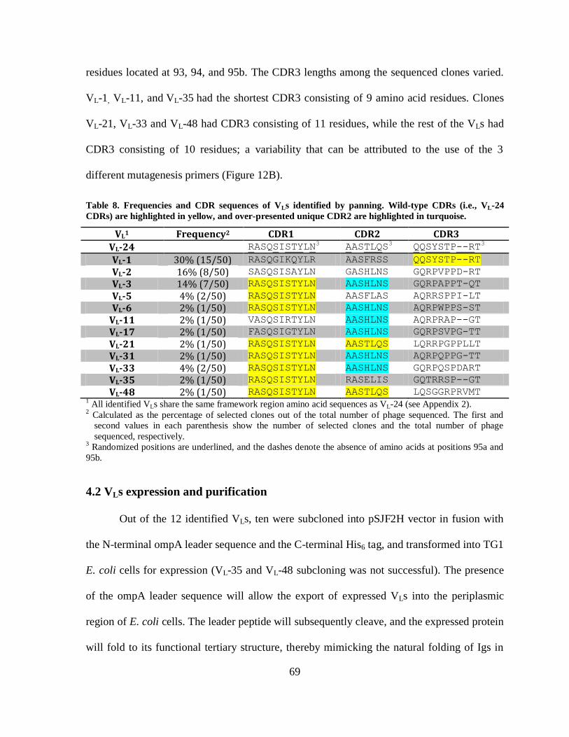
69
residues located at 93, 94, and 95b. The CDR3 lengths among the sequenced clones varied.
VL-1, VL-11, and VL-35 had the shortest CDR3 consisting of 9 amino acid residues. Clones
VL-21, VL-33 and VL-48 had CDR3 consisting of 11 residues, while the rest of the VLs had
CDR3 consisting of 10 residues; a variability that can be attributed to the use of the 3
different mutagenesis primers (Figure 12B).
Table 8. Frequencies and CDR sequences of VLs identified by panning. Wild-type CDRs (i.e., VL-24
CDRs) are highlighted in yellow, and over-presented unique CDR2 are highlighted in turquoise.
VL1 Frequency2 CDR1 CDR2 CDR3
VL-24 RASQSISTYLN3 AASTLQS
3 QQSYSTP--RT3
VL-1 30% (15/50) RASQGIKQYLR AASFRSS QQSYSTP--RT
VL-2 16% (8/50) SASQSISAYLN GASHLNS GQRPVPPD-RT
VL-3 14% (7/50) RASQSISTYLN AASHLNS GQRPAPPT-QT
VL-5 4% (2/50) RASQSISTYLN AASFLAS AQRRSPPI-LT
VL-6 2% (1/50) RASQSISTYLN AASHLNS AQRPWPPS-ST
VL-11 2% (1/50) VASQSIRTYLN AASHLNS AQRPRAP--GT
VL-17 2% (1/50) FASQSIGTYLN AASHLNS GQRPSVPG-TT
VL-21 2% (1/50) RASQSISTYLN AASTLQS LQRRPGPPLLT
VL-31 2% (1/50) RASQSISTYLN AASHLNS AQRPQPPG-TT
VL-33 4% (2/50) RASQSISTYLN AASHLNS GQRPQSPDART
VL-35 2% (1/50) RASQSISTYLN RASELIS GQTRRSP--GT
VL-48 2% (1/50) RASQSISTYLN AASTLQS LQSGGRPRVMT 1 All identified VLs share the same framework region amino acid sequences as VL-24 (see Appendix 2).
2 Calculated as the percentage of selected clones out of the total number of phage sequenced. The first and
second values in each parenthesis show the number of selected clones and the total number of phage
sequenced, respectively. 3 Randomized positions are underlined, and the dashes denote the absence of amino acids at positions 95a and
95b.
4.2 VLs expression and purification
Out of the 12 identified VLs, ten were subcloned into pSJF2H vector in fusion with
the N-terminal ompA leader sequence and the C-terminal His6 tag, and transformed into TG1
E. coli cells for expression (VL-35 and VL-48 subcloning was not successful). The presence
of the ompA leader sequence will allow the export of expressed VLs into the periplasmic
region of E. coli cells. The leader peptide will subsequently cleave, and the expressed protein
will fold to its functional tertiary structure, thereby mimicking the natural folding of Igs in
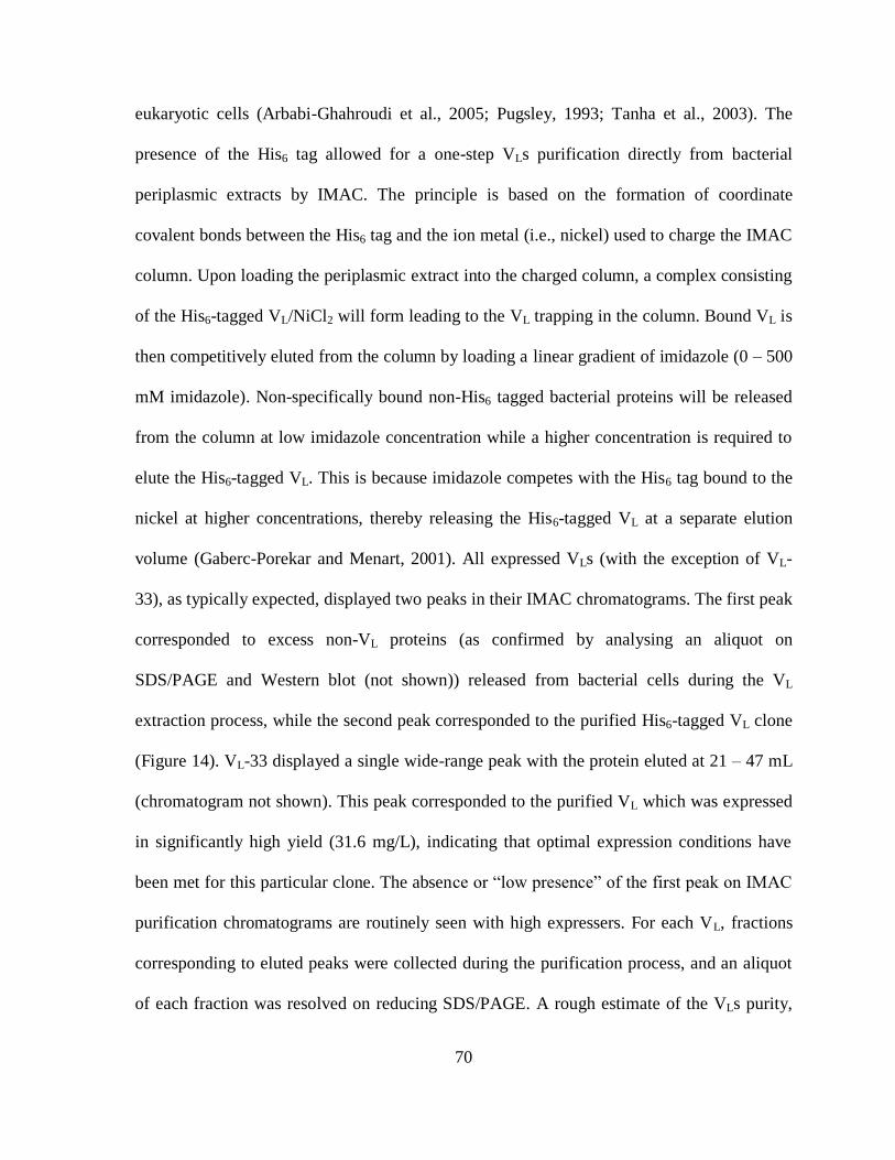
70
eukaryotic cells (Arbabi-Ghahroudi et al., 2005; Pugsley, 1993; Tanha et al., 2003). The
presence of the His6 tag allowed for a one-step VLs purification directly from bacterial
periplasmic extracts by IMAC. The principle is based on the formation of coordinate
covalent bonds between the His6 tag and the ion metal (i.e., nickel) used to charge the IMAC
column. Upon loading the periplasmic extract into the charged column, a complex consisting
of the His6-tagged VL/NiCl2 will form leading to the VL trapping in the column. Bound VL is
then competitively eluted from the column by loading a linear gradient of imidazole (0 – 500
mM imidazole). Non-specifically bound non-His6 tagged bacterial proteins will be released
from the column at low imidazole concentration while a higher concentration is required to
elute the His6-tagged VL. This is because imidazole competes with the His6 tag bound to the
nickel at higher concentrations, thereby releasing the His6-tagged VL at a separate elution
volume (Gaberc-Porekar and Menart, 2001). All expressed VLs (with the exception of VL-
33), as typically expected, displayed two peaks in their IMAC chromatograms. The first peak
corresponded to excess non-VL proteins (as confirmed by analysing an aliquot on
SDS/PAGE and Western blot (not shown)) released from bacterial cells during the VL
extraction process, while the second peak corresponded to the purified His6-tagged VL clone
(Figure 14). VL-33 displayed a single wide-range peak with the protein eluted at 21 – 47 mL
(chromatogram not shown). This peak corresponded to the purified VL which was expressed
in significantly high yield (31.6 mg/L), indicating that optimal expression conditions have
been met for this particular clone. The absence or “low presence” of the first peak on IMAC
purification chromatograms are routinely seen with high expressers. For each VL, fractions
corresponding to eluted peaks were collected during the purification process, and an aliquot
of each fraction was resolved on reducing SDS/PAGE. A rough estimate of the VLs purity,

71
molecular weight, and amount (reflected by bands intensity) contained in each eluted fraction
were determined and visualized by SDS/PAGE, which displayed pure, single protein bands
that migrated at ~ 15 kDa as expected for monomeric VLs. The concentration of purified VL
clones was determined by absorbance measurements at A280 using the extinction coefficient
and the theoretical MW (Table 9). The expression yields varied considerably among the
expressed VLs and ranged from 0.4 mg/L to 31.6 mg/L of bacterial culture. SEC analysis was
performed to determine the aggregation status of the expressed VL domains and to estimate
their apparent molecular weights (MWapps) from the standard curve of SuperdexTM
75
column established as previously described (Hussack et al., 2012). The normalized SEC
profiles of all 10 VLs (Figure 15A) displayed a sharp single symmetrical peak, which is
characteristic for non-aggregating homogeneous proteins (Graslund et al., 2008). The VL
elution volumes (Ves) ranged from 12.30 mL to 13.28 mL with mean Ve ± SEM of 12.93 mL
± 0.11 (n = 10). The mean Ve corresponded to the MWapp of 12.33 kDa, which is close to the
expected (formula) mean MWfor ± SEM of 13.86 ± 0.04. An aliquot of each SEC purified
VL was resolved on SDS/PAGE and was either stained with Coomassie brilliant blue (Figure
15B) or transferred to nitrocellulose membrane for Western blot and developed with anti-
6xHis-AP IgG conjugate (Figure 15C). All 10 VLs displayed highly pure single bands that
migrated through the gel at a molecular weight of ~ 15 kDa which is in good agreement with
their predicted MWfors. The bands’ variable intensities reflected the yield variability among
the different VL clones.
72
Table 9. Biophysical properties of the expressed VLs.
VL pI1 Ve
(mL)2
MWfor
(kDa)3
MWapp
(kDa)4
E
(M-1
cm-1
)5
A2806 Conc.
(mg/mL)
Total yield
(mg)7
VL-1 9.01 13.21 13.96 11.0 13,075 0.25 0.27 0.8
VL-2 6.76 12.30 13.80 15.7 11,585 0.43 0.51 1.5
VL-3 7.25 13.11 13.84 11.4 11,585 0.28 0.30 0.9
VL-5 8.02 13.04 13.76 11.8 11,585 0.13 0.15 0.4
VL-6 7.25 13.28 13.91 10.7 17,085 0.43 0.35 0.7
VL-11 8.69 13.11 13.71 11.4 11,585 1.08 1.28 5.8
VL-17 6.76 12.98 13.75 12.0 11,585 0.28 0.33 0.7
VL-21 8.03 12.33 14.07 15.5 11,585 0.77 0.94 4.2
VL-31 7.25 12.97 13.84 12.1 11,585 0.21 0.25 0.5
VL-33 7.25 12.92 14.00 12.3 11,585 2.49 3.00 31.6 1 Theoretical isoelectrical point, calculated at http://www.expasy.ch/tools/protparam.html.
2 VLs elution volume on Superdex
TM 75 column.
3 Formula molecular weight.
4 Apparent molecular weight, determined from Superdex
TM 75 standard curve (not shown).
5 Molar extinction coefficient at A280, calculated at http://www.expasy.ch/tools/protparam.html.
6 Protein absorbance at A280.
7 Total protein yield is given per liter of bacterial culture.

73
Figure 14. Representative purification profile of clone VL-2 by IMAC. The first eluted
peak of the purification chromatogram corresponded to bacterial proteins released during the
extraction process, while the second peak corresponded to the purified VL clone. Aliquots
obtained from fractions eluted from the second peak were loaded into reducing SDS/PAGE
and stained with Coomassie brilliant blue. M: molecular weight marker; lanes 1 – 7 contain
equal volumes (5 µL) of consecutive fractions obtained from the second peak (fractions 21 –
27).
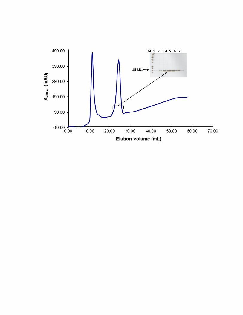
M 1 2 3 4 5 6 7
15 kDa
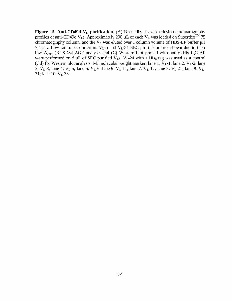
74
Figure 15. Anti-CD49d VL purification. (A) Normalized size exclusion chromatography
profiles of anti-CD49d VLs. Approximately 200 µL of each VL was loaded on SuperdexTM
75
chromatography column, and the VL was eluted over 1 column volume of HBS-EP buffer pH
7.4 at a flow rate of 0.5 mL/min. VL-5 and VL-31 SEC profiles are not shown due to their
low A280. (B) SDS/PAGE analysis and (C) Western blot probed with anti-6xHis IgG-AP
were performed on 5 μL of SEC purified VLs. VL-24 with a His6 tag was used as a control
(Ctl) for Western blot analysis. M: molecular weight marker; lane 1: VL-1; lane 2: VL-2; lane
3: VL-3; lane 4: VL-5; lane 5: VL-6; lane 6: VL-11; lane 7: VL-17; lane 8: VL-21; lane 9: VL-
31; lane 10: VL-33.
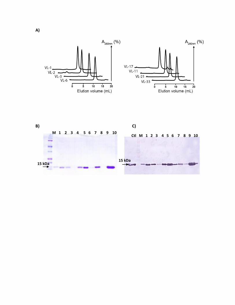
A)
B) C)
M 1 2 3 4 5 6 7 8 9 10 Ctl M 1 2 3 4 5 6 7 8 9 10
15 kDa 15 kDa
75
4.3 Binding analysis of VLs
4.3.1 VL binding to recombinant human CD49d by ELISA
The purified VLs were screened for binding to recombinant human CD49d by ELISA.
The VLs were added at a concentration of 50 µg/mL (~ 4 µM) to wells pre-coated with 0.5
µg (~ 45 nM) of recombinant CD49d in PBS, and bound VLs were detected with anti-6xHis
IgG HRP conjugate as previously described in the Methods section. The absorbance (A450 nm)
of the developed reaction measured at the end of assays is directly proportional to the amount
of VLs bound to CD49d-coated wells. A VL clone with an A450 nm greater than 0.15 was
considered positive. The positive control (~ 33 nM of mouse anti-human CD49d mAb added
to CD49d-coated wells) displayed a high A450 nm of 1.54. Controls receiving wild-type VL-24
treatment, or not receiving VL treatment displayed low A450 nm of 0.14, and 0.12, respectively
(Figure 16, control-1, and -2, respectively). All 10 VL clones showed binding to human
CD49d with A450 nm readings ranging from 0.17 – 1.21, while lower A450 nm readings were
observed for the control 2% casein-coated wells (no immobilized CD49d) ranging from 0.08
– 0.13 (Figure 16). VL-21 and VL-33 showed the highest absorbance readings (A450 nm of
1.22, and 1.20, respectively) among the screened clones. Clone VL-6 was the next one with a
high A450 nm (0.65) with its A450 nm being approximately 43% of that of the positive control.
Clones VL-2, VL-11, and VL-31 had moderate absorbance readings (A450 nm of 0.46, 0.44, and
0.38, respectively), with their A450 nm being less than one third of that of the positive control.
The rest of the clones (VL-1, VL-3, VL-17, and to a lesser extent VL-5) showed lower A450 nm
(ranging from 0.17 – 0.29) and their average A450 nm was about 18% of the positive control.
76
Figure 16. Screening VLs by ELISA for binding to recombinant human CD49d. Wells
were coated with equimolar concentrations of human recombinant CD49d (~ 45 nM) and
blocked in 2% casein. SEC purified VL clones (~ 4 µM) were added to wells and detected
with anti-6xHis IgG-HRP. Grey bars: CD49d-coated wells, treated with tested VLs; white
bars: 2% casein-blocked wells (no immobilized CD49d) treated with tested VLs. Diagonal
striped bars included the positive control: CD49d-coated wells treated with mouse anti-
human CD49d mAb, detected with goat anti-mouse IgG-HRP; control-1: CD49d-coated
wells, treated with wild-type VL-24, detected with anti-6xHis-IgG-HRP; control-2: CD49d-
coated wells, blocked with 2% casein, detected with anti-6xHis-IgG-HRP (no VL added);
control-3: 2% casein-coated wells, treated with anti-CD49d mAb, detected with goat anti-
mouse IgG-HRP; control-4: 2% casein-coated wells, treated with anti-6xHis-IgG-HRP.
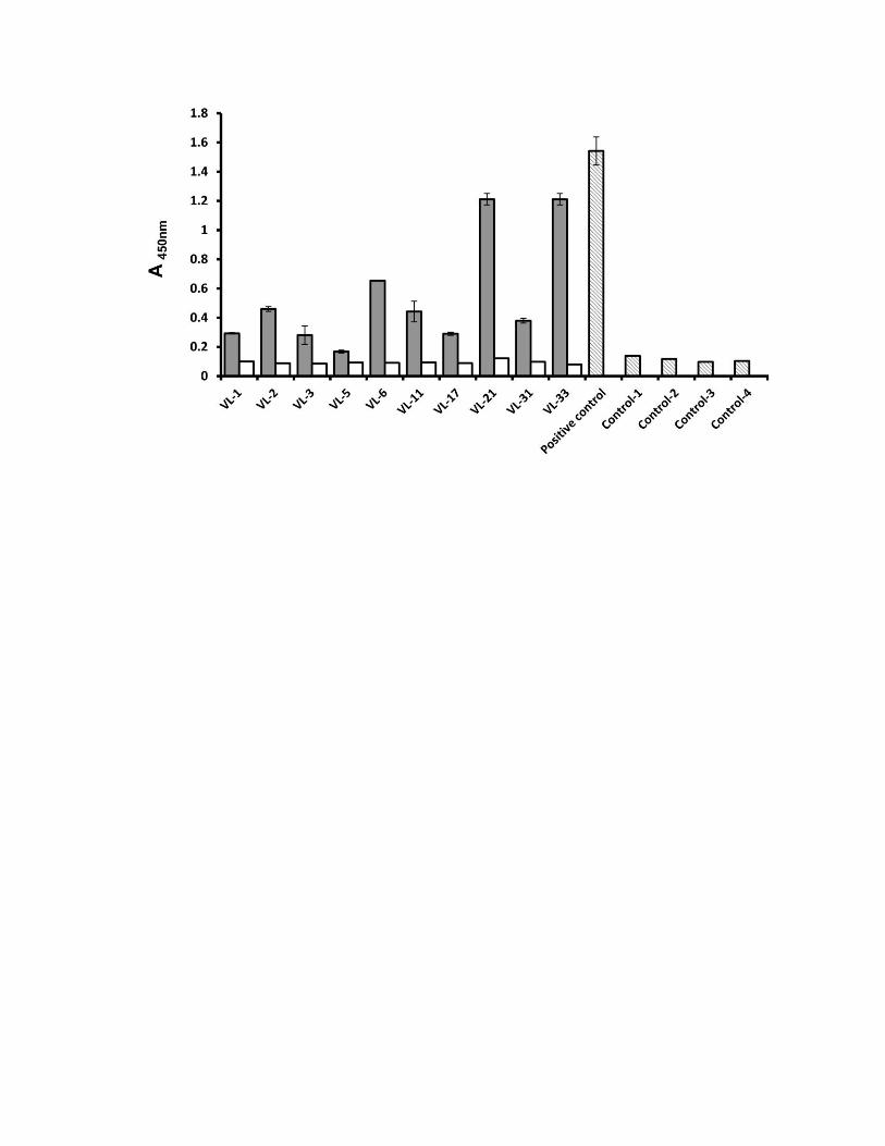
77
4.3.2 VL binding to recombinant human CD49d by surface plasmon resonance
SPR was performed to determine the binding affinity constants of expressed VLs to
CD49d. Equimolar concentrations (200 nM) of SEC purified VLs were injected over CD49d
(3,503 RUs), which was covalently immobilized to the dextran matrix on the sensorchip
channel. Increase in the refractive index (caused by the accumulation of injected VLs over
immobilized-CD49d) was measured in real time and displayed as sensorgrams in which RUs
are plotted against time. Clones VL-11, VL-17, VL-5, and VL-33 showed very low binding to
directly-immobilized-CD49d (ranging from 4 – 14 RUs), while the rest of the clones hardly
showed any binding (ranging from 1 – 3 RUs) (Figure 17A). We suspected if this was
because the immobilized CD49d was not active. To test the activity of the immobilized
CD49d, a control mAb (mouse anti-human CD49d IgG) was injected (100 nM). The IgG
showed strong binding to immobilized CD49d with an approximate KD of 2.4 nM which is in
the expected range for a specific mAb (Figure 17B). However the experimental Rmax value
was 60 RUs which is far lower than the theoretical Rmax of 2,367 RUs. This indicated that
the CD49d surface may not have been active since the experimental Rmax was less than 3%
of the theoretical Rmax.
The loss of protein activity could have been due to the treatment CD49d received
prior to immobilization (dialysis in PBS and immobilization at pH 4.0). To avoid protein
integrity and epitope exposure loss, CD49d was captured while in its provided buffer (pH
7.3) by indirect coupling through anti-c-Myc IgG as described in the Methods section. In
terms of protein activity, the experimental Rmax of the indirectly captured CD49d (187 RUs)
was no different from that obtained from the directly-immobilized-CD49d. The mouse anti-
human CD49d IgG control was injected over the captured CD49d, and again displayed low
78
experimental Rmax (60 RUs) compared to the theoretical Rmax (111,904 RUs) which led us to
the conclusion that this protein is not suitable for SPR experiments. In addition, during
CD49d capturing by anti-c-Myc IgG (at flow cell (4)), the recombinant human CD49d
protein was also injected into the other three flow cell channels of the CM5 sensorchip which
contained the following: immobilized non-relevant IgG (flow cell 1); immobilized CD49d
from the previous direct immobilization trial (flow cell 2); and blocking agent ethanolamine
as a reference channel with no protein being immobilized (flow cell 3) (Figure 17C). The
injected CD49d displayed high RUs in flow cells (1) and (2), indicating non-specific binding.
79
Figure 17. SPR sensorgrams. (A) Human recombinant CD49d was immobilized on a CM5
sensorchip and SEC purified monomeric VLs (VL-2, VL-3, VL-5, VL-6, VL-11, VL-17, 21, VL-
31, and VL-33, shown in different colors) were injected over the surface at a concentration of
200 nM. Sensorgram in red shows binding of control mouse anti-human CD49d mAb (10
nM) to the same surface. (B) Kinetic analysis of the control mouse anti-human CD49d mAb
(100 nM) gave a KD of 2.4 nM and an experimental Rmax of 60 RUs. The actual sensorgram
(black line) was fitted to the 1:1 binding model (red line). (C) Sonsorgram overlays of
CD49d (50 nM) injected over four flow cells of the CM5 sensorchip containing: immobilized
anti-c-Myc IgG (red line), immobilized non-relevant IgG (blue line), immobilized CD49d
from previous SPR trial (green line), and a flow cell channel blocked with ethanolamine as a
reference (grey line).
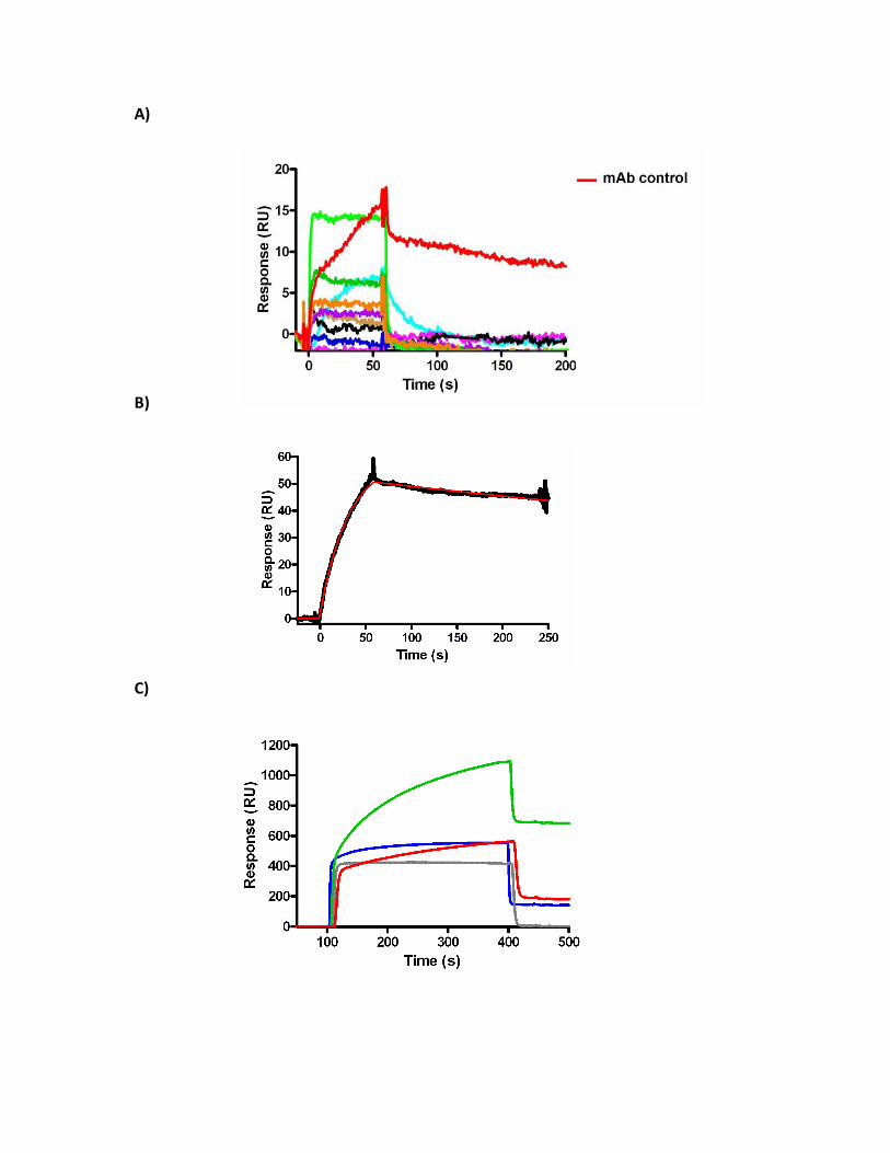
A)
B)
C)
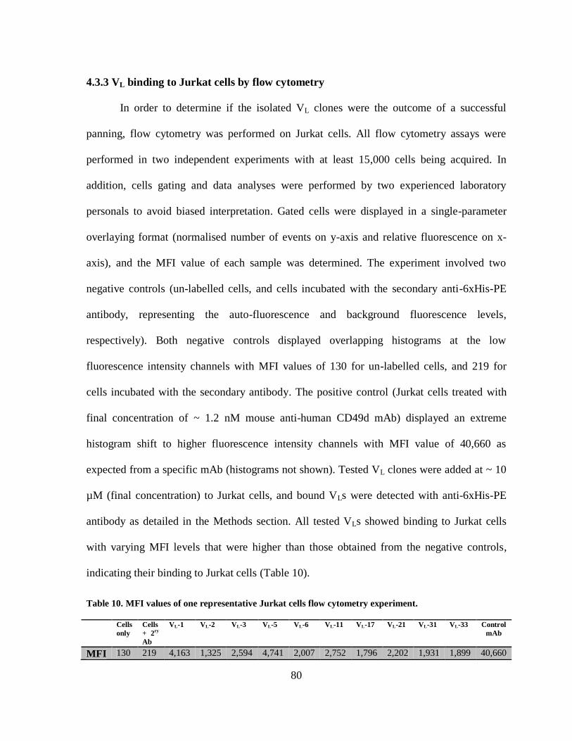
80
4.3.3 VL binding to Jurkat cells by flow cytometry
In order to determine if the isolated VL clones were the outcome of a successful
panning, flow cytometry was performed on Jurkat cells. All flow cytometry assays were
performed in two independent experiments with at least 15,000 cells being acquired. In
addition, cells gating and data analyses were performed by two experienced laboratory
personals to avoid biased interpretation. Gated cells were displayed in a single-parameter
overlaying format (normalised number of events on y-axis and relative fluorescence on x-
axis), and the MFI value of each sample was determined. The experiment involved two
negative controls (un-labelled cells, and cells incubated with the secondary anti-6xHis-PE
antibody, representing the auto-fluorescence and background fluorescence levels,
respectively). Both negative controls displayed overlapping histograms at the low
fluorescence intensity channels with MFI values of 130 for un-labelled cells, and 219 for
cells incubated with the secondary antibody. The positive control (Jurkat cells treated with
final concentration of ~ 1.2 nM mouse anti-human CD49d mAb) displayed an extreme
histogram shift to higher fluorescence intensity channels with MFI value of 40,660 as
expected from a specific mAb (histograms not shown). Tested VL clones were added at ~ 10
µM (final concentration) to Jurkat cells, and bound VLs were detected with anti-6xHis-PE
antibody as detailed in the Methods section. All tested VLs showed binding to Jurkat cells
with varying MFI levels that were higher than those obtained from the negative controls,
indicating their binding to Jurkat cells (Table 10).
Table 10. MFI values of one representative Jurkat cells flow cytometry experiment.
Cells
only
Cells
+ 2ry
Ab
VL-1 VL-2 VL-3 VL-5 VL-6 VL-11 VL-17 VL-21 VL-31 VL-33 Control
mAb
MFI 130 219 4,163 1,325 2,594 4,741 2,007 2,752 1,796 2,202 1,931 1,899 40,660
81
Clones VL-5 and VL-1 displayed the highest MFI levels, followed by clones VL-11,
VL-3, VL-21, VL-6, VL-31, and VL-33, followed by clones VL-17, and VL-2. This is
consistent with the results obtained from ELISA which showed that clones VL-6, VL-11, VL-
21, and VL-33 displayed high binding signals and clones VL-2, and VL-17 displayed
moderate binding levels. Contrarily, other VL clones did not show good agreement between
the results obtained from ELISA and Jurkat cells flow cytometry. For instance, VL-1, VL-5,
and to a lower extent VL-3 displayed high MFI levels in flow cytometry experiment, but a
low A450 nm in ELISA.
Even though all ten VLs showed binding to Jurkat cells, it is not conclusive that these
VL clones are specifically bound to CD49d in particular. In an effort to determine VLs
specificity, we explored the option of performing competitive flow cytometry using mouse
anti-human CD49d IgG as a competitor to the tested VLs. However, inconsistencies in the
obtained results and the requirement of higher concentrations of some VL clones have
altogether hampered the determination of CD49d specificity in this experiment (data not
shown); therefore, two variants of K-562 cell line were employed for this purpose (see
below).
4.3.4 VL screening for CD49d specificity using K-562 cell lines
Because the integrin CD49d is ubiquitously expressed on different mammalian cell
lines (Kim et al., 1994; Puig-Kröger et al., 2000), the erythroleukemic K-562 cell line was
chosen for this set of flow cytometry experiments due to its lack of CD49d expression (van
der Velde-Zimmerman et al., 1996). In addition, the two variants of this cell line: K562-
CD49d (K562-cells stably transfected with human CD49d cDNA) and K562-mock (K562-
82
cells transfected with the empty expression vector, pFneo), have been widely used in several
binding and functional assays as test and control cell lines, respectively (Elices et al., 1990;
Hewish et al., 2000; Parsons et al., 2008). To address the VLs’ specificity towards CD49d,
the MFI levels of VL-treated K562-CD49d cells were compared to those obtained from
K562-mock cells treated with the same VL clone. To start the analysis, acquired cells were
displayed in the forward scatter (FSC) versus side scatter (SSC) plot. The gates were defined
on the basis of the negative and positive controls used for this experiment described in the
Methods section (see Appendix 3 for gating schemes). Gated populations were subsequently
presented in histogram plots and corresponding MFI values were displayed. K562-CD49d
cells treated with the mouse anti-human CD49d IgG antibody displayed MFI ± SEM (n = 2)
of 4,081 ± 209 which was ~ 41-fold greater than what was obtained from K562-mock cells
(MFI ± SEM of 99 ± 1) treated with the same antibody, thus confirming CD49d expression
on K562-CD49d cells and its absence from the control cell line (Table 11; Figure 18A). In
addition, the wild-type VL-24 (~ 10 µM final concentration) was included as a non-relevant
VL control in order to determine the background binding level of the screened VLs (Figure
18A). Both K562-CD49d and K562-mock cells treated with VL-24 displayed overlapping
histograms with MFI ± SEM values of 590 ±110, and 390 ± 170, respectively (Table 11;
Figure 19). The close MFI levels obtained from both cell lines implicated that the VL
scaffold used for generating the library do not bind arbitrarily to CD49d, and that only VL
clones with certain CDRs combination should be able to recognize CD49d-expressing cells.
In addition, the estimated MFI ratio (MFIR; calculated as the MFI ratio of K562-CD49d to
that of K562-mock cells treated with the same VL clone) of cells treated with the wild-type
VL-24 was determined to be 1.5. Therefore, tested VL clones with MFIR of more than 1.5
83
were considered CD49d-specific (see below). Both K-562 cell line variants were treated with
~ 10 µM (final concentration) of each tested VL clone as previously described in the Methods
section.
The analysis revealed that clones VL-1, VL-6, VL-21, and VL-2 displayed the highest
fluorescence intensity levels when incubated with K562-CD49d cells (MFI ± SEM of 2,279
± 1179, 1,254.5 ± 170.5, 1,068 ± 178, and 1,035.5 ± 146.5, respectively), with relatively
lower binding levels when incubated with the control cells (corresponding to MFI ± SEM of
657.5 ± 7.5, 430.5 ± 90.5, 663.5 ± 216.5, and 693 ± 187) (Table 11; Figure 18B; Figure 19).
Similarly, clones VL-17, VL-11 and VL-33 showed moderate binding to K562-CD49d cells
(MFI ± SEM of 921.5 ± 212.5, 848.5 ± 27.5, and 774 ± 124.5, respectively) as opposed to
lower binding levels obtained from the control cells (corresponding to MFI ± SEM of 589 ±
11, 575 ± 125, and 364 ± 46). VL-3 did not display significant difference in fluorescent
intensity between the test and control K-562 cell line variants; in fact, its MFI level was close
to that of the wild-type VL-24 (Table 11; Figure 19). Overall, the pattern of MFIR ratios
obtained from K-562 cells flow cytometry was correlated with the results obtained from
ELISA. In both experiments, the clones VL-6, VL-33, and VL-21 were among the lead VLs in
terms of binding to CD49d (with MFIRs of 2.9, 2.1, and 1.6, respectively). Similarly, clones
VL-2, VL-3, and VL-11 had moderate-to-low binding signals in both ELISA and Jurkat cells
flow cytometry and displayed the lowest MFIRs in K-562 cells flow cytometry experiments
(ranging from 1.4 – 1.5). On the other hand, clone VL-1 (which displayed low absorbance
reading in ELISA) had the highest MFI level in both Jurkat cells and K-562 cells flow
cytometry experiments (with MFIR of 3.5), which suggest the ability of VL-1 to recognize
the native quaternary structure of CD49d. Another observation was that clone VL-17 which
84
displayed low binding signals in both ELISA and Jurkat cells flow cytometry, showed a
comparable MFIR (1.6) to that of VL-21, indicating its specificity.
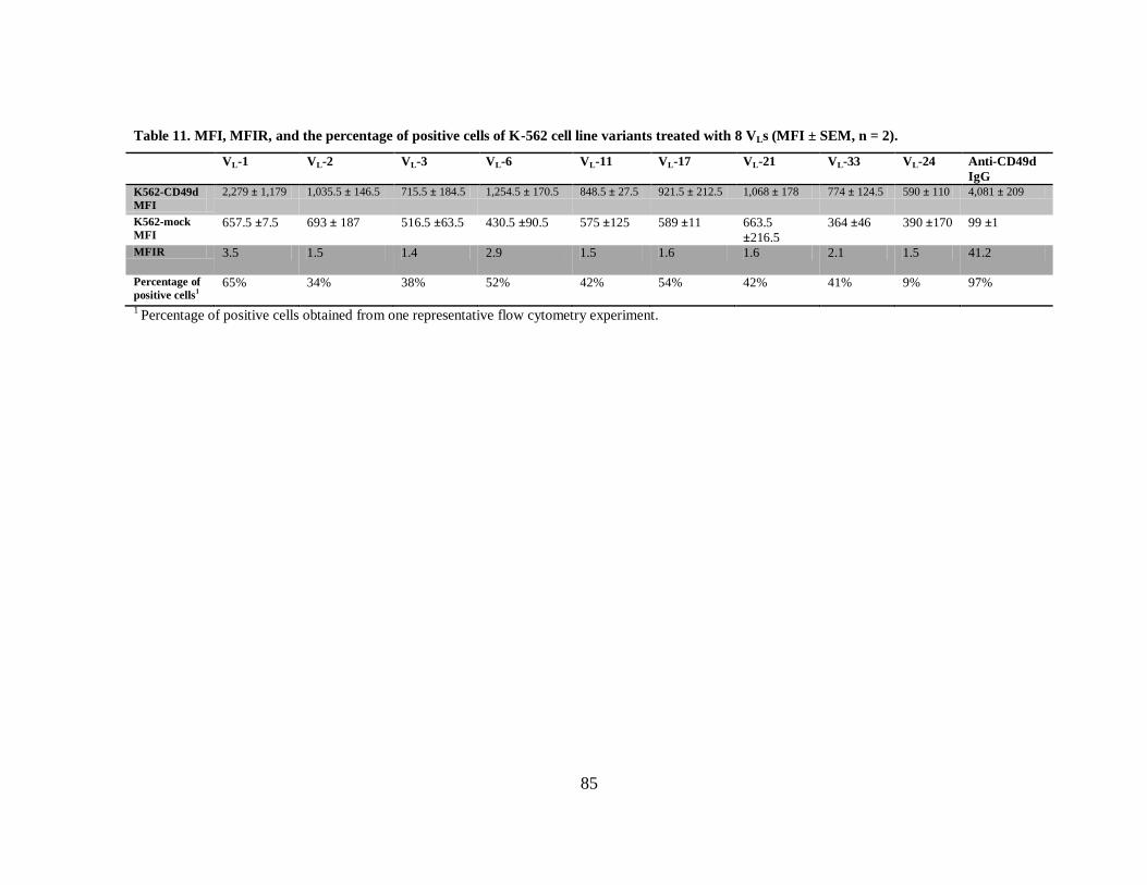
85
Table 11. MFI, MFIR, and the percentage of positive cells of K-562 cell line variants treated with 8 VLs (MFI ± SEM, n = 2).
VL-1 VL-2 VL-3 VL-6 VL-11 VL-17 VL-21 VL-33 VL-24 Anti-CD49d
IgG
K562-CD49d
MFI
2,279 ± 1,179 1,035.5 ± 146.5 715.5 ± 184.5 1,254.5 ± 170.5 848.5 ± 27.5 921.5 ± 212.5 1,068 ± 178 774 ± 124.5 590 ± 110 4,081 ± 209
K562-mock
MFI 657.5 ±7.5 693 ± 187 516.5 ±63.5 430.5 ±90.5 575 ±125 589 ±11 663.5
±216.5
364 ±46 390 ±170 99 ±1
MFIR 3.5 1.5 1.4 2.9 1.5 1.6 1.6 2.1 1.5 41.2
Percentage of
positive cells1
65% 34% 38% 52% 42% 54% 42% 41% 9% 97%
1 Percentage of positive cells obtained from one representative flow cytometry experiment.
86
Figure 18. Determining the specificity of VLs towards CD49d by flow cytometry. (A)
The wild-type VL-24 (~ 10 µM) was used to treat both K562-CD49d (test) and K562-mock
(control) cell lines and were detected with goat anti-6xHis IgG-PE. The mouse anti-human
CD49d IgG (~ 16 nM) was used to treat both cell lines (detected with goat anti-mouse IgG-
PE) to confirm CD49d presence/absence on the test and control cell lines, respectively. (B)
Histograms and MFI values of one representative flow cytometry experiment is shown.
K562-CD49d and K562-mock cells were treated with ~ 10 µM of tested VLs and bound VLs
were detected with goat anti-6xHis IgG-PE. Data is displayed in single parameter overlaying
histograms, the the vertical axis (% of Max) depicts the normalized number of events per
channel, and the horizontal axis (Comp-PE-A) represents relative fluorescence at PE-A
channel.
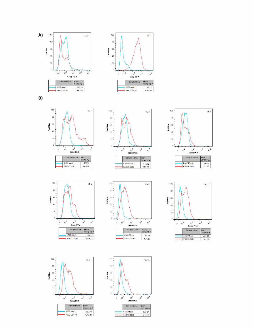
A)
B)

87
Figure 19. VLs show preferential binding to K562-CD49d cells in flow cytometry
experiments. VLs bound to K562-CD49d and K562-mock cell lines were detected with goat
anti-6xHis IgG-PE. Data represents MFI ± SEM of two independent experiments. Grey bars:
K562-CD49d cells treated with VLs (~ 10 µM) or with control antibody (Anti-CD49d IgG);
white bars: K562-mock cells treated with VLs or control antibody (Anti-CD49d IgG).
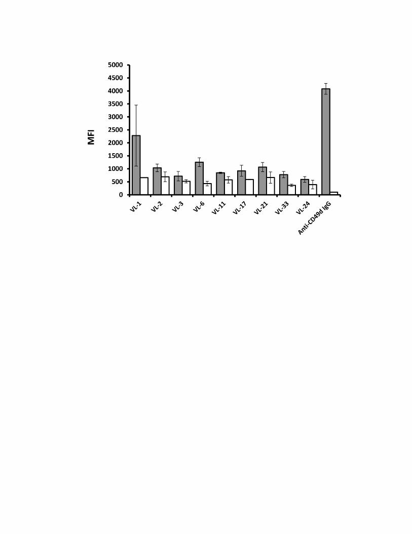
88
Because some tested VLs had weak fluorescence intensity and were not able to shift
the histogram completely to higher fluorescence intensity channels, misleading interpretation
might occur. Therefore, the percentage of positive cells (i.e., cells with bound VLs) was
determined according to Overton’s subtraction method (Overton, 1988). The method will
calculate the percentage of positive cells within gated K562-CD49d cells after background
subtraction (i.e., control cell line treated with the same antibody) automatically using
sophisticated algorithms. As expected, K562-CD49d cells treated with the specific mouse
anti-human CD49d IgG recorded ~ 97% positivity (Table 11). On the contrary, K562-CD49d
cells treated with the wild-type VL-24 showed that ~ 9% of gated cells were positive. The
domains VL-1, VL-6 and VL-17 were among the lead clones in terms of retaining high
percentage of positive cells (~ 65%, 52%, and 54%, respectively), which is in good
agreement with their increased MFIRs. Domains VL-11, VL-21 and VL-33 displayed lower
percentage of positive cells (ranging from 41 – 42%) which is consistent with their lower
MFIRs. Domains VL-2 and VL-3 displayed the least tendency to bind to K562-CD49d cells
as reflected by their low positive percentages (34% and 38%, respectively) and low MFIRs
(Table 11). It should be mention that the computed percentages of positive cells were used as
a rough measure to estimate the VLs’ specificity to CD49d. This is because the percentages
were obtained from one representative flow cytometry experiment and variability in terms of
absolute number of gated cells and/or stained cells may occur, thereby affecting the overall
positive percentages. Taken together, based on MFI, MFIRs, and positive percentage values,
VL-1, VL-6, VL-17, VL-21, and VL-33 were considered as lead CD49d-specific VLs.
89
4.4 hFc-VLs expression and purification
Three of the lead VLs (VL-1, VL-6, and VL-21) were engineered by SOE-PCR to
generate homodimeric Ab fragments. The homodimer consisted of two identical polypeptide
chains each composed of the CD49d-binding VL, fused to human IgG1 hinge region, CH2,
and CH3 (Figure 11B) (attempts to construct the remaining lead VLs failed despite repeated
subcloning and transformation trials). The aim of this engineering was to take advantage of
avidity effects and prolonged serum half-life offered by the Fc (CH2 + CH3) fragment in an
effort to improve the VLs’ therapeutic efficacy. The presence of the leader sequence
(MEFGLSWVFLVAILKGVQC) of human IgG1 (Kopetzki, 2007) prior to the VL coding
sequence allowed for directing the expressed hFc-VL into the secretory pathway of HEK293-
EBNA-1 host cells. Therefore, expressed proteins would be released into the culture
medium, eliminating the need for cell lysis for protein extraction purposes. Given that
engineered hFc-VLs lacked a tag marker, the antibody fragments were purified using protein
G affinity column. This is because the streptococcal protein G (also designated as type III Fc
receptor) is an immunoglobulin-binding protein that is capable of binding to the Fc portion
of human IgG1 with relatively high affinity (Harlow and Lane 1988), thus enabling the
purification of non-tagged hFc-VL proteins from the culture medium. The hFc-VL was
released from protein G column by loading an elution buffer of low pH (pH 2.7). The
concentration of the eluted hFc-VLs was determined by absorbance measurements at A280
using the molar extension coefficient and the theoretical MWs (Table 12).
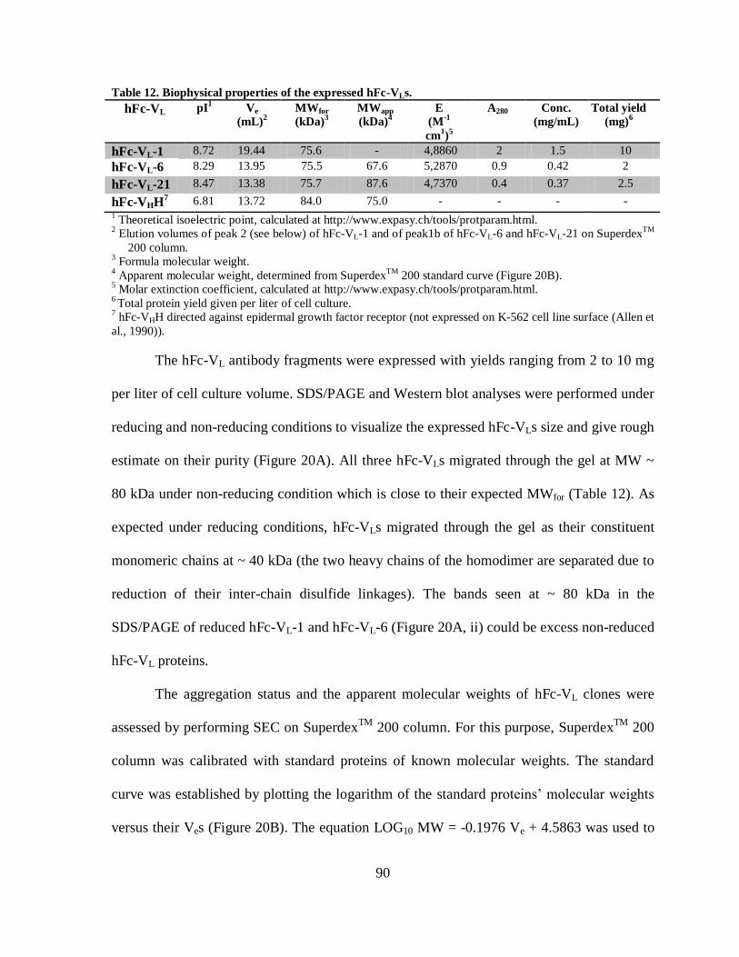
90
Table 12. Biophysical properties of the expressed hFc-VLs.
hFc-VL pI1 Ve
(mL)2
MWfor
(kDa)3
MWapp
(kDa)4
E
(M-1
cm1)
5
A280 Conc.
(mg/mL)
Total yield
(mg)6
hFc-VL-1 8.72 19.44 75.6 - 4,8860 2 1.5 10
hFc-VL-6 8.29 13.95 75.5 67.6 5,2870 0.9 0.42 2
hFc-VL-21 8.47 13.38 75.7 87.6 4,7370 0.4 0.37 2.5
hFc-VHH7 6.81 13.72 84.0 75.0 - - - -
1 Theoretical isoelectric point, calculated at http://www.expasy.ch/tools/protparam.html.
2 Elution volumes of peak 2 (see below) of hFc-VL-1 and of peak1b of hFc-VL-6 and hFc-VL-21 on Superdex
TM
200 column. 3 Formula molecular weight.
4 Apparent molecular weight, determined from Superdex
TM 200 standard curve (Figure 20B).
5 Molar extinction coefficient, calculated at http://www.expasy.ch/tools/protparam.html.
6 Total protein yield given per liter of cell culture.
7 hFc-VHH directed against epidermal growth factor receptor (not expressed on K-562 cell line surface (Allen et
al., 1990)).
The hFc-VL antibody fragments were expressed with yields ranging from 2 to 10 mg
per liter of cell culture volume. SDS/PAGE and Western blot analyses were performed under
reducing and non-reducing conditions to visualize the expressed hFc-VLs size and give rough
estimate on their purity (Figure 20A). All three hFc-VLs migrated through the gel at MW ~
80 kDa under non-reducing condition which is close to their expected MWfor (Table 12). As
expected under reducing conditions, hFc-VLs migrated through the gel as their constituent
monomeric chains at ~ 40 kDa (the two heavy chains of the homodimer are separated due to
reduction of their inter-chain disulfide linkages). The bands seen at ~ 80 kDa in the
SDS/PAGE of reduced hFc-VL-1 and hFc-VL-6 (Figure 20A, ii) could be excess non-reduced
hFc-VL proteins.
The aggregation status and the apparent molecular weights of hFc-VL clones were
assessed by performing SEC on SuperdexTM
200 column. For this purpose, SuperdexTM
200
column was calibrated with standard proteins of known molecular weights. The standard
curve was established by plotting the logarithm of the standard proteins’ molecular weights
versus their Ves (Figure 20B). The equation LOG10 MW = -0.1976 Ve + 4.5863 was used to
91
determine the MWapps of the recombinant hFc-VLs from their corresponding Ves (Figure
20B; Table 12). Both hFc-VL-6 and hFc-VL-21 were eluted mainly as single homodimeric
peaks (designated as 1b) with Ves of 13.95 mL and 13.38 mL, respectively. Thus, their Ves
deviated slightly from the hFc-VHH control which eluted at 13.72 mL (Figure 20C). The
MWapps of hFc-VL-6 and hFc-VL-21 (determined according to peak 1b) were estimated to be
67.6 kDa and 87.6 kDa, respectively, and were close to that of the pre-identified hFc-VHH
(MWapp = 75 kDa). In addition to the main homodimeric peaks of clones hFc-VL-6 and hFc-
VL-21, there were two minor peaks (designated as V0 and 1a) (Figure 20C). Both SEC
chromatograms of hFc-VL-6 and hFc-VL-21 showed that peak 1a was eluted very close to
peak1b with a mean Ve ± SEM of 11.76 mL ± 0.24 mL which corresponded to proteins with
MWapps of 183.03 kDa as determined from the SuperdexTM
200 standard curve. Regardless
of attempts to visualize the eluted protein fractions obtained from peak 1a, no bands were
detected by either Coomassie brilliant blue or silver staining of SDS/PAGE (not shown).
This may be due to the low protein concentrations loaded into the gels which perhaps were
lower than the detection limits of the stains used. Peak V0 of clones hFc-VL-6 and hFc-VL-21
(Ves 7.53 mL, and 7.36 mL, respectively) were eluted at the void volume of the column
which represents molecules of ≥ 600 kDa (Figure 20C). The V0 peak indicated that
engineered hFc-VLs have tendency to form complex structures or protein aggregates in
solution. Contrarily to hFc-VL-6 and hFc-VL-21, the control hFc-VHH displayed a single
symmetrical peak that eluted at the expected Ve for a homodimeric protein (~ 13.72 mL)
with no significant protein being eluted at the void volume of the column (Figure 20C). This
profile is consistent with the VHHs’ property of being aggregation resistant (Muyldermans et
al., 1994).
92
Clone hFc-VL-1 was an odd one. Its chromatogram displayed extremely retarded
elution peaks that eluted mainly at 19.44 mL (designated as peak 2) and 25.03 mL
(designated as peak 3) which corresponded to MWs of 5.56 kDa and 0.44 kDa, respectively
(Figure 20C). In an attempt to correct the elution behaviour of hFc-VL-1, low-salt-
concentration buffer (10 mM Tris-HCl, pH 8.0) was used to both re-equilibrate the column
and dialyse an aliquot of hFc-VL-1 against, and the SEC analysis was repeated. This was
based on the beneficial effect of low ionic strength buffers in controlling the retention
volume of antibodies with basic pIs as reported in one study that experienced similar
problem with recombinant VLs (Schiefner et al., 2011). Additionally, increasing the buffer
pH from 7.5 to 8.0, and bringing it closer to the protein’s theoretical pI would minimize the
chance of protein adsorption to the column beads by electrostatic forces (Stulik et al., 2003).
However, the use of Tris-HCl buffer did not change the SEC profile with respect to peak 2
(peak 2 eluted at 19.28 mL). In addition, this particular clone was expressed in relatively
higher yield compared to the other clones, and formed visible aggregates when stored in PBS
overnight at 4°C. To investigate if the aggregate was mediated by reversible self-association
of the protein (protein aggregate will reversibly dissociate as a response to diluted protein
concentration (Liu et al., 2005)), an aliquote of hFc-VL-1 was diluted by adding 1 mL of
PBS, mixed gently, and stored overnight at 4°C. On the following day, the protein aggregate
did not dissociate. Therefore, the aggregation status and apparent molecular weight of hFc-
VL-1 were not determined due to difficulty in speculating its late elution behavior.
Nonetheless, it is likely that for protein hFc-VL-1, fractions eluted at peak 2 corresponded to
the homodimeric protein (based on the Western blot profile), while those eluted at peak 3
probably represent a degraded protein product.
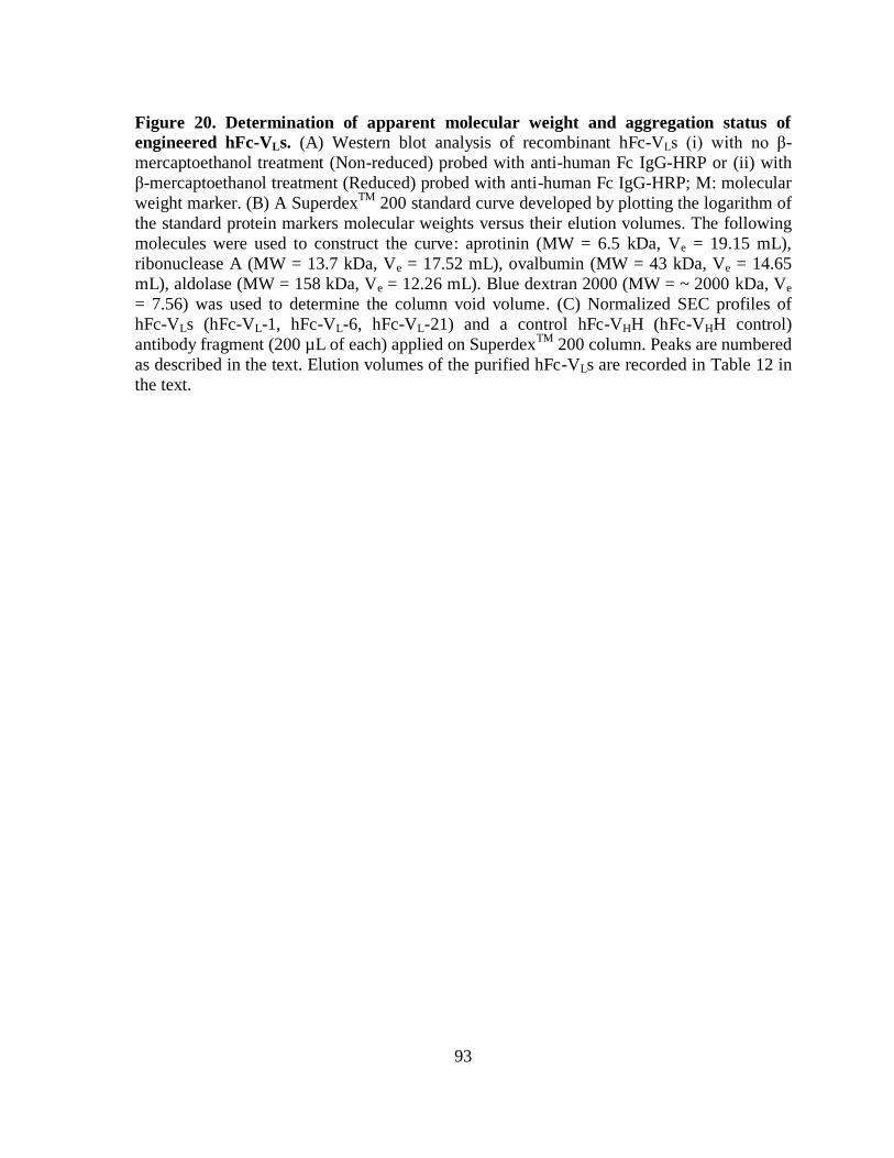
93
Figure 20. Determination of apparent molecular weight and aggregation status of
engineered hFc-VLs. (A) Western blot analysis of recombinant hFc-VLs (i) with no β-
mercaptoethanol treatment (Non-reduced) probed with anti-human Fc IgG-HRP or (ii) with
β-mercaptoethanol treatment (Reduced) probed with anti-human Fc IgG-HRP; M: molecular
weight marker. (B) A SuperdexTM
200 standard curve developed by plotting the logarithm of
the standard protein markers molecular weights versus their elution volumes. The following
molecules were used to construct the curve: aprotinin (MW = 6.5 kDa, Ve = 19.15 mL),
ribonuclease A (MW = 13.7 kDa, Ve = 17.52 mL), ovalbumin (MW = 43 kDa, Ve = 14.65
mL), aldolase (MW = 158 kDa, Ve = 12.26 mL). Blue dextran 2000 (MW = ~ 2000 kDa, Ve
= 7.56) was used to determine the column void volume. (C) Normalized SEC profiles of
hFc-VLs (hFc-VL-1, hFc-VL-6, hFc-VL-21) and a control hFc-VHH (hFc-VHH control)
antibody fragment (200 µL of each) applied on SuperdexTM
200 column. Peaks are numbered
as described in the text. Elution volumes of the purified hFc-VLs are recorded in Table 12 in
the text.
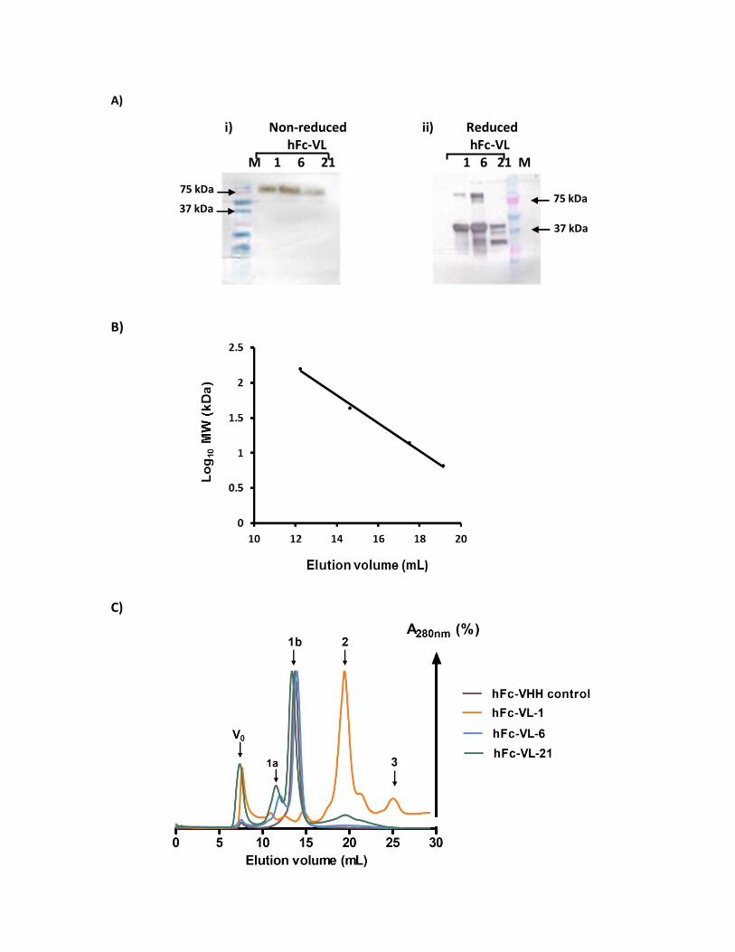
A)
i) Non-reduced ii) Reduced hFc-VL hFc-VL M 1 6 21 1 6 21 M
B)
C)
37 kDa
75 kDa 75 kDa
37 kDa
94
4.5 Engineered hFc-VLs retained specificity to CD49d on K-562 cells
SEC purified hFc-VL-1, hFc-VL-6, and hFc-VL-21 (fractions obtained from peaks 2
for hFc-VL-1, 1b for hFc-VL-6 and hFc-VL-21) were examined by flow cytometry for their
binding capability to CD49d-expressing cells. Both K-562 cell line variants were treated with
~ 1.3 µM (final concentration) of hFc-VL fragments, and binding was detected with goat
anti-human Fc-PE conjugate as detailed in the Methods section. Consistent with the previous
results, the anti-CD49d IgG control (~ 32 nM final concentration) displayed preferential
binding to K562-CD49d cells (MFI ± SEM of 8,298 ± 2) with low background binding to the
control cells (151 ± 9). The non-relevant hFc-VHH displayed binding to both cell lines,
however, with some preference for the test cells (1,320 ± 121 (test cells) vs. (851.5 ± 21.5
(control cells)). Clones hFc-VL-6 and hFc-VL-21 demonstrated CD49d-specificity since they
displayed better binding to K562-CD49d cells (MFI ± SEM of 3,282 ± 50 and 6,684 ± 121,
respectively) than to control cells (corresponding MFI ± SEM of 1,840.5 ± 260.5 and 3,312.5
± 687.5) (Table 13; Figure 21A). Although the background binding to K562-mock cells was
high, the MFIR of the homodimeric hFc-VL-6 (1.8) and hFc-VL-21 (2.0) were higher than
that of the control hFc-VHH (1.6) which indicate their specificity for CD49d. Consistent with
the MFI and MFIR results, the percentage of positive cells within the gated K562-CD49d
cell population was the highest for anti-CD49d IgG control (~ 91%), followed by hFc-VL-21
(~ 63%), and hFc-VL-6 (~ 57%), respectively. Unexpectedly, the control hFc-VHH displayed
a considerable percentage of positive cells as well (~ 41%). Upon applying the statistical
parameter on flow cytometry data files, an overlapping univariant histogram was
automatically plotted in order to display the distribution difference between the test and
control cell lines (Figure 21B). In this display mode, positive cells within the test sample are
95
displayed above the midline of the difference plot and vice versa. The clones hFc-VL-6 and
hFc-VL-21 were able of shifting the histograms of K562-CD49d cells towards high
fluorescence intensity channels with better divergence from the control histogram. On the
other hand, histograms observed for K-562 cell line variants treated with the control hFc-
VHH were less diverged (Figure 21B). The clone hFc-VL-1 showed lack of specificity to
CD49d. Similar MFI values were obtained from both the test (MFI ± SEM of 3,860.5 ±
2,560.5) and control (MFI ± SEM of 3,810 ± 2,690) cell lines. It also had low MFIR (1.0)
and low percentage of positive cells recorded within the gated test cell line population (13%).
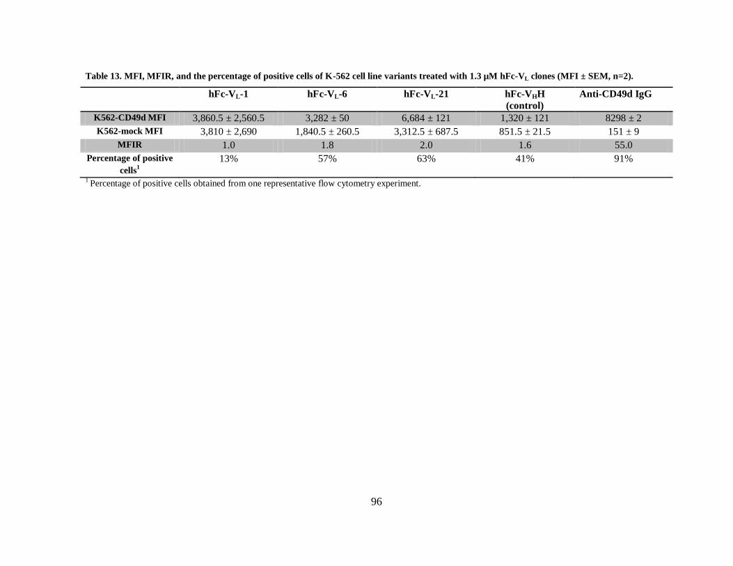
96
Table 13. MFI, MFIR, and the percentage of positive cells of K-562 cell line variants treated with 1.3 µM hFc-VL clones (MFI ± SEM, n=2).
hFc-VL-1 hFc-VL-6 hFc-VL-21 hFc-VHH
(control)
Anti-CD49d IgG
K562-CD49d MFI 3,860.5 ± 2,560.5 3,282 ± 50 6,684 ± 121 1,320 ± 121 8298 ± 2
K562-mock MFI 3,810 ± 2,690 1,840.5 ± 260.5 3,312.5 ± 687.5 851.5 ± 21.5 151 ± 9
MFIR 1.0 1.8 2.0 1.6 55.0
Percentage of positive
cells1
13% 57% 63% 41% 91%
1 Percentage of positive cells obtained from one representative flow cytometry experiment.
97
Figure 21. Binding of hFc-VLs to K-562 cell line variants measured by flow cytometry. (A) Each hFc-VL (1.3 µM) was incubated with either K562-CD49d or K562-mock cell lines.
Bound hFc-VLs were detected with goat anti-human Fc IgG-PE and measured by flow
cytometry. Grey bars: K562-CD49d cells treated with VLs or control antibody (Anti-CD49d
IgG); white bars: K562-mock cells treated with VLs or the control antibody. Data represents
MFI ± SEM of two independent experiments. (B) Histograms showing the distribution, and
the percentage of positive cells (positive %) detected within the gated K562-CD49d cell
population. K-562 cell line variants treated with the control IgG were detected with goat anti-
mouse IgG-PE. Green histogram: difference histogram; shaded histogram: K562-mock cell
line; clear histogram: K562-CD49d cell line.
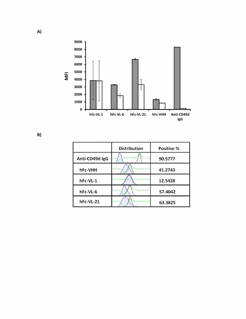
A)
B)
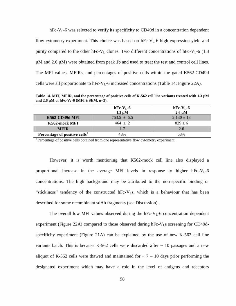
98
hFc-VL-6 was selected to verify its specificity to CD49d in a concentration dependent
flow cytometry experiment. This choice was based on hFc-VL-6 high expression yield and
purity compared to the other hFc-VL clones. Two different concentrations of hFc-VL-6 (1.3
µM and 2.6 µM) were obtained from peak 1b and used to treat the test and control cell lines.
The MFI values, MFIRs, and percentages of positive cells within the gated K562-CD49d
cells were all proportionate to hFc-VL-6 increased concentrations (Table 14; Figure 22A).
Table 14. MFI, MFIR, and the percentage of positive cells of K-562 cell line variants treated with 1.3 µM
and 2.6 µM of hFc-VL-6 (MFI ± SEM, n=2).
hFc-VL-6 1.3 µM
hFc-VL-6 2.6 µM
K562-CD49d MFI 763.5 ± 6.5 2,130 ± 13
K562-mock MFI 464 ± 2 829 ± 6
MFIR 1.7 2.6
Percentage of positive cells1 48% 63%
1 Percentage of positive cells obtained from one representative flow cytometry experiment.
However, it is worth mentioning that K562-mock cell line also displayed a
proportional increase in the average MFI levels in response to higher hFc-VL-6
concentrations. The high background may be attributed to the non-specific binding or
“stickiness” tendency of the constructed hFc-VLs, which is a behaviour that has been
described for some recombinant sdAb fragments (see Discussion).
The overall low MFI values observed during the hFc-VL-6 concentration dependent
experiment (Figure 22A) compared to those observed during hFc-VLs screening for CD49d-
specificity experiment (Figure 21A) can be explained by the use of new K-562 cell line
variants batch. This is because K-562 cells were discarded after ~ 10 passages and a new
aliquot of K-562 cells were thawed and maintained for ~ 7 – 10 days prior performing the
designated experiment which may have a role in the level of antigens and receptors
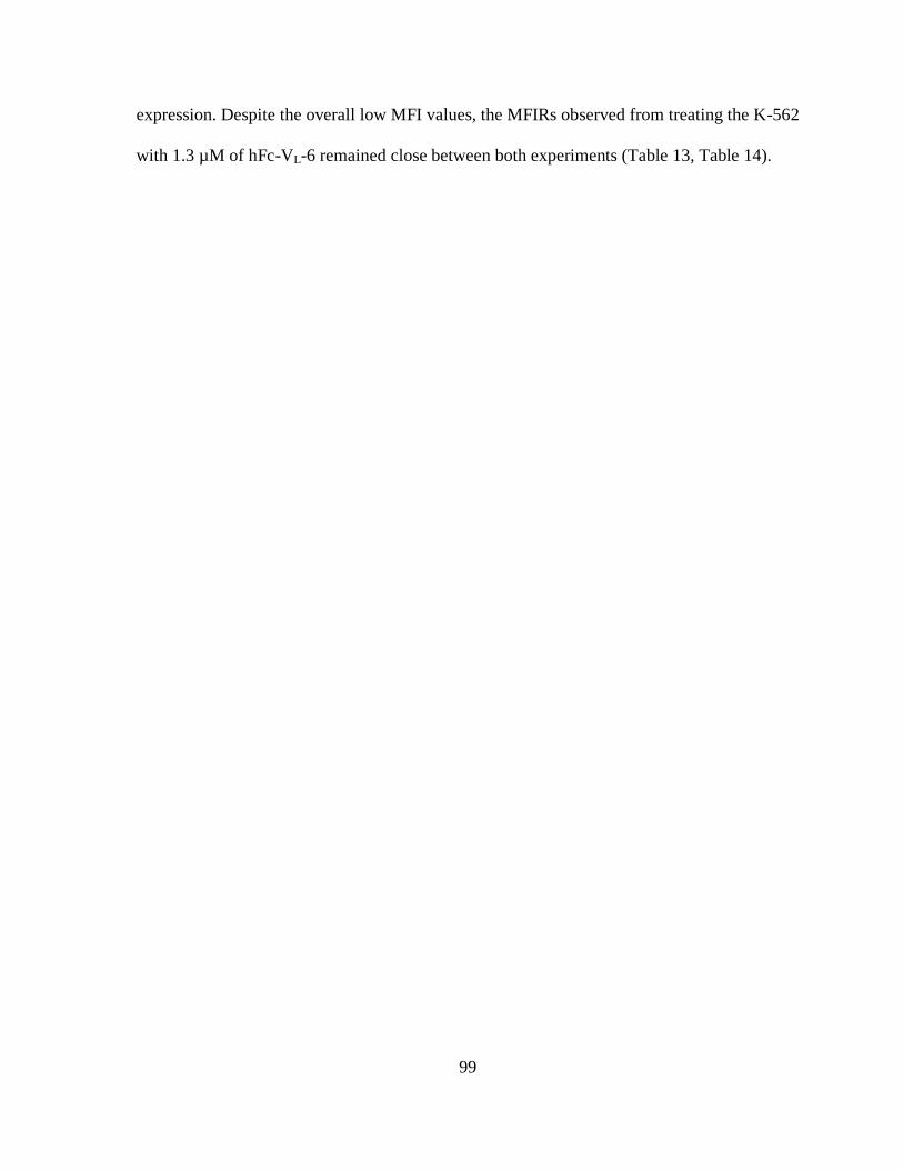
99
expression. Despite the overall low MFI values, the MFIRs observed from treating the K-562
with 1.3 µM of hFc-VL-6 remained close between both experiments (Table 13, Table 14).
100
Figure 22. Concentration dependent binding of hFc-VL-6 measured by flow cytometry.
(A) hFc-VL-6 was incubated with K-562 cell line variants at two different concentrations (1.3
μM and 2.6 μM) in a total volume of 200 µL of cell suspension. Bound hFc-VL-6 was
detected with goat anti-human Fc IgG-PE and measured by flow cytometry. Grey bars:
K562-CD49d cells treated with the VL; white bars: K562-mock cells treated with the VL.
Data represents MFI ± SEM of two independent experiments. (B) Overlaying histograms and
computed percentage (positive %) of positive cells recorded within K562-CD49d cells
treated with 1.3 μM or 2.6 μM of hFc-VL-6. The upper panel depicts the same anti-CD49d
IgG control used in Figure 21 to visualize the maximum fluorescence intensity and
divergence of saturated K562-CD49d cells. Green histogram: difference histogram; tinted
histogram: K562-mock cell line; clear histogram: K562-CD49d cell line.
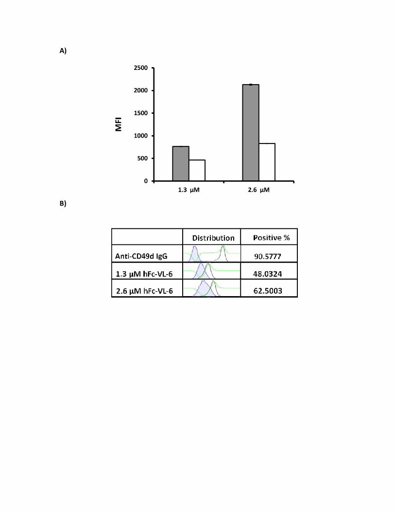
A)
B)
100
5. Discussion
MS is a chronic immune-inflammatory disorder characterized by the recruitment of
activated immune cells toward the CNS, thus leading to demyelination and neuronal damage
(Chiaravalloti and DeLuca, 2008). The disease is incurable, and 74% of patients with MS are
treated with disease-modifying therapies (DMTs) which aim at hindering the disease
progression (Karampampa et al., 2012). CD49d (α-subunit of integrin VLA-4) is an adhesion
molecule expressed on most immune cells with a critical role in mediating immune cells
adhesion and migration toward inflammatory sites (Hynes, 2002; Hogg et al., 2003).
Different studies showed that anti-CD49d mAbs have a clear-cut beneficial effect in
reducing the relapse frequency and MRI activity in patients with MS (Miller et al., 2003;
Polman et al., 2006). The potency of anti-CD49d mAbs is believed to be a consequence of
blocking the adherence of immune cells to the inflamed BBB, thereby limiting their
infiltration toward the CNS and reducing the associated inflammatory phenomenon. In
addition, cerebrospinal fluid samples obtained from patients with MS treated with anti-
CD49d mAbs showed a profound decrease in immune cell counts (Stuve et al., 2006). This
suppression was found to persist for at least 6 months after anti-CD49d mAb therapy
discontinuation, indicating that the half-life of this mAb is longer than what has been
predicted (i.e., 11 days) (Stuve et al., 2007). Although this feature may be beneficial for MS
treatment, the prolonged blockage of CD49d hampers the physiological functions mediated
by this receptor, leading to undesirable side effects (Gonzalez-Amaro et al., 2005).
Given that different epitopes of CD49d are involved in the multi-step extravasation of
activated immune cells toward the CNS (Pulido, et al., 1991), I suggest that a multi-pronged
strategy targeting CD49d by small molecular weight antibody fragments would provide an
101
efficacious alternative to the available conventional mAb therapeutic. The developed
antibody fragments (VLs and hFc-VLs) are proposed to lower the required therapeutic dose,
maintain longer remission periods, and have less advent effects.
5.1 Selection efficiency of anti-CD49d VLs
In this work, a repertoire of VL antibody fragments was constructed on the basis of
naturally occurring human VL scaffold, and was displayed in a multivalent format on the
surface of phage particles (Hussack et al., 2012). In addition to its desired biophysical
properties, the constructed VL library and the engineered hFc-VL format are predicted to
reduce the side effects associated with therapeutic anti-CD49d humanized antibodies. For
instance, it is predicted that the elicitation of neutralizing antibodies and the increased risk of
acquiring PML infection are going to be lower due to the antibody fragments’ smaller size
and feasibility of engineering. Such features would reduce the undesirable side effects
resulting from the prolonged blockage of the physiologically relevant integrin by providing
Ab fragments with the desired affinity to CD49d and the optimal serum half-life.
Anti-CD49d VLs were isolated from the library by panning against Jurkat T-cell line.
A significant advantage of whole-cell panning approach is the ability to isolate phage
particles capable of recognizing the extracellular domain of a protein target while preserved
in its natural context as a membrane-embedded protein. However, a main drawback
associated with this method is that the protein target maybe expressed in low densities, thus
reducing the likelihood of isolating target-specific phage binders. In addition, a simple
elution method by extreme pH may interfere with the library enrichment for CD49d-specific
binders, thus yielding a mixture of phage particles with no clear specificity (Huang et al.,
2012). However, the panning and elution methods applied here seem to effectively reduce
102
the drawbacks referred to earlier. For instance, Jurkat cells were validated for CD49d
expression by anti-CD49d mAb titration in flow cytometry experiment. The titration
revealed that a final concentration of 1.2 nM of anti-CD49d mAb was sufficient to saturate
CD49d expressed on Jurkat cells, and that CD49d can be detected at concentration as low as
276 pM of anti-CD49d mAb (not shown). Furthermore, our competitive elution protocol
included the addition of highly saturating concentration of anti-CD49d mAb (13 µM) to
Jurkat cells pre-incubated with the phage library. Such strategy would allow the elution of
CD49d-bound phage while minimizing the chance of disturbing phage particles bound to
other cell-surface proteins. Although this method may promote the isolation of phage clones
with low affinity to CD49d, prolonged washes with surfactant-containing buffer before
elution would improve the selection for clones with higher affinities.
The VL phage-display library was subjected to four rounds of panning, and the phage
titers were progressively tracked in all rounds. An increase in the phage recovery efficiency
was observed at the three initial rounds of selection, while lower recovery efficiency was
observed at the fourth round. This pattern is frequently seen with phage library panning, and
indicates – in some cases – that a stringent selection for phage specific clones had occur
compared to the other rounds (Porto et al., 2011; Paradis-Bleau et al., 2008). In addition,
lower recovery efficiency may be attributed to the growth rate differences between phage
clones in terms of assembly or export during phage propagation and amplification in the
bacterial host (Derda et al., 2011). This problem is more evident in libraries screened against
whole cells which possess different binding sites. In such cases, the library diversity and
enrichment are more likely to be biased towards phage clones with efficient propagation
regardless of their specificity or affinity; thereby hindering the enrichment of clones with
103
specificity or affinity advantages (Kuzmicheva et al., 2009). Another possible reason for the
low recovery efficiency is the loss of phage infectivity due to altered pIII or F-pilus display
on the surface of the phage particle or E.coli cells, respectively. In addition, technical
handling during phage purification (which included phage precipitation and multiple
filtration steps) may have an influence in phage loss. A more effective strategy for reducing
the chance of selecting aberrant clones would be through re-using the same phage eluent
directly for the second round of panning while skipping the phage amplification step;
however, such strategy may require the use of highly diverse phage libraries (Hoogenboom
et al., 1998).
A total of 12 unique VL sequences with varying frequencies of occurrence (ranging
from 30% – 2%) were retrieved after the fourth round of panning. Alignment of the predicted
amino acid sequences revealed an over-presentation of wild-type CDRs and wild-type amino
acid residues among the identified VLs (Table 8). The wild-type amino acid sequence of
CDR1 was retained by 6 VL clones, wild-type CDR1 and CDR2 were retained by 2 VL
clones, while the wild-type CDR3 was retained by the most enriched VL clone (VL-1). The
wild-type residues S28, Y32, N34, A50, and L54 were almost conserved among the
sequenced clones. Similarly, an over-presentation of the unique amino acid residues H53,
N55, and R91 was observed.
At the library level, the biased over-presentation of wild-type CDRs and amino acid
residues can be attributed to the better annealing efficiencies of the mutagenesis primers used
during library construction which compromised the library diversity (Hussack et al., 2012).
This perception is supported by the over-presentation of S28, Y32, and N34 residues located
within CDR1 of all sequenced clones (except for VL-1) encoded by the wild-type codons
104
AGC, TAT and AAT, respectively. Furthermore, the observed biased enrichment for wild-
type CDR residues can also be attributed to the incompatibility between the randomized
CDRs and the host stability or the phage replication machinery (Rodi et al., 2002).
Nonetheless, it is also possible that the VL phage library did not undergo selection for CD49d
affinity only, but for stability as well. In this case, a VL clone with wild-type amino acid
residues is more stable, and if it has high enough affinity it will be selected. This speculation
is evidenced by the over-presentation of some VL clones that retained wild-type amino acid
residues and low binding to CD49d (e.g., VL-3). In addition, such clonal variation is very
common and has been observed even between closely related amino acid sequences
(McCafferty, 1996).
5.2 Expression and SEC analysis of anti-CD49d VLs
Of the 12 isolated VLs, 10 were successfully subcloned and expressed in bacterial
system with yields ranging from 0.4 mg/L to 31.6 mg/L of bacterial culture. The variability
observed in expression yields may suggest that the expression conditions (e.g., temperature,
medium ingredients, or aeration) should be optimized for some clones. In addition, it is
widely accepted that certain amino acids of the VL domain may lower the overall folding
efficiency and stability, and thus contribute to lower production yields (Ewert et al., 2003).
This is supported by a significant number of studies which succeeded in identifying key
positions within the antibody’s variable domains that retained critical influence on the
domains’ production yields (Knappik et al., 1995; Forsberg et al., 1997; Kipriyanov et al.,
1997; Nieba et al., 1997). All 10 VLs displayed non-aggregating profiles by SEC analysis
and were eluted within the range expected for monomeric sdAbs. Although the formula
molecular weights of the 10 VL domains were similar (~ 13.8 kDa), their estimated apparent
105
molecular weights (based on their elution volumes obtained from SEC analysis) ranged from
10.7 – 15.7 kDa. Eventually, this can be attributed to the VLs amino acid sequence
composition, and since variation was only in CDRs it come down to the CDRs sequences.
For example, VL-6 and VL-21 possessed similar formula MWs of 13.9 and 14.0, respectively;
while their apparent MWs differed to a considerable extent (10.7, and 15.5, respectively). It
is possible that VL domains with estimated low MWs were relatively rich in small amino
acid residues. For instance, VL-6 possessed one Ala, and two Ser residues (small amino
acids) in CDR3, while VL-21 possessed 3 Leu residues (large hydrophobic residues). In
addition, if these folded domains expose hydrophobic residues to the solvent phase, they may
induce non-specific interactions with the column matrix, thereby resulting in their late
elution and smaller MWs (Ewert et al., 2003; Barthelemy et al., 2008). If that was the case, it
also may explain their “stickiness” and high background binding when homodimerized in the
context of Fc in flow cytometry (see below).
5.3 Binding analyses of anti-CD49d VLs
Initially, the 10 expressed VL clones were screened for human recombinant CD49d
binding in standard ELISA. Based on absorbance readings, all were capable of recognizing
immobilized CD49d with different strength. VL-21, VL-33, and VL-6, in order, demonstrated
the highest levels of binding followed by clones VL-11, VL-2, and VL-31 with intermediate
binding capabilities. The remaining VLs showed low binding. Similarly, all clones retained
binding to Jurkat cells with variable MFIs (clones VL-5, VL-1, VL-11, VL-3, and VL-21 were
among the lead binders). Although all VLs bound to CD49d in ELISA and Jurkat cells flow
cytometry, only a subpopulation of the screened VL domains retained binding to the
conformational CD49d in K-562 cells flow cytometry experiments. The clones VL-1, VL-6,
106
VL-17, VL-21, VL-33, VL-11, and VL-2 were considered as lead binders based on their
ELISA results and the increased MFI and percentage of positive cells. Contrarily VL-3
displayed low absorbance reading in ELISA and a comparable MFIR to the wild type VL-24.
The discrepancy in the VLs binding results between ELISA and K-562 cells flow
cytometry may be due to differences between the two experiments in terms of sample
preparation and antigen nature. For instance, the direct immobilization of CD49d (a trans-
membrane protein, MW ~ 150 kDa) on plastic wells by passive adsorption may lead to the
exposure of heterogeneous antigen surface among coated wells. In addition, the proportion of
CD49d that retained its native structure in ELISA, and the density of available and accessible
epitopes may differ between the two experiments. Such variability may have a role in the
inability of VL-1 and VL-17 to retain CD49d binding in ELISA while being good binders in
K-562 cells flow cytometry experiments.
Although ELISA was performed once in duplicate, the domains VL-6, VL-21, and VL-
33 were among the lead CD49d-binders in both ELISA and K-562 cells flow cytometry
experiments. According to the predicted amino acid sequence alignment, these clones shared
wild-type CDR1, the domains VL-6 and VL-33 shared the same unique over-presented
CDR2, but all three varied in their CDR3 amino acid sequences. Contrarily, the domain VL-1
possessed unique CDR1 and CDR2 amino acid sequences but a wild-type CDR3. Other than
the previously discussed possibilities of wild-type CDRs over-presentation, the notable over-
presentation of wild-type CDR1 among binding VLs may also suggest the clones’
dependency – to some extent – on CDR1 for CD49d recognition. A similar assumption was
previously suggested based on the participation of CDR1 of camelid VHH in antigen binding
along with CDR3 (Vu et al., 1997; Decanniere et al., 1999). Interestingly, the domain VL-3
107
(harboring wild-type CDR1) did not confer good binding to CD49d in K-562 cells flow
cytometry experiments, which sought further investigation to validate the role of CDR1 in
CD49d recognition. The exploration of wild-type CDRs impact on the VLs’ affinity towards
CD49d by SPR experiments was hampered due to technical difficulties associated with
CD49d nature.
5.4 Engineering homodimeric anti-CD49d hFc-VLs
Isolated VLs were validated for CD49d recognition by ELISA and flow cytometry
experiments. Due to their isolation from a synthetic library, identified VLs are speculated to
have low affinities towards CD49d. In addition, their low MW and lack of FcRn binding
would reduce their in vivo persistence (Jiang et al., 2011), and thus limiting their therapeutic
potential as CD49d short-term blocking agents. Therefore, the desirable traits of
conventional IgG were transferred into a smaller assembly by fusing the human IgG1 Fc
fragment to CD49d-specific VLs. The developed hFc-VL constructs (average MW ~ 78 kDa)
combined the advantageous properties of IgGs (long half-life and avidity) (Molema et al.,
2000) with those of sdAbs (desirable biophysical properties) (Scheuplein et al., 2010; Niu et
al., 2012). In addition, the small MW of these constructs would lower the required molar
therapeutic regiment and their fully human nature would reduce their potential
immunogenicity. In fact, these constructs mimic the structure of naturally occurring heavy-
chain antibodies which represent a major fraction of circulating functional antibodies in
camelids (Hamers-Casterman et al., 1993). The hFc-VL fragments were generated by splice
overlap extension PCR to yield a final DNA construct coding, in order, for CD49d-binding
VL, hinge, CH2, and CH3 domains. The DNA constructs were expressed in HEK293-EBNA-1
cells, and the expressed hFc-VL antibody fragments are expected to homodimerize
108
spontaneously by the disulfide bridges formed at the hinge region and CH3-CH3 non-covalent
interactions (Zhang et al., 2008). Because HEK293-EBNA-1 cells are of human origin,
glycosylation patterns of hFc-VLs are unlikely to induce immunogenicity. In addition,
glycosylation patterns are known to be critical for applications where acquiring antibody
effector functions are desirable (Abès and Teillaud, 2010). As the hFc-VLs are strictly aimed
for receptor recognition, the glycosylation patterns of the expressed antibody fragments
become less of a concern.
The three engineered hFc-VLs were expressed in yields ranging from 2 to 10 mg/L of
cell culture which were sufficient for our purposes here. However, more efficient and
manufacturing technologies should be applied in order to meet the expected demands for
therapeutics production (multiple g/L). For instant, improvement in expression vectors,
media composition, and mutagenesis can be applied to generate high expressing clones. In
addition, culture conditions optimization can also be applied for large-scale antibody
production through microbial fermentation systems (for VLs production), or by mammalian
cells bioreactors (for hFc-VLs production) (Langer, 2009). SEC analysis was performed
using SuperdexTM
200 column to determine the antibody fragments MWapps and aggregation
status. The clones hFc-VL-6 and hFc-VL-21 eluted mainly as single peaks that slightly
deviated from the pre-identified hFc-VHH control. The average MWapp of these constructs
was also in good agreement with their average predicted MWs (MWfor ~ 76 kDa). The
Western blot of the hFc-VL constructs revealed single pure bands that migrated through the
gel at ~ 80 kDa (non-reducing), and at ~ 40 kDa (reducing). It is of note to mention that the
proteins migration was a little bit greater than estimated. This can be attributed to the slow
migration of glycosylated recombinant antibodies through SDS/PAGE (Wu et al., 2001).
109
This is because in glycosylated proteins the carbohydrates do not bind to SDS (found in the
loading buffer) which will reduce the overall net charge-to-mass ratio, therefore decreasing
the protein’s mobility when migrating through the gel (Hames and Rickwood, 1990;
Westermeier, 1997).
Although the fusion constructs were produced mainly as intact homodimeric units,
they also formed aggregates in significant proportions. In order to address the non-
aggregating nature of the VLs when expressed in a monomeric form but not when
homodimerized in the context of Fc, a number of intrinsic factors (i.e., factors related to the
protein’s composition) (Mahler et al., 2009 and references therein), and extrinsic factors (i.e.,
the environmental conditions surrounding the protein) (Jungbauer and Kaar, 2007 and
references therein) should be considered. Moreover, it is possible that the VLs aggregation
tendency diminished during SEC analysis as the purified domain was diluted during the run,
while the aggregation tendency was amplified when two copies of the same antibody
fragment were assembled into one molecule of a larger format (Schaefer et al., 2010). It is
worth mentioning that the formation of dimeric or aggregating structures is a phenomenon
observed in pathological conditions of free light chain Ig fragments of the Bence-Jones type
(Stevens and Argon, 1999; Sikkink and Ramirez-Alvarado, 2008). Such aggregates are
speculated to form by the influence of the protein’s intrinsic factors such as the
intermolecular electrostatic forces, hydrophobic interaction, and non-covalent bonds exerted
by the amino acid sequence comprising the VL fragments of the Ig light chain (Kaplan et al.,
2011). The ability to express hFc-VL antibody fragments with lower aggregation tendencies
can be achieved through a number of different techniques or modifications directed towards
the VL amino acid sequence which is the root of the problem. Therefore, it is very likely that
110
when we remedy the VL domain structure (in terms of amino acid sequence and resistance to
aggregation), we will probably remedy the whole construct (see recommendations and future
direction).
An important unresolved issue was determining the aggregation status of hFc-VL-1.
Although all hFc-VL constructs migrated evenly through SDS/PAGE gel in reducing and
non-reducing conditions – which eliminates the chance of truncated protein expression –, the
clone hFc-VL-1 displayed a late elution profile during SEC analysis. In addition, the late
elution was not corrected by changing the pH and ionic strength of the running buffer.
Proteins of similar late elution profile are not unique, and different studies have reported
proteins under investigation to be non-specifically bound to the column matrix. For instance,
Barthelemy and colleges demonstrated that VHs with similar late elution profiles exhibited
good thermostability upon temperature-induced denaturation; however, possessed
irreversible unfolding behaviour which is an indicative of aggregation prone domains
(Barthelemy et al., 2008). Another case was experienced by Nasreen et al. (2006) in which
the studied protein (apolipoprotein D, associated with high-density lipoprotein) was
consistently bound to the column matrix regardless of the different buffer conditions tested.
According to the predicted three dimensional model of apolipoprotein D, the problem was
attributed to the presence of hydrophobic side chains which influenced the protein interaction
with the mobile phase during SEC analysis. The principle of separation in SEC relies on the
hydrodynamic volume (i.e., the solute physico-chemical interaction during the flow in
solution) rather than the molecular mass of the tested protein (Stulik et al., 2003). In this
sense, it is possible that due to misfolding of hFc-VL-1 during protein expression,
hydrophobic regions were exposed to the mobile phase. This exposure may lead to
111
interactions between the misfolded protein and the mobile and/or stationary phase of the
column which affected the overall hydrodynamic volume of the protein. The MW and
aggregation status of the expressed proteins can be determined through technologies
measuring the scattered light at additional angles (multi-angle light scattering or SEC-
MALS) for a more precise MW determination (Beirne et al., 2011). In addition, other
column-free analytical methods such as field flow fractionation and analytical
ultracentrifugation can be utilized for this purpose (reviewed in Liu et al., 2006). The latter
methods lack stationary phase that may influence the separation process, and covers a wide
range of molecular sizes. Because these methods rely on rigorous theoretical bases, they
provide more accurate estimations on the proteins’ shape, aggregation status, size, and
quantity (Liu et al., 2006). However, it is worth mentioning that the mean of a protein MW
or aggregation quantification may vary from one method to the other due performance and
data evaluation differences among these methods (Mahler et al., 2009).
5.5 Anti-CD49d hFc-VLs binding to CD49d-expressing cells
Consistent with their parental VLs, gel purified hFc-VL-6 and hFc-VL-21 conferred
binding to K562-CD49d cells as opposed to the negative control cell line in flow cytometry
experiments. The clones displayed 3-fold increase in the MFI ± SEM levels between the test
and control cell lines, as well an increase in the percentage of positive cells. Given that K-
562 cell line variants treated with 1.3 µM of the hFc-VL constructs displayed comparable
binding to when the cells were treated with 10 µM of parental VLs, the enhancement in
binding can be attributed to the avidity effects conferred by the constructs bivalency.
However, a more systematic analysis than the one undertook here is required to prove this.
This is because the monomeric VLs were always detected by a different antibody than that of
112
hFc-VLs (i.e., VLs were detected with anti-6xHis IgG-PE while hFc-VLs were detected with
anti-human Fc IgG-PE). In addition, definite data regarding the amount of labeling
molecules presented per each secondary antibody molecule (fluorophore : protein) may not
always be available. The option of labelling the antibodies directly using fluorescein dyes at
specific residues (e.g., lysine) can be problematic due to variable amino acid composition of
the two antibody variants – and between examined clones – requiring cautious data analysis
and experimental reproducibility. In addition, the labelling may lead to heterogeneous
population of labeled antibody fragments, in which some may lose binding to the antigen
especially if it the label resides in the CDRs.
The clone hFc-VL-1 failed to recognize CD49d specifically in this set of flow
cytometry experiments. Instead, hFc-VL-1 displayed high MFI levels when incubated with
the test cell line, and because it also displayed high background binding to the control cell
line it implied no binding when the subtraction was applied. This non-specific binding (or
stickiness) of hFc-VL-1 is consistent with the late elution profile observed during SEC
analysis. It is possible that aggregation or improper folding during hFc-VL-1 expression have
dramatically affected the required flexibility or orientation of the antibody’s binding sites to
an extent that disabled the protein’s ability to recognize its target. In general, the high
background may be attributed to the expression of Fc gamma receptor type II (FcγRII) on the
surface of K-562 cells as reported in a detailed study aiming at characterising K-562 cell line
(Chiofalo et al., 1988). The expression of such receptors may have contributed to the
increased binding of hFc-VL antibody fragments in the control cell line, and thus increasing
the background MFI levels. The lack of an appropriate human Fc blocking agent that won’t
cross-react with our primary antibody fragments (based on human IgG1 sequence) or the
113
secondary antibody (goat anti-human-Fc IgG-PE conjugate) have limited further exploration
of this possibility.
In conclusion, remarkable progress in understanding MS pathogenesis and in
investigating possible therapeutic targets has been achieved over the last decade. These
efforts led to the approval of 8 disease modifying therapies with a lot more in clinical trials
(reviewed in Miller A., 2011). The adverse effects (reviewed in Derwenskus, 2011; Miller,
2011), and high cost of existing MS therapeutics had urged the development for more
effective, tolerable and cost-effective MS therapeutics. The experimental design of this work
was based on identifying CD49d as a crucial target for MS therapeutic as proven by in vitro
models (Elices et al., 1990; Laschinger and Engelhardt 2000), MS animal models (Yednock
et al., 1992), and in human therapeutic trials (Polman et al., 2006). Although the work in this
study did not go beyond binding experiments, the isolated VLs and hFc-VLs demonstrated
specificity to CD49d-expressing cells. The two antibody formats developed in this study may
initiate a key approach for targeting multi-epitopes of large antigens with multiple binding
sites, and give rise to the generation of omniclonal human antibody fragment therapy. Such
technology has several advantages over others in the human antibody manufacturing field.
For example, the synthesis of VL repertoire for the initial screening does not require animal
immunization (as for polyclonal antibodies production) or hybridoma cultures (as mAbs
production). In addition, each antibody fragment can be characterized individually or in
controlled combinations for enhanced potency. Such approach would also increase the
likelihood of saturating the antigen due to the constructs’ smaller MWs and efficient masking
of the integrin receptor beyond conventional antibodies capability, thus suppressing the
disease aggressiveness and limiting immune cells migration more efficaciously. However,
114
the antibody fragments demonstrated propensity to aggregate and bound non-specifically.
The CDR amino acids were at the root of the aforementioned behaviour. Further
investigation of the residues that are exposed on the VLs’ surface, or affecting the protein’s
folding rates, may provide insights into how these challenges can be elucidated and would
direct the VLs’ design and construction for future studies.
5.6 Recommendations and future directions
5.6.1 On the anti-CD49d VL isolation
In order to enhance the selection efficacy for anti-CD49d VLs, we propose a number
of modifications and recommendations to the panning protocol. For instance, to overcome
the problem of isolating aggregation prone VLs, the phage library can be panned using
transient heat or acid denaturation approach (Jespers et al., 2004, Famm et al., 2008). The
idea is based on inducing the unfolding and aggregation of antibodies expressed on phage
particles by applying denaturing heat or acid. Next phage displaying antibodies that resist
aggregation are recovered, and panned against the target antigen to enrich for non-
aggregating binders. One disadvantage of this method is that phage infectivity may be
compromised during the heat or acid treatment step (Jespers et al., 2004). Although we have
successfully isolated unique VL sequences that retained CD49d binding, it is of note that
elution with polyclonal antibodies would be more efficient for isolating VLs capable of
binding diverse epitopes of CD49d. Another effective strategy to consider would be
subtractive panning (Ridgway, et al., 1999; Muruganandam et al., 2002). In such technique,
the library is pre-incubated with a non-expressing CD49d cell line (e.g., K562-mock) in
which VLs bound to common cell surface proteins are pooled down with the cells by
115
centrifugation. The depleted supernatant can be then collected and panned against CD49d-
expressing cell line (e.g., K562-CD49d cells).
5.6.2 On the efficacy enhancement of VLs and hFc-VLs
In depth analysis on the relative affinity of the isolated VLs to CD49d would be of
value. This can be achieved through optimized SPR (Smith et al., 1994; Labadia et al., 1998),
flow cytometry (Chigaev et al., 2003; Konstandin et al., 2006), or ELISA experiments
(Richards et al., 2003; Bobrovnik, 2003). In addition, epitope mapping of the antibody
fragments would provide insights on the functional properties and mechanism of action of
VLs (i.e., possessing stimulatory or inhibitory functions). These studies will also determine if
the antibodies are capable of discriminating between the high-affinity (active state), and the
low-affinity (resting state) conformations of CD49d. This knowledge will contribute to the
understanding of CD49d activation-regulation, and for integrin-based therapeutics. Once
these data are obtained, molecular biology techniques can be applied to enhance the
biophysical and pharmaceutical properties of our identified antibodies. Amino acid
substitutions at key positions of the frame work or CDRs can be applied to enhance the
antibodies affinity, expression yields, and biophysical properties (Hugo et al., 2003; Tanha et
al., 2006; Perchiacca et al., 2011). Furthermore, introducing mutations at pre-identified
amino acid positions required for the Fc-FcRn interactions would enhance the serum half-life
of the antibodies to the desired extent (Igawa et al., 2010; Roopenian and Akilesh, 2007).
Such modification would allow the blockage of CD49d interaction with VCAM-1 without
compromising the physiological role of CD49d due to prolonged serum persistence. It is
worth mentioning that for this proof-of-principle study, the VLs were fused to Fc fragment
derived from human IgG1 because of the availability of the relevant technology in house.
116
Consequently, for advanced experimental or clinical use, Fc fragment derived from human
IgG4 may provide a better alternative due to its high affinity to FcRn and lower affinities to
the different classes of human Fcγ receptors, hence reducing the chance of inducing
unnecessary effector functions (Reff and Heard, 2001; Taylor et al., 2002; Woof and Burton,
2004).
5.6.3 On the functional assays
The second set of experiments would be directed towards assessing the antibody
fragments for functional efficacy in terms of blocking CD49d-expressing cells from adhering
to the inflamed BBB using optimized in vitro and in vivo models.
6. Conclusion
The objective of this study which was to generate human CD49d-specific VLs and
their Fc fusion versions as potential therapeutics for MS has been met. The approach taken in
the study enabled the isolation of VLs capable of recognizing the quaternary structure of
CD49d. In this work, both VLs and hFc-VLs were based on human sequences and offer a
number of advantages including: (i) low immunogenicity (due to their human nature) (ii)
lower molar therapeutic regimen requirement compared to whole IgGs (iii) long serum half-
lives comparable to whole IgGs (hFc-VL) (iv) lower cost of production – especially VLs –
compared to whole IgGs (v) amenability to molecular biology modifications for enhancing
biophysical properties. In addition, the hFc-VLs engineered in this study serve as an initial
step in the development of new MS therapeutic strategy with favourable features and
potentials that surpass both chimeric and humanized mAbs.
117
References
Abès, R., & Teillaud, J. (2010). Impact of glycosylation on effector functions of therapeutic
IgG. Pharmaceuticals, 3, 146-157.
Abulrob, A., Sprong, H., Van Bergen en Henegouwen, P., & Stanimirovic, D. (2005). The
blood-brain barrier transmigrating single domain antibody: Mechanisms of transport and
antigenic epitopes in human brain endothelial cells. Journal of Neurochemistry, 95(4),
1201-1214.
Adams, G. P., Shaller, C. C., Dadachova, E., Simmons, H. H., Horak, E. M., Tesfaye, A. et
al. (2004). A single treatment of yttrium-90-labeled CHX-A"-C6.5 diabody inhibits the
growth of established human tumor xenografts in immunodeficient mice. Cancer
Research, 64(17), 6200-6206.
Aharoni, R., D. Teitelbaum, M. Sela, and R. Arnon. (1997). Copolymer 1 induces T cells of
the T helper type 2 that crossreact with myelin basic protein and suppress experimental
autoimmune encephalomyelitis. Proc. Natl. Acad. Sci. USA 94: 10821–10826.
Allen, H., Hsuan, J., Clark, S., Maziaraz, R., Waterfield, M., Flavell, R., and Haley, J.
(1990). Expression of epidermal-growth-factor in the K562 cell line by transfection.
Biochem. J. 271, 785-790.
Amzel, L. M., & Poljak, R. J. (1979). Three-dimensional structure of immunoglobulins.
Annual Review of Biochemistry, 48, 961-997.
Arbabi Ghahroudi, M., Desmyter, A., Wyns, L., Hamers, R., & Muyldermans, S. (1997).
Selection and identification of single domain antibody fragments from camel heavy-
chain antibodies. FEBS Letters, 414(3), 521-526.
Arbabi-Ghahroudi, M., Mackenzie, R., & Tanha, J. (2010). Site-directed mutagenesis for
improving biophysical properties of VH domains. Methods in Molecular Biology
(Clifton, N.J.), 634, 309-330.
Arbabi-Ghahroudi, M., Tanha, J., & MacKenzie, R. (2005). Prokaryotic expression of
antibodies. Cancer Metastasis Reviews, 24(4), 501-519.
Arbabi-Ghahroudi, M., To, R., Gaudette, N., Hirama, T., Ding, W., MacKenzie, R., &
Tanha, J. (2009). Aggregation-resistant VHs selected by in vitro evolution tend to have
disulfide-bonded loops and acidic isoelectric points. Protein Engineering, Design &
Selection : PEDS, 22(2), 59-66.
118
Arnaout, M. A., Mahalingam, B., & Xiong, J. P. (2005). Integrin structure, allostery, and
bidirectional signaling. Annual Review of Cell and Developmental Biology, 21, 381-410.
Arroyo, A. G., Yang, J. T., Rayburn, H., & Hynes, R. O. (1996). Differential requirements
for alpha4 integrins during fetal and adult hematopoiesis. Cell, 85(7), 997-1008.
Arroyo, A. G., Yang, J. T., Rayburn, H., & Hynes, R. O. (1999). Alpha4 integrins regulate
the proliferation/differentiation balance of multilineage hematopoietic progenitors in
vivo. Immunity, 11(5), 555-566.
Avasarala, J. R., Cross, A. H., Clifford, D. B., Singer, B. A., Siegel, B. A., & Abbey, E. E.
(2003). Rapid onset mitoxantrone-induced cardiotoxicity in secondary progressive
multiple sclerosis. Multiple Sclerosis (Houndmills, Basingstoke, England), 9(1), 59-62.
Babbe, H., Roers, A., Waisman, A., Lassmann, H., Goebels, N., Hohlfeld, R. et al (2000).
Clonal expansions of CD8 (+) T cells dominate the T cell infiltrate in active multiple
sclerosis lesions as shown by micromanipulation and single cell polymerase chain
reaction. The Journal of Experimental Medicine, 192(3), 393-404.
Barbas III, C., Kang, A. S., Lerner, R. A., & Benkovic, S. J. (1991). Assembly of
combinatorial antibody libraries on phage surfaces: The gene III site. Proceedings of the
National Academy of Sciences of the United States of America, 88(18), 7978-7982.
Barbass III, C., Burton, D., & Scott, J.,Silverman,G. (2004). Phage display: A laboratory
manual. Cold Spring Harbor Laboratory Press.
Barthelemy, P. A., Raab, H., Appleton, B. A., Bond, C. J., Wu, P., Wiesmann, C., & Sidhu,
S. S. (2008). Comprehensive analysis of the factors contributing to the stability and
solubility of autonomous human VH domains. The Journal of Biological Chemistry,
283(6), 3639-3654.
Beirne, J., Truchan, H., & Rao, L. (2011). Development and qualification of a size exclusion
chromatography coupled with multiangle light scattering method for molecular weight
determination of unfractionated heparin. Analytical and Bioanalytical Chemistry,
399(2), 717-725.
Berek, C., & Milstein, C. (1988). The dynamic nature of the antibody repertoire.
Immunological Reviews, 105, 5-26.
Bjartmar, C., & Trapp, B. D. (2003). Axonal degeneration and progressive neurologic
disability in multiple sclerosis. Neurotoxicity Research, 5(1-2), 157-164.
Blakemore, W. F., Summers, B. A., & Sedgwick, J. (1989). Lymphocyte-myelin sheath
interactions in acute experimental allergic encephalomyelitis. Journal of
Neuroimmunology, 23(1), 19-24.
119
Bobrovnik, S. A. (2003). Determination of antibody affinity by ELISA. theory. Journal of
Biochemical and Biophysical Methods, 57(3), 213-236.
Bonnert, T. (1993). Strategies for making antibodies by phage display. (PhD thesis,
University of Cambridge).
Boppana, S., Huang, H., Ito, K., & Dhib-Jalbut, S. (2011). Immunologic aspects of multiple
sclerosis. The Mount Sinai Journal of Medicine, New York, 78(2), 207-220.
Butcher, E. C., Williams, M., Youngman, K., Rott, L., & Briskin, M. (1999). Lymphocyte
trafficking and regional immunity. Advances in Immunology, 72, 209-253.
Calabresi, P. A., Giovannoni, G., Confavreux, C., Galetta, S. L., Havrdova, E., Hutchinson et
al. (2007). The incidence and significance of anti-natalizumab antibodies: Results from
AFFIRM and SENTINEL. Neurology, 69(14), 1391-1403.
Campbell, I. D., & Humphries, M. J. (2011). Integrin structure, activation, and interactions.
Cold Spring Harbor Perspectives in Biology, 3(3), 10.1101/cshperspect.a004994.
Chapman, A. P. (2002). PEGylated antibodies and antibody fragments for improved therapy:
A review. Advanced Drug Delivery Reviews, 54(4), 531-545.
Chiaravalloti, N. D., & DeLuca, J. (2008). Cognitive impairment in multiple sclerosis.
Lancet Neurology, 7(12), 1139-1151.
Chigaev, A., Zwartz, G., Graves, S. W., Dwyer, D. C., Tsuji, H., Foutz, T. D et al. (2003).
Alpha4beta1 integrin affinity changes govern cell adhesion. The Journal of Biological
Chemistry, 278(40), 38174-38182.
Chiofalo, M. S., Teti, G., Goust, J. M., Trifiletti, R., & La Via, M. F. (1988). Subclass
specificity of the fc receptor for human IgG on K562. Cellular Immunology, 114(2),
272-281.
Cohen, J. A., Barkhof, F., Comi, G., Hartung, H. P., Khatri, B. O., Montalban, X. et al.
(2010). Oral fingolimod or intramuscular interferon for relapsing multiple sclerosis. The
New England Journal of Medicine, 362(5), 402-415.
Colby, D. W., Garg, P., Holden, T., Chao, G., Webster, J. M., Messer, A. et al (2004).
Development of a human light chain variable domain (V(L)) intracellular antibody
specific for the amino terminus of huntingtin via yeast surface display. Journal of
Molecular Biology, 342(3), 901-912.
Cortez-Retamozo, V., Backmann, N., Senter, P. D., Wernery, U., De Baetselier, P.,
Muyldermans, S., & Revets, H. (2004). Efficient cancer therapy with a nanobody-based
conjugate. Cancer Research, 64(8), 2853-2857.
120
Davies, J., & Riechmann, L. (1996). Single antibody domains as small recognition units:
Design and in vitro antigen selection of camelized, human VH domains with improved
protein stability. Protein Engineering, 9(6), 531-537.
De Bernardis, F., Liu, H., O'Mahony, R., La Valle, R., Bartollino, S., Sandini, S. et al.
(2007). Human domain antibodies against virulence traits of candida albicans inhibit
fungus adherence to vaginal epithelium and protect against experimental vaginal
candidiasis. The Journal of Infectious Diseases, 195(1), 149-157.
Decanniere, K., Desmyter, A., Lauwereys, M., Ghahroudi, M. A., Muyldermans, S., &
Wyns, L. (1999). A single-domain antibody fragment in complex with RNase A: Non-
canonical loop structures and nanomolar affinity using two CDR loops. Structure
(London, England : 1993), 7(4), 361-370.
Derda, R., Tang, S. K., Li, S. C., Ng, S., Matochko, W., & Jafari, M. R. (2011). Diversity of
phage-displayed libraries of peptides during panning and amplification. Molecules
(Basel, Switzerland), 16(2), 1776-1803.
Derwenskus, J. (2011). Current disease-modifying treatment of multiple sclerosis. The
Mount Sinai Journal of Medicine, New York, 78(2), 161-175.
Dumoulin, M., Conrath, K., Van Meirhaeghe, A., Meersman, F., Heremans, K., Frenken, L.
G. et al. (2002). Single-domain antibody fragments with high conformational stability.
Protein Science: A Publication of the Protein Society, 11(3), 500-515.
Dumoulin, M., Last, A. M., Desmyter, A., Decanniere, K., Canet, D., Larsson, G. et al
(2003). A camelid antibody fragment inhibits the formation of amyloid fibrils by human
lysozyme. Nature, 424(6950), 783-788.
Ebbinghaus, C., Ronca, R., Kaspar, M., Grabulovski, D., Berndt, A., Kosmehl, H. et al.
(2005). Engineered vascular-targeting antibody-interferon-gamma fusion protein for
cancer therapy. International Journal of Cancer.Journal International Du Cancer,
116(2), 304-313.
Elices, M. J., Osborn, L., Takada, Y., Crouse, C., Luhowskyj, S., Hemler, M. E., & Lobb, R.
R. (1990). VCAM-1 on activated endothelium interacts with the leukocyte integrin
VLA-4 at a site distinct from the VLA-4/fibronectin binding site. Cell, 60(4), 577-584.
Els Conrath, K., Lauwereys, M., Wyns, L., & Muyldermans, S. (2001). Camel single-domain
antibodies as modular building units in bispecific and bivalent antibody constructs. The
Journal of Biological Chemistry, 276(10), 7346-7350.
Engelhardt, B. (2006). Regulation of immune cell entry into the central nervous system.
Results and Problems in Cell Differentiation, 43, 259-280.
121
Engelhardt, B., Laschinger, M., Schulz, M., Samulowitz, U., Vestweber, D., & Hoch, G.
(1998). The development of experimental autoimmune encephalomyelitis in the mouse
requires alpha4-integrin but not alpha4beta7-integrin. The Journal of Clinical
Investigation, 102(12), 2096-2105.
Ewert, S., Huber, T., Honegger, A., & Pluckthun, A. (2003). Biophysical properties of
human antibody variable domains. Journal of Molecular Biology, 325(3), 531-553.
Famm, K., Hansen, L., Christ, D., & Winter, G. (2008). Thermodynamically stable
aggregation-resistant antibody domains through directed evolution. Journal of
Molecular Biology, 376(4), 926-931.
Fellouse, F. A., Li, B., Compaan, D. M., Peden, A. A., Hymowitz, S. G., & Sidhu, S. S.
(2005). Molecular recognition by a binary code. Journal of Molecular Biology, 348(5),
1153-1162.
Ford, C., Goodman, A. D., Johnson, K., Kachuck, N., Lindsey, J. W., Lisak, R. et al. (2010).
Continuous long-term immunomodulatory therapy in relapsing multiple sclerosis:
Results from the 15-year analysis of the US prospective open-label study of glatiramer
acetate. Multiple Sclerosis (Houndmills, Basingstoke, England), 16(3), 342-350.
Forsberg, G., Forsgren, M., Jaki, M., Norin, M., Sterky, C., Enhorning, A. et al. (1997).
Identification of framework residues in a secreted recombinant antibody fragment that
control production level and localization in escherichia coli. The Journal of Biological
Chemistry, 272(19), 12430-12436.
Fox, E. J. (2004). Mechanism of action of mitoxantrone. Neurology, 63(12 Suppl 6), S15-8.
Franklin, R., and ffrench-Constant, C. (2008). Remyelination in the CNS: from biology to
therapy. Nature Reviews Neuroscience, 9: 839-855.
Gaberc-Porekar, V., & Menart, V. (2001). Perspectives of immobilized-metal affinity
chromatography. Journal of Biochemical and Biophysical Methods, 49(1-3), 335-360.
Gallatin, W. M., Weissman, I. L., & Butcher, E. C. (1983). A cell-surface molecule involved
in organ-specific homing of lymphocytes. Nature, 304(5921), 30-34.
Goldsby, R., Kindt, A., Osborne, B., & Kuby, J. (2002). Immunology (5th ed.). San
Francisco: W. H. Freeman.
Goletz, S., Christensen, P. A., Kristensen, P., Blohm, D., Tomlinson, I., Winter, G., &
Karsten, U. (2002). Selection of large diversities of antiidiotypic antibody fragments by
phage display. Journal of Molecular Biology, 315(5), 1087-1097.
122
Gonzalez-Amaro, R., Mittelbrunn, M., & Sanchez-Madrid, F. (2005). Therapeutic anti-
integrin (alpha4 and alphaL) monoclonal antibodies: Two-edged swords? Immunology,
116(3), 289-296.
Gonzalez-Amaro, R., & Sanchez-Madrid, F. (1999). Cell adhesion molecules: Selectins and
integrins. Critical Reviews in Immunology, 19(5-6), 389-429.
Graef, R. R., Anderson, G. P., Doyle, K. A., Zabetakis, D., Sutton, F. N., Liu, J. L. et al.
(2011). Isolation of a highly thermal stable lama single domain antibody specific for
staphylococcus aureus enterotoxin B. BMC Biotechnology, 11, 86.
Graslund, S., Nordlund, P., Weigelt, J., Hallberg, B. M., Bray, J., Gileadi, O., et al (2008).
Protein production and purification. Nature Methods, 5(2), 135-146.
Greenberg, A. S., Avila, D., Hughes, M., Hughes, A., McKinney, E. C., & Flajnik, M. F.
(1995). A new antigen receptor gene family that undergoes rearrangement and extensive
somatic diversification in sharks. Nature, 374(6518), 168-173.
Griffiths, A. D., Malmqvist, M., Marks, J. D., Bye, J. M., Embleton, M. J., McCafferty, J. et
al. (1993). Human anti-self antibodies with high specificity from phage display libraries.
The EMBO Journal, 12(2), 725-734.
Groot, A. J., El Khattabi, M., Sachs, N., van der Groep, P., van der Wall, E., van Diest, P. J.
et al. (2009). Reverse proteomic antibody screening identifies anti adhesive VHH
targeting VLA-3. Molecular Immunology, 46(10), 2022-2028.
Hamers-Casterman, C., Atarhouch, T., Muyldermans, S., Robinson, G., Hamers, C., Songa,
E. B. et al. (1993). Naturally occurring antibodies devoid of light chains. Nature,
363(6428), 446-448.
Hames, B., & Rickwood, D., (1990). Gel electrophoresis of proteins: A practical approach
(2nd ed.). New York: Oxford University Press.
Harlow, E., & Lane, D. (1988). Antibodies: A laboratory manual. NY, USA: Cold Spring
Harbor Laboratory.
Hemler, M. E., Huang, C., & Schwarz, L. (1987). The VLA protein family. characterization
of five distinct cell surface heterodimers each with a common 130,000 molecular weight
beta subunit. The Journal of Biological Chemistry, 262 (7), 3300-3309.
Hermeling, S., Crommelin, D. J., Schellekens, H., & Jiskoot, W. (2004). Structure-
immunogenicity relationships of therapeutic proteins. Pharmaceutical Research, 21(6),
897-903.
123
Hewish, M. J., Takada, Y., & Coulson, B. S. (2000). Integrins alpha2beta1 and alpha4beta1
can mediate SA11 rotavirus attachment and entry into cells. Journal of Virology, 74(1),
228-236.
Hickey, W. F. (2001). Basic principles of immunological surveillance of the normal central
nervous system. Glia, 36(2), 118-124.
Hmila, I., Abdallah, R. B. A., Saerens, D., Benlasfar, Z., Conrath, K., Ayeb, M. E. et al.
(2008). VHH, bivalent domains and chimeric heavy chain-only antibodies with high
neutralizing efficacy for scorpion toxin AahI'. Molecular Immunology, 45(14), 3847-
3856.
Hogg, N., Laschinger, M., Giles, K., & McDowall, A. (2003). T-cell integrins: More than
just sticking points. Journal of Cell Science, 116(Pt 23), 4695-4705.
Holliger, P., & Hudson, P. J. (2005). Engineered antibody fragments and the rise of single
domains. Nature Biotechnology, 23(9), 1126-1136.
Holt, L. J., Herring, C., Jespers, L. S., Woolven, B. P., & Tomlinson, I. M. (2003). Domain
antibodies: Proteins for therapy. Trends in Biotechnology, 21(11), 484-490.
Hong, J., Li, N., Zhang, X., Zheng, B., and Zhang, J.Z. (2005). Induction of CD4+CD25+
regulatory T cells by copolymer-I through activation of transcription factor Foxp3.
Proc. Natl Acad. Sci. USA 102, 6449–6454.
Hoogenboom, H. R. (2005). Selecting and screening recombinant antibody libraries. Nature
Biotechnology, 23(9), 1105-1116.
Hoogenboom, H.R., de Bruïne A.P., Hufton, S.E., Hoet, R.M., Arends, J-W. and Roovers,
R.C. (1998). Antibody phage display and its applications. Immunotechnology 4: 1–20.
Horga, A., Castillo, J., & Montalban, X. (2010). Fingolimod for relapsing multiple sclerosis:
An update. Expert Opinion on Pharmacotherapy, 11(7), 1183-1196.
Huang, J. X., Bishop-Hurley, S. L., & Cooper, M. A. (2012). Development of anti-infectives
using phage display: Biological agents against bacteria, viruses and parasites.
Antimicrobial Agents and Chemotherapy, (Electronically published ahead of print)
Hudson, P. J. (1998). Recombinant antibody fragments. Current Opinion in Biotechnology,
9(4), 395-402.
Hugo, N., Weidenhaupt, M., Beukes, M., Xu, B., Janson, J. C., Vernet, T., & Altschuh, D.
(2003). VL position 34 is a key determinant for the engineering of stable antibodies with
fast dissociation rates. Protein Engineering, 16(5), 381-386.
124
Humphries, M. J., Symonds, E. J., & Mould, A. P. (2003). Mapping functional residues onto
integrin crystal structures. Current Opinion in Structural Biology, 13(2), 236-243.
Hussack, G., Arbabi-Ghahroudi, M., van Faassen, H., Songer, J. G., Ng, K. K., MacKenzie,
R., & Tanha, J. (2011). Neutralization of clostridium difficile toxin A with single-
domain antibodies targeting the cell receptor binding domain. The Journal of Biological
Chemistry, 286(11), 8961-8976.
Hussack, G., Keklikian, A., Alsughayyir, J., Hanifi-Moghaddam, P., Arbabi-Ghahroudi, M.,
van Faassen, H. et al. (2012). A V(L) single-domain antibody library shows a high-
propensity to yield non-aggregating binders. Protein Engineering, Design & Selection :
PEDS, 25(6), 313-318.
Hwang, W. Y., & Foote, J. (2005). Immunogenicity of engineered antibodies. Methods (San
Diego, Calif.), 36(1), 3-10.
Hynes, R. O. (2002). Integrins: Bidirectional, allosteric signaling machines. Cell, 110(6),
673-687.
Ibbotson, G. C., Doig, C., Kaur, J., Gill, V., Ostrovsky, L., Fairhead, T., & Kubes, P. (2001).
Functional alpha4-integrin: A newly identified pathway of neutrophil recruitment in
critically ill septic patients. Nature Medicine, 7(4), 465-470.
Igawa, T., Tsunoda, H., Tachibana, T., Maeda, A., Mimoto, F., Moriyama, C. et al. (2010).
Reduced elimination of IgG antibodies by engineering the variable region. Protein
Engineering, Design & Selection : PEDS, 23(5), 385-392.
Iqbal, U., Trojahn, U., Albaghdadi, H., Zhang, J., O'Connor-McCourt, M., Stanimirovic, D.
et al. (2010). Kinetic analysis of novel mono- and multivalent VHH-fragments and their
application for molecular imaging of brain tumours. British Journal of Pharmacology,
160(4), 1016-1028.
Jacobs, L. D., Cookfair, D. L., Rudick, R. A., Herndon, R. M., Richert, J. R., Salazar, A. M.
et al. (1996). Intramuscular interferon beta-1a for disease progression in relapsing
multiple sclerosis. the multiple sclerosis collaborative research group (MSCRG). Annals
of Neurology, 39(3), 285-294.
Jespers, L., Schon, O., Famm, K., & Winter, G. (2004). Aggregation-resistant domain
antibodies selected on phage by heat denaturation. Nature Biotechnology, 22(9), 1161-
1165.
Ji, Q., Perchellet, A., & Goverman, J. M. (2010). Viral infection triggers central nervous
system autoimmunity via activation of CD8+ T cells expressing dual TCRs. Nature
Immunology, 11(7), 628-634.
125
Jiang, X. R., Song, A., Bergelson, S., Arroll, T., Parekh, B., May, K. et al. (2011). Advances
in the assessment and control of the effector functions of therapeutic antibodies. Nature
Reviews.Drug Discovery, 10(2), 101-111.
Jones, P. T., Dear, P. H., Foote, J., Neuberger, M. S., & Winter, G. (1986). Replacing the
complementarity-determining regions in a human antibody with those from a mouse.
Nature, 321(6069), 522-525.
Jungbauer, A., & Kaar, W. (2007). Current status of technical protein refolding. Journal of
Biotechnology, 128(3), 587-596.
Kabat, E., Te, W., Perry, H., Gottesman, K., & Foeller, C. (1992). Sequences of proteins of
immunological interest Diane Pub Co. MD: US. Department of Health and Human
Services, US Public Health Service.
Kaplan, B., Livneh, A., & Sela, B. A. (2011). Immunoglobulin free light chain dimers in
human diseases. TheScientificWorldJournal, 11, 726-735.
Karampampa, K., Gustavsson, A., Miltenburger, C., & Eckert, B. (2012). Treatment
experience, burden and unmet needs (TRIBUNE) in MS study: Results from five
european countries. Multiple Sclerosis (Houndmills, Basingstoke, England), 18(2
Suppl), 7-15.
Kenanova, V., E., Olafsen, ,Tove, Andersen, J.,T., Sandlie, ,Inger, & Wu, A.,M. (2010).
Engineering of the fc region for improved PK (FcRn interaction). Antibody Engineering,
411-430.
Kenanova, V., Olafsen, T., Crow, D. M., Sundaresan, G., Subbarayan, M., Carter, N. H. et al.
(2005). Tailoring the pharmacokinetics and positron emission tomography imaging
properties of anti-carcinoembryonic antigen single-chain fv-fc antibody fragments.
Cancer Research, 65(2), 622-631.
Kenanova, V., Olafsen, T., Williams, L. E., Ruel, N. H., Longmate, J., Yazaki, P. J. et al.
(2007). Radioiodinated versus radiometal-labeled anti-carcinoembryonic antigen single-
chain fv-fc antibody fragments: Optimal pharmacokinetics for therapy. Cancer
Research, 67(2), 718-726.
Kim, I., Uchiyama, H., Chauhan, D., & Anderson, K. C. (1994). Cell surface expression and
functional significance of adhesion molecules on human myeloma-derived cell lines.
British Journal of Haematology, 87(3), 483-493.
Kipriyanov, S. M., Moldenhauer, G., Martin, A. C., Kupriyanova, O. A., & Little, M. (1997).
Two amino acid mutations in an anti-human CD3 single chain fv antibody fragment that
affect the yield on bacterial secretion but not the affinity. Protein Engineering, 10(4),
445-453.
126
Knappik, A., & Pluckthun, A. (1995). Engineered turns of a recombinant antibody improve
its in vivo folding. Protein Engineering, 8(1), 81-89.
Koch-Nolte, F., Reyelt, J., Schossow, B., Schwarz, N., Scheuplein, F., Rothenburg, S. et al.
(2007). Single domain antibodies from llama effectively and specifically block T cell
ecto-ADP-ribosyltransferase ART2.2 in vivo. FASEB Journal : Official Publication of
the Federation of American Societies for Experimental Biology, 21(13), 3490-3498.
Kohler, G., & Milstein, C. (1975). Continuous cultures of fused cells secreting antibody of
predefined specificity. Nature, 256(5517), 495-497.
Konstandin, M. H., Sester, U., Klemke, M., Weschenfelder, T., Wabnitz, G. H., & Samstag,
Y. (2006). A novel flow-cytometry-based assay for quantification of affinity and avidity
changes of integrins. Journal of Immunological Methods, 310(1-2), 67-77.
Kopetzki, E. (2007). Patent No. EP1941043. Basel CH-4058, Switzerland.
Kuzmicheva, G. A., Jayanna, P. K., Sorokulova, I. B., & Petrenko, V. A. (2009). Diversity
and censoring of landscape phage libraries. Protein Engineering, Design & Selection :
PEDS, 22(1), 9-18.
Labadia, M. E., Jeanfavre, D. D., Caviness, G. O., & Morelock, M. M. (1998). Molecular
regulation of the interaction between leukocyte function-associated antigen-1 and
soluble ICAM-1 by divalent metal cations. Journal of Immunology (Baltimore, Md.:
1950), 161(2), 836-842.
Langer, E. (2009). Trends in capacity utilization for therapeutic monoclonal antibody
production. mAbs, 1(2), 141-156.
Laschinger, M., & Engelhardt, B. (2000). Interaction of alpha4-integrin with VCAM-1 is
involved in adhesion of encephalitogenic T cell blasts to brain endothelium but not in
their transendothelial migration in vitro. Journal of Neuroimmunology, 102(1), 32-43.
Lauwereys, M., Arbabi Ghahroudi, M., Desmyter, A., Kinne, J., Holzer, W., De Genst, E. et
al (1998). Potent enzyme inhibitors derived from dromedary heavy-chain antibodies.
The EMBO Journal, 17(13), 3512-3520.
Lazar, G. A., Dang, W., Karki, S., Vafa, O., Peng, J. S., Hyun, L. et al. (2006). Engineered
antibody fc variants with enhanced effector function. Proceedings of the National
Academy of Sciences of the United States of America, 103(11), 4005-4010.
Leger, O. J., Yednock, T. A., Tanner, L., Horner, H. C., Hines, D. K., Keen, S. et al. (1997).
Humanization of a mouse antibody against human alpha-4 integrin: A potential
therapeutic for the treatment of multiple sclerosis. Human Antibodies, 8(1), 3-16.
127
Ley, K., & Kansas, G. S. (2004). Selectins in T-cell recruitment to non-lymphoid tissues and
sites of inflammation. Nature Reviews.Immunology, 4(5), 325-335.
Liu, J., Andya, J. D., & Shire, S. J. (2006). A critical review of analytical ultracentrifugation
and field flow fractionation methods for measuring protein aggregation. The AAPS
Journal, 8(3), E580-9.
Liu, J., Nguyen, M. D., Andya, J. D., & Shire, S. J. (2005). Reversible self-association
increases the viscosity of a concentrated monoclonal antibody in aqueous solution.
Journal of Pharmaceutical Sciences, 94(9), 1928-1940.
Lobb, R. R., & Hemler, M. E. (1994). The pathophysiologic role of alpha 4 integrins in vivo.
The Journal of Clinical Investigation, 94(5), 1722-1728.
Luo, B. H., Carman, C. V., & Springer, T. A. (2007). Structural basis of integrin regulation
and signaling. Annual Review of Immunology, 25, 619-647.
Mahler, H. C., Friess, W., Grauschopf, U., & Kiese, S. (2009). Protein aggregation:
Pathways, induction factors and analysis. Journal of Pharmaceutical Sciences, 98(9),
2909-2934.
Mahler, S. M., Marquis, C. P., Brown, G., Roberts, A., & Hoogenboom, H. R. (1997).
Cloning and expression of human V-genes derived from phage display libraries as fully
assembled human anti-TNF alpha monoclonal antibodies. Immunotechnology : An
International Journal of Immunological Engineering, 3(1), 31-43.
Mai, K. T., Perkins, D. G., Zhang, J., & Mackenzie, C. R. (2006). ES1, a new lung
carcinoma antibody--an immunohistochemical study. Histopathology, 49(5), 515-522.
McCafferty, J. (1996). Phage display: Factors affecting panning efficiency. In B. Kay, J.
Winter & J. McCafferty (Eds.), Phage display of peptides and proteins,
A laboratory manual (pp. 261) Elsevier Inc.
Meulemans, E. V., Slobbe, R., Wasterval, P., Ramaekers, F. C., & van Eys, G. J. (1994).
Selection of phage-displayed antibodies specific for a cytoskeletal antigen by
competitive elution with a monoclonal antibody. Journal of Molecular Biology, 244(4),
353-360.
Miller D.H., Khan O.A., Sheremata W.A., Blumhardt L.D., Rice G.P., Libonati M.A.,
Willmer- Hulme A.J., Dalton C.M., Miszkiel K.A., O'Connor P.W. A controlled trial of
natalizumab for relapsing multiple sclerosis. N Engl J Med 2003; 348: 15-23.
Miller, A. E. (2011). Multiple sclerosis: Where will we be in 2020? The Mount Sinai Journal
of Medicine, New York, 78(2), 268-279.
128
Mittelbrunn, M., Molina, A., Escribese, M. M., Yanez-Mo, M., Escudero, E., Ursa, A. et al.
(2004). VLA-4 integrin concentrates at the peripheral supramolecular activation
complex of the immune synapse and drives T helper 1 responses. Proceedings of the
National Academy of Sciences of the United States of America, 101(30), 11058-11063.
Miyake, K., Weissman, I. L., Greenberger, J. S., & Kincade, P. W. (1991). Evidence for a
role of the integrin VLA-4 in lympho-hemopoiesis. The Journal of Experimental
Medicine, 173(3), 599-607.
Molema, G., Kroesen, B. J., Helfrich, W., Meijer, D. K., & de Leij, L. F. (2000). The use of
bispecific antibodies in tumor cell and tumor vasculature directed immunotherapy.
Journal of Controlled Release : Official Journal of the Controlled Release Society,
64(1-3), 229-239.
Muruganandam, A., Tanha, J., Narang, S., & Stanimirovic, D. (2002). Selection of phage-
displayed llama single-domain antibodies that transmigrate across human blood-brain
barrier endothelium. FASEB Journal : Official Publication of the Federation of
American Societies for Experimental Biology, 16(2), 240-242.
Muyldermans, S., Atarhouch, T., Saldanha, J., Barbosa, J. A., & Hamers, R. (1994).
Sequence and structure of VH domain from naturally occurring camel heavy chain
immunoglobulins lacking light chains. Protein Engineering, 7(9), 1129-1135.
Nasreen, A., Vogt, M., Kim, H. J., Eichinger, A., & Skerra, A. (2006). Solubility engineering
and crystallization of human apolipoprotein D. Protein Science : A Publication of the
Protein Society, 15(1), 190-199.
Nellis, D. F., Ekstrom, D. L., Kirpotin, D. B., Zhu, J., Andersson, R., Broadt, T. L. et al.
(2005). Preclinical manufacture of an anti-HER2 scFv-PEG-DSPE, liposome-inserting
conjugate. 1. gram-scale production and purification. Biotechnology Progress, 21(1),
205-220.
Neuberger, M. S., Williams, G. T., Mitchell, E. B., Jouhal, S. S., Flanagan, J. G., & Rabbitts,
T. H. (1985). A hapten-specific chimaeric IgE antibody with human physiological
effector function. Nature, 314(6008), 268-270.
Nieba, L., Honegger, A., Krebber, C., & Pluckthun, A. (1997). Disrupting the hydrophobic
patches at the antibody variable/constant domain interface: Improved in vivo folding
and physical characterization of an engineered scFv fragment. Protein Engineering,
10(4), 435-444.
Nieto, M., Gomez, M., Sanchez-Mateos, P., Fernandez, E., Marazuela, M., Sacedon, R. et al.
(1996). Expression of functionally active alpha 4 beta 1 integrin by thymic epithelial
cells. Clinical and Experimental Immunology, 106(1), 170-178.
129
Niu, G., Murad, Y. M., Gao, H., Hu, S., Guo, N., Jacobson, O. et al. (2012). Molecular
targeting of CEACAM6 using antibody probes of different sizes. Journal of Controlled
Release, 161(1), 18-24.
Nuttall, S. D., Irving, R. A., & Hudson, P. J. (2000). Immunoglobulin VH domains and
beyond: Design and selection of single-domain binding and targeting reagents. Current
Pharmaceutical Biotechnology, 1(3), 253-263.
O'Connell, D., Becerril, B., Roy-Burman, A., Daws, M., & Marks, J. D. (2002). Phage versus
phagemid libraries for generation of human monoclonal antibodies. Journal of
Molecular Biology, 321(1), 49-56.
Ohage, E., & Steipe, B. (1999). Intrabody construction and expression. I. the critical role of
VL domain stability. Journal of Molecular Biology, 291(5), 1119-1128.
Oksenberg, J. R., & Baranzini, S. E. (2010). Multiple sclerosis genetics--is the glass half full,
or half empty? Nature Reviews.Neurology, 6(8), 429-437.
Olafsen, T., Cheung, C. W., Yazaki, P. J., Li, L., Sundaresan, G., Gambhir, S. S. et al.
(2004). Covalent disulfide-linked anti-CEA diabody allows site-specific conjugation and
radiolabeling for tumor targeting applications. Protein Engineering, Design & Selection:
PEDS, 17(1), 21-27.
Overton, W. R. (1988). Modified histogram subtraction technique for analysis of flow
cytometry data. Cytometry, 9(6), 619-626.
Pace, C. N., Vajdos, F., Fee, L., Grimsley, G., & Gray, T. (1995). How to measure and
predict the molar absorption coefficient of a protein. Protein Science : A Publication of
the Protein Society, 4(11), 2411-2423.
Padlan, E. A. (1994). Anatomy of the antibody molecule. Molecular Immunology, 31(3),
169-217.
Paradis-Bleau, C., Lloyd, A., Sanschagrin, F., Clarke, T., Blewett, A., Bugg, T. D., &
Levesque, R. C. (2008). Phage display-derived inhibitor of the essential cell wall
biosynthesis enzyme MurF. BMC Biochemistry, 9, 33.
Parsons, M., Messent, A. J., Humphries, J. D., Deakin, N. O., & Humphries, M. J. (2008).
Quantification of integrin receptor agonism by fluorescence lifetime imaging. Journal of
Cell Science, 121(Pt 3), 265-271.
Paul, F., Dorr, J., Wurfel, J., Vogel, H. P., & Zipp, F. (2007). Early mitoxantrone-induced
cardiotoxicity in secondary progressive multiple sclerosis. Journal of Neurology,
Neurosurgery, and Psychiatry, 78(2), 198-200.
130
Paz, K., Brennan, L. A., Iacolina, M., Doody, J., Hadari, Y. R., & Zhu, Z. (2005). Human
single-domain neutralizing intrabodies directed against etk kinase: A novel approach to
impair cellular transformation. Molecular Cancer Therapeutics, 4(11), 1801-1809.
Perchiacca, J. M., Bhattacharya, M., & Tessier, P. M. (2011). Mutational analysis of domain
antibodies reveals aggregation hotspots within and near the complementarity
determining regions. Proteins, 79(9), 2637-2647.
Perelson, A. S., & Oster, G. F. (1979). Theoretical studies of clonal selection: Minimal
antibody repertoire size and reliability of self-non-self discrimination. Journal of
Theoretical Biology, 81(4), 645-670.
Persson, H., Lantto, J., & Ohlin, M. (2006). A focused antibody library for improved hapten
recognition. Journal of Molecular Biology, 357(2), 607-620.
Pincus, S. H., Shigeoka, A. O., Moe, A. A., Ewing, L. P., & Hill, H. R. (1988). Protective
efficacy of IgM monoclonal antibodies in experimental group B streptococcal infection
is a function of antibody avidity. Journal of Immunology (Baltimore, Md.: 1950),
140(8), 2779-2785.
Polman, C. H., O'Connor, P. W., Havrdova, E., Hutchinson, M., Kappos, L., Miller, D. H. et
al. (2006). A randomized, placebo-controlled trial of natalizumab for relapsing multiple
sclerosis. The New England Journal of Medicine, 354(9), 899-910.
Polman, C. H., Reingold, S. C., Edan, G., Filippi, M., Hartung, H. P., Kappos, L. et al.
(2005). Diagnostic criteria for multiple sclerosis: 2005 revisions to the "McDonald
criteria". Annals of Neurology, 58(6), 840-846.
Ponsel, D., Neugebauer, J., Ladetzki-Baehs, K., & Tissot, K. (2011). High affinity,
developability and functional size: The holy grail of combinatorial antibody library
generation. Molecules (Basel, Switzerland), 16(5), 3675-3700.
Porto, G., Giordano, R. J., Marti, L. C., Stolf, B., Pasqualini, R., Arap, W. et al. (2011).
Identification of novel immunoregulatory molecules in human thymic regulatory
CD4+CD25+ T cells by phage display. PloS One, 6(8), e21702.
Pugsley, A. P. (1993). The complete general secretory pathway in gram-negative bacteria.
Microbiological Reviews, 57(1), 50-108.
Puig-Kröger, A., Sanz-Rodriguez, F., Longo, N., Sanchez-Mateos, P., Botella, L., Teixido, J.
et al. (2000). Maturation-dependent expression and function of the CD49d integrin on
monocyte-derived human dendritic cells. Journal of Immunology (Baltimore, Md.:
1950), 165(8), 4338-4345.
Pulido, R., Elices, M. J., Campanero, M. R., Osborn, L., Schiffer, S., Garcia-Pardo, A. et al.
(1991). Functional evidence for three distinct and independently inhibitable adhesion
131
activities mediated by the human integrin VLA-4. correlation with distinct alpha 4
epitopes. The Journal of Biological Chemistry, 266(16), 10241-10245.
Rahman, Q. K., Berzins, K., Lopez, M. C., & Fernandez, C. (2003). Breaking the non-
responsiveness of C57BL/6 mice to the malarial antigen EB200--the role of carrier and
adjuvant molecules. Scandinavian Journal of Immunology, 58(4), 395-403.
Rakonjac, J., Bennett, N. J., Spagnuolo, J., Gagic, D., & Russel, M. (2011). Filamentous
bacteriophage: Biology, phage display and nanotechnology applications. Current Issues
in Molecular Biology, 13(2), 51-76.
Ramagopalan, S. V., Deluca, G. C., Degenhardt, A., & Ebers, G. C. (2008). The genetics of
clinical outcome in multiple sclerosis. Journal of Neuroimmunology, 201-202, 183-199.
Ramagopalan, S. V., Maugeri, N. J., Handunnetthi, L., Lincoln, M. R., Orton, S. M.,
Dyment, D. A. et al. (2009). Expression of the multiple sclerosis-associated MHC class
II allele HLA-DRB1*1501 is regulated by vitamin D. PLoS Genetics, 5(2), e1000369.
Ransohoff, R. M. (2007). Natalizumab for multiple sclerosis. The New England Journal of
Medicine, 356(25), 2622-2629.
Reff, M. E., & Heard, C. (2001). A review of modifications to recombinant antibodies:
Attempt to increase efficacy in oncology applications. Critical Reviews in
oncology/hematology, 40(1), 25-35.
Reiter, Y., Schuck, P., Boyd, L. F., & Plaksin, D. (1999). An antibody single-domain phage
display library of a native heavy chain variable region: Isolation of functional single-
domain VH molecules with a unique interface. Journal of Molecular Biology, 290(3),
685-698.
Richards, J., Miller, M., Abend, J., Koide, A., Koide, S., & Dewhurst, S. (2003). Engineered
fibronectin type III domain with a RGDWXE sequence binds with enhanced affinity and
specificity to human alphavbeta3 integrin. Journal of Molecular Biology, 326(5), 1475-
1488.
Ridgway, J. B., Ng, E., Kern, J. A., Lee, J., Brush, J., Goddard, A., & Carter, P. (1999).
Identification of a human anti-CD55 single-chain fv by subtractive panning of a phage
library using tumor and nontumor cell lines. Cancer Research, 59(11), 2718-2723.
Rodi, D. J., Soares, A. S., & Makowski, L. (2002). Quantitative assessment of peptide
sequence diversity in M13 combinatorial peptide phage display libraries. Journal of
Molecular Biology, 322(5), 1039-1052.
Roopenian, D. C., & Akilesh, S. (2007). FcRn: The neonatal fc receptor comes of age.
Nature Reviews.Immunology, 7(9), 715-725.
132
Rotstein, D. L., Mamdani, M., & O'Connor, P. W. (2010). Increasing use of disease
modifying drugs for MS in canada. The Canadian Journal of Neurological Sciences.Le
Journal Canadien Des Sciences Neurologiques, 37(3), 383-388.
Sambrook, J., Fritsch, E., & Maniatis, T. (1989). In Sambrook, J., E. F. Fritsch, and T.
Maniatis (Ed.), Molecular cloning: A laboratory manual. NY, USA: Cold Spring
Harbor Laboratory, Cold Spring Harbor.
Sanchez-Madrid, F., De Landazuri, M. O., Morago, G., Cebrian, M., Acevedo, A., &
Bernabeu, C. (1986). VLA-3: A novel polypeptide association within the VLA
molecular complex: Cell distribution and biochemical characterization. European
Journal of Immunology, 16(11), 1343-1349.
Saresella, M., Marventano, I., Longhi, R., Lissoni, F., Trabattoni, D., Mendozzi, L., Caputo,
D., and Clerici, M., (2008). CD4+CD25+FoxP3+PD1− regulatory T cells in acute and
stable relapsing–remitting multiple sclerosis and their modulation by therapy. FASEB
J. 22, 3500–3508.
Scalfari, A., Neuhaus, A., Degenhardt, A., Rice, G. P., Muraro, P. A., Daumer, M., & Ebers,
G. C. (2010). The natural history of multiple sclerosis: A geographically based study 10:
Relapses and long-term disability. Brain : A Journal of Neurology, 133(Pt 7), 1914-
1929.
Schaefer, J., Lindner, P., & Plückthun, A. (2010). Miniantibodies. In R. Kontermann, & S.
Dübel (Eds.), Antibody engineering (2nd ed., pp. 85-99). Berlin Heidelberg, Germany:
Springer Verlag.
Scheuplein, F., Rissiek, B., Driver, J. P., Chen, Y. G., Koch-Nolte, F., & Serreze, D. V.
(2010). A recombinant heavy chain antibody approach blocks ART2 mediated deletion
of an iNKT cell population that upon activation inhibits autoimmune diabetes. Journal
of Autoimmunity, 34(2), 145-154.
Schiefner, A., Chatwell, L., Korner, J., Neumaier, I., Colby, D. W., Volkmer, R. et al.
(2011). A disulfide-free single-domain V(L) intrabody with blocking activity towards
huntingtin reveals a novel mode of epitope recognition. Journal of Molecular Biology,
414(3), 337-355.
Schiffer, M. (1996). Molecular anatomy and the pathological expression of antibody light
chains. The American Journal of Pathology, 148(5), 1339-1344.
Serruys, B., Van Houtte, F., Verbrugghe, P., Leroux-Roels, G., & Vanlandschoot, P. (2009).
Llama-derived single-domain intrabodies inhibit secretion of hepatitis B virions in mice.
Hepatology (Baltimore, Md.), 49(1), 39-49.
Shattil, S. J., Kim, C., & Ginsberg, M. H. (2010). The final steps of integrin activation: The
end game. Nature Reviews.Molecular Cell Biology, 11(4), 288-300.
133
Shayne, G. (2007). Handbook of pharmaceutical biotechnology. NJ, USA: John Wiley and
Sons Hoboken.
Shimizu, Y., Newman, W., Tanaka, Y., & Shaw, S. (1992). Lymphocyte interactions with
endothelial cells. Immunology Today, 13(3), 106-112.
Sidhu, S. S., & Fellouse, F. A. (2006). Synthetic therapeutic antibodies. Nature Chemical
Biology, 2(12), 682-688. doi:10.1038/nchembio843
Sikkink, L. A., & Ramirez-Alvarado, M. (2008). Biochemical and aggregation analysis of
bence jones proteins from different light chain diseases. Amyloid : The International
Journal of Experimental and Clinical Investigation : The Official Journal of the
International Society of Amyloidosis, 15(1), 29-39.
Smith, B. J., Popplewell, A., Athwal, D., Chapman, A. P., Heywood, S., West, S. M. et al.
(2001). Prolonged in vivo residence times of antibody fragments associated with
albumin. Bioconjugate Chemistry, 12(5), 750-756.
Smith, G. P. (1985). Filamentous fusion phage: Novel expression vectors that display cloned
antigens on the virion surface. Science (New York, N.Y.), 228(4705), 1315-1317.
Smith, J. W., Hu, D., Satterthwait, A., Pinz-Sweeney, S., & Barbas, C. F.,3rd. (1994).
Building synthetic antibodies as adhesive ligands for integrins. The Journal of
Biological Chemistry, 269(52), 32788-32795.
Smith, K. J., & McDonald, W. I. (1999). The pathophysiology of multiple sclerosis: The
mechanisms underlying the production of symptoms and the natural history of the
disease. Philosophical Transactions of the Royal Society of London.Series B, Biological
Sciences, 354(1390), 1649-1673.
Soderlind, E., Vergeles, M., & Borrebaeck, C. A. (1995). Domain libraries: Synthetic
diversity for de novo design of antibody V-regions. Gene, 160(2), 269-272.
Stevens, F. J., & Argon, Y. (1999). Pathogenic light chains and the B-cell repertoire.
Immunology Today, 20(10), 451-457.
Stijlemans, B., Conrath, K., Cortez-Retamozo, V., Van Xong, H., Wyns, L., Senter, P. et al.
(2004). Efficient targeting of conserved cryptic epitopes of infectious agents by single
domain antibodies. african trypanosomes as paradigm. The Journal of Biological
Chemistry, 279(2), 1256-1261.
Strachan, T., & Read, A. (1999). Human molecular genetics. (2nd edition ed.). NY, USA:
Wiley-Liss.
Strober, W. (2001). Trypan blue exclusion test of cell viability. Current Protocols in
Immunology / Edited by John E.Coligan et al., Appendix 3, Appendix 3B.
134
Stulik, K., Pacakova, V., & Ticha, M. (2003). Some potentialities and drawbacks of
contemporary size-exclusion chromatography. Journal of Biochemical and Biophysical
Methods, 56(1-3), 1-13.
Stuve, O., & Bennett, J. L. (2007). Pharmacological properties, toxicology and scientific
rationale for the use of natalizumab (tysabri) in inflammatory diseases. CNS Drug
Reviews, 13(1), 79-95.
Stuve, O., Marra, C. M., Bar-Or, A., Niino, M., Cravens, P. D., Cepok, S. et al. (2006).
Altered CD4+/CD8+ T-cell ratios in cerebrospinal fluid of natalizumab-treated patients
with multiple sclerosis. Archives of Neurology, 63(10), 1383-1387.
Szynol, A., de Soet, J. J., Sieben-van Tuyl, E., Bos, J. W., & Frenken, L. G. (2004).
Bactericidal effects of a fusion protein of llama heavy-chain antibodies coupled to
glucose oxidase on oral bacteria. Antimicrobial Agents and Chemotherapy, 48(9), 3390-
3395.
Takada, Y., Strominger, J. L., & Hemler, M. E. (1987). The very late antigen family of
heterodimers is part of a superfamily of molecules involved in adhesion and
embryogenesis. Proceedings of the National Academy of Sciences of the United States of
America, 84(10), 3239-3243.
Takagi, J., & Springer, T. A. (2002). Integrin activation and structural rearrangement.
Immunological Reviews, 186, 141-163.
Tanaka, T., Lobato, M. N., & Rabbitts, T. H. (2003). Single domain intracellular antibodies:
A minimal fragment for direct in vivo selection of antigen-specific intrabodies. Journal
of Molecular Biology, 331(5), 1109-1120.
Tanha, J., Muruganandam, A., & Stanimirovic, D. (2003). Phage display technology for
identifying specific antigens on brain endothelial cells. Methods in Molecular Medicine,
89, 435-449.
Tanha, J., Nguyen, T. D., Ng, A., Ryan, S., Ni, F., & Mackenzie, R. (2006). Improving
solubility and refolding efficiency of human V(H)s by a novel mutational approach.
Protein Engineering, Design & Selection : PEDS, 19(11), 503-509.
Tanha, J., Xu, P., Chen, Z., Ni, F., Kaplan, H., Narang, S. A., & MacKenzie, C. R. (2001).
Optimal design features of camelized human single-domain antibody libraries. The
Journal of Biological Chemistry, 276(27), 24774-24780.
Taylor, L., Bachler, M., Duncan, I., Keen, S., Fallon, R., Mair, C., McDonald, T., and
Schwarz, H. (2002) In vitro and in vivo activities of OX40 (CD134)-IgG fusion protein
isoforms with different levels of immune-effector functions. Journal of Leukocyte
Biology, 72(3), 522-529.
135
Tennakoon, D. K., R. S. Mehta, S. B. Ortega, V. Bhoj, M. K. Racke, and N. J. Karandikar.
(2006). Therapeutic induction of regulatory, cytotoxic CD81 T cells in multiple
sclerosis. J. Immunol. 176: 7119–7129.
Tikunova, N. V., & Morozova, V. V. (2009). Phage display on the base of filamentous
bacteriophages: Application for recombinant antibodies selection. Acta Naturae, 1(3),
20-28.
Tonegawa, S. (1983). Somatic generation of antibody diversity. Nature, 302(5909), 575-581.
Trapp, B. D., & Nave, K. A. (2008). Multiple sclerosis: An immune or neurodegenerative
disorder? Annual Review of Neuroscience, 31, 247-269.
Trejtnar, F., & Laznicek, M. (2002). Analysis of renal handling of radiopharmaceuticals. The
Quarterly Journal of Nuclear Medicine : Official Publication of the Italian Association
of Nuclear Medicine (AIMN) [and] the International Association of Radiopharmacology
(IAR), 46(3), 181-194.
Tung, W. L., & Chow, K. C. (1995). A modified medium for efficient electrotransformation
of E. coli. Trends in Genetics : TIG, 11(4), 128-129.
Vajkoczy, P., Laschinger, M., & Engelhardt, B. (2001). Alpha4-integrin-VCAM-1 binding
mediates G protein-independent capture of encephalitogenic T cell blasts to CNS white
matter microvessels. The Journal of Clinical Investigation, 108(4), 557-565.
van den Beucken, T., van Neer, N., Sablon, E., Desmet, J., Celis, L., Hoogenboom, H. R., &
Hufton, S. E. (2001). Building novel binding ligands to B7.1 and B7.2 based on human
antibody single variable light chain domains. Journal of Molecular Biology, 310(3),
591-601.
van der Linden, R., de Geus, B., Stok, W., Bos, W., van Wassenaar, D., Verrips, T., &
Frenken, L. (2000). Induction of immune responses and molecular cloning of the heavy
chain antibody repertoire of lama glama. Journal of Immunological Methods, 240(1-2),
185-195.
van der Linden, R. H., Frenken, L. G., de Geus, B., Harmsen, M. M., Ruuls, R. C., Stok, W.
et al. (1999). Comparison of physical chemical properties of llama VHH antibody
fragments and mouse monoclonal antibodies. Biochimica Et Biophysica Acta, 1431(1),
37-46.
van der Velde-Zimmerman, D., Smits, A. J., Verdaasdonk, M. A., Rademakers, L. H.,
Werner, N., Spierings, D. C. et al. (1996). beta1-integrins dominate cell traffic of
leukemic cells in human bone-marrow stroma. International Journal of Cancer.Journal
International Du Cancer, 66(2), 225-233.
136
Vincke, C., Loris, R., Saerens, D., Martinez-Rodriguez, S., Muyldermans, S., & Conrath, K.
(2009). General strategy to humanize a camelid single-domain antibody and
identification of a universal humanized nanobody scaffold. The Journal of Biological
Chemistry, 284(5), 3273-3284.
von Andrian, U. H., & Mackay, C. R. (2000). T-cell function and migration. two sides of the
same coin. The New England Journal of Medicine, 343(14), 1020-1034.
Vu, K. B., Ghahroudi, M. A., Wyns, L., & Muyldermans, S. (1997). Comparison of llama
VH sequences from conventional and heavy chain antibodies. Molecular Immunology,
34(16-17), 1121-1131.
Walther, E. U., & Hohlfeld, R. (1999). Multiple sclerosis: Side effects of interferon beta
therapy and their management. Neurology, 53(8), 1622-1627.
Wang, S. Y., Veeramani, S., Racila, E., Cagley, J., Fritzinger, D. C., Vogel, C. W. et al.
(2009). Depletion of the C3 component of complement enhances the ability of
rituximab-coated target cells to activate human NK cells and improves the efficacy of
monoclonal antibody therapy in an in vivo model. Blood, 114(26), 5322-5330.
Wayner, E. A., Garcia-Pardo, A., Humphries, M. J., McDonald, J. A., & Carter, W. G.
(1989). Identification and characterization of the T lymphocyte adhesion receptor for an
alternative cell attachment domain (CS-1) in plasma fibronectin. The Journal of Cell
Biology, 109(3), 1321-1330.
Weiner, L (2007). Building better magic bullets-improving unconjugated monoclonal
antibody therapy for cancer. Nature Reviews Cancer, 7, 701-706.
Weinshenker, B. G., Bass, B., Rice, G. P., Noseworthy, J., Carriere, W., Baskerville, J., &
Ebers, G. C. (1989). The natural history of multiple sclerosis: A geographically based
study. I. clinical course and disability. Brain : A Journal of Neurology, 112 (Pt 1), 133-
146.
Weisel, J. W., Nagaswami, C., Vilaire, G., & Bennett, J. S. (1992). Examination of the
platelet membrane glycoprotein IIb-IIIa complex and its interaction with fibrinogen and
other ligands by electron microscopy. The Journal of Biological Chemistry, 267(23),
16637-16643.
Weisser, N. E., & Hall, J. C. (2009). Applications of single-chain variable fragment
antibodies in therapeutics and diagnostics. Biotechnology Advances, 27(4), 502-520.
Westermeier, R. (1997). Electrophoresis in practice, pp 35, 301, VCH verlagasgesellschaft
mbH, weinheim. (2nd ed.).
Winter, G., Griffiths, A. D., Hawkins, R. E., & Hoogenboom, H. R. (1994). Making
antibodies by phage display technology. Annual Review of Immunology, 12, 433-455.

137
Wolswijk, G. (2002). Oligodendrocyte precursor cells in the demyelinated multiple sclerosis
spinal cord. Brain 125 (2): 338-349.
Woof, J. M., & Burton, D. R. (2004). Human antibody-fc receptor interactions illuminated
by crystal structures. Nature Reviews.Immunology, 4(2), 89-99.
Wu, A. M., Tan, G. J., Sherman, M. A., Clarke, P., Olafsen, T., Forman, S. J., &
Raubitschek, A. A. (2001). Multimerization of a chimeric anti-CD20 single-chain fv-fc
fusion protein is mediated through variable domain exchange. Protein Engineering,
14(12), 1025-1033.
Xiong, J. P., Stehle, T., Diefenbach, B., Zhang, R., Dunker, R., Scott, D. L. et al. (2001).
Crystal structure of the extracellular segment of integrin alpha Vbeta3. Science (New
York, N.Y.), 294(5541), 339-345.
Yednock, T. A., Cannon, C., Fritz, L. C., Sanchez-Madrid, F., Steinman, L., & Karin, N.
(1992). Prevention of experimental autoimmune encephalomyelitis by antibodies against
alpha 4 beta 1 integrin. Nature, 356(6364), 63-66.
Yong, V. W., Chabot, S., Stuve, O., & Williams, G. (1998). Interferon beta in the treatment
of multiple sclerosis: Mechanisms of action. Neurology, 51(3), 682-689.
Zhang, J., Mackenzie, R., & Bell, A. (2008). Patent publication No. WO2008141449.
Zhang, J., Li, Q., Nguyen, T. D., Tremblay, T. L., Stone, E., To, R. et al. (2004b). A
pentavalent single-domain antibody approach to tumor antigen discovery and the
development of novel proteomics reagents. Journal of Molecular Biology, 341(1), 161-
169.
Zhang, J., Liu, X., Bell, A., To, R., Baral, T. N., Azizi, A. et al. (2009). Transient expression
and purification of chimeric heavy chain antibodies. Protein Expression and
Purification, 65(1), 77-82.
Zhang, J., Tanha, J., Hirama, T., Khieu, N. H., To, R., Tong-Sevinc, H. et al. (2004a).
Pentamerization of single-domain antibodies from phage libraries: A novel strategy for
the rapid generation of high-avidity antibody reagents. Journal of Molecular Biology,
335(1), 49-56.
138
CONTRIBUTION OF COLLABORATORS
Thank you to Dr. Jamshid Tanha and Artine Keklikian (NRC) for providing the synthetic VL
display library used in this study.
Thank you to Dr. Kevin Young, Dr. Yanal Murad (NRC) and Dr. Yoshikazu Takada (Dana
Farber Cancer Center) for providing Jurkat, HEK 293-EBNA-1, and K-562 mammalian cell
lines.
Thank you to Henk van Fassen and Dr. Roger MacKenzie (NRC) for conducting the SPR
experiments.
Thank you to Sonia Leclerc (NRC) who performed all DNA sequencing.
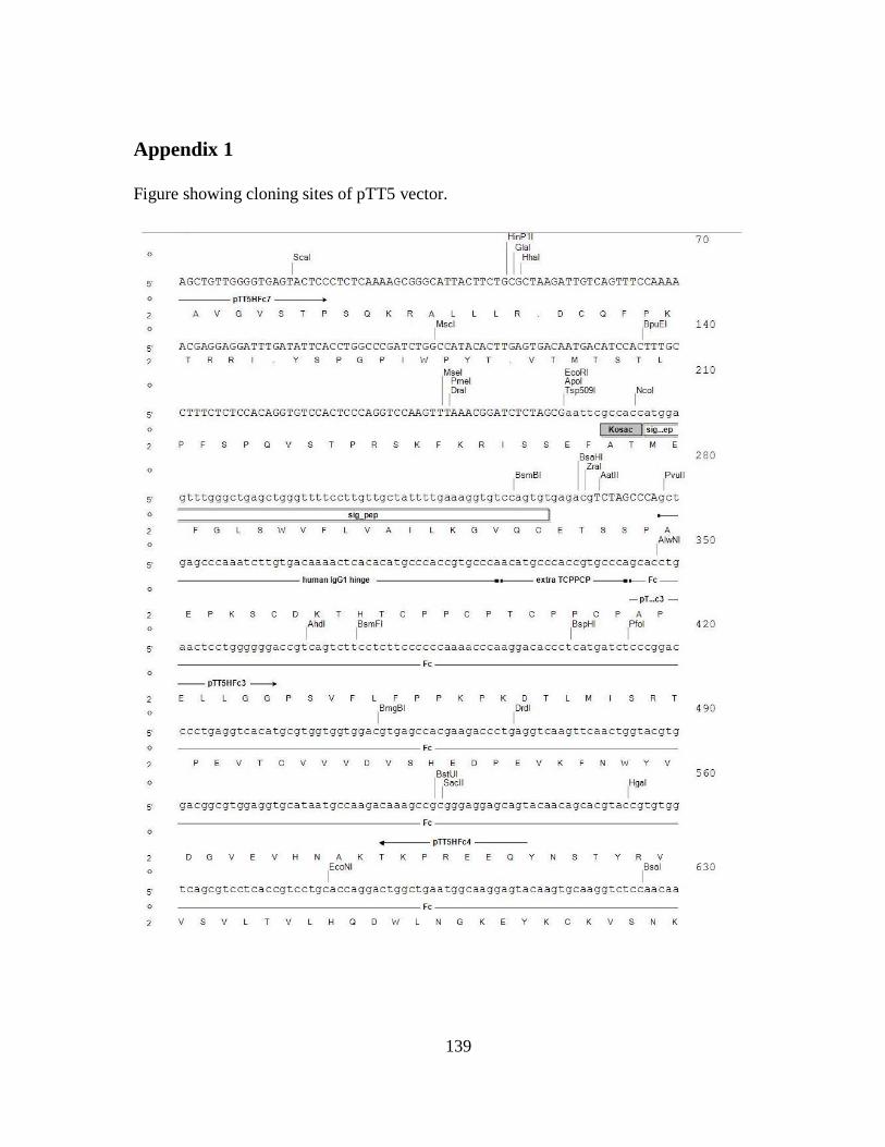
139
Appendix 1
Figure showing cloning sites of pTT5 vector.
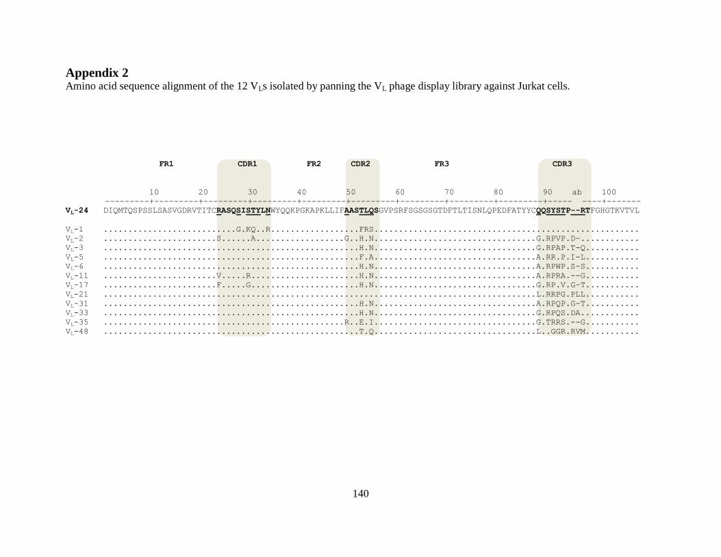
140
Appendix 2 Amino acid sequence alignment of the 12 VLs isolated by panning the VL phage display library against Jurkat cells.
FR1 CDR1 FR2 CDR2 FR3 CDR3
10 20 30 40 50 60 70 80 90 ab 100
---------+---------+---------+---------+---------+---------+---------+---------+---------+----- ----+-------
VL-24 DIQMTQSPSSLSASVGDRVTITCRASQSISTYLNWYQQKPGKAPKLLIFAASTLQSGVPSRFSGSGSGTDFTLTISNLQPEDFATYYCQQSYSTP--RTFGHGTKVTVL
VL-1 ...........................G.KQ..R..................FRS......................................................
VL-2 .......................S......A..................G..H.N.................................G.RPVP.D-............
VL-3 ....................................................H.N.................................G.RPAP.T-Q...........
VL-5 ....................................................F.A.................................A.RR.P.I-L...........
VL-6 ....................................................H.N.................................A.RPWP.S-S...........
VL-11 .......................V.....R......................H.N.................................A.RPRA.--G...........
VL-17 .......................F.....G......................H.N.................................G.RP.V.G-T...........
VL-21 ........................................................................................L.RRPG.PLL...........
VL-31 ....................................................H.N.................................A.RPQP.G-T...........
VL-33 ....................................................H.N.................................G.RPQS.DA............
VL-35 .................................................R..E.I.................................G.TRRS.--G...........
VL-48 ....................................................T.Q.................................L..GGR.RVM...........
141
Appendix 3
Representative gating schemes applied to K-562 cell line variants in flow cytometry analysis.
CD49d-expression on K562-CD49d and K562-mock cell lines, respectively, were determined by
flow cytometry experiments. Upper panels represent K562-mock cells treated with: mouse anti-
human CD49d IgG detected with goat anti-mouse IgG-PE (K562-mock + IgG) (left panel); the
secondary antibody goat anti-mouse IgG-PE (K562-mock +2ry-PE anti-mAb) (middle panel);
and the secondary antibody anti-6xHis IgG-PE (K562-mock +2ry-PE anti-His) (right panel). The
lower panels represent K562-CD49d cells treated with: mouse anti-human CD49d IgG detected
with goat anti-mouse IgG-PE (K562-CD49d + IgG) (left panel); the secondary antibody goat
anti-mouse IgG-PE (K562-CD49d +2ry-PE anti-mAb) (middle panel); and the secondary
antibody anti-6xHis IgG-PE (K562-CD49d +2ry-PE anti-His) (right panel).
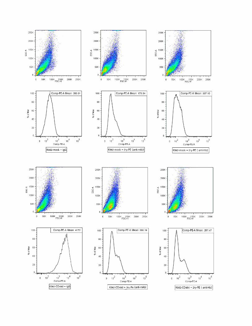
142