Embed Size (px)
Citation preview
BioMed CentralCases Journal
ss
Open AcceCase ReportGorham's disease involving the left parietal bone: a case reportVijay Parihar*, Yad Ram Yadav and Dhananjaya SharmaAddress: Assistant Professor Neurosurgery, Neurosurgery Unit, Department of General Surgery, NSCB Govt. Medical College & Hospital, Jabalpur, MP, 482002, India
Email: Vijay Parihar* - [email protected]; Yad Ram Yadav - [email protected]; Dhananjaya Sharma - [email protected]
* Corresponding author
AbstractIntroduction: Gorham's disease is a rare bone disease, is characterized by the proliferation ofthin-walled vascular channels associated with regional osteolysis. There have been fewer than 150cases reported in the literature. Shoulder and pelvic region is common site. Skull is the leastcommon site of involvement.
Case Presentation: We describe a case of a 35-year-old female of Indian origin presented withthis rare condition involving her left parietal bone. She was treated successfully with excision ofdiseased bone and cranioplasty.
Conclusion: This is the first reported case in India. We provide a review of the clinical, radiologicaland pathological diagnosis of this rare condition and describe treatment options.
IntroductionGorham disease (massive osteolysis of Gorham, vanish-ing or disappearing bone disease, Gorham-Stout syn-drome and phantom bone disease) is a rare disordercharacterized by a nonfamiliar, histologically benign vas-cular proliferation originating in bone and producing pro-gressive resorption of all or a portion of the bone [1]. Thisuncommon condition occurs sporadically and is mostoften observed in children and young adults of either sex.Involvement of almost every bone has been reported,although there is a predilection for bones that develop byintramembranous ossification, with the shoulder girdleand mandible being the most common bones affected.The lesion is typically nonexpansile and nonulcerativeand is usually monocentric but locally aggressive, withresorption of the affected bone. The vascular lesion mayspread into soft tissue and contiguous bones [1,2]. Thepathogenesis of Gorham-Stout disease remains unknown,although it has been suggested that there is an increase in
the sensitivity of osteoclast precursors to humoral factors,which promote osteoclast formation and bone resorptionand operate at the level of the bone microenvironment[3].
Case presentationA 35-year-old female of Indian origin presented with com-plaint of depression over left parietal area noticed acci-dentally about four years back. At that time the size isabout a coin. It is slowly and gradually progressive innature. At present it is about 4 × 4 cm in size. There wereno other symptoms at this time duration. There was nohistory of any trauma. Her neurological examination wasnormal. Local examination shows a well-defined non-tender rounded left parietal depression of 4 × 4 cm in sizewithout any soft tissue mass with normal overlying scalp.Plain x-ray skull was showing a well-demarcated roundedlytic area in parietal region with normal margin. (Figure1A) There is no other abnormality seen in plain x-ray. CT
Published: 22 October 2008
Cases Journal 2008, 1:258 doi:10.1186/1757-1626-1-258
Received: 4 August 2008Accepted: 22 October 2008
This article is available from: http://www.casesjournal.com/content/1/1/258
© 2008 Parihar et al; licensee BioMed Central Ltd. This is an Open Access article distributed under the terms of the Creative Commons Attribution License (http://creativecommons.org/licenses/by/2.0), which permits unrestricted use, distribution, and reproduction in any medium, provided the original work is properly cited.
Page 1 of 4(page number not for citation purposes)
Cases Journal 2008, 1:258 http://www.casesjournal.com/content/1/1/258
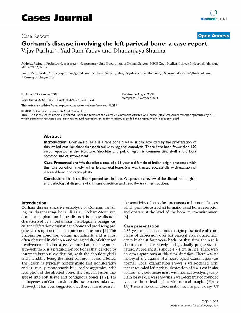
scan revealed a well-defined skull defect with completedisappearance of the central bone matrix and no evidenceof new bone formation (Figure 1B). The soft tissue wid-ows showed no associated soft tissue mass in or aroundthe calvarial defect. MRI brain showing calvarial defect tobe filled with thin soft tissues that were hypointense onboth T1-weighted and T2-weighted (Figure 1C&1D). Tc-99 m MDP bone scintigraphy reveals no bone uptake inthe resorbed site as well as around the margin. Whole-body bone scan was undertaken, which revealed no simi-lar bony lesion any where in the body. (Figure 2). The pre-operative differential diagnosis included Paget disease atdestructive stage, eosinophilic granuloma, Brown tumor,and osteolytic metastasis but none of them correlated wellwith the clinical and radiologic features.
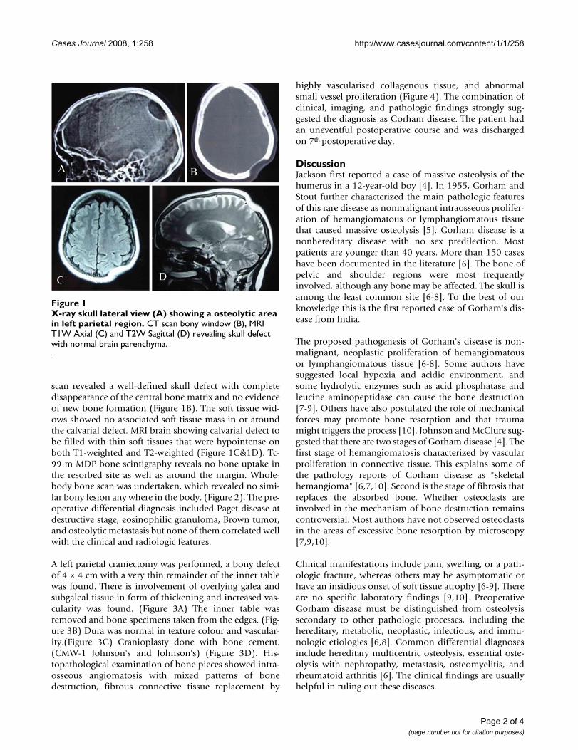
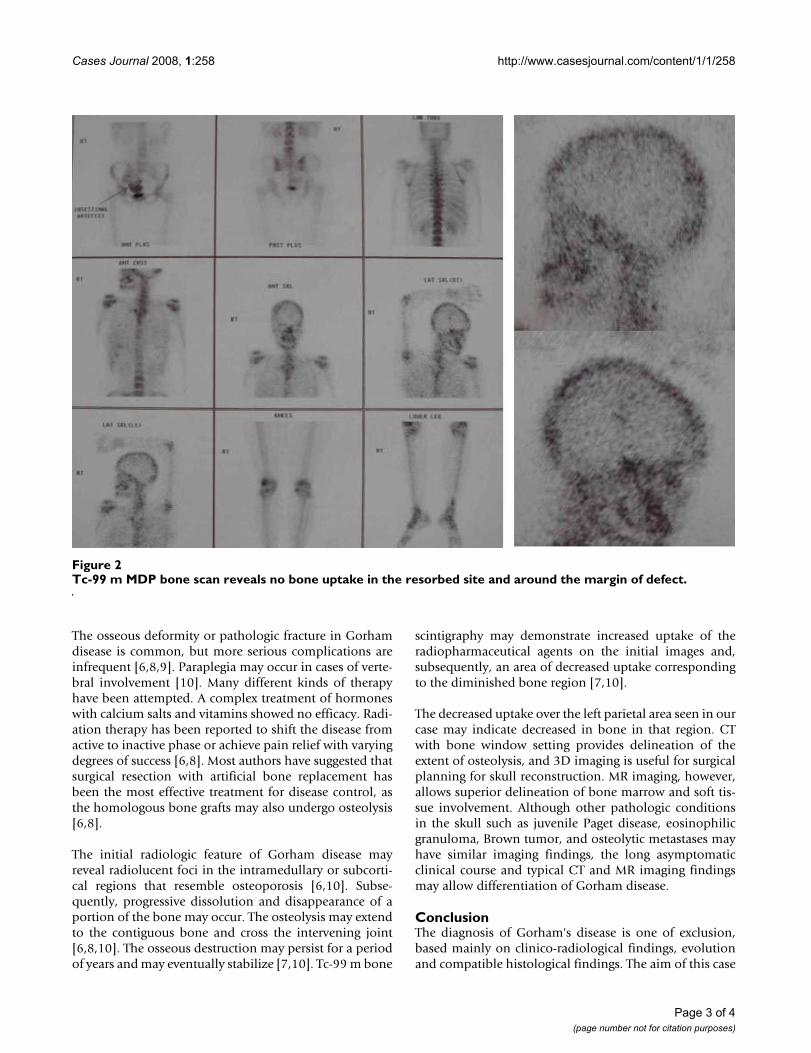
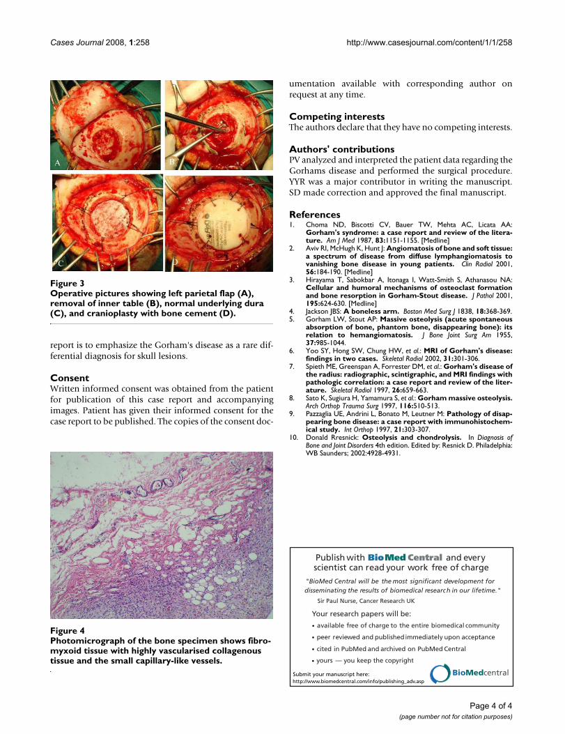
A left parietal craniectomy was performed, a bony defectof 4 × 4 cm with a very thin remainder of the inner tablewas found. There is involvement of overlying galea andsubgaleal tissue in form of thickening and increased vas-cularity was found. (Figure 3A) The inner table wasremoved and bone specimens taken from the edges. (Fig-ure 3B) Dura was normal in texture colour and vascular-ity.(Figure 3C) Cranioplasty done with bone cement.(CMW-1 Johnson's and Johnson's) (Figure 3D). His-topathological examination of bone pieces showed intra-osseous angiomatosis with mixed patterns of bonedestruction, fibrous connective tissue replacement by
highly vascularised collagenous tissue, and abnormalsmall vessel proliferation (Figure 4). The combination ofclinical, imaging, and pathologic findings strongly sug-gested the diagnosis as Gorham disease. The patient hadan uneventful postoperative course and was dischargedon 7th postoperative day.
DiscussionJackson first reported a case of massive osteolysis of thehumerus in a 12-year-old boy [4]. In 1955, Gorham andStout further characterized the main pathologic featuresof this rare disease as nonmalignant intraosseous prolifer-ation of hemangiomatous or lymphangiomatous tissuethat caused massive osteolysis [5]. Gorham disease is anonhereditary disease with no sex predilection. Mostpatients are younger than 40 years. More than 150 caseshave been documented in the literature [6]. The bone ofpelvic and shoulder regions were most frequentlyinvolved, although any bone may be affected. The skull isamong the least common site [6-8]. To the best of ourknowledge this is the first reported case of Gorham's dis-ease from India.
The proposed pathogenesis of Gorham's disease is non-malignant, neoplastic proliferation of hemangiomatousor lymphangiomatous tissue [6-8]. Some authors havesuggested local hypoxia and acidic environment, andsome hydrolytic enzymes such as acid phosphatase andleucine aminopeptidase can cause the bone destruction[7-9]. Others have also postulated the role of mechanicalforces may promote bone resorption and that traumamight triggers the process [10]. Johnson and McClure sug-gested that there are two stages of Gorham disease [4]. Thefirst stage of hemangiomatosis characterized by vascularproliferation in connective tissue. This explains some ofthe pathology reports of Gorham disease as "skeletalhemangioma" [6,7,10]. Second is the stage of fibrosis thatreplaces the absorbed bone. Whether osteoclasts areinvolved in the mechanism of bone destruction remainscontroversial. Most authors have not observed osteoclastsin the areas of excessive bone resorption by microscopy[7,9,10].
Clinical manifestations include pain, swelling, or a path-ologic fracture, whereas others may be asymptomatic orhave an insidious onset of soft tissue atrophy [6-9]. Thereare no specific laboratory findings [9,10]. PreoperativeGorham disease must be distinguished from osteolysissecondary to other pathologic processes, including thehereditary, metabolic, neoplastic, infectious, and immu-nologic etiologies [6,8]. Common differential diagnosesinclude hereditary multicentric osteolysis, essential oste-olysis with nephropathy, metastasis, osteomyelitis, andrheumatoid arthritis [6]. The clinical findings are usuallyhelpful in ruling out these diseases.
X-ray skull lateral view (A) showing a osteolytic area in left parietal regionFigure 1X-ray skull lateral view (A) showing a osteolytic area in left parietal region. CT scan bony window (B), MRI T1W Axial (C) and T2W Sagittal (D) revealing skull defect with normal brain parenchyma.
Page 2 of 4(page number not for citation purposes)
Cases Journal 2008, 1:258 http://www.casesjournal.com/content/1/1/258
The osseous deformity or pathologic fracture in Gorhamdisease is common, but more serious complications areinfrequent [6,8,9]. Paraplegia may occur in cases of verte-bral involvement [10]. Many different kinds of therapyhave been attempted. A complex treatment of hormoneswith calcium salts and vitamins showed no efficacy. Radi-ation therapy has been reported to shift the disease fromactive to inactive phase or achieve pain relief with varyingdegrees of success [6,8]. Most authors have suggested thatsurgical resection with artificial bone replacement hasbeen the most effective treatment for disease control, asthe homologous bone grafts may also undergo osteolysis[6,8].
The initial radiologic feature of Gorham disease mayreveal radiolucent foci in the intramedullary or subcorti-cal regions that resemble osteoporosis [6,10]. Subse-quently, progressive dissolution and disappearance of aportion of the bone may occur. The osteolysis may extendto the contiguous bone and cross the intervening joint[6,8,10]. The osseous destruction may persist for a periodof years and may eventually stabilize [7,10]. Tc-99 m bone
scintigraphy may demonstrate increased uptake of theradiopharmaceutical agents on the initial images and,subsequently, an area of decreased uptake correspondingto the diminished bone region [7,10].
The decreased uptake over the left parietal area seen in ourcase may indicate decreased in bone in that region. CTwith bone window setting provides delineation of theextent of osteolysis, and 3D imaging is useful for surgicalplanning for skull reconstruction. MR imaging, however,allows superior delineation of bone marrow and soft tis-sue involvement. Although other pathologic conditionsin the skull such as juvenile Paget disease, eosinophilicgranuloma, Brown tumor, and osteolytic metastases mayhave similar imaging findings, the long asymptomaticclinical course and typical CT and MR imaging findingsmay allow differentiation of Gorham disease.
ConclusionThe diagnosis of Gorham's disease is one of exclusion,based mainly on clinico-radiological findings, evolutionand compatible histological findings. The aim of this case
Tc-99 m MDP bone scan reveals no bone uptake in the resorbed site and around the margin of defectFigure 2Tc-99 m MDP bone scan reveals no bone uptake in the resorbed site and around the margin of defect.
Page 3 of 4(page number not for citation purposes)
Cases Journal 2008, 1:258 http://www.casesjournal.com/content/1/1/258
Publish with BioMed Central and every scientist can read your work free of charge
"BioMed Central will be the most significant development for disseminating the results of biomedical research in our lifetime."
Sir Paul Nurse, Cancer Research UK
Your research papers will be:
available free of charge to the entire biomedical community
peer reviewed and published immediately upon acceptance
cited in PubMed and archived on PubMed Central
yours — you keep the copyright
Submit your manuscript here:http://www.biomedcentral.com/info/publishing_adv.asp
BioMedcentral
report is to emphasize the Gorham's disease as a rare dif-ferential diagnosis for skull lesions.
ConsentWritten informed consent was obtained from the patientfor publication of this case report and accompanyingimages. Patient has given their informed consent for thecase report to be published. The copies of the consent doc-
umentation available with corresponding author onrequest at any time.
Competing interestsThe authors declare that they have no competing interests.
Authors' contributionsPV analyzed and interpreted the patient data regarding theGorhams disease and performed the surgical procedure.YYR was a major contributor in writing the manuscript.SD made correction and approved the final manuscript.
References1. Choma ND, Biscotti CV, Bauer TW, Mehta AC, Licata AA:
Gorham's syndrome: a case report and review of the litera-ture. Am J Med 1987, 83:1151-1155. [Medline]
2. Aviv RI, McHugh K, Hunt J: Angiomatosis of bone and soft tissue:a spectrum of disease from diffuse lymphangiomatosis tovanishing bone disease in young patients. Clin Radiol 2001,56:184-190. [Medline]
3. Hirayama T, Sabokbar A, Itonaga I, Watt-Smith S, Athanasou NA:Cellular and humoral mechanisms of osteoclast formationand bone resorption in Gorham-Stout disease. J Pathol 2001,195:624-630. [Medline]
4. Jackson JBS: A boneless arm. Boston Med Surg J 1838, 18:368-369.5. Gorham LW, Stout AP: Massive osteolysis (acute spontaneous
absorption of bone, phantom bone, disappearing bone): itsrelation to hemangiomatosis. J Bone Joint Surg Am 1955,37:985-1044.
6. Yoo SY, Hong SW, Chung HW, et al.: MRI of Gorham's disease:findings in two cases. Skeletal Radiol 2002, 31:301-306.
7. Spieth ME, Greenspan A, Forrester DM, et al.: Gorham's disease ofthe radius: radiographic, scintigraphic, and MRI findings withpathologic correlation: a case report and review of the liter-ature. Skeletal Radiol 1997, 26:659-663.
8. Sato K, Sugiura H, Yamamura S, et al.: Gorham massive osteolysis.Arch Orthop Trauma Surg 1997, 116:510-513.
9. Pazzaglia UE, Andrini L, Bonato M, Leutner M: Pathology of disap-pearing bone disease: a case report with immunohistochem-ical study. Int Orthop 1997, 21:303-307.
10. Donald Rresnick: Osteolysis and chondrolysis. In Diagnosis ofBone and Joint Disorders 4th edition. Edited by: Resnick D. Philadelphia:WB Saunders; 2002:4928-4931.
Operative pictures showing left parietal flap (A), removal of inner table (B), normal underlying dura (C), and cranioplasty with bone cement (D)Figure 3Operative pictures showing left parietal flap (A), removal of inner table (B), normal underlying dura (C), and cranioplasty with bone cement (D).
Photomicrograph of the bone specimen shows fibromyxoid tissue with highly vascularised collagenous tissue and the small capillary-like vesselsFigure 4Photomicrograph of the bone specimen shows fibro-myxoid tissue with highly vascularised collagenous tissue and the small capillary-like vessels.
Page 4 of 4(page number not for citation purposes)