Embed Size (px)
Citation preview
Case ReportSevere Preeclampsia in the Setting of Myasthenia Gravis
Adam J. Lake,1 Antoun Al Khabbaz,1 and Renée Keeney2
1Department of Obstetrics and Gynecology, University of Illinois College of Medicine at Rockford, 1601 Parkview Ave.,Rockford, IL 61101, USA2Department of Women, Children, and Family Health, University of Illinois College of Nursing at Rockford,1601 Parkview Ave., Rockford, IL 61101, USA
Correspondence should be addressed to Antoun Al Khabbaz; [email protected]
Received 29 October 2016; Accepted 17 January 2017; Published 9 February 2017
Academic Editor: Svein Rasmussen
Copyright © 2017 Adam J. Lake et al. This is an open access article distributed under the Creative Commons Attribution License,which permits unrestricted use, distribution, and reproduction in any medium, provided the original work is properly cited.
Myasthenia gravis (MG) is a rare autoimmune disease that leads to progressive muscle weakness and is common during femalereproductive years. The myasthenic mother and her newborn must be observed carefully, as complications during all stages ofpregnancy and the puerperium may arise suddenly. Preeclampsia is a common obstetrical condition for which magnesium sulfateis used for seizure prophylaxis. However, magnesium sulfate is strongly contraindicated in MG as it impairs already slowed nerve-muscle connections. Similarly, many first-line antihypertensive medications, including calcium channels blockers and 𝛽-blockers,may lead to MG exacerbation. This case describes the effective obstetrical management of a patient with MG who developedsevere preeclampsia. The effective use of levetiracetam and various antihypertensive medications including intravenous labetalol isdescribed. A review of the ten reported cases of MG complicated by preeclampsia is examined to aggregate observations of clinicalcare, with focus on delivery methods, anticonvulsants, and antihypertensive medications.
1. Introduction
Myasthenia gravis (MG) is an autoimmune disease inwhich antibodies most frequently target muscle nicotinicacetylcholine receptors (nAChR) or muscle-specific kinase(MuSK), which leads to the gradual attrition of neuromus-cular signals. This manifests itself as fatigue and progressiveparesis of skeletal muscle, which characteristically worsenswith exertion and improves with rest. There is an estimatedMG prevalence of 1 per 5,000 individuals in the United States[1] with maternal MG complicating 1 in 68,000 pregnancies[2]. Exacerbations of MG are termed myasthenic crises andare often precipitated by infections, antibiotics, emotionalstress, and surgery [3]. A myasthenic crisis may lead to life-threatening acute respiratory failure requiring mechanicalventilation.With improved neurocritical care protocols,mor-tality from a myasthenic crisis has improved to 5% [3]. TheMyasthenia Gravis Foundation of America (MGFA) treat-ment guidelines recommend the use of acetylcholinesterase
inhibitors, intravenous immunoglobulins, plasma exchange(PLEX), glucocorticoids, and thymectomy for myastheniatreatment [4].
Preeclampsia is a systemic disorder characterized by new-onset hypertension, proteinuria, and end-organ damage after20-week gestation and complicates 2–8% of pregnancies inthe United States [5]. Treatment of hypertension in thesetting of preeclampsia may be approached aggressively withmultiple antihypertensive medications to achieve adequateblood pressure control.Magnesium sulfate, shown to be supe-rior to other anticonvulsants, is used frequently for seizureprophylaxis [6]. Both MG and preeclampsia have specifictreatment guidelines, which are often sufficient for adequatecontrol of each disease. However, management of preeclamp-sia with magnesium sulfate and commonly used antihyper-tensive medications, such as 𝛽-blockers and calcium channelblockers, is contraindicated in MG as it may exacerbateMG symptomatology and precipitate a myasthenic crisis. Anassociation between MG and preeclampsia prevalence has
HindawiCase Reports in Obstetrics and GynecologyVolume 2017, Article ID 9204930, 5 pageshttps://doi.org/10.1155/2017/9204930
2 Case Reports in Obstetrics and Gynecology
not been demonstrated in the English literature. There is apaucity of reports describing treatment of preeclampsia inpatients with MG.
We present a pregnancy complicated by preexisting MGand the later development of severe preeclampsia withdescription of novel clinical management with intravenouslevetiracetam and labetalol. A review of the English literatureis presented as well, describing experiences with this rareclinical scenario.
2. Presentation of Case
A 28-year-old G3P2002 patient at 34-week gestation wasadmitted to the labor and delivery suite with a diagnosisof preeclampsia. The patient had two prior uncomplicatedspontaneous vaginal deliveries. Her pregnancy was dated byultrasound at 8-week gestation. The patient is known to haveMGmanagedwith pyridostigmine 30.0mg orally, three timesa day. She had a thymectomy six years prior to this preg-nancy. She had an uncomplicated prenatal course. Her bloodpressure during pregnancy ranged from 108 to 132mmHgsystolic and 67 to 88mmHg diastolic. She has a history ofpreexisting prehypertension and was on no antihypertensivetreatment. Upon arrival to the hospital, the patient’s initialblood pressure was in the range of 170–180mmHg systolicand 100–110mmHg diastolic. She was in no acute distressand reported no clinically significant edema, right upperquadrant pain, weakness, dyspnea, diplopia, or ptosis. Heronly notable symptom was a new-onset, mild headache.She denied experiencing contractions, leakage of fluid pervagina, or vaginal bleeding, and she reported normal fetalmovements. Workup for preeclampsia showed an elevatedprotein/creatinine ratio of 0.7, a slightly elevated uric acidof 5.6mg/dL, creatinine of 0.74mg/dL, normal liver enzymes(AST 26U/L, ALT 11U/L), and platelet count 152 × 103/𝜇L.
She was started on intravenous levetiracetam (1.0 g intra-venous bolus for seizure prophylaxis). She was given mul-tiple doses of 5.0mg intravenous hydralazine to treat herhypertension, which had minimal effect. Her blood pressurewas as high as 229/117mmHg. The patient was then givenlabetalol intravenously resulting in better control of her bloodpressure with noMG exacerbation noted.Within a few hoursof admission, the patient developed mild clonus and herheadache increased in severity. The patient’s cervix at thispoint was unfavorable so the decision was taken to proceedwith Cesarean section under spinal anesthesia. Surgery wasperformed without complications, resulting in the birth ofa healthy male newborn with a birth weight of 1865 g andAPGAR scores of 8 at one minute and 9 at five minutes. Thenewborn was followed up closely and showed no signs oftransient neonatal MG.
In the postpartum period, levetiracetam was continuedat a dose of 500mg intravenously every 12 hours and wasdiscontinued 2 days postpartum.The only notable side effectswere somnolence and fatigue.These symptoms resolved afterdiscontinuation of levetiracetam. Hypertension was treatedwith methyldopa and hydralazine. Clonidine was added onpostpartum day 2. The patient was discharged home onpostpartum day 4 on methyldopa and clonidine in stable
condition with blood pressure in the prehypertensive range.Thepatient hadnomyasthenic crisis in either the intrapartumor the postpartumperiods. Shewas weaned off her antihyper-tensivemedications postpartum andwas normotensive at hersix-week postpartum visit.
3. Discussion
MG exacerbations are common in pregnancy, perhaps due toincreases in blood volume and changes in the renal clearanceofmedications [17].MG is associated with other autoimmunediseases such as rheumatoid arthritis and thyroid dysfunctionbut is not related to any increase in the prevalence ofpreeclampsia [18, 19].The clinical course ofMG in pregnancyis variable, with an improvement of symptoms in 29% ofpregnancies, worsening in 41%, and no change in 30% [20].Pregnant patients with MG must be watched closely dueto its unpredictable course in pregnancy. Chorioamnionitis,postpartum endometritis, and mastitis should be treatedeffectively as they can precipitate a myasthenic crisis [17].Ideally, patients with MG should have optimization of theirmyasthenic symptoms prior to attempting to conceive [4].Transient neonatal myasthenia gravis is due to transplacentalpassage ofmaternal IgG autoantibodies [17] andpresentswithmask-like facies, a weakened cry, and impaired ability to suck.Clinicians recommend neonatal observation for a minimumof 2 days prior to discharge [4, 21]. Breastfeeding is notcontraindicated in MG unless the patient is in a myastheniccrisis [15].
A comprehensive literature review of published cases ofMG and preeclampsia in the English literature publishedfrom 1976 to 2016 using a MEDLINE search is presented(Table 1). Ten case reports have been published. A varietyof medications have been implicated in inducing myastheniccrises and should be avoided in all MG patients (Table 2)[17, 22]. Paradoxically, this includes the initiation of corti-costeroid treatment, which causes exacerbations in 50% ofpatients [3]. These cases provide insight into the modes ofdelivery, anticonvulsants, and antihypertensive medicationsused for this clinical situation.
Delivery was accomplished by vaginal delivery in fourof the reported cases; the other six patients delivered byCesarean section. Surgical deliverymay lead to exacerbationsof MG and should only be performed for maternal or fetalindications [11]. For this case and previous cases, Cesareansections were considered beneficial due to concerns of wors-ening maternal status from preeclampsia [12] with consid-eration taken that surgery itself could cause a myastheniccrisis. Regional anesthesia is preferred over general anesthesiafor both vaginal and surgical deliveries. Of the reportedcases, seven patients received regional anesthesia. One ofthose patients had profound vagal bradycardia followingneuraxial anesthetic blockage, which required atropine andephedrine to resolve [14]. General anesthesia with intubationwas performed in only one case, and prolonged ventilationwas needed postpartum. In that case, the muscle relaxantpancuronium was used. Muscle relaxants, both depolarizingand nondepolarizing, are also to be avoided as MG patientsare quite sensitive to their effects [21].

Case Reports in Obstetrics and Gynecology 3
Table1:Cu
rrentm
yasthenic-preecla
mpsiacasesintheE
nglishliterature,MED
LINEsearch.
(a)
Author(s)
Mother’s
age,
gravidity
and
parity,and
gesta
tionalage
atadmission
Mod
eof
delivery
Anesthesia
Hypertensivetreatment
Anticon
vulsa
nttre
atment
Com
plications
Coh
enetal.[7]
(1976)
37yo,G
3P1,term
Spon
taneou
svaginald
elivery
Spinalanesthesia
Furosemidea
ndmethydo
paMagnesiu
msulfate
(IM
injection)
With
in10
minutes
ofIM
magnesiu
msulfate,the
patie
nthadam
yasthenicc
risisbu
timproved
quickly
with
1.0gcalcium
glucon
ate,0.4m
gatropine,and
10.0mgedroph
onium.
Duff
[8](1979)
26yo,G
2P1,
37weeks
Cesarean
section
Generalanesthesia
(thiopenton
e,scoline,
pancuron
ium,and
nitro
usoxide)
Poor
controlw
ithmethyldop
a,diazoxide,
reserpine,and
furosemide
Diazepam
“for
sedatio
n”Th
epatient
requ
ired16
hourso
fventilator
supp
ort
becauseo
frespiratory
insufficiency
after
general
anesthesia.
Duff
(1979)
36yo,G
2P1,
36weeks
Vaginald
elivery
after
labo
rindu
ction
Spinalanesthesia
Ephedrine(no
evidence
foru
securrently
)Non
ereported
MGdidno
timproveinthep
ostpartum
perio
d,so
the
patie
ntwas
startedon
prednisone.Th
epatient
was
discharged
fivew
eeks
postp
artum
with
improvem
ent
ofMGsymptom
s.Brogan
and
Corcoran[9]
(1983)
37yo,G
1P0,
32weeks
Cesarean
section
Spinalanesthesia
Non
ereported
Non
ereported
Nocomplications
noted.
Bashuk
and
Krendel[10](1990)19
yo,G
1P0,term
Spon
taneou
svaginald
elivery
Non
enoted
Non
ereported
Magnesiu
msulfateIV
(4.0gon
ce)a
ndIM
(5.0gq4
h)
Weakn
essw
orsenedwith
each
IMinjectionandshe
becameq
uadripleg
ic.O
ncetreatmentw
assto
pped,
sher
egainedmuscle
strengthwith
inad
ay.
(b)
Author(s)
Mother’s
age,
gravidity
and
parity,and
gesta
tionalage
atadmission
Mod
eof
delivery
Anesthesia
Hypertensivetreatment
Anticon
vulsa
nttre
atment
Clinicalpresentatio
n
Benshu
shan
etal.
[11](19
94)
31yo,G
1P0,
31weeks
Vaginald
elivery
after
labo
rindu
ctionand
artifi
cialrupture
ofmem
branes
Non
enoted
Methyldop
aand
hydralazine,lateru
sing
furosemidew
ithlittle
diuresis
Non
ereported
Delivery
complicated
byhemorrhage.Th
epatient
was
transfe
rred
totheICU
ford
yspn
ea,oliguria,and
weakn
essw
here
treatmentw
assta
rted
with
dopamine,
furosemide,IV
corticosteroids,andIV
pyrid
ostig
mine.
Thep
atient
was
discharged
tendays
later.
DiSpiezio
Sardoet
al.[12]
27yo,G
2P0,
37weeks
Cesarean
section
Spinalanesthesia
Methyldop
aNon
ereported
Thep
atient
was
diagno
sedwith
severe
preecla
mpsia
complicated
byHEL
LPsynd
rome.Th
epatient
was
discharged
ondaysix
postp
artum.
Ham
aoui
and
Mercado
[13]
(200
9)31
yo,G
7P3,
27weeks
Cesarean
section
Spinalanesthesia
(bup
ivacaine)
Hydralazine,m
etop
rolol,
losartan,amlodipine,
andlabetaol.L
abetalol
drip
controlledBP
.Non
ereported
Threed
aysp
ostpartum,the
patie
ntdevelopedHEL
LPsynd
romea
ndmyasthenice
xacerbation.
Ozcan
etal.[14]
(2015)
34yo,G
1P0,
36weeks
Cesarean
section
Spinalanesthesia
(bup
ivacaine
and
fentanyl)
Enalapril
Non
ereported
Thep
atient
becameb
radycardic(42b
pm)a
fterspinal
anesthesia,requirin
gatropine
andephedrineto
resolve.Po
stpartum
perio
drequ
iredenalapril,w
ithincreaseddo
seof
pyrid
ostig
minea
ndIV
immun
oglobu
linto
resolves
ymptom
s.Th
epatient
was
discharged
8days
after
theC
esareansection.
Sikk
aetal.[15]
(2015)
25yo,G
1P0,
36weeks
Cesarean
section
Spinalanesthesia
Non
ereported
Non
ereported
Nocomplications
noted.
4 Case Reports in Obstetrics and Gynecology
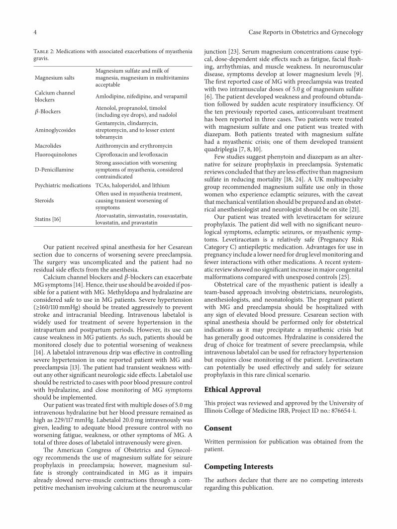
Table 2: Medications with associated exacerbations of myastheniagravis.
Magnesium saltsMagnesium sulfate and milk ofmagnesia, magnesium in multivitaminsacceptable
Calcium channelblockers Amlodipine, nifedipine, and verapamil
𝛽-Blockers Atenolol, propranolol, timolol(including eye drops), and nadolol
AminoglycosidesGentamycin, clindamycin,streptomycin, and to lesser extenttobramycin
Macrolides Azithromycin and erythromycinFluoroquinolones Ciprofloxacin and levofloxacin
D-PenicillamineStrong association with worseningsymptoms of myasthenia, consideredcontraindicated
Psychiatric medications TCAs, haloperidol, and lithium
SteroidsOften used in myasthenia treatment,causing transient worsening ofsymptoms
Statins [16] Atorvastatin, simvastatin, rosuvastatin,lovastatin, and pravastatin
Our patient received spinal anesthesia for her Cesareansection due to concerns of worsening severe preeclampsia.The surgery was uncomplicated and the patient had noresidual side effects from the anesthesia.
Calcium channel blockers and 𝛽-blockers can exacerbateMGsymptoms [14].Hence, their use should be avoided if pos-sible for a patient with MG. Methyldopa and hydralazine areconsidered safe to use in MG patients. Severe hypertension(≥160/110mmHg) should be treated aggressively to preventstroke and intracranial bleeding. Intravenous labetalol iswidely used for treatment of severe hypertension in theintrapartum and postpartum periods. However, its use cancause weakness in MG patients. As such, patients should bemonitored closely due to potential worsening of weakness[14]. A labetalol intravenous drip was effective in controllingsevere hypertension in one reported patient with MG andpreeclampsia [13]. The patient had transient weakness with-out any other significant neurologic side effects. Labetalol useshould be restricted to cases with poor blood pressure controlwith hydralazine, and close monitoring of MG symptomsshould be implemented.
Our patient was treated first withmultiple doses of 5.0mgintravenous hydralazine but her blood pressure remained ashigh as 229/117mmHg. Labetalol 20.0mg intravenously wasgiven, leading to adequate blood pressure control with noworsening fatigue, weakness, or other symptoms of MG. Atotal of three doses of labetalol intravenously were given.
The American Congress of Obstetrics and Gynecol-ogy recommends the use of magnesium sulfate for seizureprophylaxis in preeclampsia; however, magnesium sul-fate is strongly contraindicated in MG as it impairsalready slowed nerve-muscle contractions through a com-petitive mechanism involving calcium at the neuromuscular
junction [23]. Serum magnesium concentrations cause typi-cal, dose-dependent side effects such as fatigue, facial flush-ing, arrhythmias, and muscle weakness. In neuromusculardisease, symptoms develop at lower magnesium levels [9].The first reported case of MG with preeclampsia was treatedwith two intramuscular doses of 5.0 g of magnesium sulfate[6]. The patient developed weakness and profound obtunda-tion followed by sudden acute respiratory insufficiency. Ofthe ten previously reported cases, anticonvulsant treatmenthas been reported in three cases. Two patients were treatedwith magnesium sulfate and one patient was treated withdiazepam. Both patients treated with magnesium sulfatehad a myasthenic crisis; one of them developed transientquadriplegia [7, 8, 10].
Few studies suggest phenytoin and diazepam as an alter-native for seizure prophylaxis in preeclampsia. Systematicreviews concluded that they are less effective thanmagnesiumsulfate in reducing mortality [18, 24]. A UK multispecialtygroup recommended magnesium sulfate use only in thosewomen who experience eclamptic seizures, with the caveatthatmechanical ventilation should be prepared and anobstet-rical anesthesiologist and neurologist should be on site [21].
Our patient was treated with levetiracetam for seizureprophylaxis. The patient did well with no significant neuro-logical symptoms, eclamptic seizures, or myasthenic symp-toms. Levetiracetam is a relatively safe (Pregnancy RiskCategory C) antiepileptic medication. Advantages for use inpregnancy include a lower need for drug levelmonitoring andfewer interactions with other medications. A recent system-atic review showedno significant increase inmajor congenitalmalformations compared with unexposed controls [25].
Obstetrical care of the myasthenic patient is ideally ateam-based approach involving obstetricians, neurologists,anesthesiologists, and neonatologists. The pregnant patientwith MG and preeclampsia should be hospitalized withany sign of elevated blood pressure. Cesarean section withspinal anesthesia should be performed only for obstetricalindications as it may precipitate a myasthenic crisis buthas generally good outcomes. Hydralazine is considered thedrug of choice for treatment of severe preeclampsia, whileintravenous labetalol can be used for refractory hypertensionbut requires close monitoring of the patient. Levetiracetamcan potentially be used effectively and safely for seizureprophylaxis in this rare clinical scenario.
Ethical Approval
This project was reviewed and approved by the University ofIllinois College of Medicine IRB, Project ID no.: 876654-1.
Consent
Written permission for publication was obtained from thepatient.
Competing Interests
The authors declare that there are no competing interestsregarding this publication.

Case Reports in Obstetrics and Gynecology 5
References
[1] L. H. Phillips II, “The epidemiology of myasthenia gravis,”Annals of the New York Academy of Sciences, vol. 998, pp. 407–412, 2003.
[2] F. F. Schade and M. P. Foley, “Pregnancy in myasthenia gravis,”American Journal of Obstetrics andGynecology, vol. 63, no. 5, pp.1154–1156, 1952.
[3] D. A. Godoy, L. J. V. de Mello, L. Masotti, and M. Di Napoli,“Themyasthenic patient in crisis: an update of themanagementinNeurointensive care unit,”Arquivos de Neuro-Psiquiatria, vol.71, no. 9 A, pp. 627–639, 2013.
[4] D. B. Sanders, G. I. Wolfe, M. Benatar et al., “Internationalconsensus guidance for management of myasthenia gravis:executive summary,”Neurology, vol. 87, no. 4, pp. 419–425, 2016.
[5] L. Duley, “The global impact of pre-eclampsia and eclampsia,”Seminars in Perinatology, vol. 33, no. 3, pp. 130–137, 2009.
[6] L. Duley, A. M. Gulmezoglu, D. J. Henderson-Smart et al.,“Magnesium sulphate and other anticonvulsants for womenwith pre-eclampsia,” The Cochrane Database of SystematicReviews, no. 11, Article ID CD000025, 2010.
[7] B. A. Cohen, R. S. London, and P. J. Goldstein, “Myastheniagravis and preeclampsia,” Obstetrics and Gynecology, vol. 48,supplement 1, pp. 35S–37S, 1976.
[8] C. B. Duff, “Preeclampsia and the patient with myastheniagravis,” Obstetrics and Gynecology, vol. 54, no. 3, pp. 355–358,1979.
[9] M. Brogan and D. J. D. Corcoran, “Myasthenia gravis and pre-eclampsia,” Irish Medical Journal, vol. 76, no. 2, pp. 84–85, 1983.
[10] R. G. Bashuk and D. A. Krendel, “Myasthenia gravis presentingas weakness after magnesium administration,” Muscle andNerve, vol. 13, no. 8, pp. 708–712, 1990.
[11] A. Benshushan, N. Rojansky, and D. Weinstein, “Myastheniagravis and preeclampsia,” Israel Journal of Medical Sciences, vol.30, no. 3, pp. 229–233, 1994.
[12] A. Di Spiezio Sardo, A. Taylor, M. Pellicano et al., “MyastheniaandHELLP syndrome,” European Journal of Obstetrics Gynecol-ogy and Reproductive Biology, vol. 116, no. 1, pp. 108–111, 2004.
[13] A. Hamaoui and R. Mercado, “Association of preeclampsia andmyasthenia: a case report,” Journal of Reproductive Medicine,vol. 54, no. 9, pp. 587–590, 2009.
[14] J. Ozcan, I. F. Balson, and A. T. Dennis, “New diagnosismyasthenia gravis and preeclampsia in late pregnancy,” BMJCase Reports, vol. 2015, Article ID e208323, 2015.
[15] P. Sikka, B. Joshi, N. Aggarwal, V. Suri, and H. Bhagat, “Dis-tinguishingmyasthenia exacerbation from severe preeclampsia:a diagnostic and therapeutic challenge,” Journal of Clinical andDiagnostic Research, vol. 9, no. 8, pp. QD05–QD06, 2015.
[16] J. Gale andH. V. Danesh-Meyer, “Statins can inducemyastheniagravis,” Journal of Clinical Neuroscience, vol. 21, no. 2, pp. 195–197, 2014.
[17] M. Varner, “Myasthenia gravis and pregnancy,” Clinical Obstet-rics and Gynecology, vol. 56, no. 2, pp. 372–381, 2013.
[18] W. C. Plauche, “Myasthenia gravis in pregnancy,” AmericanJournal of Obstetrics and Gynecology, vol. 88, pp. 404–409, 1964.
[19] B. Piura, “The association of preeclampsia and myastheniagravis: double trouble,” Israel Journal of Medical Sciences, vol.30, no. 3, pp. 243–244, 1994.
[20] A. P. Batocchi, L. Majolini, A. Evoli, M.M. Lino, C.Minisci, andP. Tonali, “Course and treatment of myasthenia gravis duringpregnancy,” Neurology, vol. 52, no. 3, pp. 447–452, 1999.
[21] F. Norwood, M. Dhanjal, M. Hill et al., “Myasthenia in preg-nancy: best practice guidelines from a UKmultispecialty work-ing group,” Journal of Neurology, Neurosurgery and Psychiatry,vol. 85, no. 5, pp. 538–543, 2014.
[22] A. Elsais, T. H. Popperud, Ø. Melien, and E. Kerty, “Medika-menter somkan utløse og forverremyasthenia gravis,”Tidsskriftfor Den Norske Legeforening, vol. 133, no. 3, pp. 296–299, 2013(Norwegian).
[23] D. H. Jenkinson, “The nature of the antagonism betweencalcium and magnesium ions at the neuromuscular junction,”The Journal of physiology, vol. 138, no. 3, pp. 434–444, 1957.
[24] J. N. Mueksch and W. A. Stevens, “Undiagnosed myastheniagravis masquerading as eclampsia,” International Journal ofObstetric Anesthesia, vol. 16, no. 4, pp. 379–382, 2007.
[25] S. A. Chaudhry, G. Jong, and G. Koren, “The fetal safety ofLevetiracetam: a systematic review,” Reproductive Toxicology,vol. 46, pp. 40–45, 2014.

Submit your manuscripts athttps://www.hindawi.com
Stem CellsInternational
Hindawi Publishing Corporationhttp://www.hindawi.com Volume 2014
Hindawi Publishing Corporationhttp://www.hindawi.com Volume 2014
MEDIATORSINFLAMMATION
of
Hindawi Publishing Corporationhttp://www.hindawi.com Volume 2014
Behavioural Neurology
EndocrinologyInternational Journal of
Hindawi Publishing Corporationhttp://www.hindawi.com Volume 2014
Hindawi Publishing Corporationhttp://www.hindawi.com Volume 2014
Disease Markers
Hindawi Publishing Corporationhttp://www.hindawi.com Volume 2014
BioMed Research International
OncologyJournal of
Hindawi Publishing Corporationhttp://www.hindawi.com Volume 2014
Hindawi Publishing Corporationhttp://www.hindawi.com Volume 2014
Oxidative Medicine and Cellular Longevity
Hindawi Publishing Corporationhttp://www.hindawi.com Volume 2014
PPAR Research
The Scientific World JournalHindawi Publishing Corporation http://www.hindawi.com Volume 2014
Immunology ResearchHindawi Publishing Corporationhttp://www.hindawi.com Volume 2014
Journal of
ObesityJournal of
Hindawi Publishing Corporationhttp://www.hindawi.com Volume 2014
Hindawi Publishing Corporationhttp://www.hindawi.com Volume 2014
Computational and Mathematical Methods in Medicine
OphthalmologyJournal of
Hindawi Publishing Corporationhttp://www.hindawi.com Volume 2014
Diabetes ResearchJournal of
Hindawi Publishing Corporationhttp://www.hindawi.com Volume 2014
Hindawi Publishing Corporationhttp://www.hindawi.com Volume 2014
Research and TreatmentAIDS
Hindawi Publishing Corporationhttp://www.hindawi.com Volume 2014
Gastroenterology Research and Practice
Hindawi Publishing Corporationhttp://www.hindawi.com Volume 2014
Parkinson’s Disease
Evidence-Based Complementary and Alternative Medicine
Volume 2014Hindawi Publishing Corporationhttp://www.hindawi.com