Embed Size (px)
Citation preview
Editorial
Introduction
Case Report
articulation at its base [9]. Due to benign nature of pelvic rib, it is necessary to diagnose accurately. Asymptomatic The pelvic digit, pelvic rib or pelvic finger is an unusual lesion will require no treatment and accurate diagnosis congenital anomaly in which there is bone formation in the
soft tissue around the normal skeleton of pelvis. This lesion ic pelvic digit we report a is benign and usually asymptomatic and for this reason is
symptomatic case which required surgical intervention.detected incidentally in majority of cases [1-10]. Plain radiographs show a bony structure similar to rib or a finger with a clear cortex and medulla arising from pelvis. Sometimes there may be a characteristic pseudo- A 57-year-old man presented with a long history of groin
may avoid unnecessary interventions [11]. Most reports have described an asymptomat
CASE REPORT
The Pelvic Digit: A Rare Congenital Anomaly as a
Cause of Hip Pain1* 1Jesús Moreta-Suárez ,Oskar Sáez de Ugarte-Sobrón , 1 1Alberto Sánchez-Sobrino , José Luis Martínez-De Los Mozos
Abstract
Introduction:
Case Report:
Conclusions:
Keywords:
The pelvic digit or pelvic rib is an unusual congenital anomaly with a finger or rib like
bone formation in soft tissues around normal pelvic skeleton. This is a benign lesion and mostly an
Incidental finding on radiographs. Most reported cases are asymptomatic and do not require
intervention. We report a case of symptomatic pelvic rib that required surgical excision.
57-year-old man presented with a long history of pain and functional limitation in
his right hip. On plain radiographs a fusiform bony structure adjacent to the acetabulum was noted.
The imaging tests (MRI and CT) suggested the diagnosis of pelvic digit. We performed surgical
excision of the lesion through anterior Smith-Peterson approach. The histopathology showed
presence of corticomedullary structure. After surgery the patient's symptoms were relieved.
Pelvic digit, pelvic rib, iliac rib, bone growth and development
A
It is important to recognize this lesion on plain radiographs and to confirm by CT scan
and make differential diagnosis. In the majority of cases the pelvic digit is asymptomatic and no
treatments is needed. However in cases where symptoms can be attributed to pelvic digit an
excision will relieve the pain and disability.
1Department of Orthopaedic Surgery and Trauma
Galdakao-Usansolo Hospital. Barrio Labeaga s/n
48960 Usansolo (Vizcaya) Spain
Fax: 944007132.
Address of Correspondence
Dr Jesús Moreta-Suárez
Department of Orthopaedic Surgery and Trauma
Galdakao-Usansolo Hospital
Barrio Labeaga s/n 48960 Usansolo (Vizcaya). Spain
Fax: 944007132
Email: [email protected]
Journal of Orthopaedic Case Reports 2012 Oct-Dec;2(4):19-22
Dr. Jesus Moreta
MDDr. Oskar Sáez de Ugarte
MD
Dr Alberto Sánchez
MD, PhD
Dr. Jose Luis Martínez-
de los Mozos
MD
19
Author’s Photo Gallery
Copyright © 2012 by Journal of Orthpaedic Case ReportsJournal of Orthopaedic Case Reports | pISSN 2250-0685 | Available on
This is an Open Access article distributed under the terms of the Creative Commons Attribution Non-Commercial License (http://creativecommons.org/licenses/by-nc/3.0) which permits unrestricted non-commercial use, distribution, and reproduction in any medium, provided the original work is properly cited.
www.jocr.co.in
What to Learn from this Article?Insight into existence of entity called Pelvic digit or Pelvic horn
How to differentiate Pelvic digit from lesions like osteochondroma?
pain for several years. There was no history of trauma or Initially, non-steroidal anti-inflammatory injury to groin. The pain exacerbated by walking and medications were prescribed with no symptomatic prolonged sitting. Since last few months his pain had improvement. Surgical excision of the pelvic rib was increased with loss of movement at the hip. Physical planned with consent of the patient. Anterior approach to examination revealed slight asymmetry in the region of the hip (Smith-Petersen) was used. Tensor fascia latae and the iliac bone compared with the contralateral side, with gluteus minimus required to be partially detached to no signs of inflammation or palpable mass. Hip range of completely expose the 'rib' till the base. The 'rib' was motion was decreased with flexion limited to 90º, and resected from its base and samples send for abduction of 15º. Patient walked with Trendelenburg gait histopathology (Fig. 2). Histopathological examination possibly because of pain. Conventional radiographs of showed the presence of corticomedullary structure the hip (Fig.1A) revealed the presence of a bony similar to a rib, with no accompanying cartilage; this protuberance at the right anterior inferior iliac spine, finding was consistent with a pelvic digit.several centimeters in length close to the right acetabulum, The pain disappeared gradually after surgery, with no signs of joint degeneration.Suspecting a giant and a year later the patient remains asymptomatic, with an osteochondroma which could be in the process of improvement in flexion (130°) and abduction (35°) of the malignant transformation we performed a magnetic hip; the radiograph (Fig. 2b) shows no radiological signs resonance imaging (MRI) which showed the existence of a of recurrence after one year (Fig. 3).bone tumor with benign appearance, but excluding the osteochondroma as a diagnostic possibility, because there was no peripheral cartilage. Subsequently a CT scan (Fig. In medical literature, few cases of pelvic digits or pelvic 1B) revealed a bony lesion (6 cm long and 3 cm thick) with ribs have been reported. This lesion was first reported by a well-defined cortical bone and a heterogeneous zone in Sullivan and Cornwell [1] in 1974 in a 15-year-old girl the junction with the ilium suggesting a pseudo- with a bone formation curved caudad toward the right articulation. With these studies we could establish the side or the distal sacral vertebra but was not directly diagnosis of pelvic digit instead of an osteochondroma. attached to the sacrum. The histological work-up revealed
Discussion
www.jocr.co.inMoreta et al
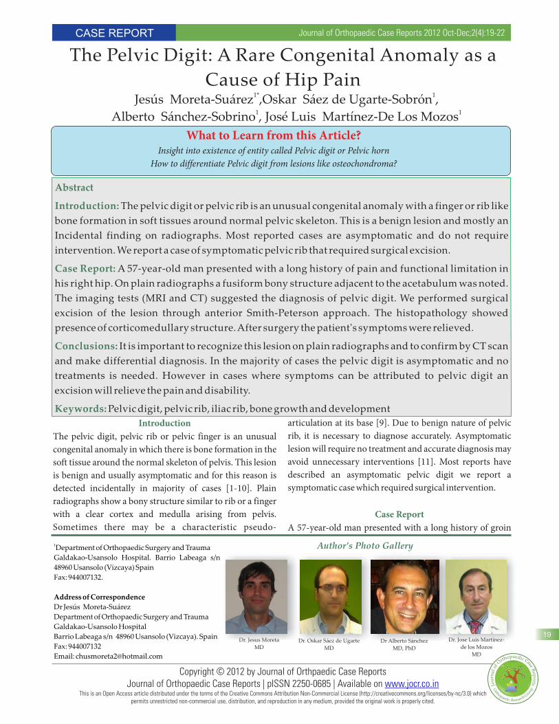
Figure 1. . Conventional radiography showed the pelvic digit adjacent to the right hip. . Computed tomography including the coronal view (up), the axial view (down) and the tridimensional reconstruction.
AB
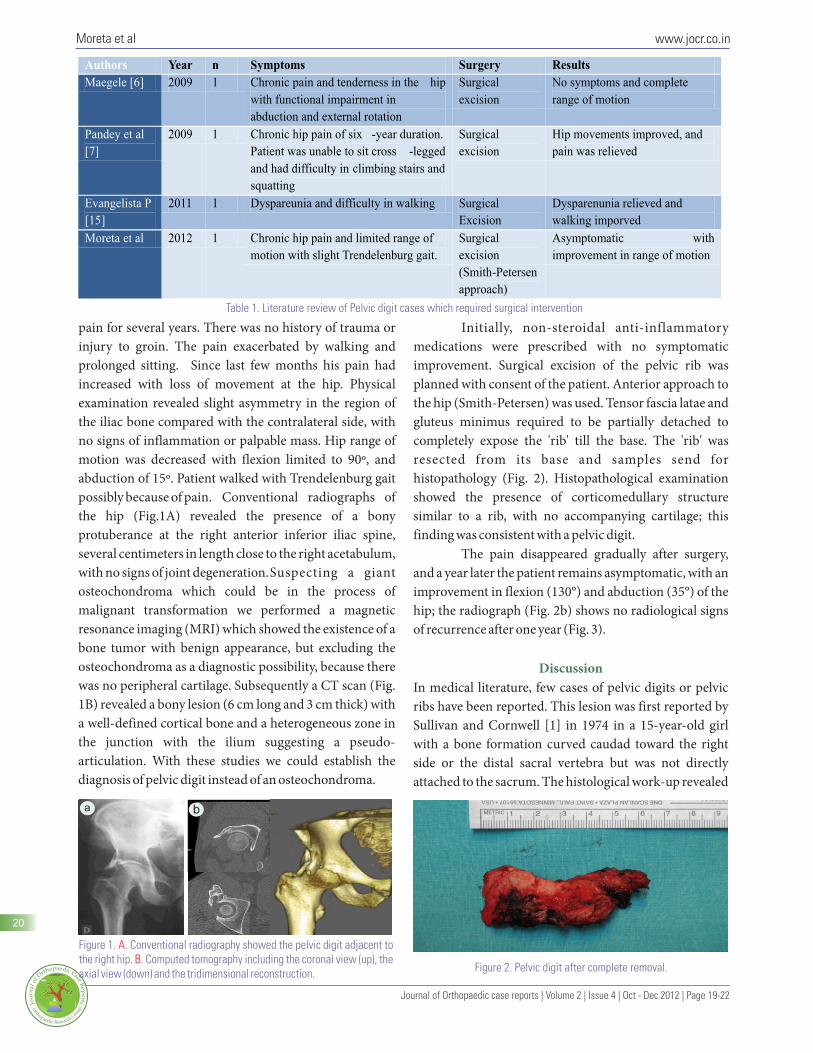
Figure 2. Pelvic digit after complete removal.
Table 1. Literature review of Pelvic digit cases which required surgical intervention
Journal of Orthopaedic case reports | Volume 2 | Issue 4 | Oct - Dec 2012 | Page 19-22
20
Authors Year n Symptoms Surgery Results
Maegele [6] 2009 1 Chronic pain and tenderness in the hip
with functional impairment in
abduction and external rotation
Surgical
excision
No symptoms and complete
range of motion
Pandey et al
[7]
2009 1 Chronic hip pain of six -year duration.
Patient was unable to sit cross -legged
and had difficulty in climbing stairs and
squatting
Surgical
excision
Hip movements improved, and
pain was relieved
Evangelista P [15]
2011 1 Dyspareunia and difficulty in walking Surgical
Excision
Dysparenunia relieved and
walking imporved Moreta et al 2012 1 Chronic hip pain and limited range of
motion with slight Trendelenburg gait.
Surgical
excision
(Smith-Petersen
approach)
Asymptomatic with
improvement in range of motion
formation, Fong disease (Nail-Patella Syndrome) and also bony avulsion because it can generate an ossification in its path [6,10]. Osteochondroma is continuous with underlying bone (CT scan is very useful to reveal this feature) and a typical cartilaginous cap may be present (MRI is the best imaging technique for this purpose) [12]. Myositis ossificans is related to history of trauma and the radiological appearance shows heterogeneous density without well-corticated structure. Additionally, CT scan would confirm the absence of cortical bone [13]. Finally, Fong disease is an entity that is inherited as an autosomal dominant, with nail and skeletal anomalies, where the appearance of "iliac horns", usually bilateral, is considered pathognomonic [14].
a structure similar to a rib. These authors thought that the The pelvic digit is a casual radiological finding in majority anomaly had originated from the first coccygeal vertebra. of cases. It is usually asymptomatic and does not require Subsequently, other authors hypothesized that the intervention. But sometimes it can generate mechanical anomaly could arise from the displacement of a rib or pain or functional impairment by proximity to joints like sternal center, or from the ossification center at the the hip or other structures of perineum. In that case a anterior superior iliac spine [2]. However, the most surgical resection could be performed. To best of our plausible explanation seems to be a failure of apoptosis of knowledge, there are only four reported cases of the mesenchyme for the anterior and lateral costal symptomatic pelvic digit[5,6,7,15] and three of them process. In the third week of embryological development, required surgical intervention (Table 1). Thus these can cells of the mesoderm capable of bone formation migrate occasionally cause pain and disability and surgeons should from the primitive streak to the area of the future coccyx, be aware of this particular entity.pelvis and abdominal wall. In normal development of the rib, the posterior zone of the rib creates as a costal process attaching with the vertebral body, but in the pelvis, the
This case has a particular interest because the pelvic digit is costal process mesenchyma degenerates as a consequence usually asymptomatic and is often incidentally detected of the process of apoptosis [3]. A failure of this and, for this reason, in the majority of cases surgical programmed cell death would make the formation of intervention is not needed. The current patient presented bone possible in these areas, developing a shape and a chronic pain and functional impairment of his hip and histological structure similar to a rib.
Some authors have described the morphology of this bone protuberance as a pelvic rib[1], while others prefer to define it as a pelvic digit[2,3,4] because this lesion can develop one or more pseudo-articulations, resembling the appearance of the phalanges of the fingers. In our case, there are no pseudo-articulations in the body of the ossification, but one pseudo-articulation was at the site of origin in the pelvis. There have been reports in other anatomical locations: ilium, sacrum, coccyx, pubic symphysis, last rib, hip, and even in the abdominal wall
surgical removal restored good range of motion with relief [6]. Moreover, cases of bilateral lesions have been
of his symptoms. It is important to recognize this lesion by described [4]. Recognition of this benign lesion could be
plain radiographs and to confirm by CT scan and make done with plain radiographs and CT scan [5,8,9] shows
differential diagnosis, which should include other causes the presence of cortical bone and confirms the diagnosis.
of heterotopic bone formation and bone tumors.Differential diagnosis of a pelvic digit comprises osteochondroma, myositis ossificans or heterotopic bone
Conclusion
www.jocr.co.inVijay PG, Joseph MV
Journal of Orthopaedic case reports | Volume 2 | Issue 4 | Oct - Dec 2012 | Page 19-22
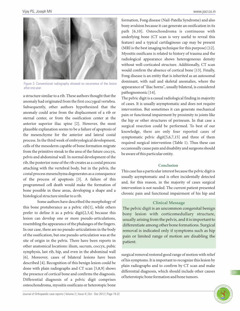
Figure 3. Conventional radiography showed no recurrence of the lesion after one year.
Clinical Message
The pelvic digit is an uncommon congenital benign
bony lesion with corticomedullary structure,
usually arising from the pelvis, and it is important to
differentiate among other bone formations. Surgical
removal is indicated only if symptoms such as hip
pain or limited range of motion are disabling the
patient.
21

References 8. Nguyen VD, Matthes JD, Wunderlich CC. The pelvic digit: CT correlation and review of the literature. Comput Med 1. Sullivan D, Cornwell WS. Pelvic rib. Report of a case. Imaging Graph 1990;14:127–31. Radiology.1974;110(2): 355-7.9. Van Breuseghem I. The pelvic digit: a harmless “eleventh” 2. Granieri GF, Bacarini L. The pelvic digit: five new examples finger. Br J Radiol. 2006;79:e106-7.of an unusual anomaly. Skeletal Radiol. 1996;25:723-6.10. Rijal L, Nepal P. Multiple Pelvic digits: a rare congenital 3. McGlone BS, Hamilton S, FitzGerald MJT. Pelvic digit: an anomaly. Eur J Orthop Surg Traumatol. 2010;20:411-3.uncommon developmental anomaly. Eur Radiol. 2000;10:89-11. Hamilton S. Pelvic digit. Br J Radiol 1985;58:1010–1.91.
12. Mavrogenis AF, Papagelopoulos PJ, Soucacos PN. 4. Greenspan A, Norman A. The “Pelvic Digit”, an unusual Skeletal osteochondromas revisited. Orthopedics. 2008 developmental anomaly. Skeletal Radiol. 1982;9:118-22.Oct;31(10)5. Lohan DG, Chang PC, Motamedi K, Seeger LL. Can you 13. Tyler P, Saifuddin A. The imaging of myositis ossificans. point to where the abnormality is?: Pelvic digit, a case report. Semin Musculoskelet Radiol. 2010 Jun;14(2):201-16.Eur J Radiol. 2009:Extra 72:e141-3.
14. Das CJ, Debnath J. Nail patella syndrome. Indian J 6. Maegele M. Pelvic digit as a rare cause of chronic hip pain Pediatr. 2009 Oct;76(10):1077.and functional impairment: a case report and review of the
literatura. J Med Case Reports. 2009;3:139. 15. Evangelista P, Evangelista G. Dyspareunia associated with a pelvic digit. JSCR 2011. 12:7.7. Pandey V, Thakur AS, Acharya KK, Rao PS. The pelvic digit
''eleventh finger". Indian J Orthop 2009;43:97-8.
www.jocr.co.inVijay PG, Joseph MV
Conflict of Interest: Nil Source of Support: None
How to Cite this Article:Moreta-Suárez J, Sáez de Ugarte-Sobrón O, Sánchez-Sobrino A, Martínez-De Los Mozos J. The Pelvic Digit: a Rare Congenital Anomaly as a cause of Hip Pain. Journal of Orthopaedic Case Reports. 2012 Oct-Dec;2(4):19-22
Journal of Orthopaedic case reports | Volume 2 | Issue 4 | Oct - Dec 2012 | Page 19-22
22


![Imaging of Pelvic Lymph Nodes - link.springer.com · The external iliac and obturator nodes are most commonly affected in endometrial cancer [26, 27•]. When the tumor is confined](https://img.pdfslide.us/doc/110x75/5ed5521912a6d6201a657fcf/imaging-of-pelvic-lymph-nodes-link-the-external-iliac-and-obturator-nodes-are.jpg)