Embed Size (px)
Citation preview
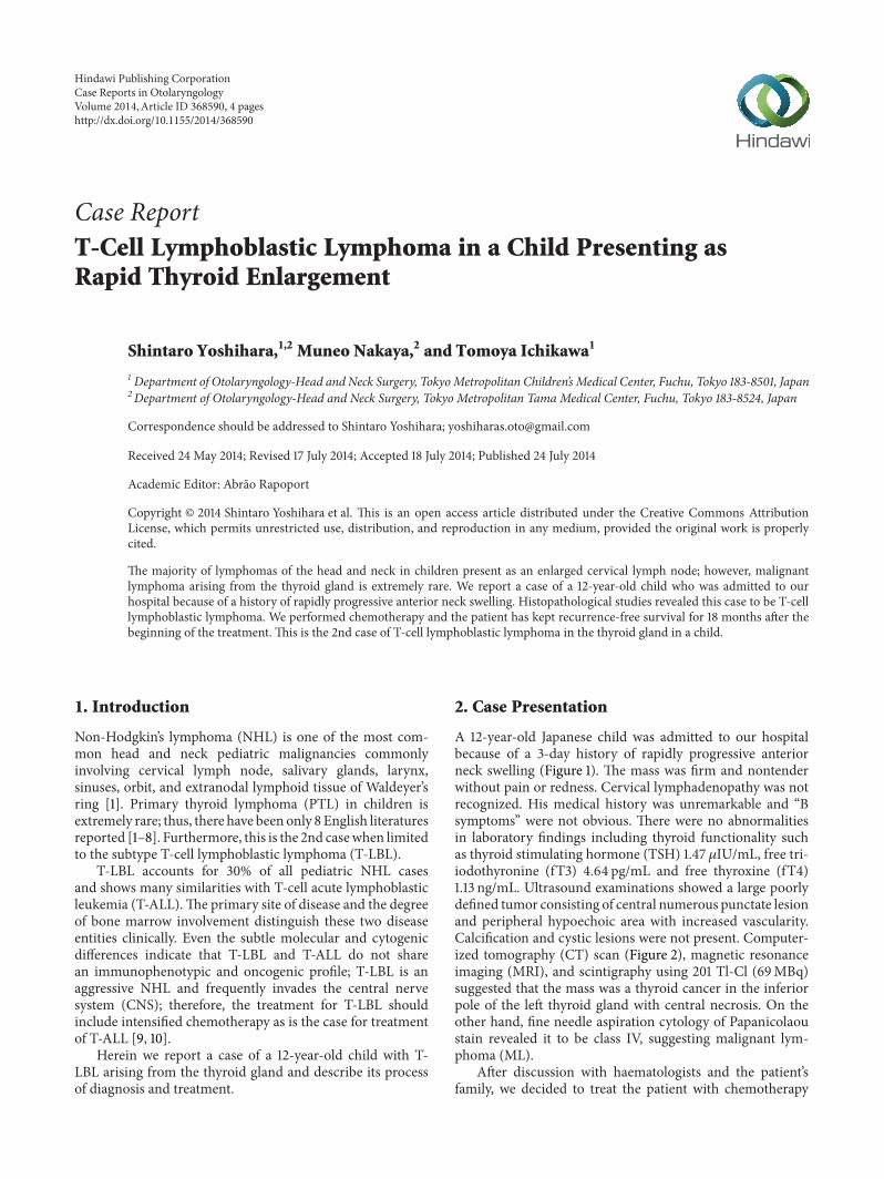
Case ReportT-Cell Lymphoblastic Lymphoma in a Child Presenting asRapid Thyroid Enlargement
Shintaro Yoshihara,1,2 Muneo Nakaya,2 and Tomoya Ichikawa1
1 Department of Otolaryngology-Head and Neck Surgery, TokyoMetropolitan Children’s Medical Center, Fuchu, Tokyo 183-8501, Japan2Department of Otolaryngology-Head and Neck Surgery, Tokyo Metropolitan Tama Medical Center, Fuchu, Tokyo 183-8524, Japan
Correspondence should be addressed to Shintaro Yoshihara; [email protected]
Received 24 May 2014; Revised 17 July 2014; Accepted 18 July 2014; Published 24 July 2014
Academic Editor: Abrao Rapoport
Copyright © 2014 Shintaro Yoshihara et al. This is an open access article distributed under the Creative Commons AttributionLicense, which permits unrestricted use, distribution, and reproduction in any medium, provided the original work is properlycited.
The majority of lymphomas of the head and neck in children present as an enlarged cervical lymph node; however, malignantlymphoma arising from the thyroid gland is extremely rare. We report a case of a 12-year-old child who was admitted to ourhospital because of a history of rapidly progressive anterior neck swelling. Histopathological studies revealed this case to be T-celllymphoblastic lymphoma. We performed chemotherapy and the patient has kept recurrence-free survival for 18 months after thebeginning of the treatment. This is the 2nd case of T-cell lymphoblastic lymphoma in the thyroid gland in a child.
1. Introduction
Non-Hodgkin’s lymphoma (NHL) is one of the most com-mon head and neck pediatric malignancies commonlyinvolving cervical lymph node, salivary glands, larynx,sinuses, orbit, and extranodal lymphoid tissue of Waldeyer’sring [1]. Primary thyroid lymphoma (PTL) in children isextremely rare; thus, there have been only 8 English literaturesreported [1–8]. Furthermore, this is the 2nd casewhen limitedto the subtype T-cell lymphoblastic lymphoma (T-LBL).
T-LBL accounts for 30% of all pediatric NHL casesand shows many similarities with T-cell acute lymphoblasticleukemia (T-ALL).The primary site of disease and the degreeof bone marrow involvement distinguish these two diseaseentities clinically. Even the subtle molecular and cytogenicdifferences indicate that T-LBL and T-ALL do not sharean immunophenotypic and oncogenic profile; T-LBL is anaggressive NHL and frequently invades the central nervesystem (CNS); therefore, the treatment for T-LBL shouldinclude intensified chemotherapy as is the case for treatmentof T-ALL [9, 10].
Herein we report a case of a 12-year-old child with T-LBL arising from the thyroid gland and describe its processof diagnosis and treatment.
2. Case Presentation
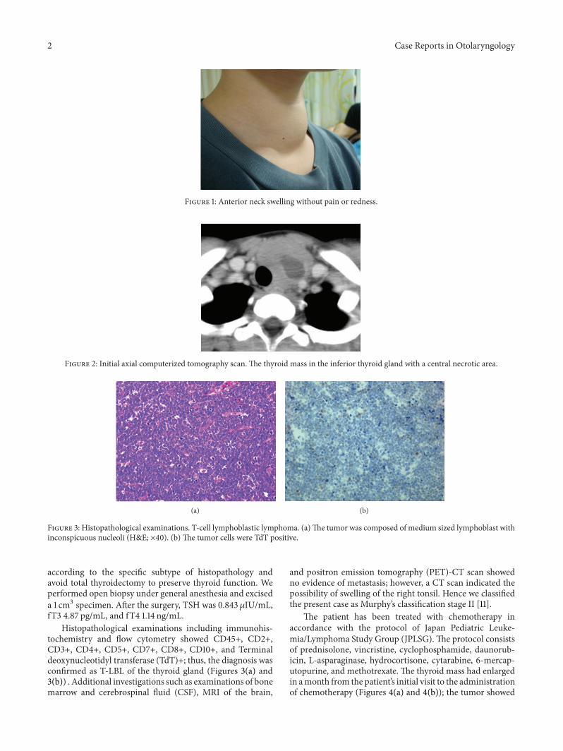
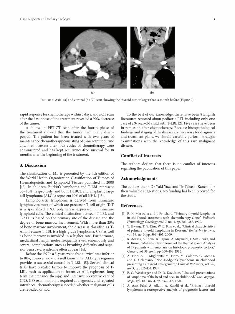
A 12-year-old Japanese child was admitted to our hospitalbecause of a 3-day history of rapidly progressive anteriorneck swelling (Figure 1). The mass was firm and nontenderwithout pain or redness. Cervical lymphadenopathy was notrecognized. His medical history was unremarkable and “Bsymptoms” were not obvious. There were no abnormalitiesin laboratory findings including thyroid functionality suchas thyroid stimulating hormone (TSH) 1.47 𝜇IU/mL, free tri-iodothyronine (fT3) 4.64 pg/mL and free thyroxine (fT4)1.13 ng/mL. Ultrasound examinations showed a large poorlydefined tumor consisting of central numerous punctate lesionand peripheral hypoechoic area with increased vascularity.Calcification and cystic lesions were not present. Computer-ized tomography (CT) scan (Figure 2), magnetic resonanceimaging (MRI), and scintigraphy using 201 Tl-Cl (69MBq)suggested that the mass was a thyroid cancer in the inferiorpole of the left thyroid gland with central necrosis. On theother hand, fine needle aspiration cytology of Papanicolaoustain revealed it to be class IV, suggesting malignant lym-phoma (ML).
After discussion with haematologists and the patient’sfamily, we decided to treat the patient with chemotherapy
Hindawi Publishing CorporationCase Reports in OtolaryngologyVolume 2014, Article ID 368590, 4 pageshttp://dx.doi.org/10.1155/2014/368590
2 Case Reports in Otolaryngology
Figure 1: Anterior neck swelling without pain or redness.
Figure 2: Initial axial computerized tomography scan. The thyroid mass in the inferior thyroid gland with a central necrotic area.
(a) (b)
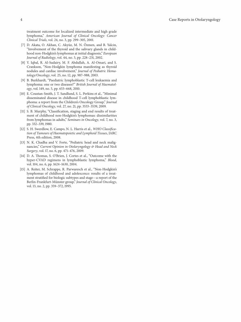
Figure 3: Histopathological examinations. T-cell lymphoblastic lymphoma. (a)The tumor was composed of medium sized lymphoblast withinconspicuous nucleoli (H&E; ×40). (b) The tumor cells were TdT positive.
according to the specific subtype of histopathology andavoid total thyroidectomy to preserve thyroid function. Weperformed open biopsy under general anesthesia and exciseda 1 cm3 specimen. After the surgery, TSH was 0.843𝜇IU/mL,fT3 4.87 pg/mL, and fT4 1.14 ng/mL.
Histopathological examinations including immunohis-tochemistry and flow cytometry showed CD45+, CD2+,CD3+, CD4+, CD5+, CD7+, CD8+, CD10+, and Terminaldeoxynucleotidyl transferase (TdT)+; thus, the diagnosis wasconfirmed as T-LBL of the thyroid gland (Figures 3(a) and3(b)) . Additional investigations such as examinations of bonemarrow and cerebrospinal fluid (CSF), MRI of the brain,
and positron emission tomography (PET)-CT scan showedno evidence of metastasis; however, a CT scan indicated thepossibility of swelling of the right tonsil. Hence we classifiedthe present case as Murphy’s classification stage II [11].
The patient has been treated with chemotherapy inaccordance with the protocol of Japan Pediatric Leuke-mia/Lymphoma Study Group (JPLSG).The protocol consistsof prednisolone, vincristine, cyclophosphamide, daunorub-icin, L-asparaginase, hydrocortisone, cytarabine, 6-mercap-utopurine, and methotrexate. The thyroid mass had enlargedin amonth from the patient’s initial visit to the administrationof chemotherapy (Figures 4(a) and 4(b)); the tumor showed
Case Reports in Otolaryngology 3
(a) (b)
Figure 4: Axial (a) and coronal (b) CT scan showing the thyroid tumor larger than a month before (Figure 2).
rapid response for chemotherapywithin 5 days, and aCT scanafter the first phase of the treatment revealed a 90% decreaseof the tumor.
A follow-up PET-CT scan after the fourth phase ofthe treatment showed that the tumor had totally disap-peared. The patient has been treated with two years ofmaintenance chemotherapy consisting of 6-mercaputopurineand methotrexate after four cycles of chemotherapy wereadministered and has kept recurrence-free survival for 18months after the beginning of the treatment.
3. Discussion
The classification of ML is presented by the 4th edition ofthe World Health Organization Classification of Tumors ofHaematopoietic and Lymphoid Tissues published in 2008[12]. In children, Burkitt’s lymphoma and T-LBL represent30–40%, respectively, and both DLBCL and anaplastic largecell lymphoma (ALCL) represent 10% of all NHLs [13].
Lymphoblastic lymphoma is derived from immaturelymphocytes most of which are precursor T-cell origin. TdTis a specialized DNA polymerase expressed in immaturelymphoid cells. The clinical distinction between T-LBL andT-ALL is based on the primary site of the disease and thedegree of bone marrow involvement. With more than 25%of bone marrow involvement, the disease is classified as T-ALL. Because T-LBL is a high-grade lymphoma, CSF as wellas bone marrow is involved in a higher rate. Furthermore,mediastinal lymph nodes frequently swell enormously andseveral complications such as breathing difficulty and supe-rior vena cava syndrome often appear [14].
Before the 1970’s a 5-year event-free survival was inferiorto 10%; however, now it is well known that ALL-type regimenprovides a successful control in T-LBL [15]. Several clinicaltrials have revealed factors to improve the prognosis of T-LBL, such as application of intensive ALL regimens, longterm maintenance therapy, and intensive preventive care ofCNS. CFS examination is required at diagnosis, and repeatedintrathecal chemotherapy is needed whether malignant cellsare revealed or not.
To the best of our knowledge, there have been 8 Englishliteratures reported about pediatric PTL including only onecase of a 9-year-old child with T-LBL [2]. Five cases have beenin remission after chemotherapy. Because histopathologicalfindings and staging of the disease are necessary for diagnosisand treatment plans, we should carefully perform strategicexaminations with the knowledge of this rare malignantdisease.
Conflict of Interests
The authors declare that there is no conflict of interestsregarding the publication of this paper.
Acknowledgments
The authors thank Dr Yuki Yuza and Dr Takashi Kaneko fortheir valuable suggestions. No funding has been received forthe study.
References
[1] R. K. Marwaha and J. Pritchard, “Primary thyroid lymphomain childhood: treatment with chemotherapy alone,” PediatricHematology-Oncology, vol. 7, no. 4, pp. 383–388, 1990.
[2] Y. Hwang, T. Y. Kim, W. B. Kim et al., “Clinical characteristicsof primary thyroid lymphoma in Koreans,” Endocrine Journal,vol. 56, no. 3, pp. 399–405, 2009.
[3] K. Aozasa, A. Inoue, K. Tajima, A. Miyauchi, F. Matsuzuka, andK. Kuma, “Malignant lymphomas of the thyroid gland: Analysisof 79 patients with emphasis on histologic prognostic factors,”Cancer, vol. 58, no. 1, pp. 100–104, 1986.
[4] A. Fiorillo, R. Migliorati, M. Fiore, M. Caldore, G. Menna,and L. Celentano, “Non-Hodgkin’s lymphoma in childhoodpresenting as thyroid enlargement,” Clinical Pediatrics, vol. 26,no. 3, pp. 152–154, 1987.
[5] E. C. Weisberger and D. D. Davidson, “Unusual presentationsof lymphoma of the head and neck in childhood,”The Laryngo-scope, vol. 100, no. 4, pp. 337–342, 1990.
[6] A. Aziz Belal, A. Allam, A. Kandil et al., “Primary thyroidlymphoma: a retrospective analysis of prognostic factors and
4 Case Reports in Otolaryngology
treatment outcome for localized intermediate and high gradelymphoma,” American Journal of Clinical Oncology: CancerClinical Trials, vol. 24, no. 3, pp. 299–305, 2001.
[7] D. Akata, O. Akhan, C. Akyuz, M. N. Ozmen, and B. Yalcin,“Involvement of the thyroid and the salivary glands in child-hood non-Hodgkin’s lymphomas at initial diagnosis,” EuropeanJournal of Radiology, vol. 44, no. 3, pp. 228–231, 2002.
[8] Y. Iqbal, R. Al-Sudairy, M. F. Abdullah, A. Al-Omari, and S.Crankson, “Non-Hodgkin lymphoma manifesting as thyroidnodules and cardiac involvement,” Journal of Pediatric Hema-tology/Oncology, vol. 25, no. 12, pp. 987–988, 2003.
[9] B. Burkhardt, “Paediatric lymphoblastic T-cell leukaemia andlymphoma: one or two diseases?” British Journal of Haematol-ogy, vol. 149, no. 5, pp. 653–668, 2010.
[10] E. Coustan-Smith, J. T. Sandlund, S. L. Perkins et al., “Minimaldisseminated disease in childhood T-cell lymphoblastic lym-phoma: a report from the Children’s Oncology Group,” Journalof Clinical Oncology, vol. 27, no. 21, pp. 3533–3539, 2009.
[11] S. B. Murphy, “Classification, staging and end results of treat-ment of childhood non-Hodgkin’s lymphomas: dissimilaritiesfrom lymphomas in adults,” Seminars in Oncology, vol. 7, no. 3,pp. 332–339, 1980.
[12] S. H. Swerdlow, E. Campo, N. L. Harris et al., WHO Classifica-tion of Tumours of Haematopoietic and Lymphoid Tissues, IARCPress, 4th edition, 2008.
[13] N. K. Chadha and V. Forte, “Pediatric head and neck malig-nancies,” Current Opinion in Otolaryngology & Head and NeckSurgery, vol. 17, no. 6, pp. 471–476, 2009.
[14] D. A. Thomas, S. O’Brien, J. Cortes et al., “Outcome with thehyper-CVAD regimens in lymphoblastic lymphoma,” Blood,vol. 104, no. 6, pp. 1624–1630, 2004.
[15] A. Reiter, M. Schrappe, R. Parwaresch et al., “Non-Hodgkin’slymphomas of childhood and adolescence: results of a treat-ment stratified for biologic subtypes and stage—a report of theBerlin-Frankfurt-Munster group,” Journal of Clinical Oncology,vol. 13, no. 2, pp. 359–372, 1995.

Submit your manuscripts athttp://www.hindawi.com
Stem CellsInternational
Hindawi Publishing Corporationhttp://www.hindawi.com Volume 2014
Hindawi Publishing Corporationhttp://www.hindawi.com Volume 2014
MEDIATORSINFLAMMATION
of
Hindawi Publishing Corporationhttp://www.hindawi.com Volume 2014
Behavioural Neurology
EndocrinologyInternational Journal of
Hindawi Publishing Corporationhttp://www.hindawi.com Volume 2014
Hindawi Publishing Corporationhttp://www.hindawi.com Volume 2014
Disease Markers
Hindawi Publishing Corporationhttp://www.hindawi.com Volume 2014
BioMed Research International
OncologyJournal of
Hindawi Publishing Corporationhttp://www.hindawi.com Volume 2014
Hindawi Publishing Corporationhttp://www.hindawi.com Volume 2014
Oxidative Medicine and Cellular Longevity
Hindawi Publishing Corporationhttp://www.hindawi.com Volume 2014
PPAR Research
The Scientific World JournalHindawi Publishing Corporation http://www.hindawi.com Volume 2014
Immunology ResearchHindawi Publishing Corporationhttp://www.hindawi.com Volume 2014
Journal of
ObesityJournal of
Hindawi Publishing Corporationhttp://www.hindawi.com Volume 2014
Hindawi Publishing Corporationhttp://www.hindawi.com Volume 2014
Computational and Mathematical Methods in Medicine
OphthalmologyJournal of
Hindawi Publishing Corporationhttp://www.hindawi.com Volume 2014
Diabetes ResearchJournal of
Hindawi Publishing Corporationhttp://www.hindawi.com Volume 2014
Hindawi Publishing Corporationhttp://www.hindawi.com Volume 2014
Research and TreatmentAIDS
Hindawi Publishing Corporationhttp://www.hindawi.com Volume 2014
Gastroenterology Research and Practice
Hindawi Publishing Corporationhttp://www.hindawi.com Volume 2014
Parkinson’s Disease
Evidence-Based Complementary and Alternative Medicine
Volume 2014Hindawi Publishing Corporationhttp://www.hindawi.com