Embed Size (px)
Citation preview
Case ReportSupraglottic Kaposi’s Sarcoma in HIV-Negative Patients:Case Report and Literature Review
Ela A. Server,1 Yusuf M. Durna,1 Ozgur Yigit,1 and Erol R. Bozkurt2
1Department of Otolaryngology, Istanbul Research and Training Hospital, 34540 Istanbul, Turkey2Department of Pathology, Istanbul Research and Training Hospital, 34540 Istanbul, Turkey
Correspondence should be addressed to Yusuf M. Durna; [email protected]
Received 3 December 2015; Revised 15 March 2016; Accepted 18 May 2016
Academic Editor: Kamal Morshed
Copyright © 2016 Ela A. Server et al. This is an open access article distributed under the Creative Commons Attribution License,which permits unrestricted use, distribution, and reproduction in any medium, provided the original work is properly cited.
This paper presents a case report of an HIV-negative, supraglottic Kaposi’s sarcoma patient. The 80-year-old male patient wasadmittedwith complaints of hoarseness, difficulty in swallowing, and a stinging sensation in his throat for approximately sixmonths.The endoscopic larynx examination revealed a lesion which had completely infiltrated the epiglottis, reached right aryepiglotticfold, was vegetating, pink and purple in color, multilobular, fragile, and shaped like a bunch of grapes, and partially blocked thebleeding airway passage. The case was discussed by the hospital’s head-neck cancer committee and a surgery decision was made.A tracheotomy was performed under local anesthesia before the operation due to respiratory distress and endotracheal intubationdifficulty. Direct laryngoscopy showed that themasswas limited in the supraglottic area, had invaded the entire left aryepiglottic foldand one-third of the front right aryepiglottic fold, and completely covered epiglottis. It should be remembered that although rare,Kaposi’s sarcoma may be encountered in larynx malignancy cases. Disease-free survival may be achieved through local excisionand postoperative radiotherapy.
1. Introduction
Kaposi’s sarcoma is a vascular tumor that was first describedby Kaposi in 1872 [1] and has a low potential of malignancy.Kaposi’s sarcoma is caused by human herpesvirus 8 (HHV-8)and often seen in Human Immunodeficiency Virus- (HIV-)infected patients [2, 3]. Its multifocal localization areas havebeen defined. It is frequently found in the lower extremities,facial skin, and genital and oropharyngeal mucosa but mayalso appear in gastrointestinal and respiratory tract mucosa,in addition to other less common areas, such as the larynx.This uncommon location is usually related to HIV [4, 5].Thispaper presents a case report and literature review of a rarecase of supraglottic, epiglottis, and aryepiglottic fold-derived,HIV-negative, HHV-8 positive Kaposi’s sarcoma.
2. Case Report
The 80-year-oldmale patient complained of hoarseness and astinging sensation in his throat for approximately six monthsand was then transferred to our clinic.The endoscopic larynx
examination revealed a lesion that completely infiltrated theepiglottis and reached the right aryepiglottic fold. It wasvegetating, pink and purple in color,multilobular, fragile, andshaped like a bunch of grapes and was covering the bleedingairway passage (Figure 1).
Before arriving at our clinic, a punch biopsy was doneand suggested a diagnosis of Kaposi’s sarcoma. The patientreported that he used to smoke approximately fifty packsof cigarettes per year but had not smoked in the last fiveyears. He had no history of systemic disease or infection.Nothing was remarkable in routine blood analyses. ELISAtests for HIV-1, HBV, and HCV were negative. A positronemission tomography (PET-CT) showed that no other areaswere affected. The case was discussed by our hospital’s head-neck cancer committee and a surgery decision was made. Atracheotomy was performed under local anesthesia beforethe operation due to respiratory distress and endotrachealintubation difficulty. The direct laryngoscopy showed thatthe mass was limited in the supraglottic area, had invadedthe entire left aryepiglottic fold and one-third of the frontright aryepiglottic fold, and completely covered epiglottis.
Hindawi Publishing CorporationCase Reports in OtolaryngologyVolume 2016, Article ID 1818304, 3 pageshttp://dx.doi.org/10.1155/2016/1818304
2 Case Reports in Otolaryngology
Figure 1: Pink and purple colored mass with lobular contours onthe epiglottis laryngeal surface near the aryepiglottic fold.
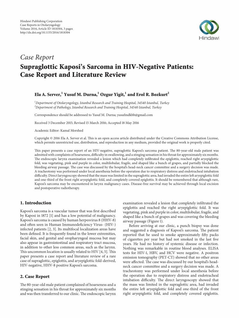
Figure 2: One week after endolaryngeal laser surgery, glottic levelswere intact and white-colored granulation tissues were observed.
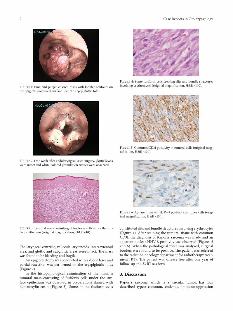
Figure 3: Tumoral mass consisting of fusiform cells under the sur-face epithelium (original magnification, H&E ×40).
The laryngeal ventricle, vallecula, arytenoids, interarytenoidarea, and glottic and subglottic areas were intact. The masswas found to be bleeding and fragile.
An epiglottectomy was conducted with a diode laser andpartial resection was performed on the aryepiglottic folds(Figure 2).
In the histopathological examination of the mass, atumoral mass consisting of fusiform cells under the sur-face epithelium was observed in preparations stained withhematoxylin-eosin (Figure 3). Some of the fusiform cells
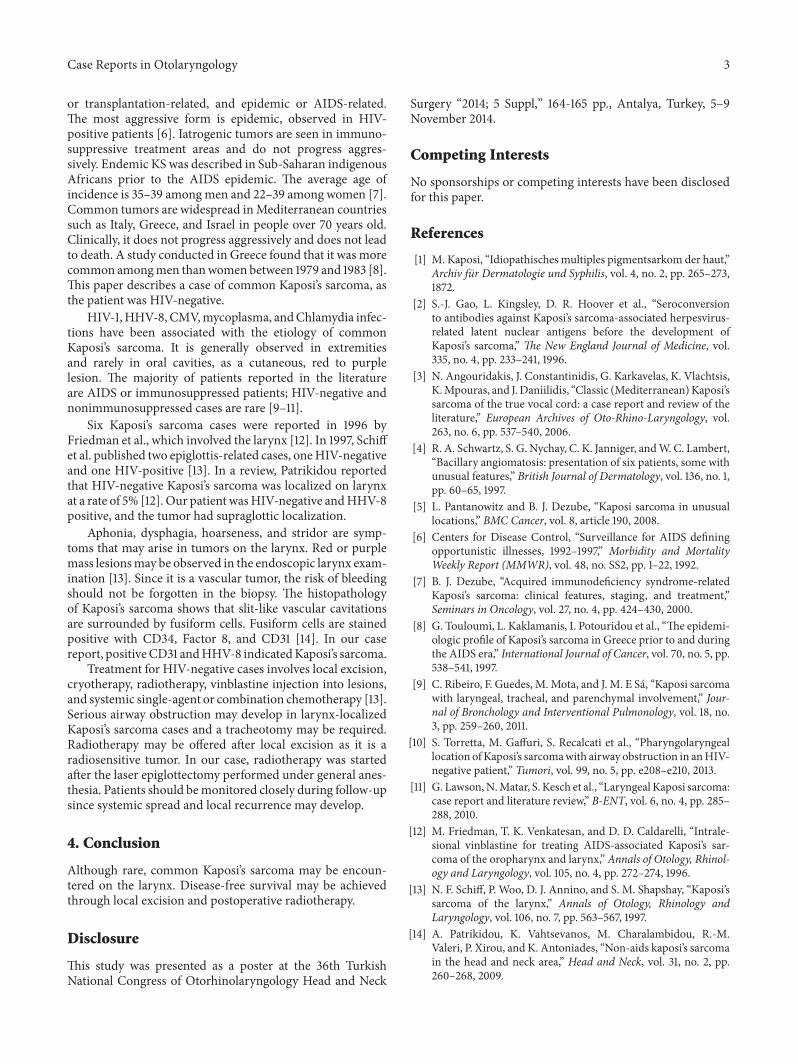
Figure 4: Some fusiform cells creating slits and bundle structuresinvolving erythrocytes (original magnification, H&E ×100).
Figure 5: Common CD31 positivity in tumoral cells (original mag-nification, H&E ×100).
Figure 6: Apparent nuclear HHV-8 positivity in tumor cells (orig-inal magnification, H&E ×100).
constituted slits and bundle structures involving erythrocytes(Figure 4). After staining the tumoral tissue with commonCD31, the diagnosis of Kaposi’s sarcoma was made and anapparent nuclear HHV-8 positivity was observed (Figures 5and 6). When the pathological piece was analyzed, surgicalborders were found to be positive. The patient was referredto the radiation oncology department for radiotherapy treat-ment (RT). The patient was disease-free after one year offollow-up and 33 RT sessions.
3. Discussion
Kaposi’s sarcoma, which is a vascular tumor, has fourdescribed types: common, endemic, immunosuppression
Case Reports in Otolaryngology 3
or transplantation-related, and epidemic or AIDS-related.The most aggressive form is epidemic, observed in HIV-positive patients [6]. Iatrogenic tumors are seen in immuno-suppressive treatment areas and do not progress aggres-sively. Endemic KS was described in Sub-Saharan indigenousAfricans prior to the AIDS epidemic. The average age ofincidence is 35–39 amongmen and 22–39 among women [7].Common tumors are widespread inMediterranean countriessuch as Italy, Greece, and Israel in people over 70 years old.Clinically, it does not progress aggressively and does not leadto death. A study conducted in Greece found that it was morecommon amongmen thanwomen between 1979 and 1983 [8].This paper describes a case of common Kaposi’s sarcoma, asthe patient was HIV-negative.
HIV-1,HHV-8, CMV,mycoplasma, andChlamydia infec-tions have been associated with the etiology of commonKaposi’s sarcoma. It is generally observed in extremitiesand rarely in oral cavities, as a cutaneous, red to purplelesion. The majority of patients reported in the literatureare AIDS or immunosuppressed patients; HIV-negative andnonimmunosuppressed cases are rare [9–11].
Six Kaposi’s sarcoma cases were reported in 1996 byFriedman et al., which involved the larynx [12]. In 1997, Schiffet al. published two epiglottis-related cases, oneHIV-negativeand one HIV-positive [13]. In a review, Patrikidou reportedthat HIV-negative Kaposi’s sarcoma was localized on larynxat a rate of 5% [12]. Our patient wasHIV-negative andHHV-8positive, and the tumor had supraglottic localization.
Aphonia, dysphagia, hoarseness, and stridor are symp-toms that may arise in tumors on the larynx. Red or purplemass lesionsmay be observed in the endoscopic larynx exam-ination [13]. Since it is a vascular tumor, the risk of bleedingshould not be forgotten in the biopsy. The histopathologyof Kaposi’s sarcoma shows that slit-like vascular cavitationsare surrounded by fusiform cells. Fusiform cells are stainedpositive with CD34, Factor 8, and CD31 [14]. In our casereport, positiveCD31 andHHV-8 indicatedKaposi’s sarcoma.
Treatment for HIV-negative cases involves local excision,cryotherapy, radiotherapy, vinblastine injection into lesions,and systemic single-agent or combination chemotherapy [13].Serious airway obstruction may develop in larynx-localizedKaposi’s sarcoma cases and a tracheotomy may be required.Radiotherapy may be offered after local excision as it is aradiosensitive tumor. In our case, radiotherapy was startedafter the laser epiglottectomy performed under general anes-thesia. Patients should bemonitored closely during follow-upsince systemic spread and local recurrence may develop.
4. Conclusion
Although rare, common Kaposi’s sarcoma may be encoun-tered on the larynx. Disease-free survival may be achievedthrough local excision and postoperative radiotherapy.
Disclosure
This study was presented as a poster at the 36th TurkishNational Congress of Otorhinolaryngology Head and Neck
Surgery “2014; 5 Suppl,” 164-165 pp., Antalya, Turkey, 5–9November 2014.
Competing Interests
No sponsorships or competing interests have been disclosedfor this paper.
References
[1] M. Kaposi, “Idiopathisches multiples pigmentsarkom der haut,”Archiv fur Dermatologie und Syphilis, vol. 4, no. 2, pp. 265–273,1872.
[2] S.-J. Gao, L. Kingsley, D. R. Hoover et al., “Seroconversionto antibodies against Kaposi’s sarcoma-associated herpesvirus-related latent nuclear antigens before the development ofKaposi’s sarcoma,” The New England Journal of Medicine, vol.335, no. 4, pp. 233–241, 1996.
[3] N. Angouridakis, J. Constantinidis, G. Karkavelas, K. Vlachtsis,K.Mpouras, and J.Daniilidis, “Classic (Mediterranean)Kaposi’ssarcoma of the true vocal cord: a case report and review of theliterature,” European Archives of Oto-Rhino-Laryngology, vol.263, no. 6, pp. 537–540, 2006.
[4] R. A. Schwartz, S. G. Nychay, C. K. Janniger, andW. C. Lambert,“Bacillary angiomatosis: presentation of six patients, some withunusual features,” British Journal of Dermatology, vol. 136, no. 1,pp. 60–65, 1997.
[5] L. Pantanowitz and B. J. Dezube, “Kaposi sarcoma in unusuallocations,” BMC Cancer, vol. 8, article 190, 2008.
[6] Centers for Disease Control, “Surveillance for AIDS definingopportunistic illnesses, 1992–1997,” Morbidity and MortalityWeekly Report (MMWR), vol. 48, no. SS2, pp. 1–22, 1992.
[7] B. J. Dezube, “Acquired immunodeficiency syndrome-relatedKaposi’s sarcoma: clinical features, staging, and treatment,”Seminars in Oncology, vol. 27, no. 4, pp. 424–430, 2000.
[8] G. Touloumi, L. Kaklamanis, I. Potouridou et al., “The epidemi-ologic profile of Kaposi’s sarcoma in Greece prior to and duringthe AIDS era,” International Journal of Cancer, vol. 70, no. 5, pp.538–541, 1997.
[9] C. Ribeiro, F. Guedes, M.Mota, and J. M. E Sa, “Kaposi sarcomawith laryngeal, tracheal, and parenchymal involvement,” Jour-nal of Bronchology and Interventional Pulmonology, vol. 18, no.3, pp. 259–260, 2011.
[10] S. Torretta, M. Gaffuri, S. Recalcati et al., “Pharyngolaryngeallocation ofKaposi’s sarcomawith airway obstruction in anHIV-negative patient,” Tumori, vol. 99, no. 5, pp. e208–e210, 2013.
[11] G. Lawson,N.Matar, S. Kesch et al., “Laryngeal Kaposi sarcoma:case report and literature review,” B-ENT, vol. 6, no. 4, pp. 285–288, 2010.
[12] M. Friedman, T. K. Venkatesan, and D. D. Caldarelli, “Intrale-sional vinblastine for treating AIDS-associated Kaposi’s sar-coma of the oropharynx and larynx,” Annals of Otology, Rhinol-ogy and Laryngology, vol. 105, no. 4, pp. 272–274, 1996.
[13] N. F. Schiff, P. Woo, D. J. Annino, and S. M. Shapshay, “Kaposi’ssarcoma of the larynx,” Annals of Otology, Rhinology andLaryngology, vol. 106, no. 7, pp. 563–567, 1997.
[14] A. Patrikidou, K. Vahtsevanos, M. Charalambidou, R.-M.Valeri, P. Xirou, and K. Antoniades, “Non-aids kaposi’s sarcomain the head and neck area,” Head and Neck, vol. 31, no. 2, pp.260–268, 2009.

Submit your manuscripts athttp://www.hindawi.com
Stem CellsInternational
Hindawi Publishing Corporationhttp://www.hindawi.com Volume 2014
Hindawi Publishing Corporationhttp://www.hindawi.com Volume 2014
MEDIATORSINFLAMMATION
of
Hindawi Publishing Corporationhttp://www.hindawi.com Volume 2014
Behavioural Neurology
EndocrinologyInternational Journal of
Hindawi Publishing Corporationhttp://www.hindawi.com Volume 2014
Hindawi Publishing Corporationhttp://www.hindawi.com Volume 2014
Disease Markers
Hindawi Publishing Corporationhttp://www.hindawi.com Volume 2014
BioMed Research International
OncologyJournal of
Hindawi Publishing Corporationhttp://www.hindawi.com Volume 2014
Hindawi Publishing Corporationhttp://www.hindawi.com Volume 2014
Oxidative Medicine and Cellular Longevity
Hindawi Publishing Corporationhttp://www.hindawi.com Volume 2014
PPAR Research
The Scientific World JournalHindawi Publishing Corporation http://www.hindawi.com Volume 2014
Immunology ResearchHindawi Publishing Corporationhttp://www.hindawi.com Volume 2014
Journal of
ObesityJournal of
Hindawi Publishing Corporationhttp://www.hindawi.com Volume 2014
Hindawi Publishing Corporationhttp://www.hindawi.com Volume 2014
Computational and Mathematical Methods in Medicine
OphthalmologyJournal of
Hindawi Publishing Corporationhttp://www.hindawi.com Volume 2014
Diabetes ResearchJournal of
Hindawi Publishing Corporationhttp://www.hindawi.com Volume 2014
Hindawi Publishing Corporationhttp://www.hindawi.com Volume 2014
Research and TreatmentAIDS
Hindawi Publishing Corporationhttp://www.hindawi.com Volume 2014
Gastroenterology Research and Practice
Hindawi Publishing Corporationhttp://www.hindawi.com Volume 2014
Parkinson’s Disease
Evidence-Based Complementary and Alternative Medicine
Volume 2014Hindawi Publishing Corporationhttp://www.hindawi.com