Embed Size (px)
Citation preview

Int J Clin Exp Pathol 2015;8(3):3299-3303www.ijcep.com /ISSN:1936-2625/IJCEP0004992
Case Report Spindle cell lipoma of the wrist, occurring in a distinctly rare location: a case report with review of literature
Keisuke Akaike1,2, Yoshiyuki Suehara2, Tatsuya Takagi2, Kazuo Kaneko2, Atsushi Yamashita1, Takashi Yao1, Tsuyoshi Saito1
Departments of 1Human Pathology, 2Orthopaedic Surgery, School of Medicine, Juntendo University, 2-1-1 Hongo, Bunkyo-ku Tokyo 113-8421, Japan
Received December 17, 2014; Accepted February 21, 2015; Epub March 1, 2015; Published March 15, 2015
Abstract: Spindle cell lipoma (SCL) is a rare, benign adipocytic tumor commonly arising in the upper neck, back, and shoulder regions. To the best of our knowledge, only one case of SCL of the wrist has previously been reported. We herein report a rare case of SCL arising at the wrist. A 77-year-old man presented with a 4-year history of a mass in the right wrist. Radiography showed no significant findings, and magnetic resonance imaging demonstrated the presence of a mass on the radial dorsal side of the right wrist. Needle biopsy suggested the tumor was SCL, and total excision was performed. Macroscopically, the tumor was circumscribed by fibrous membrane with a yellowish to partly white surface. Histologically, the tumor was composed of mature adipocytes and proliferation of the less atypical spindle cells in a ropey-like collagen background. Immunohistochemically, the tumor cells showed diffuse and strong expression for CD34. The final diagnosis of SCL was made on the basis of these pathological and radio-logical findings. The patient was successfully treated and shows no evidence of disease at 3 months after surgery.
Keywords: Spindle cell lipoma, wrist
Introduction
Spindle cell lipoma (SCL) is a benign adipocytic tumor first described in 1975 [1, 2]. According to the recent edition of the World Health Organization (WHO) Classification of Tumours of Soft Tissue and Bone, lipomas are catego-rized into 11 types [3]. SCL is rare, accounting for approximately 1.5% of all lipomas [4]. Histologically, SCL is classically composed of mature fat cells, bland spindle cells with low mitotic activity, ropey-like collagen, and myxoid matrix. By immunohistochemistry, spindle cell positivity for CD34 has been used as a diag-nostic marker for SCL [5, 6]. However, despite these pathological characteristics, a definite diagnosis is sometimes difficult because of tumor variants with proliferation of spindle cells and scant adipocytes [6]. Differential diagnosis is important for tumors that share histological features with SCL such as well-differentiated sclerosing liposarcoma (WSLS) and spindle cell liposarcoma (SCLS) [7, 8]. In contrast to well-differentiated liposarcoma that occurs in vari-ous locations, SCL mainly occurs in subcutane-
ous regions of the upper back, neck, or shoul-der, and occurrence in other sites is uncommon [1, 3]. Previous reports document SCL arising in rare sites such as the face, forehead, upper arm, and thigh [2, 4, 5]. However, there are only a few reports of SCL of the wrist and hand. To the best of our knowledge, only one case of SCL of the wrist has been presented in the literature [5]. We herein report a distinctly rare case of SCL arising in the right wrist that was treated with complete excision.
Case report
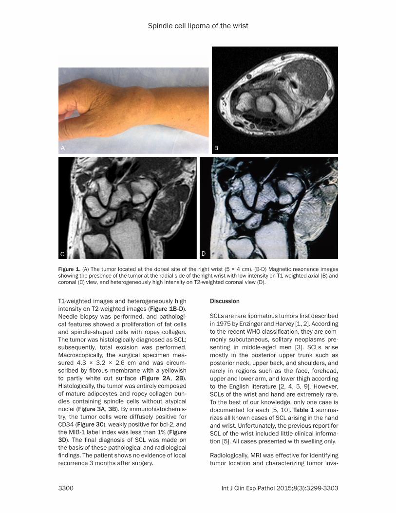
A 77-year-old man presented with a 4-year his-tory of a mass of the right wrist that did not cause pain and had grown gradually during semiannual observation. Physical examination revealed a soft mass (5 × 4 cm) with good mobility (Figure 1A). The patient did not have motor or sensory disturbance. Although radiog-raphy showed no significant findings, magnetic resonance imaging (MRI) demonstrated the presence of a mass on the radial and dorsal side of the right wrist with low intensity on
Spindle cell lipoma of the wrist
3300 Int J Clin Exp Pathol 2015;8(3):3299-3303
T1-weighted images and heterogeneously high intensity on T2-weighted images (Figure 1B-D). Needle biopsy was performed, and pathologi-cal features showed a proliferation of fat cells and spindle-shaped cells with ropey collagen. The tumor was histologically diagnosed as SCL; subsequently, total excision was performed. Macroscopically, the surgical specimen mea-sured 4.3 × 3.2 × 2.6 cm and was circum-scribed by fibrous membrane with a yellowish to partly white cut surface (Figure 2A, 2B). Histologically, the tumor was entirely composed of mature adipocytes and ropey collagen bun-dles containing spindle cells without atypical nuclei (Figure 3A, 3B). By immunohistochemis-try, the tumor cells were diffusely positive for CD34 (Figure 3C), weakly positive for bcl-2, and the MIB-1 label index was less than 1% (Figure 3D). The final diagnosis of SCL was made on the basis of these pathological and radiological findings. The patient shows no evidence of local recurrence 3 months after surgery.
Discussion
SCLs are rare lipomatous tumors first described in 1975 by Enzinger and Harvey [1, 2]. According to the recent WHO classification, they are com-monly subcutaneous, solitary neoplasms pre-senting in middle-aged men [3]. SCLs arise mostly in the posterior upper trunk such as posterior neck, upper back, and shoulders, and rarely in regions such as the face, forehead, upper and lower arm, and lower thigh according to the English literature [2, 4, 5, 9]. However, SCLs of the wrist and hand are extremely rare. To the best of our knowledge, only one case is documented for each [5, 10]. Table 1 summa-rizes all known cases of SCL arising in the hand and wrist. Unfortunately, the previous report for SCL of the wrist included little clinical informa-tion [5]. All cases presented with swelling only.
Radiologically, MRI was effective for identifying tumor location and characterizing tumor inva-
Figure 1. (A) The tumor located at the dorsal site of the right wrist (5 × 4 cm). (B-D) Magnetic resonance images showing the presence of the tumor at the radial side of the right wrist with low intensity on T1-weighted axial (B) and coronal (C) view, and heterogeneously high intensity on T2-weighted coronal view (D).

Spindle cell lipoma of the wrist
3301 Int J Clin Exp Pathol 2015;8(3):3299-3303
sion. However, in tumors that consist of low-fat or fat-free components, SCL can be difficult to diagnose using MRI [11, 12]. Khashper et al. reported that fat-free components of SCL with isointensity to muscle on T1-weighted images and heterogeneously variable intensity to fat on T2-weighted images resembled those of other soft-tissue tumors such as schwannomas, neu-rofibromas, and liposarcomas. In our case, MRI showed low intensity on T1-weighted images and heterogeneously high intensity on T2-weighted images, which are not typically characteristic of lipomatous tumors. Because the wrist is an uncommon location for SCL, radiologists are not likely to consider SCL as a differential diagnosis for this tumor. Fibrolipomatous hamartoma of the nerve is another possible radiological diagnosis for the mass, although it rarely arises from the radial side of the wrist [13, 14]. In one report, 9 out of 26 patients with fibrolipomatous hamartoma had neurologic symptoms of pain or paresthe-sias [13]. However, the current patient did not have any neurologic symptoms.
Among the histological mimics for SCL, liposar-coma, especially WSLS and SCLS variants, is an important differential diagnosis. According to the WHO classification, well-differentiated liposarcoma is divided into 3 subtypes: lipoma-like, sclerosing, and inflammatory [3]. Among liposarcoma subtypes, WSLS is second in fre-quency to the lipoma-like subtype and is histo-logically characterized by dense stromal fibro-sis with scattered bizarre cells and proliferation of mature adipose tissue with lipoblasts at vari-ous levels [8, 15]. Similarly, SCL consists of mature fat cells and spindle shaped cells with collagen matrix. Lipoblasts and floret cells are commonly absent in SCL. Immunohistoche- mically, CD34 expression is diffuse and strong in SCL [5, 6], while weak or absent in WSLS.
SCLS must also be differentiated. SCLS was previously categorized as a well-differentiated liposarcoma [16] but was excluded from the category of well-differentiated liposarcoma in the recent WHO classification [3]. SCL and SCLS share similar immunohistochemical and
Figure 2. During the surgery, it was noted that the lobulated tumor was encapsulated without adhesion to the sur-rounding soft tissue. The tumor size was 4.3 × 3.2 × 2.6 cm (A). The cut surface of the resected tumor shows a yellow component divided by a white septal wall (B).

Spindle cell lipoma of the wrist
3302 Int J Clin Exp Pathol 2015;8(3):3299-3303
molecular genetic characteristics. In one study examining 18 SCLs, all tumors were found to lack Rb-1 expression [17]. In another study, all SCLSs examined showed Rb-1 deletion and at least focal CD34 expression [18]. However, no SCLSs showed amplification of MDM2 and/or CDK4, which is characteristic of well-differenti-ated liposarcoma, establishing SCLS as an atypical counterpart of SCL rather than includ-ing it in the category of well-differentiated lipo-sarcoma [18].
Because of similarities between SCL and SCLS, differential diagnosis should be made carefully. Histologically, SCLS contains atypical spindle cells with enlarged nuclei, a variable amount of
atypical adipocytes, and vacuolated lipoblasts [18]. The present case did not contain atypical cells with enlarged and pleomorphic nuclei, and lipoblasts were not seen throughout the tumor. These findings led us to the final diagnosis of SCL.
In summary, we identified a case of SCL of the wrist. This location is extremely rare for SCL, representing only the second case of SCL aris-ing at the wrist.
Acknowledgements
This work was supported in part by a Grant-in-Aid for General Scientific Research from the
Figure 3. (A, B) Histologically, the tumor was composed of mature adipocytes and proliferation of the less atypical spindle cells in a ropey-like collagen background. Original magnification: ×40 (A) and ×200 (B). The tumor cells were diffusely positive for CD34 (C) and the MIB-1 label index was less than 1% (D).
Table 1. Reported cases of spindle cell lipoma at the wrist and handNo Author Age Sex Location aspect Size (cm) Treatment Outcome1 Templeton et al. [5] 40 M Wrist - 2.4 - -2 Varatharaj et al. [10] 34 F Palm volar 4.0 resection NED/12mos3 Present case 77 M Wrist dorsal 4.2 resection NED/3mos

Spindle cell lipoma of the wrist
3303 Int J Clin Exp Pathol 2015;8(3):3299-3303
Ministry of Education, Science, Sports and Culture (#26670286 to Tsuyoshi Saito and # 25861342 to Yoshiyuki Suehara), Tokyo, Japan.
Disclosure of conflict of interest
None.
Adress correspondence to: Dr. Tsuyoshi Saito, De- partment of Human Pathology, School of Medicine, Juntendo University, 2-1-1 Hongo, Bunkyo-ku Tokyo, Japan. Tel: +81-3-3813-3111; Fax: +81-3-3813-3428; E-mail: [email protected]
References
[1] Goldblum JR, Weiss SW, Folpe AL. Enzinger and weiss’s soft tissue tumors. 2013; p456-63.
[2] Enzinger FM, Harvey DA. Spindle cell lipoma. Cancer 1975; 36: 1852-59.
[3] WHO classification of tumours of soft tissue and bone. Fletcher CDM, Bridge JA, Hogen-doorn PCW, Mertens F, editors. World Health Organization International Agency for Re-search on Cancer. IARC Press; 2013. pp. 33-6.
[4] Fletcher CD, Martin-Bates E. Spindle cell lipo-ma: a clinicopathological study with some orig-inal observations. Histopathology 1987; 11: 803-17.
[5] Templeton SF, Solomon AR Jr. Spindle cell li-poma is strongly CD34 positive. An immuno-histochemical study. J Cutan Pathol 1996; 23: 546-50.
[6] Billings SD, Folpe AL. Diagnostically Chall- enging Spindle Cell lipomas: a report of 34 “low-fat” and “fat-free” variants. Am J Derma-topathol 2007; 29: 437-42.
[7] Dei Tos AP, Mentzel T, Newman PL, Fletcher CD. Spindle cell liposarcoma, a hitherto unrec-ognized variant of liposarcoma. Analysis of six cases. Am J Surg Pathol 1994; 18: 913-21.
[8] Shattuck MC, Victor TA. Cytologic features of well-differentiated sclerosing liposarcoma in aspirated samples. Acta Cytol 1988; 32: 896-901.
[9] Angervall L, Dahl I, Kindblom LG, Säve-Söder-bergh. Spindle cell lipoma. Acta Pathol Micro-biol Scand A 1976; 84: 477-87.
[10] Mounasamy V, Thacker M, Bugnone AN, Hum-ble SP, Pitcher JD, Temple HT. Spindle cell li-poma of the hand. Eur J Orthop Surg Traumatol 2006; 16: 248-50.
[11] Kirwadi A, Abdul-Halim R, Fernando M, High-land A, Kotnis N. MR imaging features of spin-dle cell lipoma. Skeletal Radiol 2014; 43: 191-6.
[12] Khashper A, Zheng J, Nahal A, Discepola F. Im-aging characteristics of spindle cell lipoma and its variants. Skeletal Radiol 2014; 43: 591-7.
[13] Silverman TA, Enzinger FM. Fibrolipomatous hamartoma of nerve. A clinicopathologic anal-ysis of 26 cases. Am J Surg Pathol 1985; 9: 7-14.
[14] Okubo T, Saito T, Mitomi H, Takagi T, Torigoe T, Suehara Y, Katagiri H, Murata H, Takahashi M, Ito I, Yao T, Kaneko K. Intraneural lipomatous tumor of the median nerve: Three case reports with a review of literature. Int J Surg Case Rep 2012; 3: 407-11.
[15] Bestic JM, Kransdorf MJ, White LM, Bridges MD, Murphey MD, Peterson JJ, Garner HW. Sclerosing variant of well-differentiated lipo-sarcoma: relative prevalence and spectrum of CT and MRI features. AJR Am J Roentgenol 2013; 201: 154-61.
[16] WHO Classification Tumours of Soft Tissue and Bone. Fletcher CDM, Unni KK, Mertens F, edi-tors. World Health Organization International Agency for Research On Cancer. IARC Press; 2002. pp. 35-7.
[17] Chen BJ, Mariño-Enríquez A, Fletcher CD, Hor-nick JL. Loss of retinoblastoma protein expres-sion in spindle cell/pleomorphic lipomas and cytogenetically related tumors: an immunohis-tochemical study with diagnostic implications. Am J Surg Pathol 2012; 36: 1119-28.
[18] Mentzel T, Palmedo G, Kuhnen C. Well-differ-entiated spindle cell liposarcoma (‘atypical spindle cell lipomatous tumor’) does not be-long to the spectrum of atypical lipomatous tumor but has a close relationship to spindle cell lipoma: clinicopathologic, immunohisto-chemical, and molecular analysis of six cases. Mod Pathol 2010; 23: 729-36.
![Large buccal fat pad lipoma: A rare case report...gland lipoma in 2 cases, angiolipoma in 2 cases, and spindle cell lipoma in 3 cases [10]. The most common presentation of BFP lipoma](https://img.pdfslide.us/doc/110x75/5e610a1252021369db53e163/large-buccal-fat-pad-lipoma-a-rare-case-report-gland-lipoma-in-2-cases-angiolipoma.jpg)


![Case Report Spindle Cell Lipoma of the Soft Palatedownloads.hindawi.com/journals/criot/2015/813240.pdftumours manifest as asymptomatic, slow-growing submu-cosal nodules [ , ]. Approximately](https://img.pdfslide.us/doc/110x75/5f0c0cb77e708231d4337f95/case-report-spindle-cell-lipoma-of-the-soft-tumours-manifest-as-asymptomatic-slow-growing.jpg)