Embed Size (px)
Citation preview
Int J Clin Exp Pathol 2014;7(12):8996-9001www.ijcep.com /ISSN:1936-2625/IJCEP0002499
Case ReportOvarian malignant mixed germ cell tumor with clear cell carcinoma in a postmenopausal woman
Xiu-Jie Yu1, Lin Zhang1, Zai-Ping Liu2, Yi-Quan Shi1, Yi-Xin Liu1
1Department of Pathology, Tianjin Central Hospital of Gynecology and Obstetrics, Tianjin, China; 2Department of Pathology and Laboratory Medicine, Dalhousie University, Halifax, Nova Scoria, Canada
Received September 14, 2014; Accepted November 1, 2014; Epub December 1, 2014; Published December 15, 2014
Abstract: Malignant germ cell tumors of the ovary are very rare and account for about 2-5% of all ovarian tumors of germ origin. Most patients are adolescent and young women, approximately two-thirds of them are under 20 years of age, occasionally in postmenopausal women. But clear cell carcinoma usually occurs in older patients (median age: 57-year old), and closely related with endometriosis. Here we report a case of a 55-year old woman with right ovarian mass that discovered by B ultrasonic. Her serum levels of human chorionic gonadotropin (hCG) and α-fetoprotein (AFP) were elevated. Pathological examination revealed the tumor to be a mixed germ cell tumor (yolk sac tumor, embryonal carcinoma and mature teratoma) with clear cell carcinoma in a background of endometriosis. Immunohistochemical staining showed SALL4 and PLAP were positive in germ cell tumor area, hCG, CD30 and OCT4 were positive in epithelial-like cells and giant synctiotrophoblastic cells, AFP, AAT, CD117 and Glyp3 were posi-tive in yolk sac component, EMA and CK7 were positive in clear cell carcinoma, CD10 was positive in endometrial cells of endometriotic area. She was treated with surgery followed by seven courses of chemotherapy. She is well and serum levels of hCG and AFP have been decreased to normal levels.
Keywords: Mixed germ cell tumor, ovary, clear cell carcinoma
Introduction
Mixed germ cell tumors of the ovary are rare malignant neoplasms containing combinations of two or more types of germ cell element [1, 2], such as dysgerminoma combined with terato-ma, yolk sac tumor, choriocarcinoma, embryo-nal carcinoma, or polyembryoma, as well as any other possible combination of these tumor types. Like other malignant germ cell tumor, these tumors occur in the first four decades most frequently in children and adolescents and are rare thereafter. But ovarian clear cell carcinoma usually occurs in patients over 50 years old (median age: 57-year old), and can arise from endometriosis (35.9%) [3].
Herein we reported the case of a 55-year-old woman with right ovarian mixed germ tumor (composed of yolk sac tumor, embryonal carci-noma and mature teratoma) combination of clear cell carcinoma in a background of en- dometriosis.
Case report
The patient was a 55-year-old woman and had postmenopausal for 5 years. Recently, she pre-sented with right lower abdomen discomfort and weakness of both lower limbs. Her past medical history was unremarkable except for dysmenorrheal and multiple uterine myomata. She had no surgery in the past. Physical exami-nation revealed an irregular, nontender lower abdominal tumor at right rear uterus. B ultra-sound examination found a solid and cystic neoplasm of right ovary (5.1 cm × 5.0 cm × 3.4 cm) and multiple myomata of uterus (the bi ggest measuring 2.5 cm × 2.0 cm × 1.8 cm). Her preoperative serum tumor marker levels were CA199: 14.24 U/ml, CEA: 1.39 ng/ml, CA- 125: 15.45 U/ml, HE4: 1.11 pmol/L, AFP: 47.5 ng/ml, and HCG: 831.6 mIU/ml. These results strongly favored malignant ovarian germ cell tumor.
The patient then consented for surgical opera-tion performed. The histological findings of
Ovarian malignant mixed germ cell tumor
8997 Int J Clin Exp Pathol 2014;7(12):8996-9001
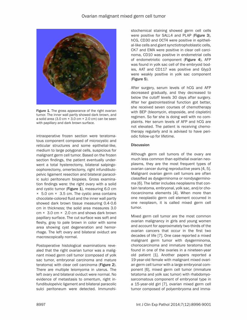
intraoperative frozen section were teratoma-tous component composed of microcystic and reticular structures and some epithelial-like, medium to large polygonal cells, suspicious for malignant germ cell tumor. Based on the frozen section findings, the patient eventually under-went a total hysterectomy, bilateral salpingo-oophorectomy, omertectomy, right infundibulo-pelvic ligament resection and bilateral paracol-ic sulci peritoneum biopsies. Gross examina-tion findings were: the right ovary with a solid and cystic tumor (Figure 1), measuring 6.0 cm × 5.0 cm × 3.5 cm. The cystic area contains chocolate-colored fluid and the inner wall partly showed dark brown tissue measuring 0.4-0.6 cm in thickness; the solid area measures 3.0 cm × 3.0 cm × 2.0 cm and shows dark brown papillary surface. The cut surface was soft and fleshy, gray to pale brown in color with some area showing cyst degeneration and hemor-rhage. The left ovary and bilateral oviduct are macroscopically normal.
Postoperative histological examinations reve- aled that the right ovarian tumor was a malig-nant mixed germ cell tumor (composed of yolk sac tumor, embryonal carcinoma and mature teratoma) with clear cell carcinoma (Figure 2). There are multiple leiomyoma in uterus. The left ovary and bilateral oviduct were normal. No evidence of metastasis to omentum, right in- fundibulopelvic ligament and bilateral paracolic sulci peritoneum were detected. Immunohi-
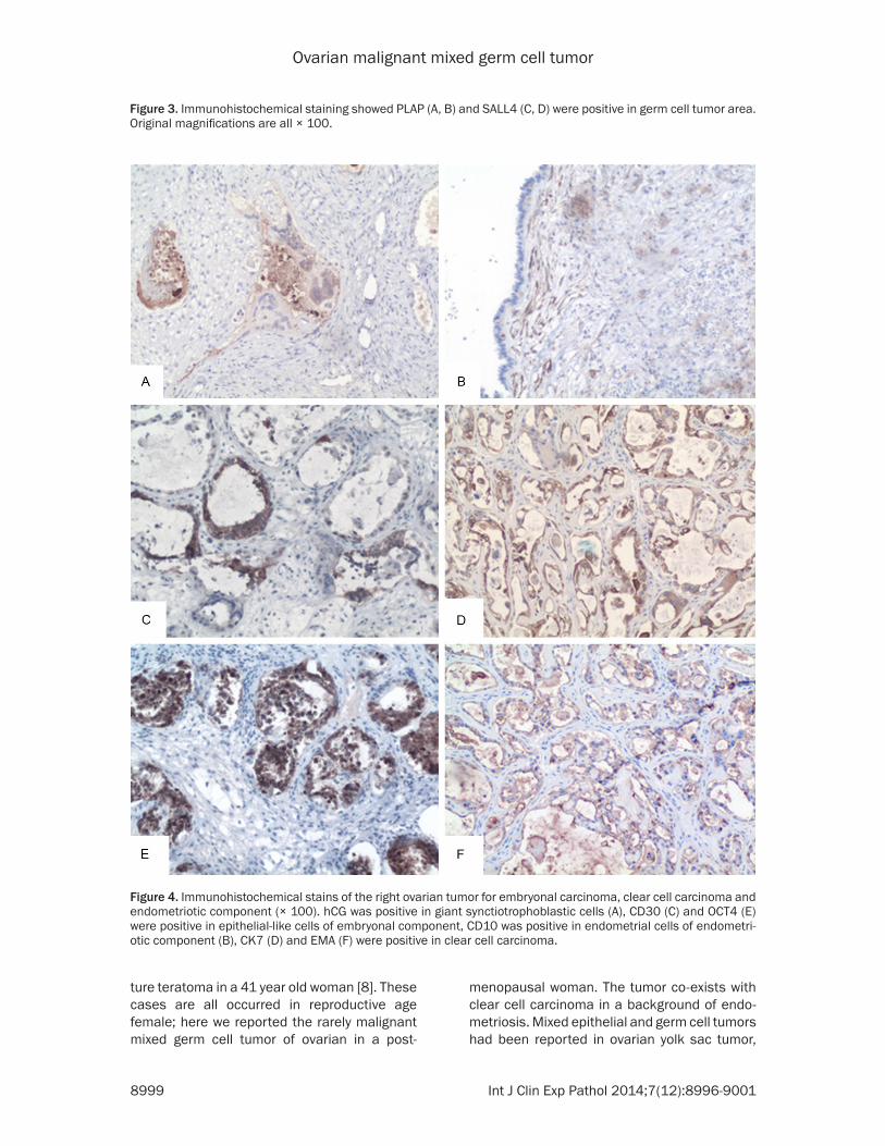
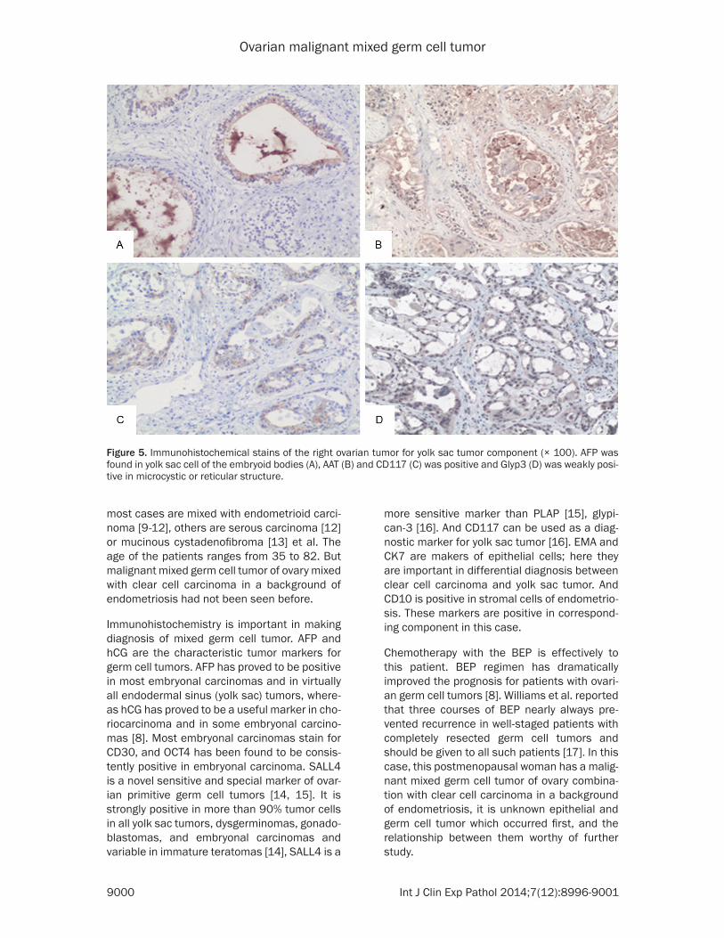
stochemical staining showed germ cell cells were positive for SALL4 and PLAP (Figure 3), hCG, CD30 and OCT4 were positive in epitheli-al-like cells and giant synctiotrophoblastic cells, CK7 and EMA were positive in clear cell carci-noma, CD10 was positive in endometrial cells of endometriotic component (Figure 4), AFP was found in yolk sac cell of the embryoid bod-ies, AAT and CD117 was positive and Glyp3 were weakly positive in yolk sac component (Figure 5).
After surgery, serum levels of hCG and AFP decreased gradually, and they decreased to below the cutoff levels 30 days after surgery. After her gastrointestinal function got better, she received seven courses of chemotherapy with BEP (bleomycin, etoposide, and cisplatin) regimen. So far she is doing well with no com-plaints. Her serum levels of AFP and hCG are not elevated. The patient is receiving chemo-therapy regularly and is advised to have peri-odic follow-up for lifetime.
Discussion
Although germ cell tumors of the ovary are much less common than epithelial ovarian neo-plasms, they are the most frequent types of ovarian cancer during reproductive years [4, 5]. Malignant ovarian germ cell tumors are often classified as dysgerminoma or nondysgermino-ma [6]. The latter includes neoplasms that con-tain teratoma, embryonal, yolk sac, and/or cho-riocarcinoma elements [4]. When more than one neoplastic germ cell element occurred in one neoplasm, it is called mixed germ cell tumor.
Mixed germ cell tumor are the most common ovarian malignancy in girls and young women and account for approximately two-thirds of the ovarian cancers that occur in the first two decades of life [7]. One case reported a mixed malignant germ tumor with dysgerminoma, choriocarcinoma and immature teratoma that found in one of the ovaries in a nineteen-year old patient [1]. Another papers reported a 19-year-old female with malignant mixed ovari-an germ cell tumor with a large embryonal com-ponent [6], mixed germ cell tumor (immature tetatoma and yolk sac tumor) with rhabdomyo-sarcomatous component of embryonal type in a 15-year-old girl [7], ovarian mixed germ cell tumor composed of polyembryoma and imma-
Figure 1. The gross appearance of the right ovarian tumor. The inner wall partly showed dark brown, and a solid area (3.0 cm × 3.0 cm × 2.0 cm) can be seen with papillary and dark brown surface.

Ovarian malignant mixed germ cell tumor
8998 Int J Clin Exp Pathol 2014;7(12):8996-9001
Figure 2. Histopathological found the right ovarian tumor was a malignant mixed germ cell tumor (composed of yolk sac tumor (A), embryonal carcinoma (B) and mature teratoma (C)) with clear cell carcinoma (D) by H&E staining. Original magnifications are all × 100.
Ovarian malignant mixed germ cell tumor
8999 Int J Clin Exp Pathol 2014;7(12):8996-9001
ture teratoma in a 41 year old woman [8]. These cases are all occurred in reproductive age female; here we reported the rarely malignant mixed germ cell tumor of ovarian in a post-
menopausal woman. The tumor co-exists with clear cell carcinoma in a background of endo-metriosis. Mixed epithelial and germ cell tumors had been reported in ovarian yolk sac tumor,
Figure 3. Immunohistochemical staining showed PLAP (A, B) and SALL4 (C, D) were positive in germ cell tumor area. Original magnifications are all × 100.
Figure 4. Immunohistochemical stains of the right ovarian tumor for embryonal carcinoma, clear cell carcinoma and endometriotic component (× 100). hCG was positive in giant synctiotrophoblastic cells (A), CD30 (C) and OCT4 (E) were positive in epithelial-like cells of embryonal component, CD10 was positive in endometrial cells of endometri-otic component (B), CK7 (D) and EMA (F) were positive in clear cell carcinoma.
Ovarian malignant mixed germ cell tumor
9000 Int J Clin Exp Pathol 2014;7(12):8996-9001
most cases are mixed with endometrioid carci-noma [9-12], others are serous carcinoma [12] or mucinous cystadenofibroma [13] et al. The age of the patients ranges from 35 to 82. But malignant mixed germ cell tumor of ovary mixed with clear cell carcinoma in a background of endometriosis had not been seen before.
Immunohistochemistry is important in making diagnosis of mixed germ cell tumor. AFP and hCG are the characteristic tumor markers for germ cell tumors. AFP has proved to be positive in most embryonal carcinomas and in virtually all endodermal sinus (yolk sac) tumors, where-as hCG has proved to be a useful marker in cho-riocarcinoma and in some embryonal carcino-mas [8]. Most embryonal carcinomas stain for CD30, and OCT4 has been found to be consis-tently positive in embryonal carcinoma. SALL4 is a novel sensitive and special marker of ovar-ian primitive germ cell tumors [14, 15]. It is strongly positive in more than 90% tumor cells in all yolk sac tumors, dysgerminomas, gonado-blastomas, and embryonal carcinomas and variable in immature teratomas [14], SALL4 is a
more sensitive marker than PLAP [15], glypi-can-3 [16]. And CD117 can be used as a diag-nostic marker for yolk sac tumor [16]. EMA and CK7 are makers of epithelial cells; here they are important in differential diagnosis between clear cell carcinoma and yolk sac tumor. And CD10 is positive in stromal cells of endometrio-sis. These markers are positive in correspond-ing component in this case.
Chemotherapy with the BEP is effectively to this patient. BEP regimen has dramatically improved the prognosis for patients with ovari-an germ cell tumors [8]. Williams et al. reported that three courses of BEP nearly always pre-vented recurrence in well-staged patients with completely resected germ cell tumors and should be given to all such patients [17]. In this case, this postmenopausal woman has a malig-nant mixed germ cell tumor of ovary combina-tion with clear cell carcinoma in a background of endometriosis, it is unknown epithelial and germ cell tumor which occurred first, and the relationship between them worthy of further study.
Figure 5. Immunohistochemical stains of the right ovarian tumor for yolk sac tumor component (× 100). AFP was found in yolk sac cell of the embryoid bodies (A), AAT (B) and CD117 (C) was positive and Glyp3 (D) was weakly posi-tive in microcystic or reticular structure.

Ovarian malignant mixed germ cell tumor
9001 Int J Clin Exp Pathol 2014;7(12):8996-9001
Disclosure of conflict of interest
None.
Address correspondence to: Dr. Yixin Liu, Depart- ment of Pathology, Tianjin Central Hospital of Gy- necology and Obstetrics, Tianjin 300100, China. Tel: +86-22-58287667; E-mail: [email protected]
References
[1] Sviracević B, Sedlar S, Malobabić D, Cuk D. Mixed malignant germ cell tumor of ovary. Med Pregl 2011; 64: 93-95.
[2] Zuntová A, Sumerauer D, Teslík L, Kabícková E, Koutecký J. Mixed germ cell tumors of the ova-ry in childhood and adolescence. Cesk Patol 2004; 40: 92-101.
[3] Koshiyama M, Matsumura N, Konishi I. Resent concepts of ovarian carcinogenesis: type I and type II. Biomed Res Int 2014; 2014: 934261.
[4] Viana LS, Tsunoda AT, Nunes JS, Fregnani JH, Vieira MA, Borges AK, Andrade CE, Serrano SV. Preservation of Pregnancy in a Patient With Acute Abdominal Pain Secondary to Advanced and Hemorrhagic Yolk Sac Tumor of the Right Ovary. J Clin Oncol 2011; 29: 758-762.
[5] Quirk JT, Natarajan N. Ovarian cancer incide- nce in the United States, 1992-1999. Gynecol Oncol 2005; 97: 519-523.
[6] Moniaga NC, Randall LM. Malignant mixed ovarian germ cell tumor with embryonal com-ponent. J Pediatr Adolesc Gynecol 2011; 24: e1-3.
[7] Bel Haj Salah M, Brahim EB, Zidi YS, Tangour M, Kilani H, Chatti-Dey S. Mixed germ cell tu-mor of the ovary with rhabdomyosarcomatous component. A case report. Ann Pathol 2010; 30: 394-397.
[8] Takemori M, Nishimura R, Yamasaki M, Ka- wabe Y, Hasegawa K. Ovarian mixed germ cell tumor composed of polyembryoma and imma-ture teratoma. Gynecol Oncol 1998; 69: 260-263.
[9] Abe A, Furumoto H, Yoshida K, Nishimura M, Irahara M, Kudo E, Sano T. A case of ovarian endometrioid adenocarcinoma with a yolk sac tumor component. Int J Gynecol Cancer 2008; 18: 168-172.
[10] Hong DG, Chong GO, Seong WJ, Lee YS, Cho YL, Park JY, Chae JM, Park IS. A case of ovarian endometrioid adenocarcinoma with yolk sac tumor in a 35-year-old woman. Eur J Gynaecol Oncol 2010; 31: 471-474.
[11] Nogales FF, Bergeron C, Carvia RE, Alvaro T, Fulwood HR. Ovarian endometrioid tumors with yolk sac tumor component, an unusual form of ovarian neoplasm. Analysis of six cas-es. Am J Surg Pathol 1996; 20: 1056-1066.
[12] Roth LM, Talerman A, Levy T, Sukmanov O, Czernobilsky B. Ovarian yolk sac tumors in older women arising from epithelial ovarian tu-mors or with no detectable epithelial compo-nent. Int J Gynecol Pathol 2011; 30: 442-451.
[13] Mazur MT, Talbot WH Jr, Talerman A. En- dodermal sinus tumor and mucinous cystade-nofibroma of the ovary. Occurrence in an 82-year-old woman. Cancer 1988; 62: 2011-2015.
[14] Cao D, Guo S, Allan RW, Molberg KH, Peng Y. SALL4 is a novel sensitive and special marker of ovarian primitive germ cell tumors and is particularly useful in distinguishing yolk sac tu-mor from clear cell carcinoma. AM J Surg Pathol 2009; 33: 894-904.
[15] Wang F, Liu A, Peng Y, Rakheja D, Wei L, Xue D, Allan RW, Molberg KH, Li J, Cao D. Diagnostic utility of SALL4 in extragonadal yolk sac tu-mors: an immunohistochemical study of 59 cases with comparison to placental-like alka-line phosphatase, alpha-fetoprotein, and glypi-can-3. AM J Surg Pathol 2009; 33: 1529-39.
[16] Trinh DT, Shibata K, Hirosawa T, Umezu T, Mizuno M, Kajiyama H, Kikkawa F. Diagnostic utility of CD117, CD133, SALL4, OCT4, TCL1 and glypican-3 in malignant germ cell tumors of the ovary. J Obstet Gynaecol Res 2012; 38: 841-8.
[17] Williams S, Blessing JA, Liao SY, Ball H, Hanjani P. Adjuvant therapy of ovarian germ cell tumors with cisplatin, etoposide, and bleomycin: a trial of the Gynecologic Oncology Group. J Clin Oncol 1994; 12: 701-706.
![Epithelial borderline ovarian tumor: Diagnosis and ...€¦ · Borderline ovarian tumor (BOT) was first described in 1929 by Taylor [1], as the tumor having a pathological and clinical](https://img.pdfslide.us/doc/110x75/5f07430e7e708231d41c1cf4/epithelial-borderline-ovarian-tumor-diagnosis-and-borderline-ovarian-tumor.jpg)