Embed Size (px)
Citation preview
21
INTRODUCTION
Intracranial germ cell tumors (GCTs) are divided into ger-minomas and non-germinomatous GCTs (NGGCTs) accord-ing to the histologic components and the degree of differen-tiation [1]. NGGCTs constitute approximately one-third of intracranial GCTs, and exhibited worse prognoses than ger-minomas [1-3].
Multimodal approaches, including chemotherapy, radio-therapy, and surgical resection, have been attempted in many combinations to improve the dismal prognoses of intracrani-al NGGCTs [1,4-9]. Herein, we present the fulminant course of an intracranial NGGCT. Despite the standard chemother-apy and radiotherapy, a cerebellar nodule was detected, and the cerebrospinal fluid (CSF) seeding rapidly progressed into the whole cistern subsequently.
A Case of Nongerminomatous Germ Cell Tumor with Fulminant Course Concomitant Leptomeningeal MetastasisYoun-Beom Jeong1, Kyu-Chang Wang1, Ji Hoon Phi1, Ji Yeoun Lee1,2, Jung-Eun Cheon3, Hyoung Jin Kang4,5, Il Han Kim6, Seung-Ki Kim1
Divisions of 1Pediatric Neurosurgery, 3Pediatric Radiology, Departments of 2Anatomy, 4Pediatrics, 6Radiation Oncology, Seoul National University Children’s Hospital, Seoul National University College of Medicine, Seoul, Korea5Cancer Research Institute, Seoul National University Hospital, Seoul National University College of Medicine, Seoul, Korea
Received November 12, 2015Revised December 17, 2015Accepted January 18, 2016
CorrespondenceSeung-Ki KimDivision of Pediatric Neurosurgery, Seoul National University Children’s Hospital, Seoul National University College of Medicine, 101 Daehak-ro, Jongno-gu, Seoul 03080, KoreaTel: +82-2-2072-3084Fax: +82-2-744-8459E-mail: [email protected]
We present the case of a 9-year-old boy with a non-germinomatous germ cell tumor (NGGCT) in the pineal gland that exhibited a fulminant course following chemo- and radiotherapy. After the detection of the tiny cerebellar enhancing nodule at the end of chemo- and radiotherapy, tumor seeding pro-gressed rapidly into the entire cisternal space. We herein report a rare case of NGGCT with fulminant clinical course of concomitant cerebellar seeding, with review of literature.
Key Words Germ cell tumor; Pinealoma; Neoplasm metastasis; Cerebellar neoplasm; Cerebrospinal fluid; Tumor markers.
CASE REPORT
Initial presentation and managementA 9-year-old boy presented diplopia for 15 days. In neuro-
logic examination, he had an upward gaze limitation and sluggish light reflexes in both eyes. Magnetic resonance im-aging (MRI) from an outside hospital revealed a 3 cm mass in the pineal gland. The mass exhibited a low signal intensity on T1-weighted imaging (T1WI) and a high signal intensity on T2-weighted imaging (T2WI). Contrast-enhanced T1WI revealed a strong enhancement of the lesion (Fig. 1). There was no evidence of leptomeningeal seeding in the spine on MRI. A tumor marker study revealed remarkably elevated level of alpha-fetoprotein (AFP) in both the serum (10,500 ng/mL) and CSF (8,300 ng/mL). However, the level of human chorionic gonadotropin in the serum was within the normal range.
Based on the MRI and tumor marker study [1], the pineal mass was diagnosed as a NGGCT. Therefore, the patient un-derwent chemotherapy and radiotherapy.
CASE REPORT Brain Tumor Res Treat 2016;4(1):21-25 / pISSN 2288-2405 / eISSN 2288-2413http://dx.doi.org/10.14791/btrt.2016.4.1.21
This is an Open Access article distributed under the terms of the Creative Commons Attribution Non-Commercial License (http://creativecommons.org/licenses/by-nc/3.0) which permits unrestricted non-commercial use, distribution, and reproduction in any medium, provided the original work is properly cited.Copyright © 2016 The Korean Brain Tumor Society, The Korean Society for Neuro-Oncology, and The Korean Society for Pediatric Neuro-Oncology
22 Brain Tumor Res Treat 2016;4(1):21-25
A Fulminant Case of Germ Cell Tumor
ChemotherapyHe underwent chemotherapy using the Korean Society of
Pediatric Neuro-Oncology (KSPNO) protocol for high-risk GCTs (KSPNO-G052). He was treated with 2 cycles of alter-nating regimens (A and B). Regimen A was composed of car-boplatin (450 mg/m2) on day 0, etoposide (150 mg/m2) on days 0, 1, and 2, and bleomycin (15 mg/m2) on day 2. Regi-men B was composed of etoposide (150 mg/m2) on days 0, 1, and 2, cyclophosphamide (2,000 mg/m2) on days 0, and 1, and bleomycin (15 mg/m2) on day 2.
After 4th chemotherapy, the serum AFP level decreased to 94 ng/mL, the CSF AFP level decreased to 312 ng/mL, and an
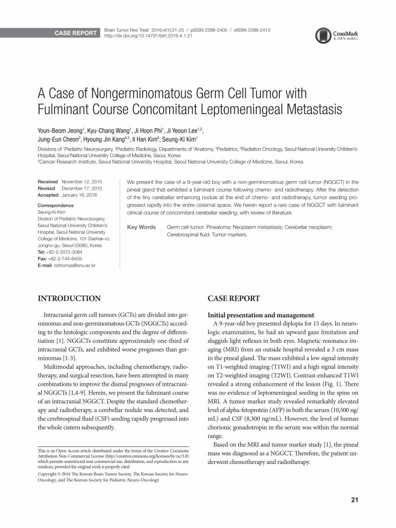
MRI revealed a decrease in the size of mass to 1.75 cm (Fig. 2A). There was no evidence of leptomeningeal seeding in MRI (Fig. 2A), and CSF cytology was negative.
RadiotherapyOne month after chemotherapy, he underwent radiothera-
py. The initial radiation was administered to the craniospinal axis at a dose of 30.6 Gy divided into 17 fractions over 3 weeks and subsequently to the pineal gland area at 23.4 Gy divided into 13 fractions over 3 weeks.
One month after the last radiotherapy, the serum AFP level decreased from 1,194 ng/mL to 187.2 ng/mL. MRI revealed a
Fig. 1. Initial images. MR image showing the pineal mass, which is hypointense on T1- (A) and hyperintense on T2-weighted images (B). The mass is strongly enhanced following the injection of gadolinium (C: axial, D: sagittal image).
A
C
B
D
YB Jeong et al.
23
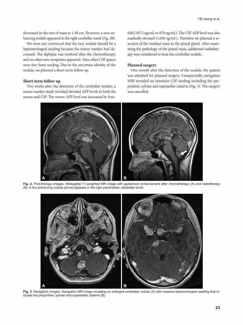
decreased in the size of mass to 1.38 cm. However, a new en-hancing nodule appeared in the right cerebellar tonsil (Fig. 2B).
We were not convinced that the tiny nodule should be a leptomeningeal seeding because the tumor marker had de-creased. The diplopia was resolved after the chemotherapy, and no other new symptoms appeared. Also, other CSF spaces were free from seeding. Due to the uncertain identity of the nodule, we planned a short-term follow up.
Short-term follow upTwo weeks after the detection of the cerebellar nodule, a
tumor marker study revealed elevated AFP levels in both the serum and CSF. The serum AFP level was increased by four-
fold (187.2 ng/mL to 879 ng/mL). The CSF AFP level was also markedly elevated (1,050 ng/mL). Therefore we planned a re-section of the residual mass in the pineal gland. After exam-ining the pathology of the pineal mass, additional radiother-apy was considered to treat the cerebellar nodule.
Planned surgeryOne month after the detection of the nodule, the patient
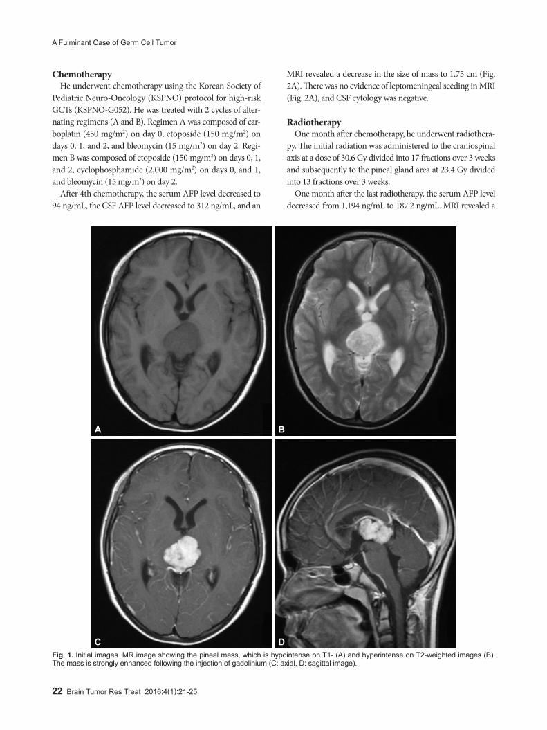
was admitted for planned surgery. Unexpectedly, navigation MRI revealed an extensive CSF seeding including the pre-pontine, sylvian and suprasellar cisterns (Fig. 3). The surgery was cancelled.
Fig. 2. Post-therapy images. Midsagittal T1-weighted MR image with gadolinium enhancement after chemotherapy (A) and radiotherapy (B). A tiny enhancing nodule (arrow) appears in the right paramedian cerebellar tonsil.
A B
Fig. 3. Navigation images. Navigation MR image revealing an enlarged cerebellar nodule (A) with massive leptomeningeal seeding that in-cludes the prepontine, sylvian and suprasellar cisterns (B).
A B
24 Brain Tumor Res Treat 2016;4(1):21-25
A Fulminant Case of Germ Cell Tumor
Further treatmentHe was scheduled for treatment according to the KSPNO
protocol for relapsed/disseminated intracranial GCT (KSPNO-S-053). This protocol includes salvage chemotherapy with al-ternating A and B regimens for 4 cycles, tandem high-dose chemotherapy and autologous stem cell transplantation (HD-CT/ASCT). Regimen A is composed of cisplatin (60 mg/m2) on day 0; etoposide (50 mg/m2) on days 0, 1, and 2; cyclo-phosphamide (750 mg/m2) on days 1 and 2; and vincristine (1.5 mg/m2) on days 0 and 7. Regimen B is composed of car-boplatin (200 mg/m2) on days 0 and 1; etoposide (50 mg/m2) on days 0, 1, 2, 3, and 4; ifosfamide (750 mg/m2) on days 0, 1, 2, 3, and 4; and vincristine (1.5 mg/m2) on days 0 and 7 [10].
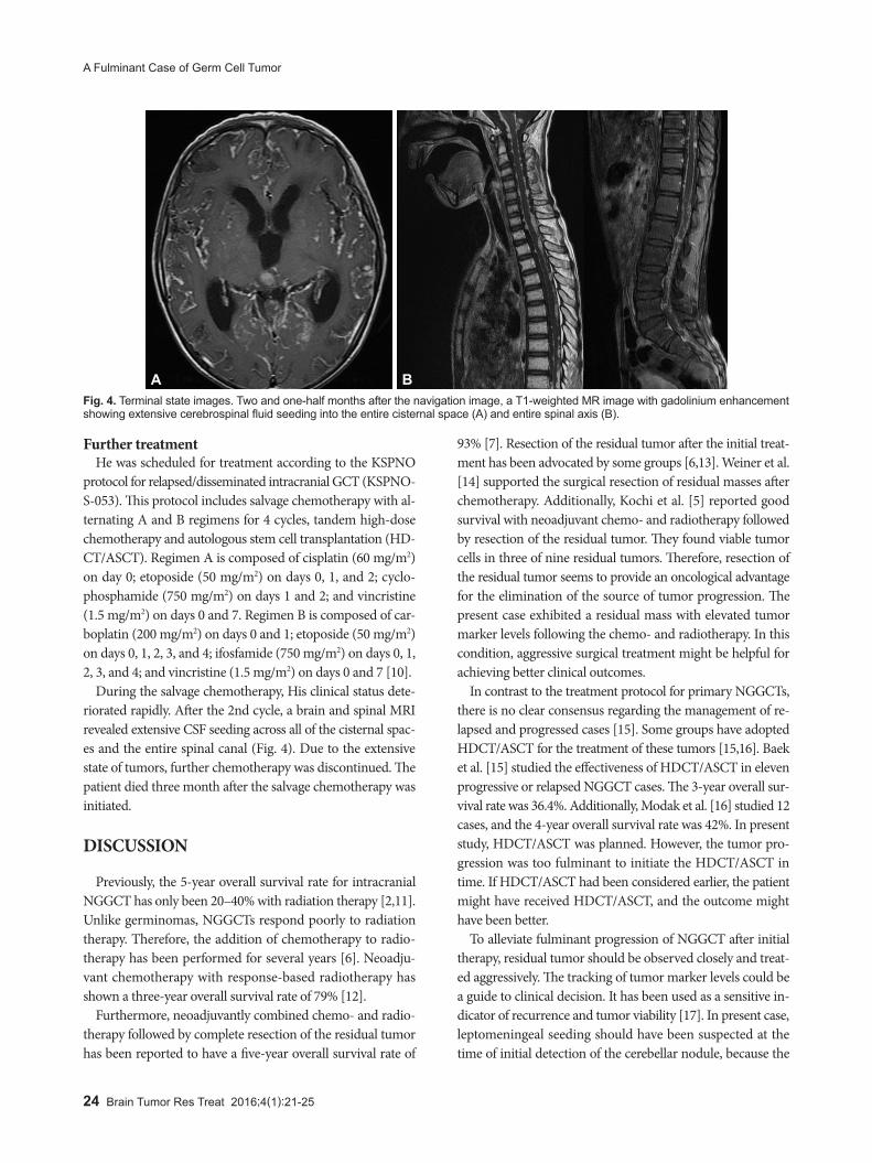
During the salvage chemotherapy, His clinical status dete-riorated rapidly. After the 2nd cycle, a brain and spinal MRI revealed extensive CSF seeding across all of the cisternal spac-es and the entire spinal canal (Fig. 4). Due to the extensive state of tumors, further chemotherapy was discontinued. The patient died three month after the salvage chemotherapy was initiated.
DISCUSSION
Previously, the 5-year overall survival rate for intracranial NGGCT has only been 20–40% with radiation therapy [2,11]. Unlike germinomas, NGGCTs respond poorly to radiation therapy. Therefore, the addition of chemotherapy to radio-therapy has been performed for several years [6]. Neoadju-vant chemotherapy with response-based radiotherapy has shown a three-year overall survival rate of 79% [12].
Furthermore, neoadjuvantly combined chemo- and radio-therapy followed by complete resection of the residual tumor has been reported to have a five-year overall survival rate of
93% [7]. Resection of the residual tumor after the initial treat-ment has been advocated by some groups [6,13]. Weiner et al. [14] supported the surgical resection of residual masses after chemotherapy. Additionally, Kochi et al. [5] reported good survival with neoadjuvant chemo- and radiotherapy followed by resection of the residual tumor. They found viable tumor cells in three of nine residual tumors. Therefore, resection of the residual tumor seems to provide an oncological advantage for the elimination of the source of tumor progression. The present case exhibited a residual mass with elevated tumor marker levels following the chemo- and radiotherapy. In this condition, aggressive surgical treatment might be helpful for achieving better clinical outcomes.
In contrast to the treatment protocol for primary NGGCTs, there is no clear consensus regarding the management of re-lapsed and progressed cases [15]. Some groups have adopted HDCT/ASCT for the treatment of these tumors [15,16]. Baek et al. [15] studied the effectiveness of HDCT/ASCT in eleven progressive or relapsed NGGCT cases. The 3-year overall sur-vival rate was 36.4%. Additionally, Modak et al. [16] studied 12 cases, and the 4-year overall survival rate was 42%. In present study, HDCT/ASCT was planned. However, the tumor pro-gression was too fulminant to initiate the HDCT/ASCT in time. If HDCT/ASCT had been considered earlier, the patient might have received HDCT/ASCT, and the outcome might have been better.
To alleviate fulminant progression of NGGCT after initial therapy, residual tumor should be observed closely and treat-ed aggressively. The tracking of tumor marker levels could be a guide to clinical decision. It has been used as a sensitive in-dicator of recurrence and tumor viability [17]. In present case, leptomeningeal seeding should have been suspected at the time of initial detection of the cerebellar nodule, because the
Fig. 4. Terminal state images. Two and one-half months after the navigation image, a T1-weighted MR image with gadolinium enhancement showing extensive cerebrospinal fluid seeding into the entire cisternal space (A) and entire spinal axis (B).
A B

YB Jeong et al.
25
level of AFP had not normalized. Retrospectively, it seems that urgent application of active treatment such as surgical re-section or HDCT/ASCT may have improved the outcome of the patient.
The treatment outcomes of NGGCTs are much worse than those of germinomas. Even when the initial therapy encom-passes chemotherapy and radiotherapy, some NGGCTs can progress rapidly. To identify fulminant NGGCTs in advance, tumor marker studies and imaging to search for evidence of seeding are necessary. If the tumor marker is not normalized after the initial therapy and tumor seeding is suspected based on imaging, active management, including resective surgery or HDCT/ASCT, should be considered.
Conflicts of InterestThe authors have no financial conflicts of interest.
AcknowledgmentsThis research was supported by a grant from the Korea Health Technol-
ogy R&D Project through the Korea Health Industry Development Insti-tute (KHIDI) funded by the Ministry of Health & Welfare of the Republic of Korea (grant number: HI12C0066).
REFERENCES
1. Echevarría ME, Fangusaro J, Goldman S. Pediatric central nervous system germ cell tumors: a review. Oncologist 2008;13:690-9.
2. Jennings MT, Gelman R, Hochberg F. Intracranial germ-cell tumors: natural history and pathogenesis. J Neurosurg 1985;63:155-67.
3. Jooma R, Kendall BE. Diagnosis and management of pineal tumors. J Neurosurg 1983;58:654-65.
4. Kim JW, Kim WC, Cho JH, et al. A multimodal approach including craniospinal irradiation improves the treatment outcome of high-risk intracranial nongerminomatous germ cell tumors. Int J Radiat Oncol Biol Phys 2012;84:625-31.
5. Kochi M, Itoyama Y, Shiraishi S, Kitamura I, Marubayashi T, Ushio Y. Successful treatment of intracranial nongerminomatous malignant germ cell tumors by administering neoadjuvant chemotherapy and ra-diotherapy before excision of residual tumors. J Neurosurg 2003;
99:106-14.6. Millard NE, Dunkel IJ. Advances in the management of central ner-
vous system germ cell tumors. Curr Oncol Rep 2014;16:393.7. Nakamura H, Makino K, Kochi M, Ushio Y, Kuratsu J. Evaluation of
neoadjuvant therapy in patients with nongerminomatous malignant germ cell tumors. J Neurosurg Pediatr 2011;7:431-8.
8. Ogawa K, Toita T, Nakamura K, et al. Treatment and prognosis of pa-tients with intracranial nongerminomatous malignant germ cell tu-mors: a multiinstitutional retrospective analysis of 41 patients. Cancer 2003;98:369-76.
9. Robertson PL, DaRosso RC, Allen JC. Improved prognosis of intracra-nial non-germinoma germ cell tumors with multimodality therapy. J Neurooncol 1997;32:71-80.
10. Park JE, Kang J, Yoo KH, et al. Efficacy of high-dose chemotherapy and autologous stem cell transplantation in patients with relapsed me-dulloblastoma: a report on the Korean Society for Pediatric Neuro-On-cology (KSPNO)-S-053 study. J Korean Med Sci 2010;25:1160-6.
11. Hoffman HJ, Otsubo H, Hendrick EB, et al. Intracranial germ-cell tu-mors in children. J Neurosurg 1991;74:545-51.
12. Kretschmar C, Kleinberg L, Greenberg M, Burger P, Holmes E, Wharam M. Pre-radiation chemotherapy with response-based radiation therapy in children with central nervous system germ cell tumors: a report from the Children’s Oncology Group. Pediatr Blood Cancer 2007;48:285-91.
13. Souweidane MM, Krieger MD, Weiner HL, Finlay JL. Surgical man-agement of primary central nervous system germ cell tumors: pro-ceedings from the Second International Symposium on Central Ner-vous System Germ Cell Tumors. J Neurosurg Pediatr 2010;6:125-30.
14. Weiner HL, Lichtenbaum RA, Wisoff JH, et al. Delayed surgical resec-tion of central nervous system germ cell tumors. Neurosurgery 2002; 50:727-33; discussion 733-4.
15. Baek HJ, Park HJ, Sung KW, et al. Myeloablative chemotherapy and autologous stem cell transplantation in patients with relapsed or pro-gressed central nervous system germ cell tumors: results of Korean Soci-ety of Pediatric Neuro-Oncology (KSPNO) S-053 study. J Neurooncol 2013;114:329-38.
16. Modak S, Gardner S, Dunkel IJ, et al. Thiotepa-based high-dose chemo-therapy with autologous stem-cell rescue in patients with recurrent or progressive CNS germ cell tumors. J Clin Oncol 2004;22:1934-43.
17. Allen JC, Nisselbaum J, Epstein F, Rosen G, Schwartz MK. Alphafeto-protein and human chorionic gonadotropin determination in cerebro-spinal fluid. An aid to the diagnosis and management of intracranial germ-cell tumors. J Neurosurg 1979;51:368-74.