Embed Size (px)
Citation preview
Bremmer et al. Diagnostic Pathology 2012, 7:140http://www.diagnosticpathology.org/content/7/1/140
CASE REPORT Open Access
Leiomyoma of the tunica albuginea, a case reportof a rare tumour of the testis and review ofthe literatureFelix Bremmer1*, Felix J Kessel2, Carl L Behnes1, Lutz Trojan2 and Elmar Heinrich2
Abstract
Background: Leiomyomas are benign tumours that originate from smooth muscles. They are often seen in theuterus, but also in the renal pelvis, bladder, spermatic cord, epididymis, prostate, scrotum or the glans penis.Leiomyomas of the tunica albuginea are extremely rare.
Case presentation: A 59-year-old white male has noted an asymptomatic tumour on the right side of his scrotalsac for several years. This tumour has increased slowly and caused local scrotal pain. An inguinal incision wasperformed, in which the hypoplastic testis, the epididymis and the tumour could be easily mobilized.Macroscopically the tumour showed a solid round nonencapsulated whorling cut surface. Histologically thediagnosis of a leiomyoma was made.
Conclusion: We report here a very interesting and rare case of a leiomyoma of the tunica albuginea. Leiomyomascan be a possible differential diagnosis in this area.
Virtual Slides: http://www.diagnosticpathology.diagnomx.eu/vs/2585095378537599
Keywords: Leiomyoma, Tunica albuginea, Immunohistochemistry
BackgroundLeiomyomas are benign tumours that originate fromsmooth muscles cells and are often found as benignlesions arising in the uterus [1,2]. But there are also beenseen cases of leiomyomas of the renal pelvis, bladder,spermatic cord, epididymis, prostate, scrotum and theglans penis [1,3-6]. Rare cases of a primary ovarian leio-myoma [7] , leiomyoma of the testis [8] or leiomyoma ofthe kidney have been also reported [9]. Leiomyomas ofthe tunica albuginea are extremely rare, and to our knowl-edge only five cases have been reported so far [10-15].In case of a bilateral leiomyoma so far only two casesare reported [14]. Here we present a case of a leiomyomaof the tunica albuginea.
* Correspondence: [email protected] of Pathology, University Medical Centre Göttingen,Robert-Koch-Str. 40, Göttingen 37075, GermanyFull list of author information is available at the end of the article
© 2012 Bremmer et al.; licensee BioMed CentrCommons Attribution License (http://creativecreproduction in any medium, provided the or
Case HistoryClinical featuresA 59-year-old white male has noted an asymptomatictumour on the right side of his scrotal sac for severalyears. Since the size of this tumour has increased andlately sometimes even caused local scrotal pain, he waspresented to the clinic of urology.Physical examination revealed a solid tumour, approxi-
mately 5 cm in diameter, on the right scrotal side. Thetestis on this side felt unremarkable, though it seemed tobe very small. Inguinal lymph nodes were not palpable.Ultrasound of the scrotum revealed a tumour with
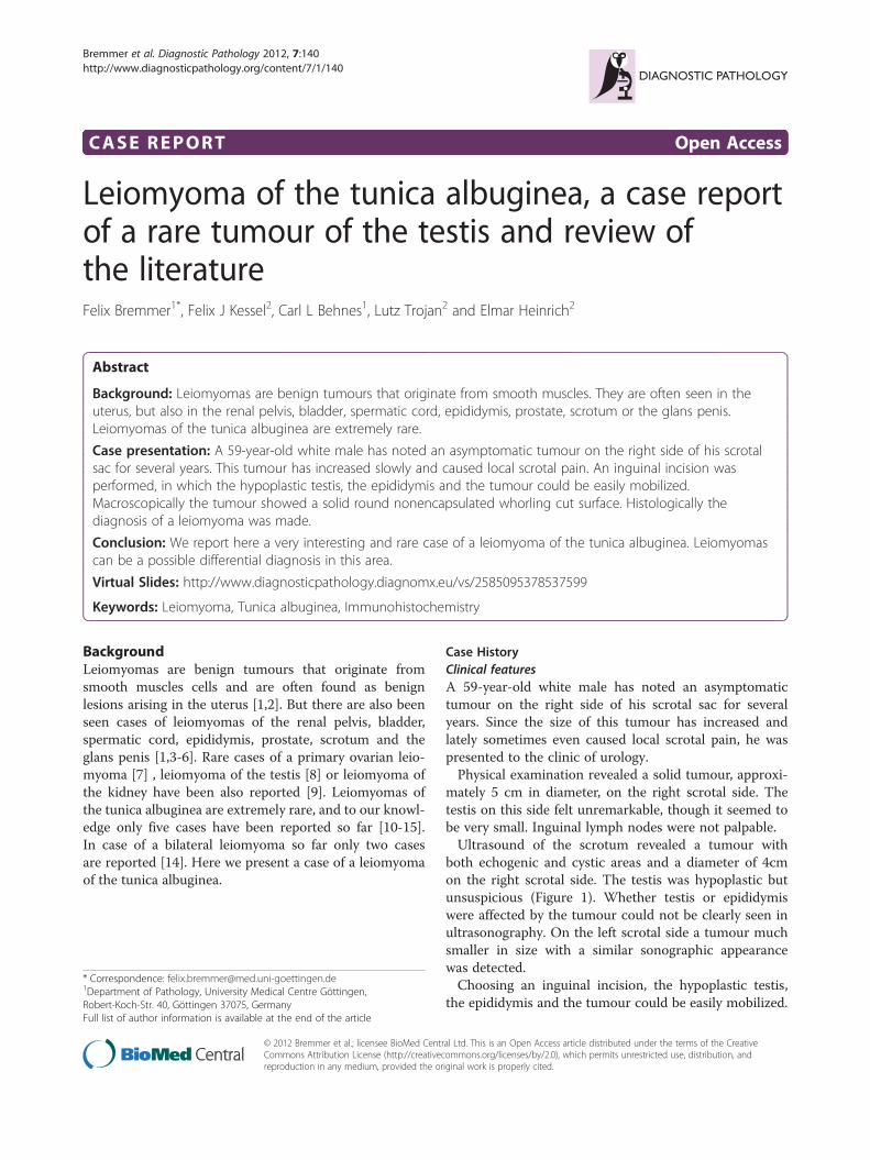
both echogenic and cystic areas and a diameter of 4cmon the right scrotal side. The testis was hypoplastic butunsuspicious (Figure 1). Whether testis or epididymiswere affected by the tumour could not be clearly seen inultrasonography. On the left scrotal side a tumour muchsmaller in size with a similar sonographic appearancewas detected.Choosing an inguinal incision, the hypoplastic testis,
the epididymis and the tumour could be easily mobilized.
al Ltd. This is an Open Access article distributed under the terms of the Creativeommons.org/licenses/by/2.0), which permits unrestricted use, distribution, andiginal work is properly cited.
A B
Figure 1 In sonographic examination beside a little hypoplastic but unremarkable testis (white arrow) a 5 cm in diamater tumourcould be seen (black arrows) (A). The tumour shows echogenic and cystic areas (B).
Bremmer et al. Diagnostic Pathology 2012, 7:140 Page 2 of 4http://www.diagnosticpathology.org/content/7/1/140
The resection of the tumour was accomplished withoutharming testis and epididymis. Testis and epididymiswere replaced into the scrotum. The postoperativecourse was uneventful.
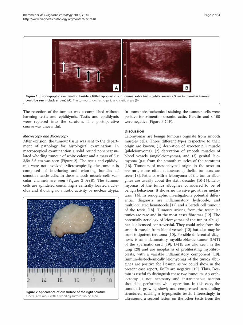
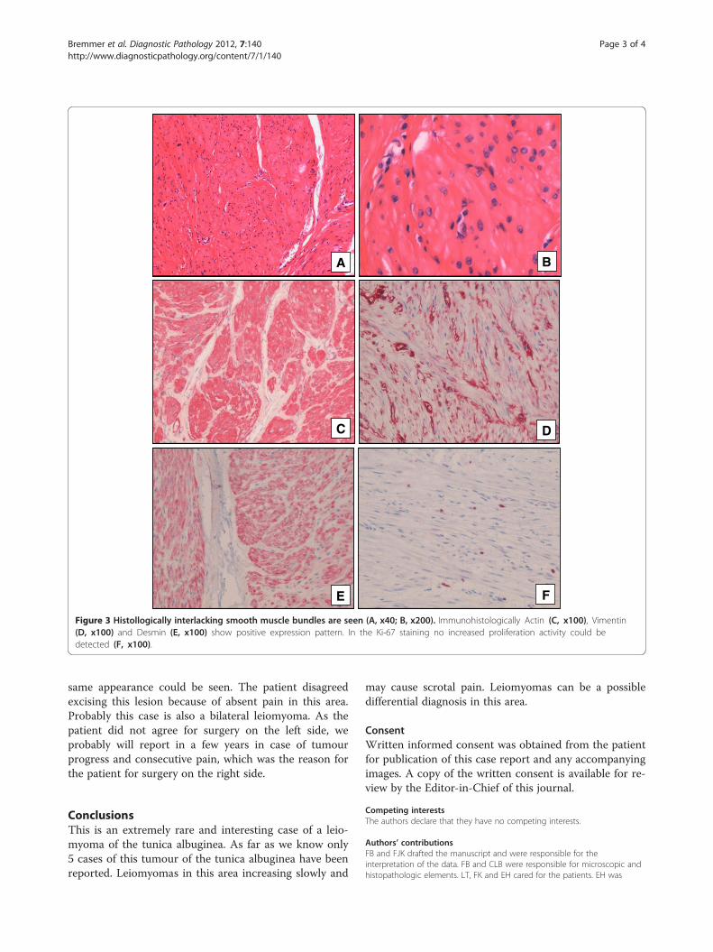
Macroscopy and MicroscopyAfter excision, the tumour tissue was sent to the depart-ment of pathology for histological examination. Inmacroscopical examinantion a solid round nonencapsu-lated whorling tumour of white colour and a mass of 5 x3,5x 3.5 cm was seen (Figure 2). The testis and epididy-mis were not involved. Microscopically, the tumour iscomposed of interlacing and whorling bundles ofsmooth muscle cells. In these smooth muscle cells vas-cular channels are seen (Figure 3 A+B). The tumourcells are spindeled containing a centrally located nucle-olus and showing no mitotic activity or nuclear atypia.
Figure 2 Appearance of cut surface of the right scrotum.A nodular tumour with a whorling surface can be seen.
In immunohsitochemical staining the tumour cells werepositive for vimentin, desmin, actin. Keratin and s-100were negative (Figure 3 C-F).
DiscussionLeiomyomas are benign tumours orginate from smoothmuscles cells. Three different types respective to theirorigin are known; (1) derivation of arrector pili muscle(piloleiomyoma), (2) derevation of smooth muscles ofblood vessels (angioleiomyoma), and (3) genital leio-myoma (p.e. from the smooth muscles of the scrotum)[16]. Tumours of mesenchymal origin in the scrotumare rare, more often cutaneous epithelial tumours areseen [13]. Patients with a leiomyoma of the tunica albu-ginea are usually about the sixth decades [10-15]. Leio-myomas of the tunica albuginea considered to be ofbenign behaviour. It shows no invasive growth or metas-tasis [14]. In sonographic investigations potential differ-ential diagnosis are inflammatory hydrocele, andmultiloculated hematocele [17] and a Sertoli cell tumourof the testis [18]. Tumours arising from the testiculartunics are rare and in the most cases fibromas [12]. Thepotentially aetiology of leiomyomas of the tunica albugi-nea is discussed controversial. They could arise from thesmooth muscle from blood vessels [12] but also may befrom totipotent teratoma [10]. Possible differential diag-nosis is an inflammatory myofibroblastic tumor (IMT)of the spermatic cord [19]. IMTs are also seen in thelung [20] and are neoplasms of proliferating myofibro-blasts, with a variable inflammatory component [19].Immunohistochemically leiomyomas of the tunica albu-ginea are positive for Desmin as we could show in thepresent case report, IMTs are negative [19]. Thus, Des-min is useful to distinguish these two tumours. An orch-iectomy is not necessary and instantaneous sectionshould be performed while operation. In this case, thetumour is growing slowly and compressed surroundingstructures, causing a hypoplastic testis. Interestingly inultrasound a second lesion on the other testis from the
A B
C D
E F
Figure 3 Histollogically interlacking smooth muscle bundles are seen (A, x40; B, x200). Immunohistologically Actin (C, x100), Vimentin(D, x100) and Desmin (E, x100) show positive expression pattern. In the Ki-67 staining no increased proliferation activity could bedetected (F, x100).
Bremmer et al. Diagnostic Pathology 2012, 7:140 Page 3 of 4http://www.diagnosticpathology.org/content/7/1/140
same appearance could be seen. The patient disagreedexcising this lesion because of absent pain in this area.Probably this case is also a bilateral leiomyoma. As thepatient did not agree for surgery on the left side, weprobably will report in a few years in case of tumourprogress and consecutive pain, which was the reason forthe patient for surgery on the right side.
ConclusionsThis is an extremely rare and interesting case of a leio-myoma of the tunica albuginea. As far as we know only5 cases of this tumour of the tunica albuginea have beenreported. Leiomyomas in this area increasing slowly and
may cause scrotal pain. Leiomyomas can be a possibledifferential diagnosis in this area.
ConsentWritten informed consent was obtained from the patientfor publication of this case report and any accompanyingimages. A copy of the written consent is available for re-view by the Editor-in-Chief of this journal.
Competing interestsThe authors declare that they have no competing interests.
Authors’ contributionsFB and FJK drafted the manuscript and were responsible for theinterpretation of the data. FB and CLB were responsible for microscopic andhistopathologic elements. LT, FK and EH cared for the patients. EH was
Bremmer et al. Diagnostic Pathology 2012, 7:140 Page 4 of 4http://www.diagnosticpathology.org/content/7/1/140
responsible for the critical revision of the manuscript. All authors read andapproved the final manuscript.
AcknowledgementsNone
Author details1Department of Pathology, University Medical Centre Göttingen,Robert-Koch-Str. 40, Göttingen 37075, Germany. 2Department of Urology,University Medical Center Göttingen, Robert-Koch-Str. 40, Göttingen 37075,Germany.
Received: 9 August 2012 Accepted: 27 September 2012Published: 9 October 2012
References1. Belis JA, Post GJ, Rochman SC, Milam DF: Genitourinary leiomyomas.
Urology 1979, 13(4):424–429.2. Robboy SJ, Bentley RC, Butnor K, Anderson MC: Pathology and
pathophysiology of uterine smooth-muscle tumors. Environmental healthperspectives 2000, 108(Suppl 5):779–784.
3. Borri A, Nesi G, Bencini L, Pernice LM: Bizarre leiomyoma of theepididymis. A case report. Minerva urologica e nefrologica = The Italianjournal of urology and nephrology 2000, 52(1):29–31.
4. Redman JF, Liang X, Ferguson MA, Savell VH: Leiomyoma of the glanspenis in a child. The Journal of urology 2000, 164(3 Pt 1):791.
5. Rosen Y, Ambiavagar PC, Vuletin JC, Macchia RJ: Atypical leiomyoma ofprostate. Urology 1980, 15(2):183–185.
6. Park JW, Jeong BC, Seo SI, Jeon SS, Kwon GY, Lee HM: Leiomyoma of theurinary bladder: a series of nine cases and review of the literature.Urology 2010, 76(6):1425–1429.
7. Tomas D, Lenicek T, Tuckar N, Puljiz Z, Ledinsky M, Kruslin B: Primaryovarian leiomyoma associated with endometriotic cyst presenting withsymptoms of acute appendicitis: a case report. Diagnostic pathology 2009,4:25.
8. Kullolli VS, Kullolli S, Pawar S, Gautam D: Leiomyoma of testis -a casereport. The Indian journal of surgery 2011, 73(3):233–235.
9. Brunocilla E, Pultrone CV, Schiavina R, Vagnoni V, Caprara G, Martorana G:Renal leiomyoma: Case report and literature review. Canadian UrologicalAssociation journal = Journal de l'Association des urologues du Canada 2012,6(2):E87–E90.
10. Albert PS, Mininberg DT: Leiomyoma of the tunica albuginea. The Journalof urology 1972, 107(5):869–871.
11. Aus G, Boiesen PT: Bilateral leiomyoma of the tunica albuginea.Case report. Scandinavian journal of urology and nephrology 1991,25(1):79–80.
12. Chiaramonte RM: Leiomyoma of tunica albuginea of testis. Urology 1988,31(4):344–345.
13. Giyanani VL, Hennigan DB, Fowler M, Sanders TJ: Sonographicfindings in leiomyoma of postorchiectomy scrotum. Urology 1985,25(2):204–206.
14. Lia-Beng T, Wei-Wuang H, Biing-Rorn C, Chia-Chun T: Bilateral synchronousleiomyomas of the testicular tunica albuginea. A case report andreview of the literature. International urology and nephrology 1996,28(4):549–552.
15. Mak CW, Tzeng WS, Chou CK, Chen CY, Chang JM, Tzeng CC: Leiomyomaarising from the tunica albuginea of the testis: sonographic findings.Journal of clinical ultrasound: JCU 2004, 32(6):309–311.
16. Newman PL, Fletcher CD: Smooth muscle tumours of the externalgenitalia: clinicopathological analysis of a series. Histopathology 1991,18(6):523–529.
17. Cunningham JJ: Sonographic findings in clinically unsuspected acute andchronic scrotal hematoceles. AJR American journal of roentgenology 1983,140(4):749–752.
18. Cunningham JJ: Echographic findings in Sertoli cell tumor of the testis.Journal of clinical ultrasound: JCU 1981, 9(6):341–342.
19. Yee CH, To KF, Hou SM, Ng CF: Inflammatory myofibroblastictumor of spermatic cord in undescended testis. Urology 2009,73(6):e1429–e1412. 1423.
20. Hammas N, Chbani L, Rami M, Boubbou M, Benmiloud S, Bouabdellah Y,Tizniti S, Hida M, Amarti A: A rare tumor of the lung: inflammatorymyofibroblastic tumor. Diagnostic pathology 2012, 7(1):83.
doi:10.1186/1746-1596-7-140Cite this article as: Bremmer et al.: Leiomyoma of the tunica albuginea,a case report of a rare tumour of the testis and review of the literature.Diagnostic Pathology 2012 7:140.
Submit your next manuscript to BioMed Centraland take full advantage of:
• Convenient online submission
• Thorough peer review
• No space constraints or color figure charges
• Immediate publication on acceptance
• Inclusion in PubMed, CAS, Scopus and Google Scholar
• Research which is freely available for redistribution
Submit your manuscript at www.biomedcentral.com/submit
![Porcine vesical acellular matrix graft of tunica albuginea for penile … · 2016-08-26 · sue [2, 3]. The acellular matrix, using urinary tract tis-sue or small intestinal submucosa](https://img.pdfslide.us/doc/110x75/5f9142224c3f14202461bc23/porcine-vesical-acellular-matrix-graft-of-tunica-albuginea-for-penile-2016-08-26.jpg)


![4thy lecture [وضع التوافق] - kau.edu.sakau.edu.sa/Files/140/Files/29546_4thy lecture.pdf · epididymal cyst Tumor Epididymitis Hydrocele Hematocele Torsion Epididymitis](https://img.pdfslide.us/doc/110x75/5c852ee009d3f2ea4b8c2a98/4thy-lecture-kauedusakauedusafiles140files295464thy.jpg)