Embed Size (px)
Citation preview
Choi et al. Multidisciplinary Respiratory Medicine 2014, 9:55http://www.mrmjournal.com/content/9/1/55
CASE REPORT Open Access
Application of veno-arterial-venous extracorporealmembrane oxygenation in differential hypoxiaJoon Hyouk Choi1, Su Wan Kim2*, Young Uck Kim1, Song-Yi Kim1, Ki-Seok Kim1, Seung-Jae Joo1
and Jung Seok Lee3
Abstract
Veno-arterial extracorporeal membrane oxygenation (ECMO) through the femoral vein and artery may causedifferential hypoxia, i.e., lower PaO2 in the upper body than in the lower body, because of normal cardiac outputwith severe impairment of pulmonary function. Hereby, we report the diagnosis and the treatment of differentialhypoxia caused by veno-arterial ECMO. A 39-year-old man received cardiopulmonary resuscitation from a cardiacarrest due to acute myocardial infarction. Even after more than 30 min of resuscitation, spontaneous circulationhad not resumed. Next, we performed veno-arterial ECMO through the femoral artery and vein, and the patientrecovered consciousness on the second day of ECMO. On day 5 of ECMO, he lost consciousness again and presenteda generalized tonic-clonic seizure, and an electroencephalogram showed delta waves suggesting diffuse cerebralcortical dysfunction. While an echocardiogram revealed improvements in myocardial function, a follow up chestradiograph showed increasing massive parenchymal infiltrations, and gas analysis of blood from the right radial arteryrevealed severe hypoxemia. These findings indicated a definite diagnosis of differential hypoxia, and therefore, weinserted a 17-Fr cannula into the left subclavian vein as a return cannula. The patient’s consciousness and pulmonaryinfiltrations were improved 2 days after veno-arterial-venous ECMO, and the electroencephalogram showednormal findings. To our knowledge, this is the first report of successful clinical management of differential hypoxia.We suggest that veno-arterial-venous ECMO could be the treatment of choice for differential hypoxia resulting fromveno-arterial ECMO.
Keywords: Brain, Extracorporeal membrane oxygenation, Lungs, Pulmonary function
BackgroundFemoral veno-arterial extracorporeal membrane oxygenation(VA ECMO) may cause differential hypoxia (lower PaO2 inthe upper body than in the lower body, i.e., two-circulationsyndrome) because of normal cardiac output with severeimpairment of pulmonary function [1]. The phenomenonof differential hypoxia has been introduced theoretically;however, to our knowledge, thus far no definitiveclinical report has been published on the managementof differential hypoxia.Hereby, we report a case of differential hypoxia resulting
from VA ECMO and its successful treatment using
* Correspondence: [email protected] of Thoracic and Cardiovascular Surgery, Jeju National UniversityHospital, Jeju National University School of Medicine, Aran 13 gil 15, Jeju-si,Jeju Special Self-Governing Province 690-767, KoreaFull list of author information is available at the end of the article
© 2014 Choi et al.; licensee BioMed Central LtCommons Attribution License (http://creativecreproduction in any medium, provided the orDedication waiver (http://creativecommons.orunless otherwise stated.
veno-arterial-venous (VAV) ECMO in a patient withrespiratory failure resulting from cardiac arrest.
Case presentationA 39-year-old man presented with chest pain that hadlasted for 90 minutes. Electrocardiography revealed STsegment depression in leads V1–6 (Figure 1A). Fiveminutes after his arrival at the emergency department,he lost consciousness and displayed indications of aseizure. The electrocardiogram showed alternate ventriculartachycardia and ventricular fibrillation. Defibrillationand cardiopulmonary resuscitation were immediatelyperformed. Although resuscitation was performed formore than 30 minutes, spontaneous circulation had notrecovered. A VA ECMO system (EBS, Capiox EmergencyBypass System; Terumo Inc., Tokyo, Japan) was set upwith a 21-Fr drainage cannula (DLP, Medtronic Inc.,Minneapolis, MN) from the left femoral vein and a 17-Fr
d. This is an Open Access article distributed under the terms of the Creativeommons.org/licenses/by/4.0), which permits unrestricted use, distribution, andiginal work is properly credited. The Creative Commons Public Domaing/publicdomain/zero/1.0/) applies to the data made available in this article,
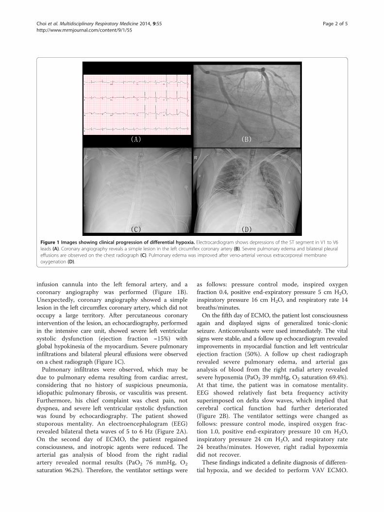
Figure 1 Images showing clinical progression of differential hypoxia. Electrocardiogram shows depressions of the ST segment in V1 to V6leads (A). Coronary angiography reveals a simple lesion in the left circumflex coronary artery (B). Severe pulmonary edema and bilateral pleuraleffusions are observed on the chest radiograph (C). Pulmonary edema was improved after veno-arterial venous extracorporeal membraneoxygenation (D).
Choi et al. Multidisciplinary Respiratory Medicine 2014, 9:55 Page 2 of 5http://www.mrmjournal.com/content/9/1/55
infusion cannula into the left femoral artery, and acoronary angiography was performed (Figure 1B).Unexpectedly, coronary angiography showed a simplelesion in the left circumflex coronary artery, which did notoccupy a large territory. After percutaneous coronaryintervention of the lesion, an echocardiography, performedin the intensive care unit, showed severe left ventricularsystolic dysfunction (ejection fraction =15%) withglobal hypokinesia of the myocardium. Severe pulmonaryinfiltrations and bilateral pleural effusions were observedon a chest radiograph (Figure 1C).Pulmonary infiltrates were observed, which may be
due to pulmonary edema resulting from cardiac arrest,considering that no history of suspicious pneumonia,idiopathic pulmonary fibrosis, or vasculitis was present.Furthermore, his chief complaint was chest pain, notdyspnea, and severe left ventricular systolic dysfunctionwas found by echocardiography. The patient showedstuporous mentality. An electroencephalogram (EEG)revealed bilateral theta waves of 5 to 6 Hz (Figure 2A).On the second day of ECMO, the patient regainedconsciousness, and inotropic agents were reduced. Thearterial gas analysis of blood from the right radialartery revealed normal results (PaO2 76 mmHg, O2
saturation 96.2%). Therefore, the ventilator settings were
as follows: pressure control mode, inspired oxygenfraction 0.4, positive end-expiratory pressure 5 cm H2O,inspiratory pressure 16 cm H2O, and respiratory rate 14breaths/minutes.On the fifth day of ECMO, the patient lost consciousness
again and displayed signs of generalized tonic-clonicseizure. Anticonvulsants were used immediately. The vitalsigns were stable, and a follow up echocardiogram revealedimprovements in myocardial function and left ventricularejection fraction (50%). A follow up chest radiographrevealed severe pulmonary edema, and arterial gasanalysis of blood from the right radial artery revealedsevere hypoxemia (PaO2 39 mmHg, O2 saturation 69.4%).At that time, the patient was in comatose mentality.EEG showed relatively fast beta frequency activitysuperimposed on delta slow waves, which implied thatcerebral cortical function had further deteriorated(Figure 2B). The ventilator settings were changed asfollows: pressure control mode, inspired oxygen frac-tion 1.0, positive end-expiratory pressure 10 cm H2O,inspiratory pressure 24 cm H2O, and respiratory rate24 breaths/minutes. However, right radial hypoxemiadid not recover.These findings indicated a definite diagnosis of differen-
tial hypoxia, and we decided to perform VAV ECMO.
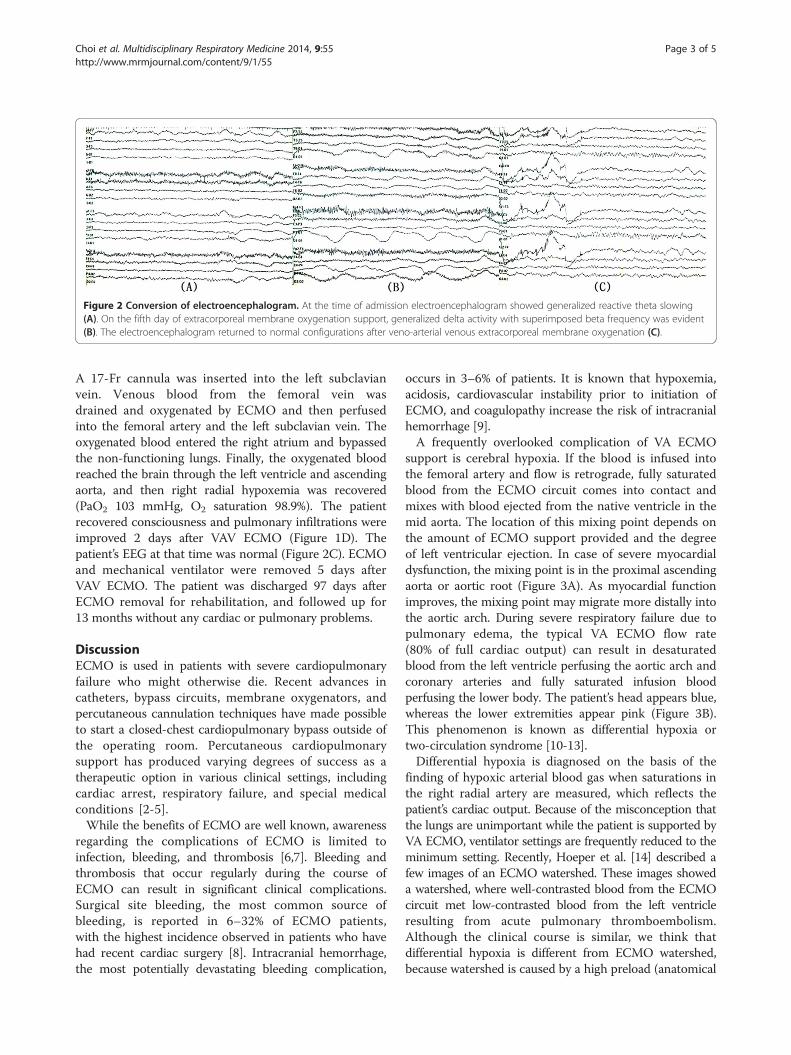
Figure 2 Conversion of electroencephalogram. At the time of admission electroencephalogram showed generalized reactive theta slowing(A). On the fifth day of extracorporeal membrane oxygenation support, generalized delta activity with superimposed beta frequency was evident(B). The electroencephalogram returned to normal configurations after veno-arterial venous extracorporeal membrane oxygenation (C).
Choi et al. Multidisciplinary Respiratory Medicine 2014, 9:55 Page 3 of 5http://www.mrmjournal.com/content/9/1/55
A 17-Fr cannula was inserted into the left subclavianvein. Venous blood from the femoral vein wasdrained and oxygenated by ECMO and then perfusedinto the femoral artery and the left subclavian vein. Theoxygenated blood entered the right atrium and bypassedthe non-functioning lungs. Finally, the oxygenated bloodreached the brain through the left ventricle and ascendingaorta, and then right radial hypoxemia was recovered(PaO2 103 mmHg, O2 saturation 98.9%). The patientrecovered consciousness and pulmonary infiltrations wereimproved 2 days after VAV ECMO (Figure 1D). Thepatient’s EEG at that time was normal (Figure 2C). ECMOand mechanical ventilator were removed 5 days afterVAV ECMO. The patient was discharged 97 days afterECMO removal for rehabilitation, and followed up for13 months without any cardiac or pulmonary problems.
DiscussionECMO is used in patients with severe cardiopulmonaryfailure who might otherwise die. Recent advances incatheters, bypass circuits, membrane oxygenators, andpercutaneous cannulation techniques have made possibleto start a closed-chest cardiopulmonary bypass outside ofthe operating room. Percutaneous cardiopulmonarysupport has produced varying degrees of success as atherapeutic option in various clinical settings, includingcardiac arrest, respiratory failure, and special medicalconditions [2-5].While the benefits of ECMO are well known, awareness
regarding the complications of ECMO is limited toinfection, bleeding, and thrombosis [6,7]. Bleeding andthrombosis that occur regularly during the course ofECMO can result in significant clinical complications.Surgical site bleeding, the most common source ofbleeding, is reported in 6–32% of ECMO patients,with the highest incidence observed in patients who havehad recent cardiac surgery [8]. Intracranial hemorrhage,the most potentially devastating bleeding complication,
occurs in 3–6% of patients. It is known that hypoxemia,acidosis, cardiovascular instability prior to initiation ofECMO, and coagulopathy increase the risk of intracranialhemorrhage [9].A frequently overlooked complication of VA ECMO
support is cerebral hypoxia. If the blood is infused intothe femoral artery and flow is retrograde, fully saturatedblood from the ECMO circuit comes into contact andmixes with blood ejected from the native ventricle in themid aorta. The location of this mixing point depends onthe amount of ECMO support provided and the degreeof left ventricular ejection. In case of severe myocardialdysfunction, the mixing point is in the proximal ascendingaorta or aortic root (Figure 3A). As myocardial functionimproves, the mixing point may migrate more distally intothe aortic arch. During severe respiratory failure due topulmonary edema, the typical VA ECMO flow rate(80% of full cardiac output) can result in desaturatedblood from the left ventricle perfusing the aortic arch andcoronary arteries and fully saturated infusion bloodperfusing the lower body. The patient’s head appears blue,whereas the lower extremities appear pink (Figure 3B).This phenomenon is known as differential hypoxia ortwo-circulation syndrome [10-13].Differential hypoxia is diagnosed on the basis of the
finding of hypoxic arterial blood gas when saturations inthe right radial artery are measured, which reflects thepatient’s cardiac output. Because of the misconception thatthe lungs are unimportant while the patient is supported byVA ECMO, ventilator settings are frequently reduced to theminimum setting. Recently, Hoeper et al. [14] described afew images of an ECMO watershed. These images showeda watershed, where well-contrasted blood from the ECMOcircuit met low-contrasted blood from the left ventricleresulting from acute pulmonary thromboembolism.Although the clinical course is similar, we think thatdifferential hypoxia is different from ECMO watershed,because watershed is caused by a high preload (anatomical
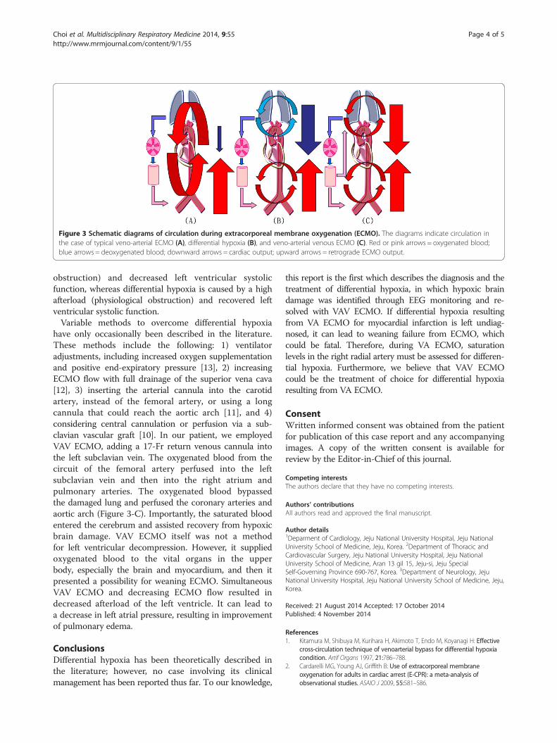
Figure 3 Schematic diagrams of circulation during extracorporeal membrane oxygenation (ECMO). The diagrams indicate circulation inthe case of typical veno-arterial ECMO (A), differential hypoxia (B), and veno-arterial venous ECMO (C). Red or pink arrows = oxygenated blood;blue arrows = deoxygenated blood; downward arrows = cardiac output; upward arrows = retrograde ECMO output.
Choi et al. Multidisciplinary Respiratory Medicine 2014, 9:55 Page 4 of 5http://www.mrmjournal.com/content/9/1/55
obstruction) and decreased left ventricular systolicfunction, whereas differential hypoxia is caused by a highafterload (physiological obstruction) and recovered leftventricular systolic function.Variable methods to overcome differential hypoxia
have only occasionally been described in the literature.These methods include the following: 1) ventilatoradjustments, including increased oxygen supplementationand positive end-expiratory pressure [13], 2) increasingECMO flow with full drainage of the superior vena cava[12], 3) inserting the arterial cannula into the carotidartery, instead of the femoral artery, or using a longcannula that could reach the aortic arch [11], and 4)considering central cannulation or perfusion via a sub-clavian vascular graft [10]. In our patient, we employedVAV ECMO, adding a 17-Fr return venous cannula intothe left subclavian vein. The oxygenated blood from thecircuit of the femoral artery perfused into the leftsubclavian vein and then into the right atrium andpulmonary arteries. The oxygenated blood bypassedthe damaged lung and perfused the coronary arteries andaortic arch (Figure 3-C). Importantly, the saturated bloodentered the cerebrum and assisted recovery from hypoxicbrain damage. VAV ECMO itself was not a methodfor left ventricular decompression. However, it suppliedoxygenated blood to the vital organs in the upperbody, especially the brain and myocardium, and then itpresented a possibility for weaning ECMO. SimultaneousVAV ECMO and decreasing ECMO flow resulted indecreased afterload of the left ventricle. It can lead toa decrease in left atrial pressure, resulting in improvementof pulmonary edema.
ConclusionsDifferential hypoxia has been theoretically described inthe literature; however, no case involving its clinicalmanagement has been reported thus far. To our knowledge,
this report is the first which describes the diagnosis and thetreatment of differential hypoxia, in which hypoxic braindamage was identified through EEG monitoring and re-solved with VAV ECMO. If differential hypoxia resultingfrom VA ECMO for myocardial infarction is left undiag-nosed, it can lead to weaning failure from ECMO, whichcould be fatal. Therefore, during VA ECMO, saturationlevels in the right radial artery must be assessed for differen-tial hypoxia. Furthermore, we believe that VAV ECMOcould be the treatment of choice for differential hypoxiaresulting from VA ECMO.
ConsentWritten informed consent was obtained from the patientfor publication of this case report and any accompanyingimages. A copy of the written consent is available forreview by the Editor-in-Chief of this journal.
Competing interestsThe authors declare that they have no competing interests.
Authors’ contributionsAll authors read and approved the final manuscript.
Author details1Deparment of Cardiology, Jeju National University Hospital, Jeju NationalUniversity School of Medicine, Jeju, Korea. 2Department of Thoracic andCardiovascular Surgery, Jeju National University Hospital, Jeju NationalUniversity School of Medicine, Aran 13 gil 15, Jeju-si, Jeju SpecialSelf-Governing Province 690-767, Korea. 3Department of Neurology, JejuNational University Hospital, Jeju National University School of Medicine, Jeju,Korea.
Received: 21 August 2014 Accepted: 17 October 2014Published: 4 November 2014
References1. Kitamura M, Shibuya M, Kurihara H, Akimoto T, Endo M, Koyanagi H: Effective
cross-circulation technique of venoarterial bypass for differential hypoxiacondition. Artif Organs 1997, 21:786–788.
2. Cardarelli MG, Young AJ, Griffith B: Use of extracorporeal membraneoxygenation for adults in cardiac arrest (E-CPR): a meta-analysis ofobservational studies. ASAIO J 2009, 55:581–586.
Choi et al. Multidisciplinary Respiratory Medicine 2014, 9:55 Page 5 of 5http://www.mrmjournal.com/content/9/1/55
3. Chen YS, Chao A, Yu HY, Ko WJ, Wu IH, Chen RJ, Huang SC, Lin FY, Wang SS:Analysis and results of prolonged resuscitation in cardiac arrest patientsrescued by extracorporeal membrane oxygenation. J Am Coll Cardiol 2003,41:197–203.
4. Peek GJ, Mugford M, Tiruvoipati R, Wilson A, Allen E, Thalanany MM, Hibbert CL,Truesdale A, Clemens F, Cooper N, Firmin RK, Elbourne D: Efficacy and economicassessment of conventional ventilatory support versus extracorporealmembrane oxygenation for severe adult respiratory failure (CESAR):a multicenter randomised controlled trial. Lancet 2009, 374:1351–1363.
5. Lee JH, Kim SW: Successful management of warfarin-exacerbated diffusealveolar hemorrhage using an extracorporeal membrane oxygenation.Multidiscip Respir Med 2013, 8:16.
6. Guillermo M, Alain V: Extracorporeal membrane oxygenation in adults.Cont Edu Anaesth Crit Care and Pain 2012, 12:57–61.
7. Zangrillo A, Landoni G, Biondi ZG, Greco M, Greco T, Frati G, Patroniti N,Antonelli M, Pesenti A, Pappalardo F: A meta-analysis of complicationsand mortality of extracorporeal membrane oxygenation. Crit Care Resusc2013, 15:172–178.
8. Organization ELS: Registry Report. Ann Arbor: University of Michigan; 2010.9. Bulas D, Glass P: Neonatal ECMO: neuroimaging and neurodevelopmental
outcome. Seminars Perinatalogy 2005, 29:58–65.10. Strickland R, Frantzis P: Royal Adelaide Hospital General ICU ECMO Guidelines.
http://www.icuadelaide.com.au/files/manual_ecmo.pdf.11. Min HK, Lee YT: Role of Percutaneous Cardiopulmonary Support (PCPS) in
Patients with Unstable Hemodynamics During the Peri-Coronary-InterventionPeriod. In Coronary Angiography - The Need for Improvement in Medical andInterventional Therapy. Edited by Branislav B. Rijeka: InTech; 2011:135.
12. Bartlett RH, Zwischenberger JB: Management of Blood Flow and GasExchange during ECLS. In Extracorporeal Cardiopulmonary Support in CriticalCare. Edited by Annich G, MacLaren G. Michigan: Extracorporeal LifeSupporting Organization; 2012:152.
13. Haft J, Firmin R: Adult Cardiac Support. In Extracorporeal CardiopulmonarySupport in Critical Care. Edited by Annich G, MacLaren G. Michigan:Extracorporeal Life Supporting Organization; 2012:327–328.
14. Hoeper MM, Tudorache I, Kühn C, Marsch G, Hartung D, Wiesner O,Boenisch O, Haverich A, Hinrich J: Extracorporeal membrane oxygenationwatershed. Circ 2014, 130:864–865.
doi:10.1186/2049-6958-9-55Cite this article as: Choi et al.: Application of veno-arterial-venousextracorporeal membrane oxygenation in differential hypoxia. MultidisciplinaryRespiratory Medicine 2014 9:55.
Submit your next manuscript to BioMed Centraland take full advantage of:
• Convenient online submission
• Thorough peer review
• No space constraints or color figure charges
• Immediate publication on acceptance
• Inclusion in PubMed, CAS, Scopus and Google Scholar
• Research which is freely available for redistribution
Submit your manuscript at www.biomedcentral.com/submit