Embed Size (px)
Citation preview
CASE REPORT Open Access
A rare case of recurrent ovarian cancerpresenting as a round ligament metastasisShinichi Togami*, Tomoyasu Kato, Takateru Oi, Mitsuya Ishikawa, Takashi Onda, Shun-ichi Ikeda andTakahiro Kasamatsu
Abstract
We report a rare case of recurrent ovarian cancer presenting as a round ligament metastasis. A 44-year-old womanpresented with a lower abdominal mass. Computed tomography showed a pelvic mass. Primary surgery wasperformed. A histopathological examination showed an ovarian serous adenocarcinoma of Stage IIIb. The patientreceived 6 cycles of paclitaxel and carboplatin. Almost 2 years after the initial operation, the patient noticed a leftinguinal mass. Computed tomography showed a left inguinal mass, 18 mm in size. An excisional biopsy wasperformed and the tumor was found to originate in the left round ligament. A histopathological examinationshowed serous adenocarcinoma and there was no evidence of lymph node tissue. Recurrence of ovarian cancer inthe round ligament is extremely rare. This unique case suggests, however, that the round ligament in rare casesmay be a recurrence site for ovarian cancer, and that accurate differentiation including confirmation by diagnosticimaging and excisional biopsy, is necessary for a definitive pathological diagnosis.
Keywords: diagnosis, inguinal mass, ovarian cancer, recurrence, round ligament
BackgroundThe majority of women with ovarian cancer presentwith advanced stage disease. A complete clinical remis-sion after surgical cytoreduction and platinum-basedchemotherapy can be achieved in 80-90% of thesepatients. Despite this, 70-90% of patients will developrecurrent disease [1]. Fifty-five percent of the firstrelapse cases were found at the pelvis or abdomen [2].There was a wide variety among the other recurrentsites, such as, retroperitoneal nodes, liver or spleen,brain, and bone [2,3]. We experienced a case with soli-tary recurrence at the left round ligament. To the bestof our knowledge, this is the first report of recurrentovarian cancer occurring in the round ligament.
CaseA 44-year-old woman presented with a 1.5-year historyof progressive enlargement of a lower abdominal mass.On physical examination, a pelvic mass was noted.Transvaginal ultrasound revealed bilateral adnexalmasses (left: 80*58 mm, right: 54*40 mm) with solid
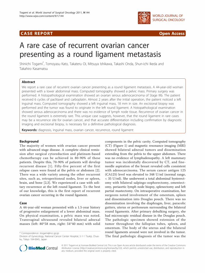
components in the pelvic cavity. Computed tomography(CT) (Figure 1) and magnetic resonance imaging (MRI)showed bilateral adnexal tumors and disseminationextending from the pelvis to the upper abdomen. Therewas no evidence of lymphadenopathy. A left mammarytumor was incidentally discovered by CT, and fine-needle aspiration of the breast revealed cells consistentwith adenocarcinoma. The serum cancer antigen 125(CA125) level was elevated to 348 U/ml (normal range,< 35 U/ml). She underwent a total abdominal hysterect-omy with bilateral salpingo-oophorectomy, omentect-omy, periaortic lymph node biopsy, splenectomy and leftpartial mastectomy. On intraoperative examination, hersurgeons noted involvement of the omentum, spleenand dissemination into Douglas pouch. There was nodissemination involving the diaphragm, liver, paracolicgutters, uterus or peritoneum surrounding the bilateralround ligaments. After primary debulking surgery, shehad microscopic residual disease in the Douglas pouch.The pathologic specimen showed extension of thetumor throughout the fallopian tubes, spleen, andomentum. The body of the uterus and the bilateralround ligaments around were not involved in the tumor.The final pathologic diagnosis of the tumor was the
* Correspondence: [email protected] of Gynecology, National Cancer Center Hospital, 5-1-1 Tsukiji, Chuo-ku, Tokyo 104-0045, Japan
Togami et al. World Journal of Surgical Oncology 2011, 9:144http://www.wjso.com/content/9/1/144 WORLD JOURNAL OF
SURGICAL ONCOLOGY
© 2011 Togami et al; licensee BioMed Central Ltd. This is an Open Access article distributed under the terms of the Creative CommonsAttribution License (http://creativecommons.org/licenses/by/2.0), which permits unrestricted use, distribution, and reproduction inany medium, provided the original work is properly cited.
International Federation of Gynecology and Obstetrics(FIGO) Stage IIIb ovarian serous adenocarcinoma andleft breast invasive ductal carcinoma.Initially, the patient received 6 cycles of paclitaxel (175
mg/m2) and carboplatin (AUC = 6) for ovarian cancerin April 2009. In August 2009, her serum CA125 levelsdeclined to 5 U/ml. She then received adjuvant radio-therapy (WB 50Gy/25 f) for breast cancer in October2009. After that, she received routine follow-up fromher gynecologic oncologist.Almost 2 years after her initial ovarian cancer opera-
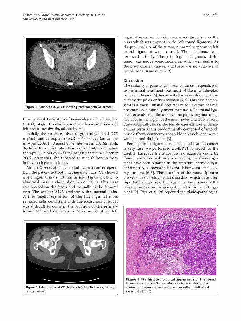
tion, the patient noticed a left inguinal mass. CT showeda left inguinal mass, 18 mm in size (Figure 2), but noabnormal mass in chest, abdomen or pelvis. This masswas located on the fascia and medially to the femoralvein. The serum CA125 level was within normal limits.A fine-needle aspiration of the left inguinal massrevealed cells consistent with adenocarcinoma, but itwas difficult to confirm the location of the primarylesion. She underwent an excision biopsy of the left
inguinal mass. An incision was made directly over themass which was present in the left round ligament. Atthe proximal site of the tumor, a normally appearing leftround ligament was exposed. Then the mass wasremoved entirely. The pathological diagnosis of thetumor was serous adenocarcinoma, which was similar tothe prior ovarian cancer, and there was no evidence oflymph node tissue (Figure 3).
DiscussionThe majority of patients with ovarian cancer responds wellto the initial treatment, but most of them will developrecurrent disease [4]. Recurrent disease involves most fre-quently the pelvis or the abdomen [2,3]. This case demon-strates a most unusual recurrence for ovarian cancer,presenting as a round ligament metastasis. The round liga-ment extends from the uterus, through the inguinal canal,and ends in the region of the mons pubis and labia majora.Embryologically, this is the female equivalent of guberna-culums testis and is predominantly composed of smoothmuscle fibers, connective tissue, blood vessels, and nerveswith a mesothelial coating [5].Because round ligament recurrence of ovarian cancer
is very rare, we performed a MEDLINE search of theEnglish language literature, but no example could befound. Some unusual tumors involving the round liga-ment have been reported in the literature: dermoid cyst,endometriosis, mesothelial cyst, leiomyoma and leio-myosarcoma [6-8]. These tumors of the round ligamentare very rare developmental disorders, which have beenreported as case reports. Especially, leiomyoma is themost common tumor associated with the round liga-ment [9]. Patil et al. [9] reported the clinicopathological
Figure 1 Enhanced axial CT showing bilateral adnexal tumors.
Figure 2 Enhanced axial CT shows a left inguinal mass, 18 mmin size (arrow).
Figure 3 The histopathological appearance of the roundligament recurrence: Serous adenocarcinoma exists in thecontext of fibrous connective tissue, including small bloodvessels. (H&E, ×40).
Togami et al. World Journal of Surgical Oncology 2011, 9:144http://www.wjso.com/content/9/1/144
Page 2 of 3

features of 55 cases of women with inguinal smoothmuscle tumors of women. Histologically, 23 tumorswere considered as leiomyomas, and five tumors arosein the round ligament. In contrast to the leiomyomas,none of the leiomyosarcomas were associated with theround ligament. Indeed, leiomyosarcomas of the roundligament of the uterus are extremely rare and there isonly 1 case report published of leiomyosarcoma arisingin the round ligament of the uterus [6]. Recurrenttumors of the round ligament are also rare. We found 1case report of recurrent endometrial cancer stage Ia, ori-ginating in the round ligament [10]. The 5 cm solidmass was located near the right superficial inguinal ring.Resection of the round ligament mass and dissection ofthe retroperitoneal node were performed, and one posi-tive obturator node was found. They concluded thatpatients presenting with a round ligament recurrenceshould have a thorough work-up with pelvic lymphnode evaluation. In our case, CT showed no evidence ofpelvic lymph node swelling, so we performed only theround ligament mass resection to confirm the pathologi-cal diagnosis. Postoperative PET/CT scan revealed nohot spot, showing that metastasis to the round ligamentwas solitary.Two possible pathways of metastasis to the round
ligament have been considered: The lymphatic and thevascular pathway. Pathologically, the tumor showed vas-cular infiltration but no evidence of lymph node tissue,but it is difficult to determine whether the metastasis ispathway whether lymphatic or vascular. Ovarian cancergenerally metastasizes via the lymphatic system or byperitoneal dissemination [11]. Lymphatic vessels enterand travel along the round ligament to reach the ingu-inal region, and the most likely hypothesis is that themicroscopic tumor metastasized to the round ligamentthrough a lymphatic pathway.There are some reports of solitary splenic metastasis
of ovarian cancer after surgical remission [11-13]. Interms of treatment, splenectomy was performed in allcases and adjuvant chemotherapy was administered inmost cases. The decision as to whether adjuvant che-motherapy is indicated must be carefully considered ineach case.
ConclusionsThis case presents an unusual example of a recurrencesite for ovarian cancer. Although solitary ovarian can-cer recurrence at the round ligament is extremely rare,it should be included in the differential diagnosis forany patient with a past history of ovarian cancer. Theround ligament has the potential to be a site of occur-rence of various tumors. Among them, benign tumorssuch as leiomyoma are often seen in the round liga-ment. This unique case suggests that the round
ligament in rare cases may be a site of recurrence inovarian cancer, and indicates that accurate differentia-tion, including confirmation with diagnostic imagingand excisional biopsy, are necessary because the subse-quent treatment depends significantly on the patholo-gical results.
Consent statementInformed consent was obtained from the patient forpublication of this case report and accompanyingimages. A copy of the written consent is available forreview by the Editor-in-Chief of this journal.
Authors’ contributionsST, TK and TOi have operated this case. MI, TO, SI and TK have assisted toanalyze all data. All authors read and approved the final manuscript.
Competing interestsThe authors declare that they have no competing interests.
Received: 19 August 2011 Accepted: 7 November 2011Published: 7 November 2011
References1. Shih KK, Chi DS, Barakat RR, Leitao MM Jr: Beyond tertiary cytoreduction in
patients with recurrent epithelial ovarian, fallopian tube, or primaryperitoneal cancer. Gynecol Oncol 116:364-369.
2. Ushijima K: Treatment for recurrent ovarian cancer-at first relapse. JOncol 2010:497429.
3. Gadducci A, Fuso L, Cosio S, Landoni F, Maggino T, Perotto S, Sartori E,Testa A, Galletto L, Zola P: Are surveillance procedures of clinical benefitfor patients treated for ovarian cancer?: A retrospective Italianmulticentric study. Int J Gynecol Cancer 2009, 19:367-374.
4. Martin LP, Schilder RJ: Management of recurrent ovarian carcinoma:current status and future directions. Semin Oncol 2009, 36:112-125.
5. Warshauer DM, Mandel SR: Leiomyoma of the extraperitoneal roundligament: CT demonstration. Clin Imaging 1999, 23:375-376.
6. Kirkham JC, Nero CJ, Tambouret RH, Yoon SS: Leiomyoma andleiomyosarcoma arising from the round ligament of the uterus. J AmColl Surg 2008, 207:452.
7. Kim BM, Lee JY, Han YH, Kim SY, Seo JW, Kim YH, Cha SJ, Hur G, Joo M,Lee ES: Mesothelial cyst of the round ligament mimicking a metastasis: acase report. Korean J Radiol 11:364-367.
8. Kaleli B, Aktan E, Bayramoglu H, Alatas E: Mature cystic teratoma in roundligament: case report. Eur J Obstet Gynecol Reprod Biol 1997, 74:195-196.
9. Patil DT, Laskin WB, Fetsch JF, Miettinen M: Inguinal smooth muscletumors in women-a dichotomous group consisting of Mullerian-typeleiomyomas and soft tissue leiomyosarcomas: an analysis of 55 cases.Am J Surg Pathol 35:315-324.
10. Koonings PP, Schlaerth JB: Recurrent endometrial cancer arising in theround ligament. Eur J Gynaecol Oncol 1993, 14:311-313.
11. Furukawa N: Solitary splenic metastasis of ovarian cancer. Arch GynecolObstet 2007, 275:499-502.
12. Yoshioka R, Okabayashi T, Nishimori I, Maeda N, Sugimoto T, Kohsaki T,Onishi S, Fukaya T, Kobayashi M, Hanazaki K: A long-survived case withsolitary splenic metastasis from ovarian carcinoma. Surg Technol Int 2008,17:192-194.
13. Izuishi K, Sano T, Usuki H, Okano K, Masaki T, Kushida Y, Suzuki Y: Isolatedsplenic metastasis of ovarian cancer 20 years after operation: a casereport and literature review. Tumori 96:784-786.
doi:10.1186/1477-7819-9-144Cite this article as: Togami et al.: A rare case of recurrent ovarian cancerpresenting as a round ligament metastasis. World Journal of SurgicalOncology 2011 9:144.
Togami et al. World Journal of Surgical Oncology 2011, 9:144http://www.wjso.com/content/9/1/144
Page 3 of 3