Embed Size (px)
Citation preview
Case ReportOligodendroglioma Arising in Mature Cystic Teratoma
Betül Ünal,1 Faruk Güleç,2 and Murat Fedele2
1 Department of Pathology, Medical Faculty, Akdeniz University, Antalya, Turkey2Department of Pathology, Antalya Training and Research Hospital, Antalya, Turkey
Correspondence should be addressed to Betul Unal; [email protected]
Received 16 January 2014; Accepted 19 February 2014; Published 16 March 2014
Academic Editors: F. Micci, S. Ohno, and Y. Yokoyama
Copyright © 2014 Betul Unal et al. This is an open access article distributed under the Creative Commons Attribution License,which permits unrestricted use, distribution, and reproduction in any medium, provided the original work is properly cited.
Background. Development of neuroepithelial tumors from mature cystic teratoma is very rare. We present a case of oligoden-droglioma developing inside mature cystic teratoma. Case. Eighteen-years-old female, right adnexal mass with solid and cysticareas was detected. Sections showed all three germ layers. Also, a tumoral lesion was observed in a glial fibrillarymatrix. Tumor wascomposed of monotonous, uniform cells which have oval-round nucleus, perinuclear halo, and indistinct cytoplasm. GFAP, EGFR,P53 were positive. Conclusions.We diagnosed oligodendroglioma arising frommature cystic teratoma. There was no recurrence atthe end of 13 months followup. The number of cases which have been reported in the literature is only a few.
1. Introduction
Mature cystic teratoma is the most common neoplasia of theovary and it originates from all three germ layers (endoderm,mesoderm, and ektoderm). Ovarian teratomas account for25% of all ovarian tumors [1]. Malignancies can develop fromteratomas in the elderly, especially after the fifth decade. Themost common malignancy originating from mature cysticteratoma is squamous cell carcinoma [2]. Additionally, ade-nocarcinoma, undifferentiated carcinoma, sarcoma, papillarycarcinoma, and malignant melanoma can also develop [3].But the development of neuroepithelial tumors is very rare.We present a case of oligodendroglioma developing insidemature cystic teratoma.
2. Case Report
An eighteen-year-old female was admitted to our hospitalwith abdominal pain. On physical examination, on theright side of the abdomen a palpable mass was detected.Ultrasonography showed right adnexal mass with solid andcystic areas inside. After laparotomy and oophorectomy,pathological examination was performed.
2.1. Gross Evaluation. Oophorectomy material had smoothsurface and consisted of solid and cystic areas. On the cut
surfacemature adipose tissue, bone, cartilage, and hair and, inan area of about 6 cm soft, gray-pink, solid-microcystic lesionwere observed.
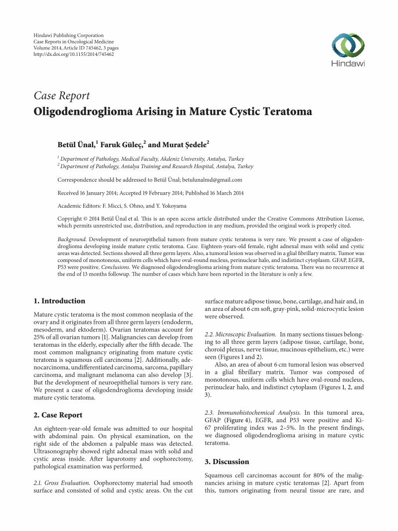
2.2. Microscopic Evaluation. Inmany sections tissues belong-ing to all three germ layers (adipose tissue, cartilage, bone,choroid plexus, nerve tissue, mucinous epithelium, etc.) wereseen (Figures 1 and 2).
Also, an area of about 6 cm tumoral lesion was observedin a glial fibrillary matrix. Tumor was composed ofmonotonous, uniform cells which have oval-round nucleus,perinuclear halo, and indistinct cytoplasm (Figures 1, 2, and3).
2.3. Immunohistochemical Analysis. In this tumoral area,GFAP (Figure 4), EGFR, and P53 were positive and Ki-67 proliferating index was 2–5%. In the present findings,we diagnosed oligodendroglioma arising in mature cysticteratoma.
3. Discussion
Squamous cell carcinomas account for 80% of the malig-nancies arising in mature cystic teratomas [2]. Apart fromthis, tumors originating from neural tissue are rare, and
Hindawi Publishing CorporationCase Reports in Oncological MedicineVolume 2014, Article ID 745462, 3 pageshttp://dx.doi.org/10.1155/2014/745462
2 Case Reports in Oncological Medicine
Figure 1: Tumoral lesion inside mature cystic teratoma with glialfibrillary matrix.
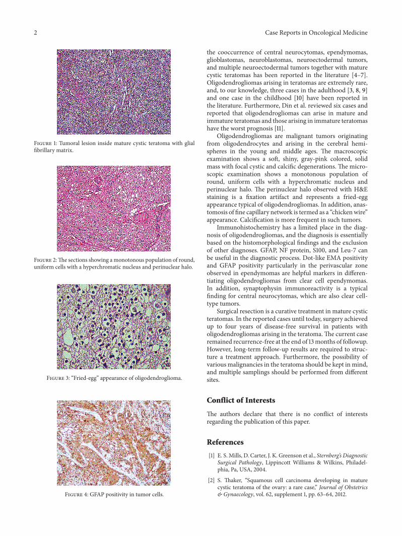
Figure 2:The sections showing amonotonous population of round,uniform cells with a hyperchromatic nucleus and perinuclear halo.
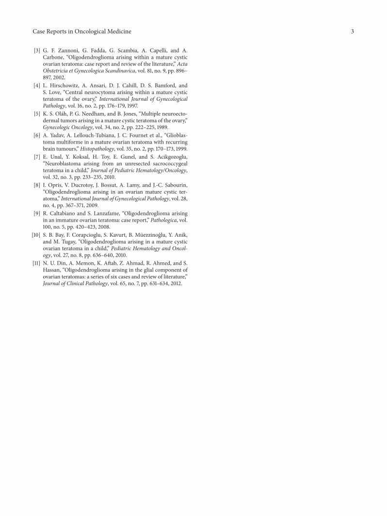
Figure 3: “Fried-egg” appearance of oligodendroglioma.
Figure 4: GFAP positivity in tumor cells.
the cooccurrence of central neurocytomas, ependymomas,glioblastomas, neuroblastomas, neuroectodermal tumors,and multiple neuroectodermal tumors together with maturecystic teratomas has been reported in the literature [4–7].Oligodendrogliomas arising in teratomas are extremely rare,and, to our knowledge, three cases in the adulthood [3, 8, 9]and one case in the childhood [10] have been reported inthe literature. Furthermore, Din et al. reviewed six cases andreported that oligodendrogliomas can arise in mature andimmature teratomas and those arising in immature teratomashave the worst prognosis [11].
Oligodendrogliomas are malignant tumors originatingfrom oligodendrocytes and arising in the cerebral hemi-spheres in the young and middle ages. The macroscopicexamination shows a soft, shiny, gray-pink colored, solidmass with focal cystic and calcific degenerations. The micro-scopic examination shows a monotonous population ofround, uniform cells with a hyperchromatic nucleus andperinuclear halo. The perinuclear halo observed with H&Estaining is a fixation artifact and represents a fried-eggappearance typical of oligodendrogliomas. In addition, anas-tomosis of fine capillary network is termed as a “chickenwire”appearance. Calcification is more frequent in such tumors.
Immunohistochemistry has a limited place in the diag-nosis of oligodendrogliomas, and the diagnosis is essentiallybased on the histomorphological findings and the exclusionof other diagnoses. GFAP, NF protein, S100, and Leu-7 canbe useful in the diagnostic process. Dot-like EMA positivityand GFAP positivity particularly in the perivascular zoneobserved in ependymomas are helpful markers in differen-tiating oligodendrogliomas from clear cell ependymomas.In addition, synaptophysin immunoreactivity is a typicalfinding for central neurocytomas, which are also clear cell-type tumors.
Surgical resection is a curative treatment in mature cysticteratomas. In the reported cases until today, surgery achievedup to four years of disease-free survival in patients witholigodendrogliomas arising in the teratoma.The current caseremained recurrence-free at the end of 13months of followup.However, long-term follow-up results are required to struc-ture a treatment approach. Furthermore, the possibility ofvariousmalignancies in the teratoma should be kept inmind,and multiple samplings should be performed from differentsites.
Conflict of Interests
The authors declare that there is no conflict of interestsregarding the publication of this paper.
References
[1] E. S. Mills, D. Carter, J. K. Greenson et al., Sternberg’s DiagnosticSurgical Pathology, Lippincott Williams & Wilkins, Philadel-phia, Pa, USA, 2004.
[2] S. Thaker, “Squamous cell carcinoma developing in maturecystic teratoma of the ovary: a rare case,” Journal of Obstetrics& Gynaecology, vol. 62, supplement 1, pp. 63–64, 2012.
Case Reports in Oncological Medicine 3
[3] G. F. Zannoni, G. Fadda, G. Scambia, A. Capelli, and A.Carbone, “Oligodendroglioma arising within a mature cysticovarian teratoma: case report and review of the literature,” ActaObstetricia et Gynecologica Scandinavica, vol. 81, no. 9, pp. 896–897, 2002.
[4] L. Hirschowitz, A. Ansari, D. J. Cahill, D. S. Bamford, andS. Love, “Central neurocytoma arising within a mature cysticteratoma of the ovary,” International Journal of GynecologicalPathology, vol. 16, no. 2, pp. 176–179, 1997.
[5] K. S. Olah, P. G. Needham, and B. Jones, “Multiple neuroecto-dermal tumors arising in a mature cystic teratoma of the ovary,”Gynecologic Oncology, vol. 34, no. 2, pp. 222–225, 1989.
[6] A. Yadav, A. Lellouch-Tubiana, J. C. Fournet et al., “Glioblas-toma multiforme in a mature ovarian teratoma with recurringbrain tumours,”Histopathology, vol. 35, no. 2, pp. 170–173, 1999.
[7] E. Unal, Y. Koksal, H. Toy, E. Gunel, and S. Acikgozoglu,“Neuroblastoma arising from an unresected sacrococcygealteratoma in a child,” Journal of Pediatric Hematology/Oncology,vol. 32, no. 3, pp. 233–235, 2010.
[8] I. Opris, V. Ducrotoy, J. Bossut, A. Lamy, and J.-C. Sabourin,“Oligodendroglioma arising in an ovarian mature cystic ter-atoma,” International Journal of Gynecological Pathology, vol. 28,no. 4, pp. 367–371, 2009.
[9] R. Caltabiano and S. Lanzafame, “Oligodendroglioma arisingin an immature ovarian teratoma: case report,” Pathologica, vol.100, no. 5, pp. 420–423, 2008.
[10] S. B. Bay, F. Corapcioglu, S. Kavurt, B. Muezzinoglu, Y. Anik,and M. Tugay, “Oligodendroglioma arising in a mature cysticovarian teratoma in a child,” Pediatric Hematology and Oncol-ogy, vol. 27, no. 8, pp. 636–640, 2010.
[11] N. U. Din, A. Memon, K. Aftab, Z. Ahmad, R. Ahmed, and S.Hassan, “Oligodendroglioma arising in the glial component ofovarian teratomas: a series of six cases and review of literature,”Journal of Clinical Pathology, vol. 65, no. 7, pp. 631–634, 2012.

Submit your manuscripts athttp://www.hindawi.com
Stem CellsInternational
Hindawi Publishing Corporationhttp://www.hindawi.com Volume 2014
Hindawi Publishing Corporationhttp://www.hindawi.com Volume 2014
MEDIATORSINFLAMMATION
of
Hindawi Publishing Corporationhttp://www.hindawi.com Volume 2014
Behavioural Neurology
EndocrinologyInternational Journal of
Hindawi Publishing Corporationhttp://www.hindawi.com Volume 2014
Hindawi Publishing Corporationhttp://www.hindawi.com Volume 2014
Disease Markers
Hindawi Publishing Corporationhttp://www.hindawi.com Volume 2014
BioMed Research International
OncologyJournal of
Hindawi Publishing Corporationhttp://www.hindawi.com Volume 2014
Hindawi Publishing Corporationhttp://www.hindawi.com Volume 2014
Oxidative Medicine and Cellular Longevity
Hindawi Publishing Corporationhttp://www.hindawi.com Volume 2014
PPAR Research
The Scientific World JournalHindawi Publishing Corporation http://www.hindawi.com Volume 2014
Immunology ResearchHindawi Publishing Corporationhttp://www.hindawi.com Volume 2014
Journal of
ObesityJournal of
Hindawi Publishing Corporationhttp://www.hindawi.com Volume 2014
Hindawi Publishing Corporationhttp://www.hindawi.com Volume 2014
Computational and Mathematical Methods in Medicine
OphthalmologyJournal of
Hindawi Publishing Corporationhttp://www.hindawi.com Volume 2014
Diabetes ResearchJournal of
Hindawi Publishing Corporationhttp://www.hindawi.com Volume 2014
Hindawi Publishing Corporationhttp://www.hindawi.com Volume 2014
Research and TreatmentAIDS
Hindawi Publishing Corporationhttp://www.hindawi.com Volume 2014
Gastroenterology Research and Practice
Hindawi Publishing Corporationhttp://www.hindawi.com Volume 2014
Parkinson’s Disease
Evidence-Based Complementary and Alternative Medicine
Volume 2014Hindawi Publishing Corporationhttp://www.hindawi.com


![Verrucous carcinoma arising from a previous cystic lesion: a ......occurred at OC [7]. Primary intraosseous squamous cell carcinoma refers to carcinoma originated in the jaw as primary](https://img.pdfslide.us/doc/110x75/60ffb8f3283ea60750318493/verrucous-carcinoma-arising-from-a-previous-cystic-lesion-a-occurred-at.jpg)