Embed Size (px)
Citation preview
J Ayub Med Coll Abbottabad 2014;26(3)
http://www.ayubmed.edu.pk/JAMC/26-3/Nawaz.pdf 404
CASE REPORT NEUROBLASTOMA IN HORSESHOE KIDNEY
Gul Nawaz, Shujah Muhammad, Muhammad Imran Jamil, Durre Shohab, Iftikhar Ali Khan, Muhammad Athar Khawaja, Shahnawar Gulzar, Mian Khalid Akber, Ayaz Khan,
Ijaz Hussain , Saeed Akhter Department of Urology and Kidney Transplant, Pakistan Kidney Institute, Shifa International Hospital, Islamabad, Pakistan
Neuroblastoma is one of the commonest malignancy of childhood. Neuroblatoma in horseshoe kidney is an extremely rare condition. There is only one case of this tumour occurring in horseshoe kidney described in the literature. Recently we successfully treated a boy with neuroblastoma in horseshoe kidney. Keywords: Neuroblastoma, Horseshoe kidney
J Ayub Med Coll Abbottabad 2014;26(3):404–5
INTRODUCTION Neuroblastoma is the fourth most common malignancy of the childhood and is the most common intra-abdominal malignancy of infancy.1 However, the occurrence of this tumour in horseshoe kidney is extremely rare. There is only one case of this tumour occurring in horseshoe kidney described in the literature.2 Patients can be treated with surgery alone or by multimodal approach depending upon stage of disease.
CASE REPORT A 2 years old healthy boy presented with painless haematuria of two months duration. On abdominal examination, a hard, non-tender mass was palpable in the left lumber region. Patient’s complete blood count and renal function tests were within normal limits except haematuria on urine routine analysis.
CT scan showed a horseshoe kidney having a large, poorly defined, infiltrative mass in left renal and suprarenal region encasing the left renal vessels with enlarged regional lymph nodes (Figure-1). USG guided tumor biopsy turned out to be neuroblastoma.
After 4 cycles of neo-adjuvant chemotherapy with etoposide, carboplatin and doxorubicin, patient was planned for surgery. The kidney was approached through a transverse abdominal incision. Tumour was localized to the left hemi-kidney, which was extending into the isthmus. Left hemi-nephrectomy, isthmectomy and right partial nephrectomy was performed to get tumour free margins based on frozen sections. This was followed by a repair of calyces over a 4.7 Fr DJ stent. The patient had an uneventful post-operative course. Serum creatinine was 0.99 mg/dL at the time of discharge, on 7th postoperative day.
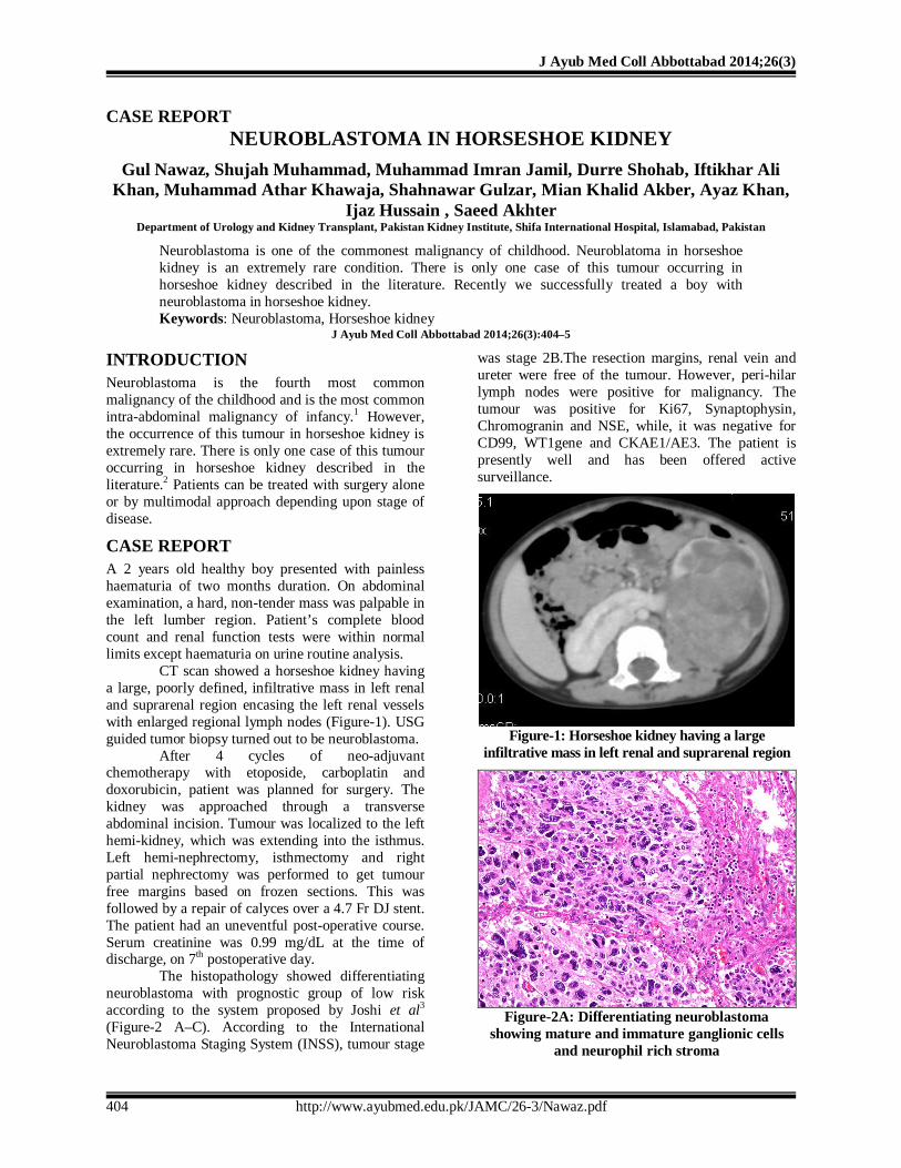
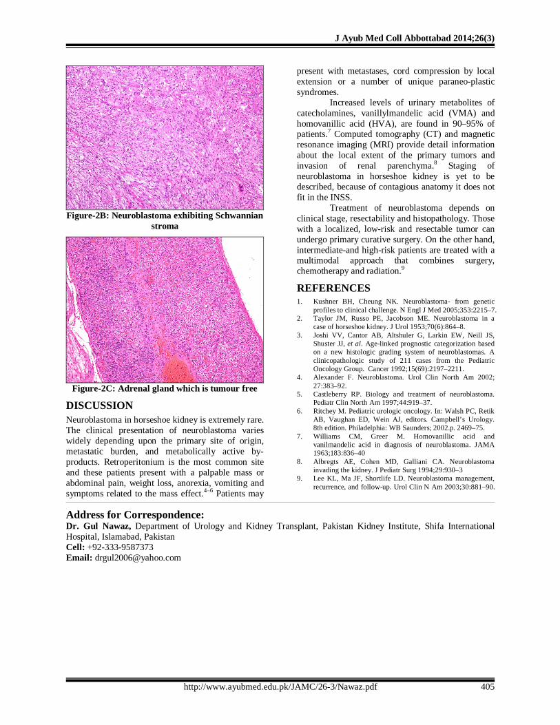
The histopathology showed differentiating neuroblastoma with prognostic group of low risk according to the system proposed by Joshi et al3 (Figure-2 A–C). According to the International Neuroblastoma Staging System (INSS), tumour stage
was stage 2B.The resection margins, renal vein and ureter were free of the tumour. However, peri-hilar lymph nodes were positive for malignancy. The tumour was positive for Ki67, Synaptophysin, Chromogranin and NSE, while, it was negative for CD99, WT1gene and CKAE1/AE3. The patient is presently well and has been offered active surveillance.
Figure-1: Horseshoe kidney having a large
infiltrative mass in left renal and suprarenal region
Figure-2A: Differentiating neuroblastoma
showing mature and immature ganglionic cells and neurophil rich stroma
J Ayub Med Coll Abbottabad 2014;26(3)
http://www.ayubmed.edu.pk/JAMC/26-3/Nawaz.pdf 405
Figure-2B: Neuroblastoma exhibiting Schwannian
stroma
Figure-2C: Adrenal gland which is tumour free
DISCUSSION Neuroblastoma in horseshoe kidney is extremely rare. The clinical presentation of neuroblastoma varies widely depending upon the primary site of origin, metastatic burden, and metabolically active by-products. Retroperitonium is the most common site and these patients present with a palpable mass or abdominal pain, weight loss, anorexia, vomiting and symptoms related to the mass effect.4–6 Patients may
present with metastases, cord compression by local extension or a number of unique paraneo-plastic syndromes.
Increased levels of urinary metabolites of catecholamines, vanillylmandelic acid (VMA) and homovanillic acid (HVA), are found in 90–95% of patients.7 Computed tomography (CT) and magnetic resonance imaging (MRI) provide detail information about the local extent of the primary tumors and invasion of renal parenchyma.8 Staging of neuroblastoma in horseshoe kidney is yet to be described, because of contagious anatomy it does not fit in the INSS.
Treatment of neuroblastoma depends on clinical stage, resectability and histopathology. Those with a localized, low-risk and resectable tumor can undergo primary curative surgery. On the other hand, intermediate-and high-risk patients are treated with a multimodal approach that combines surgery, chemotherapy and radiation.9
REFERENCES 1. Kushner BH, Cheung NK. Neuroblastoma- from genetic
profiles to clinical challenge. N Engl J Med 2005;353:2215–7. 2. Taylor JM, Russo PE, Jacobson ME. Neuroblastoma in a
case of horseshoe kidney. J Urol 1953;70(6):864–8. 3. Joshi VV, Cantor AB, Altshuler G, Larkin EW, Neill JS,
Shuster JJ, et al. Age-linked prognostic categorization based on a new histologic grading system of neuroblastomas. A clinicopathologic study of 211 cases from the Pediatric Oncology Group. Cancer 1992;15(69):2197–2211.
4. Alexander F. Neuroblastoma. Urol Clin North Am 2002; 27:383–92.
5. Castleberry RP. Biology and treatment of neuroblastoma. Pediatr Clin North Am 1997;44:919–37.
6. Ritchey M. Pediatric urologic oncology. In: Walsh PC, Retik AB, Vaughan ED, Wein AJ, editors. Campbell’s Urology. 8th edition. Philadelphia: WB Saunders; 2002.p. 2469–75.
7. Williams CM, Greer M. Homovanillic acid and vanilmandelic acid in diagnosis of neuroblastoma. JAMA 1963;183:836–40
8. Albregts AE, Cohen MD, Galliani CA. Neuroblastoma invading the kidney. J Pediatr Surg 1994;29:930–3
9. Lee KL, Ma JF, Shortlife LD. Neuroblastoma management, recurrence, and follow-up. Urol Clin N Am 2003;30:881–90.
Address for Correspondence: Dr. Gul Nawaz, Department of Urology and Kidney Transplant, Pakistan Kidney Institute, Shifa International Hospital, Islamabad, Pakistan Cell: +92-333-9587373 Email: [email protected]


![Neuroblastoma: Biology and Therapy · neuroblastoma tumors and is the most consistently reported abnormality.[1,2] Cytogenetic analysis of near-diploid neuroblastoma tumors and cell](https://img.pdfslide.us/doc/110x75/5d4ce04a88c9930e558b554a/neuroblastoma-biology-and-therapy-neuroblastoma-tumors-and-is-the-most-consistently.jpg)