Embed Size (px)
Citation preview
Case ReportHaemorrhagic Presentation of a Craniopharyngioma ina Pregnant Woman
Cesare Zoia,1 Andrea Cattalani,2 Elena Turpini,2 Viola Marta Custodi,1 Marco Benazzo,3
Fabio Pagella,3 Paolo Carena,3 Elisabetta Lovati,4 Pietro Lucotti,4 and Paolo Gaetani1
1 Department of Neurosurgery, IRCCS Fondazione Policlinico San Matteo, Viale Golgi 19, 27100 Pavia, Italy2 Neurosurgery, Department of Clinical Surgical Diagnostic and Pediatric Science, University of Pavia, Viale Golgi 19, 27100 Pavia, Italy3 Department of Otorhinolaryngology, IRCCS Fondazione Policlinico San Matteo, Viale Golgi 19, 27100 Pavia, Italy4 First Department of Medicine, IRCCS Fondazione Policlinico San Matteo, Viale Golgi 19, 27100 Pavia, Italy
Correspondence should be addressed to Cesare Zoia; [email protected]
Received 14 May 2014; Accepted 7 July 2014; Published 5 August 2014
Academic Editor: Jorge C. Kattah
Copyright © 2014 Cesare Zoia et al. This is an open access article distributed under the Creative Commons Attribution License,which permits unrestricted use, distribution, and reproduction in any medium, provided the original work is properly cited.
Objective. Craniopharyngioma is a rare tumour, and, consequently, acute clinical presentation and diagnosis, during pregnancy, ofthis pathology are quite difficult to find. Only few cases are reported in the literature, and no one describes these two conditions inassociation.Methods.Wereport a particular case of craniopharyngiomapresenting both of the above conditions.Results.Thepatientwas successfully operated with endoscopic technique.Conclusions.Rare and difficult cases, created by the superposition of differentclinical conditions, needmultidisciplinarymanagement, with collaboration, integration, and cooperation between differentmedicalspecialists.
1. Introduction
Craniopharyngioma is a rare tumour, with incidence of 0,13cases per 100,000 people/year [1], and it is the mostly benignepithelial tumour of the sellar and suprasellar region. In1857 Zenker first described pathological findings of cellsclusters similar to squamous epithelium in the hypothalamic-pituitary region [2]. The term “craniopharyngioma” wascoined in 1931 by Charles Frazier and further popularized byHarvey Cushing who described craniopharyngiomas as “themost formidable of intracranial tumours” [3]. Two principalpatterns of craniopharyngioma are recognized: papillaryand adamantinomatous. This latter pattern is made up bynests and cords of stratified squamous epithelium which arereplaced by a layer of columnar cells on the outskirts, and itis characterized by the presence of dystrophic calcificationsand cysts containing “motor-oil-like” fluid (brown-yellowcholesterol-rich fluid). The papillary pattern is made up bypapillary squamous epithelium, and it is generally withoutcalcifications or cysts [4, 5].
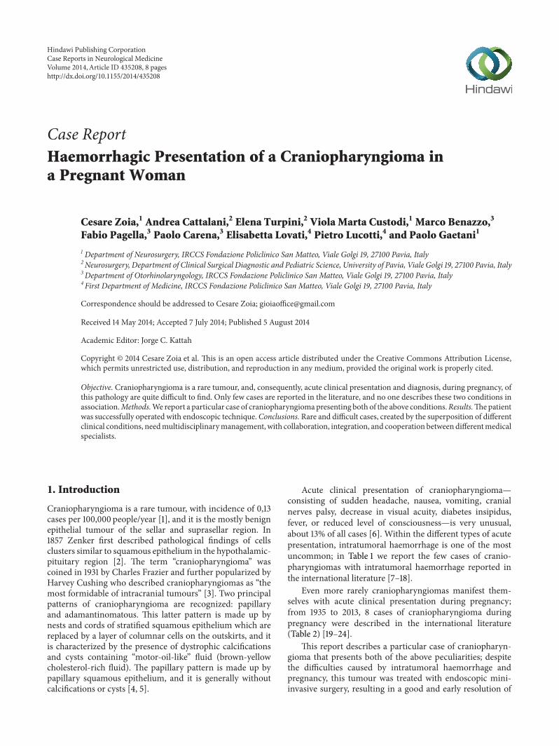
Acute clinical presentation of craniopharyngioma—consisting of sudden headache, nausea, vomiting, cranialnerves palsy, decrease in visual acuity, diabetes insipidus,fever, or reduced level of consciousness—is very unusual,about 13% of all cases [6]. Within the different types of acutepresentation, intratumoral haemorrhage is one of the mostuncommon; in Table 1 we report the few cases of cranio-pharyngiomas with intratumoral haemorrhage reported inthe international literature [7–18].
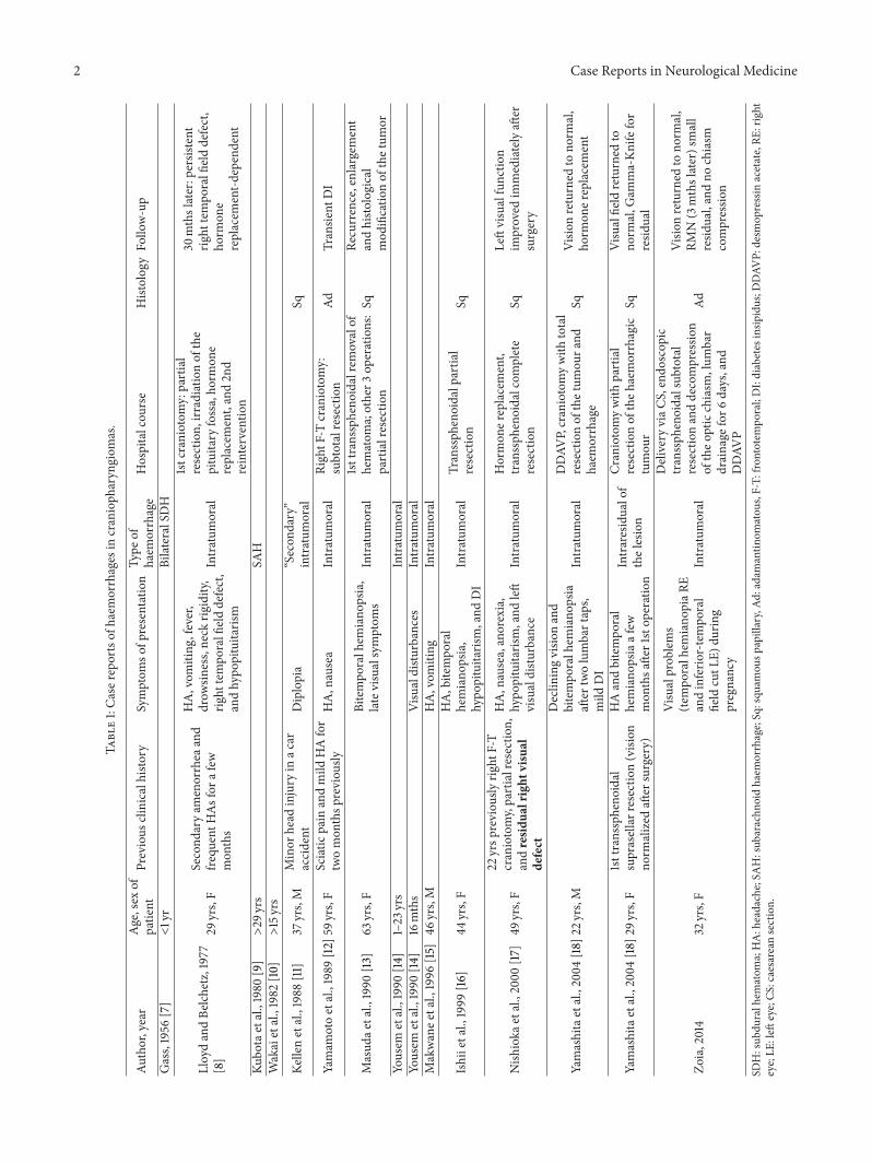
Even more rarely craniopharyngiomas manifest them-selves with acute clinical presentation during pregnancy;from 1935 to 2013, 8 cases of craniopharyngioma duringpregnancy were described in the international literature(Table 2) [19–24].
This report describes a particular case of craniopharyn-gioma that presents both of the above peculiarities; despitethe difficulties caused by intratumoral haemorrhage andpregnancy, this tumour was treated with endoscopic mini-invasive surgery, resulting in a good and early resolution of
Hindawi Publishing CorporationCase Reports in Neurological MedicineVolume 2014, Article ID 435208, 8 pageshttp://dx.doi.org/10.1155/2014/435208
2 Case Reports in Neurological Medicine
Table1:Ca
serepo
rtso
fhaemorrhages
incranioph
aryngiom
as.
Author,year
Age,sex
ofpatie
ntPrevious
clinicalh
istory
Symptom
sofp
resentation
Type
ofhaem
orrhage
Hospitalcou
rse
Histolog
yFo
llow-up
Gass,1956
[7]
<1yr
BilateralSDH
LloydandBe
lchetz,1977
[8]
29yrs,F
Second
aryam
enorrhea
and
frequ
entH
Asfora
few
mon
ths
HA,vom
iting
,fever,
drow
siness,neck
rigidity,
right
tempo
ralfielddefect,
andhypo
pituitaris
m
Intratum
oral
1stcraniotom
y:partial
resection,
irradiatio
nof
the
pituitary
fossa,ho
rmon
ereplacem
ent,and2n
dreinterventio
n
30mthslater:persistent
right
tempo
ralfi
elddefect,
horm
one
replacem
ent-d
ependent
Kubo
taetal.,1980
[9]>29
yrs
SAH
Wakaietal.,1982
[10]>15yrs
Kellenetal.,1988
[11]
37yrs,M
Minor
head
injury
inac
araccident
Diplopia
“Secon
dary”
intratum
oral
Sq
Yamam
otoetal.,1989
[12]
59yrs,F
Sciatic
pain
andmild
HAfor
twomon
thsp
reviou
slyHA,nausea
Intratum
oral
RightF
-Tcraniotomy:
subtotalresection
AdTransie
ntDI
Masud
aetal.,1990
[13]
63yrs,F
Bitempo
ralh
emiano
psia,
latevisualsymptom
sIntratum
oral
1sttransspheno
idalremovalof
hematom
a;other3
operations:
partialresectio
nSq
Recurrence,enlargement
andhisto
logical
mod
ificatio
nof
thetum
orYo
usem
etal.,1990
[14]
1–23
yrs
Intratum
oral
Yousem
etal.,1990
[14]
16mths
Visualdistu
rbances
Intratum
oral
Makwanee
tal.,1996
[15]
46yrs,M
HA,vom
iting
Intratum
oral
Ishiietal.,1999
[16]
44yrs,F
HA,bitempo
ral
hemiano
psia,
hypo
pituitaris
m,and
DI
Intratum
oral
Transsph
enoidalp
artia
lresection
Sq
Nish
ioka
etal.,2000
[17]
49yrs,F
22yrsp
reviou
slyrig
htF-T
craniotomy,partialresectio
n,andresid
ualright
visual
defect
HA,nausea,anorexia,
hypo
pituitaris
m,and
left
visualdistu
rbance
Intratum
oral
Hormon
ereplacement,
transsph
enoidalcom
plete
resection
SqLeftvisualfunctio
nim
proved
immediatelyaft
ersurgery
Yamashitaetal.,2004
[18]
22yrs,M
Declin
ingvisio
nand
bitempo
ralh
emiano
psia
after
twolumbartaps,
mild
DI
Intratum
oral
DDAV
P,craniotomywith
total
resectionof
thetum
oura
ndhaem
orrhage
SqVisio
nreturned
tono
rmal,
horm
oner
eplacement
Yamashitaetal.,2004
[18]
29yrs,F
1sttransspheno
idal
suprasellarresectio
n(vision
norm
alized
after
surgery)
HAandbitempo
ral
hemiano
psiaafew
mon
thsa
fter1stop
eration
Intraresidualof
thelesion
Craniotomywith
partial
resectionof
theh
aemorrhagic
tumou
rSq
Visualfield
returned
tono
rmal,G
amma-Kn
ifefor
resid
ual
Zoia,2014
32yrs,F
Visualprob
lems
(tempo
ralh
emiano
piaR
Eandinferio
r-tempo
ral
field
cutL
E)du
ring
pregnancy
Intratum
oral
Delivery
viaC
S,endo
scop
ictranssph
enoidalsub
total
resectionanddecompressio
nof
theo
pticchiasm
,lum
bar
drainage
for6
days,and
DDAV
P
Ad
Visio
nreturned
tono
rmal,
RMN(3mthslater)small
resid
ual,andno
chiasm
compressio
n
SDH:sub
duralh
ematom
a;HA:headache;SA
H:sub
arachn
oidhaem
orrhage;Sq:squ
amou
spapillary,Ad
:adamantin
omatou
s,F-T:
frontotem
poral;DI:diabetes
insip
idus;D
DAV
P:desm
opressin
acetate,RE
:right
eye;LE
:left
eye;CS
:caesarean
section.
Case Reports in Neurological Medicine 3
Table 2: Case reports of craniopharyngiomas during pregnancy.
Authors, year Age of patient Symptoms of presentation Hospital course Follow-up
Fischer, 1935 [19] ? Bitemporal HA at 20wkgestation Therapeutic abortion Patient became blind
Sachs et al., 1978 [20] 24 yrs Visual problems and HA at28wk gestation
Tumour resected, DDAVPtreatment, and normalterm delivery
Vision returned to normal
Van der Wildt et al., 1980 [21] 24 yrs DI at 20wk gestationDDAVP treatment, deliveryat 36wk, and tumourresected postpartum
Vision returned to normal
Hiett and Barton, 1990 [22] 22 yrs DI at 27wk gestation, HA,and visual problems
DDAVP treatment, deliveryat 34wk, and tumourresected postpartum
Vision improved aftertumour resection
Johnson et al., 1993 [19] 27 yrs Visual problems, HA in 2ndtrimester
Tumour resection, normalterm delivery
Vision near normal aftertumor resection
Maniker and Krieger, 1996[23] 35 yrs Visual problems, HA at
8wk gestation
2 transsphenoidalresections, healthy deliveryvia CS at 33wk
Vision returned to normal
Aydin et al., 1999 [25] 19 yrs Visual problems and HA at20wk gestation
Transsphenoidal resection,delivery at term
Vision returned to normal,2nd resection duringsubsequent pregnancy 4 yrslater
Magge et al., 2001 [24] 39 yrs Visual problems, DI, andsevere fatigue at 6wk
Abortion, F-T craniotomywith suprasellar resection,and intranasal DDAVP
Small inferior temporalquadrantanopsia in LE, 2ndpregnancy with newbitemporal field cut thatdisappeared after normalvaginal delivery, DI intreatment with DDAVP,and thyroid hormonereplacement
Zoia, 2014 32 yrs
Visual problems (temporalhemianopsia RE andinferior-temporal field cutLE) at 30wk + 1 gestation
Cortisol replacement,delivery via CS at 33wk + 3,endoscopic transsphenoidalsubtotal resection anddecompression of the opticchiasm, lumbar drainagefor 6 days, and DDAVP
Vision returned to normal,no hormone deficiencies;RMN (3 months later)shows small residual withno chiasm compression
HA: headache; DI: diabetes insipidus; CS: caesarean section; F-T: frontotemporal; DDAVP: desmopressin acetate; RE: right eye; LE: left eye.
the mother’s new symptoms without negative consequencesfor the newborn.
2. Case Report
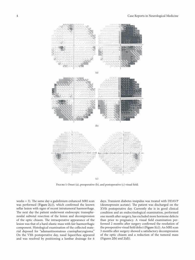
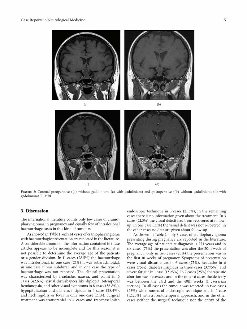
The patient, a 32-year-old woman, became pregnant forthe first time in her life. At the 30 weeks + 1 mark of atill-then-normal pregnancy, she presented a sharp bilateraldecline in visual acuity. A careful study of her clinical historyonly noted obesity and Hashimoto’s thyroiditis treated withchronic hormone replacement. Then she was assessed by theophthalmologist: a visual field examination (Figure 1(a)) doc-umented temporal hemianopia in the right eye and inferior-temporal field cut in the left eye. Subsequently she underwentmagnetic resonance imaging (MRI) scan, which showed anextra-axial lesion in the intra- and suprasellar regions, isoin-tense in T1-weighted sequence (Figure 2(a)), and isohyperin-tense in long-TR sequences; these neuroradiological findings
could be indicative of pituitary apoplexy, macroadenoma, orcraniopharyngioma with signs of intratumoral haemorrhage.This clinical and radiological picture made early surgerymandatory. Neurosurgeons and obstetricians jointly evalu-ated the case and decided, in agreement with the patient, topostpone the operation until a more advanced gestational ageto safeguard the fetal well-being.The patient underwent RDS(respiratory distress syndrome) prophylaxis and maternal-fetal welfare monitoring; endocrinological examination ofhormone levels confirmed diagnosis of central hypocorti-solism. As a consequence the patient underwent adequatehormone replacement: Cortone Acetate 37.5mg/day. At 33weeks + 2 of gestation, the patient complained of a furtherdecline in visual acuity; thus, she underwent a new visualfield examination (Figure 1(b)) that turned out to be worsethan the previous one. For this reason, after a new collegialevaluation, she underwent delivery via caesarean section (33
4 Case Reports in Neurological Medicine
(a)
(b)
(c)
Figure 1: Onset (a), preoperative (b), and postoperative (c) visual field.
weeks + 3). The same day a gadolinium enhanced MRI scanwas performed (Figure 2(c)), which confirmed the knownsellar lesion with signs of recent intratumoral haemorrhage.The next day the patient underwent endoscopic transsphe-noidal subtotal resection of the lesion and decompressionof the optic chiasm. The intraoperative appearance of thelesion was that of a hard-elastic mass with fair haemorrhagiccomponent. Histological examination of the collected mate-rial deposed for “adamantinomatous craniopharyngioma.”On the VIth postoperative day, nasal liquorrhea appearedand was resolved by positioning a lumbar drainage for 6
days. Transient diabetes insipidus was treated with DDAVP(desmopressin acetate). The patient was discharged on theXVth postoperative day. Currently she is in good clinicalcondition and an endocrinological examination, performedonemonth after surgery, has excludedmore hormone defectsthan prior to pregnancy. A visual field examination per-formed 2 months after surgery confirmed the resolution ofthe preoperative visual field defect (Figure 1(c)). AnMRI scan3 months after surgery showed a satisfactory decompressionof the optic chiasm and a reduction of the tumoral mass(Figures 2(b) and 2(d)).
Case Reports in Neurological Medicine 5
(a) (b)
(c) (d)
Figure 2: Coronal preoperative ((a) without gadolinium; (c) with gadolinium) and postoperative ((b) without gadolinium; (d) withgadolinium) T1 MRI.
3. Discussion
The international literature counts only few cases of cranio-pharyngiomas in pregnancy and equally few of intralesionalhaemorrhage cases in this kind of tumours.
As showed in Table 1, only 14 cases of craniopharyngiomawith haemorrhagic presentation are reported in the literature.A considerable amount of the information contained in thesearticles appears to be incomplete and for this reason it isnot possible to determine the average age of the patientsor a gender division. In 11 cases (78.5%) the haemorrhagewas intralesional, in one case (7.1%) it was subarachnoidal,in one case it was subdural, and in one case the type ofhaemorrhage was not reported. The clinical presentationwas characterized by headache, nausea, and vomit in 6cases (42.6%), visual disturbances like diplopia, bitemporalhemianopsia, and other visual symptoms in 8 cases (56.8%,),hypopituitarism and diabetes insipidus in 4 cases (28.4%),and neck rigidity or fever in only one case (7.1%). Surgicaltreatment was transcranial in 4 cases and transnasal with
endoscopic technique in 3 cases (21.3%); in the remainingcases there is no information given about the treatment. In 3cases (21.3%) the visual deficit had been recovered at follow-up; in one case (7.1%) the visual deficit was not recovered; inthe other cases no data are given about follow-up.
As shown in Table 2, only 8 cases of craniopharyngiomapresenting during pregnancy are reported in the literature.The average age of patients at diagnosis is 27.1 years and insix cases (75%) the presentation was after the 20th week ofpregnancy; only in two cases (25%) the presentation was inthe first 10 weeks of pregnancy. Symptoms of presentationwere visual disturbances in 6 cases (75%), headache in 6cases (75%), diabetes insipidus in three cases (37.75%), andsevere fatigue in 1 case (12.25%). In 2 cases (25%) therapeuticabortion was necessary and in the other 6 cases the deliverywas between the 33rd and the 40th weeks (1 caesariansection). In all cases the tumour was resected: in two cases(25%) with transnasal endoscopic technique and in 1 case(12.25%) with a frontotemporal approach, and in the othercases neither the surgical technique nor the entity of the
6 Case Reports in Neurological Medicine
removal is reported. At follow-up, in six cases the patient hadrecovered the visual deficit after delivery and operation, inone case (12.25%) the patient became blind, and in one casean inferotemporal quadrantopsy was reported.
The case we report is of particular relevance because itis the first to include both of these rarely associated clini-cal features. Such combination requires a multidisciplinaryassessment to allow choosing the most suitable treatmentfrom different specialistic points of view: neurosurgical, neu-roradiological, endocrinological, obstetrical, gynaecological,otolaryngological, and ophthalmological.
The role of the first specialist observing the patientis crucial: his task is to correctly identify the location ofthe problem, in order to set an appropriate diagnostic andtherapeutic plan. In the case we report, the first specialistto assess the patient was the ophthalmologist. Bitemporalhemianopia or temporal field cuts in the examination ofthe visual field are often suggestive of expansive lesionsnear the optic chiasm and sellar region. A suspicion ofsuch nature needs neuroradiological evidence, obtainable byMRI. In this case, since the patient was pregnant, MRI wasperformed without contrast: it showed the presence of anextra-axial lesion in the sellar and suprasellar regions. Asdescribed by Jagannathan et al., there are many diseases thatcan develop in these anatomical sites: pituitary adenomas,meningiomas, metastases, abscesses, aneurysms, pituitaryapoplexy, and Rathke’s cleft cyst in the sellar region andaneurysms, teratomas, hypothalamic gliomas, meningiomas,and epidermoid and dermoid cysts in the suprasellar regions[26]. In the case we report, neuroradiologist formulated threediagnostic hypotheses: pituitary apoplexy,macroadenoma, orcraniopharyngioma with signs of intratumoral haemorrhage.At this point, the patient was assessed by a neurosurgeon andan endocrinologist. The neurosurgeon pointed out that thepatient needed a surgical intervention as soon as possible, inorder to obtain adequate decompression of the optic chiasmand to treat visual impairment. The endocrinologist stressedthe importance of a comprehensive study of the hormonalstatus, to discover and correct any subclinical deficiency.Both of them requested close collaboration with obstetrics.At first, hormonal changes during pregnancy affected theendocrinological treatment. Furthermore, intervention ofthe neurosurgeon was subject to the timing of delivery;despite the visual disturbances requiring treatment as soonas possible, in this case the most important elements to keepin mind were maternal well-being and fetal growth. So opticchiasm decompression was done a few hours after deliveryby caesarean section. In the end, the endocrinologist alsohad the task of avoiding acute adrenal insufficiency in apatient subjected to two surgical stresses in a few hours. Thesurgery, in cooperation with ENT surgeons, was performedusing the endoscopic endonasal transsphenoidal approachwith the “two nostrils four hands technique” [27]. Sellar floorreconstruction was not needed.
The choice of endoscopic treatment was based on thegreat benefits reported by the international literature duringthe last years [28–38]: minimal invasiveness, reduced postop-erative recovery period, and minimal psychological impact
on patient, surgical outcomes, and complication rate similarto those of the classic microscopic technique.
In this case, a patient with a delicate endocrinologicalbalance underwent 2 surgical procedures in few hours: use ofendoscopic minimally invasive technique reduced the conse-quences of this particular surgical stress. Reduced recoverydays and early ability in taking care of the newborn alsoresulted in the patient’s more positive psychological reactionto her own disease.
In our opinion, this case represents another excellentdemonstration of the versatility of the endoscopic technique,which can be used in complex clinical conditions andconsidered a gold standard in the surgery of the sellar andparaphrase regions.
The role of the various specialists remains important dur-ing follow-up of the patient: the ENT surgeon endoscopicallychecks for good repair of the wound; the neuroradiologistcarries out the necessary radiological postoperative scans(CT in the early postoperative period and MRI 3 monthslater); the endocrinologist checks the hormonal balance andsets adequate hormone replacement; the ophthalmologistcarries out field examinations in the postoperative period.Therefore, the collaboration remains critical among thesedifferent medical figures during follow-up.
4. Conclusion
We report what, to the best of our knowledge, is the first casein the literature in which two rare features of craniopharyn-giomas overlapped at the onset; such synergistic coexistencecreated a clinical condition difficult to diagnose and manage.
In this particular situation, once diagnosis of sellar lesionhad been obtained, all medical and surgical treatmentsundertaken were found to be necessary and, in particular,endoscopic minimally invasive surgery was considered thefirst choice. In our opinion this technique represents the goldstandard in surgical approach to sellar and parasellar regionsand is indispensable in cases like the reported one.
Rare and difficult cases, created by the overlapping ofdifferent clinical conditions, need multidisciplinary manage-ment, with collaboration and cooperation between manymedical specialists: careful preoperative planning, minimallyinvasive surgery with less possible complications, and ade-quate follow-up are obtained only with close support andcollaboration among all specialists involved.
Conflict of Interests
The authors report no conflict of interests concerning thematerials or methods used in this study or findings specifiedin this paper.
References
[1] G. R. Bunin, T. S. Surawicz, P. A. Witman, S. Preston-Martin, F.Davis, and J. M. Bruner, “The descriptive epidemiology of cran-iopharyngioma,” Journal of Neurosurgery, vol. 89, no. 4, pp. 547–551, 1998.
Case Reports in Neurological Medicine 7
[2] F. A. Zenker, “Enorme Cystenbildung im Gehirn, vom Hir-nanhang ausgehend,” Archiv fur Pathologische Anatomie undPhysiologie und fur Klinische Medicin, vol. 12, no. 4-5, pp. 454–466, 1857.
[3] G. Barkhoudarian and E. R. Laws, “Craniopharyngioma: his-tory,” Pituitary, vol. 16, no. 1, pp. 1–8, 2013.
[4] J. C. Fernandez-Miranda, P. A. Gardner, C. H. Snyderman etal., “Craniopharyngioma: a pathologic, clinical, and surgicalreview,” Head and Neck, vol. 34, no. 7, pp. 1036–1044, 2012.
[5] V. Kumar, A. Abbas, and N. Fausto, Robbins e Cotran Le basipatologiche delle malattie, vol. 2, Elsevier, 2009.
[6] E.H.Nielsen, J. O. Jørgensen, P. Bjerre et al., “Acute presentationof craniopharyngioma in children and adults in a Danishnational cohort,” Pituitary, vol. 16, no. 4, pp. 528–535, 2013.
[7] H. H. GASS, “Large calcified craniopharyngioma and bilateralsubdural hematomata present at birth; survey of neonatal braintumors.,” Journal of neurosurgery, vol. 13, no. 5, pp. 514–519, 1956.
[8] M. H. Lloyd and P. E. Belchetz, “The clinical features and man-agement of pituitary apoplexy,” Postgraduate Medical Journal,vol. 53, no. 616, pp. 82–85, 1977.
[9] T. Kubota, H. Fujii, K. Ikeda, H. Ito, S. Yamamoto, and I.Nakanishi, “A case of intraventricular craniopharyngioma withsubarachnoid hemorrhage,” Neurological Surgery, vol. 8, no. 5,pp. 495–501, 1980.
[10] S. Wakai, K. Yamakawa, S. Manaka, and K. Takakura, “Spon-taneous intracranial hemorrhage caused by brain tumor: itsincidence and clinical significance,” Neurosurgery, vol. 10, no.4, pp. 437–444, 1982.
[11] R. I. Kellen, R. M. Burde, and F. J. Hodges III, “Occultpituitary apoplexy associatedwith craniopharyngioma,” Journalof Clinical Neuro-Ophthalmology, vol. 8, no. 2, pp. 99–104, 1988.
[12] T. Yamamoto, S. Yoneda, and N. Funatsu, “Spontaneous haem-orrhage in craniopharyngioma,” Journal of Neurology Neuro-surgery and Psychiatry, vol. 52, no. 6, pp. 803–804, 1989.
[13] R. Masuda, E. Tsukamoto, S. Takeda, S. Furuichi, S. Endo, andA. Takaku, “An elderly case of recurrent craniopharyngiomasuffering from hemorrhage,” Neurological Surgery, vol. 18, no.12, pp. 1151–1155, 1990.
[14] D. M. Yousem, J. A. Arrington, A. J. Kumar, and R. N. Bryan,“Bright lesions on sellar/parasellar T1-weighted scans,” ClinicalImaging, vol. 14, no. 2, pp. 99–105, 1990.
[15] U. K. Makwane, A. K. Singh, V. Puri, and S. Kumar, “Primarybrain tumors presenting as intra cerebral haemorrhage,” Journalof Association of Physicians of India, vol. 44, no. 10, pp. 729–733,1996.
[16] K. Ishii, M. Isono, S. Hori, Y. Kinba, and T. Mori, “A case ofcraniopharyngioma with intratumoral hemorrhage,” Neurolog-ical Surgery, vol. 27, no. 1, pp. 73–77, 1999.
[17] H. Nishioka, H. Ito, J. Haraoka, T. Hashimoto, and Y. Kato,“Repeated hemorrhage in ciliated craniopharyngioma—casereport,” Neurologia Medico-Chirurgica, vol. 40, no. 6, pp. 324–328, 2000.
[18] S. Yamashita, Y. Matsumoto, K. Kunishio, and S. Nagao,“Craniopharyngiomas with intratumoral hemorrhage: two casereports,”NeurologiaMedico-Chirurgica, vol. 44, no. 1, pp. 43–46,2004.
[19] R. J. Johnson Jr., R. M. Voorhies, M. Witkin, A. G. RobichauxIII, and W. A. Broussard Jr., “Fertility following excision ofa symptomatic craniopharyngioma during pregnancy: casereport,” Surgical Neurology, vol. 39, no. 4, pp. 257–262, 1993.
[20] B. P. Sachs, S. K. Smith, J. Cassar, and B. van Iddekinge, “Rapidenlargement of carniopharyngioma in pregnancy,” British Jour-nal of Obstetrics and Gynaecology, vol. 85, no. 7, pp. 577–578,1978.
[21] B. van derWildt, J. I. M. Drayer, and T. K. A. B. Eskes, “Diabetesinsipidus in pregnancy as a first sign of a craniopharyngioma,”European Journal of Obstetrics Gynecology and ReproductiveBiology, vol. 10, no. 4, pp. 269–274, 1980.
[22] A. K. Hiett and J. R. Barton, “Diabetes insipidus associated withcraniopharyngioma in pregnancy,” Obstetrics and Gynecology,vol. 76, no. 5, pp. 982–984, 1990.
[23] A. H. Maniker and A. J. Krieger, “Rapid recurrence of cranio-pharyngioma during pregnancy with recovery of vision: a casereport,” Surgical Neurology, vol. 45, no. 4, pp. 324–327, 1996.
[24] S. N. Magge, M. Brunt, and R. M. Scott, “Craniopharyngiomapresenting during pregnancy 4 years after a normal magneticresonance imaging scan: case report,” Neurosurgery, vol. 49, no.4, pp. 1014–1017, 2001.
[25] Y. Aydin, S. M. Can, A. Gulkilik, O. Turkmenoglu, C. Alatli,and I. Ziyal, “Rapid enlargement and recurrence of a preexistingintrasellar craniopharyngioma during the course of two preg-nancies: case report,” Journal of Neurosurgery, vol. 91, no. 2, pp.322–324, 1999.
[26] J. Jagannathan, A. S. Dumont, J. A. Jane Jr., and E. R. Laws Jr.,“Pediatric sellar tumors: diagnostic procedures and manage-ment,” Neurosurgical Focus, vol. 18, no. 6, 2005.
[27] P. Castelnuovo, A. Pistochini, and D. Locatelli, “Differentsurgical approaches to the sellar region: focusing on the “twonostrils four hands technique”,” Rhinology, vol. 44, no. 1, pp. 2–7, 2006.
[28] T. S. Higgins, C. Courtemanche, D. Karakla et al., “Analysis oftransnasal endoscopic versus transseptal microscopic approachfor excision of pituitary tumors,” The American Journal ofRhinology, vol. 22, no. 6, pp. 649–652, 2008.
[29] B. Rotenberg, S. Tam, W. H. A. Ryu, and N. Duggal, “Micro-scopic versus endoscopic pituitary surgery: a systematic review,”The Laryngoscope, vol. 120, no. 7, pp. 1292–1297, 2010.
[30] J. K. Goudakos, K. D. Markou, and C. Georgalas, “Endoscopicversus microscopic trans-sphenoidal pituitary surgery: a sys-tematic review and meta-analysis,” Clinical Otolaryngology, vol.36, no. 3, pp. 212–220, 2011.
[31] A. Tabaee, V. K. Anand, Y. Barron et al., “Endoscopic pituitarysurgery: a systematic review and meta-analysis,” Journal ofNeurosurgery, vol. 111, no. 3, pp. 545–554, 2009.
[32] D. Y. Cho and W. R. Liau, “Comparison of endonasal endo-scopic surgery and sublabial microsurgery for prolactinomas,”Surgical Neurology, vol. 58, no. 6, pp. 371–375, 2002.
[33] A. R. Dehdashti, A. Ganna, K. Karabatsou, and F. Gentili, “Pureendoscopic endonasal approach for pituitary adenomas: earlysurgical results in 200 patients and comparison with previousmicrosurgical series,”Neurosurgery, vol. 62, no. 5, pp. 1006–1015,2008.
[34] H. D. Jho, “Endoscopic transsphenoidal surgery,” Journal ofNeuro-Oncology, vol. 54, no. 2, pp. 187–195, 2001.
[35] R. T. Netea-Maier, E. J. van Lindert, M. den Heijer et al.,“Transsphenoidal pituitary surgery via the endoscopic tech-nique: results in 35 consecutive patients with Cushing’s disease,”European Journal of Endocrinology, vol. 154, no. 5, pp. 675–684,2006.
[36] B. W. O’Malley Jr., M. S. Grady, B. C. Gabel et al., “Comparisonof endoscopic and microscopic removal of pituitary adenomas:

8 Case Reports in Neurological Medicine
single-surgeon experience and the learning curve.,”Neurosurgi-cal focus, vol. 25, no. 6, article E10, 2008.
[37] G. Frank, E. Pasquini, G. Farneti et al., “The endoscopic versusthe traditional approach in pituitary surgery,”Neuroendocrinol-ogy, vol. 83, no. 3-4, pp. 240–248, 2006.
[38] M. Berker, D. B. Hazer, T. Yu cel et al., “Complications ofendoscopic surgery of the pituitary adenomas: analysis of 570patients and review of the literature,” Pituitary, vol. 15, no. 3, pp.288–300, 2012.

Submit your manuscripts athttp://www.hindawi.com
Stem CellsInternational
Hindawi Publishing Corporationhttp://www.hindawi.com Volume 2014
Hindawi Publishing Corporationhttp://www.hindawi.com Volume 2014
MEDIATORSINFLAMMATION
of
Hindawi Publishing Corporationhttp://www.hindawi.com Volume 2014
Behavioural Neurology
EndocrinologyInternational Journal of
Hindawi Publishing Corporationhttp://www.hindawi.com Volume 2014
Hindawi Publishing Corporationhttp://www.hindawi.com Volume 2014
Disease Markers
Hindawi Publishing Corporationhttp://www.hindawi.com Volume 2014
BioMed Research International
OncologyJournal of
Hindawi Publishing Corporationhttp://www.hindawi.com Volume 2014
Hindawi Publishing Corporationhttp://www.hindawi.com Volume 2014
Oxidative Medicine and Cellular Longevity
Hindawi Publishing Corporationhttp://www.hindawi.com Volume 2014
PPAR Research
The Scientific World JournalHindawi Publishing Corporation http://www.hindawi.com Volume 2014
Immunology ResearchHindawi Publishing Corporationhttp://www.hindawi.com Volume 2014
Journal of
ObesityJournal of
Hindawi Publishing Corporationhttp://www.hindawi.com Volume 2014
Hindawi Publishing Corporationhttp://www.hindawi.com Volume 2014
Computational and Mathematical Methods in Medicine
OphthalmologyJournal of
Hindawi Publishing Corporationhttp://www.hindawi.com Volume 2014
Diabetes ResearchJournal of
Hindawi Publishing Corporationhttp://www.hindawi.com Volume 2014
Hindawi Publishing Corporationhttp://www.hindawi.com Volume 2014
Research and TreatmentAIDS
Hindawi Publishing Corporationhttp://www.hindawi.com Volume 2014
Gastroenterology Research and Practice
Hindawi Publishing Corporationhttp://www.hindawi.com Volume 2014
Parkinson’s Disease
Evidence-Based Complementary and Alternative Medicine
Volume 2014Hindawi Publishing Corporationhttp://www.hindawi.com


![Dhf (Dengue Haemorrhagic Fever)[1]](https://img.pdfslide.us/doc/110x75/577c86e01a28abe054c2ee69/dhf-dengue-haemorrhagic-fever1.jpg)