Embed Size (px)
Citation preview
- 54 - Jaffna Medical Journal
Introduction
Tuberculosis (TB) is one of the most important causes of morbidity and mortality. Incidence of tuberculosis in Sri Lanka fell gradually from 66 cases per 100,000 people in 2004 to 64 cases per 100,000 people in 2018. This case report highlights the importance of systematic clinical and radiological correlation with microbiology leading to the diagnosis of a common, but lethal condition, pulmonary tuberculosis in an unsuspected young female patient.
Case presentation
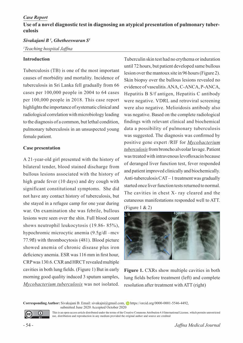
A 21-year-old girl presented with the history of bilateral tender, blood stained discharge from bullous lesions associated with the history of high grade fever (10 days) and dry cough with significant constitutional symptoms. She did not have any contact history of tuberculosis, but she stayed in a refugee camp for one year during war. On examination she was febrile, bullous lesions were seen over the shin. Full blood count shows neutrophil leukocytosis (19.86- 85%), hypochromic microcytic anemia (9.5g/dl –mcv 77.9fl) with thrombocytosis (481). Blood picture showed anemia of chronic disease plus iron deficiency anemia. ESR was 116 mm in first hour, CRP was 130.6. CXR and HRCT revealed multiple cavities in both lung fields. (Figure 1) But in early morning good quality induced 3 sputum samples, Mycobacterium tuberculosis was not isolated.
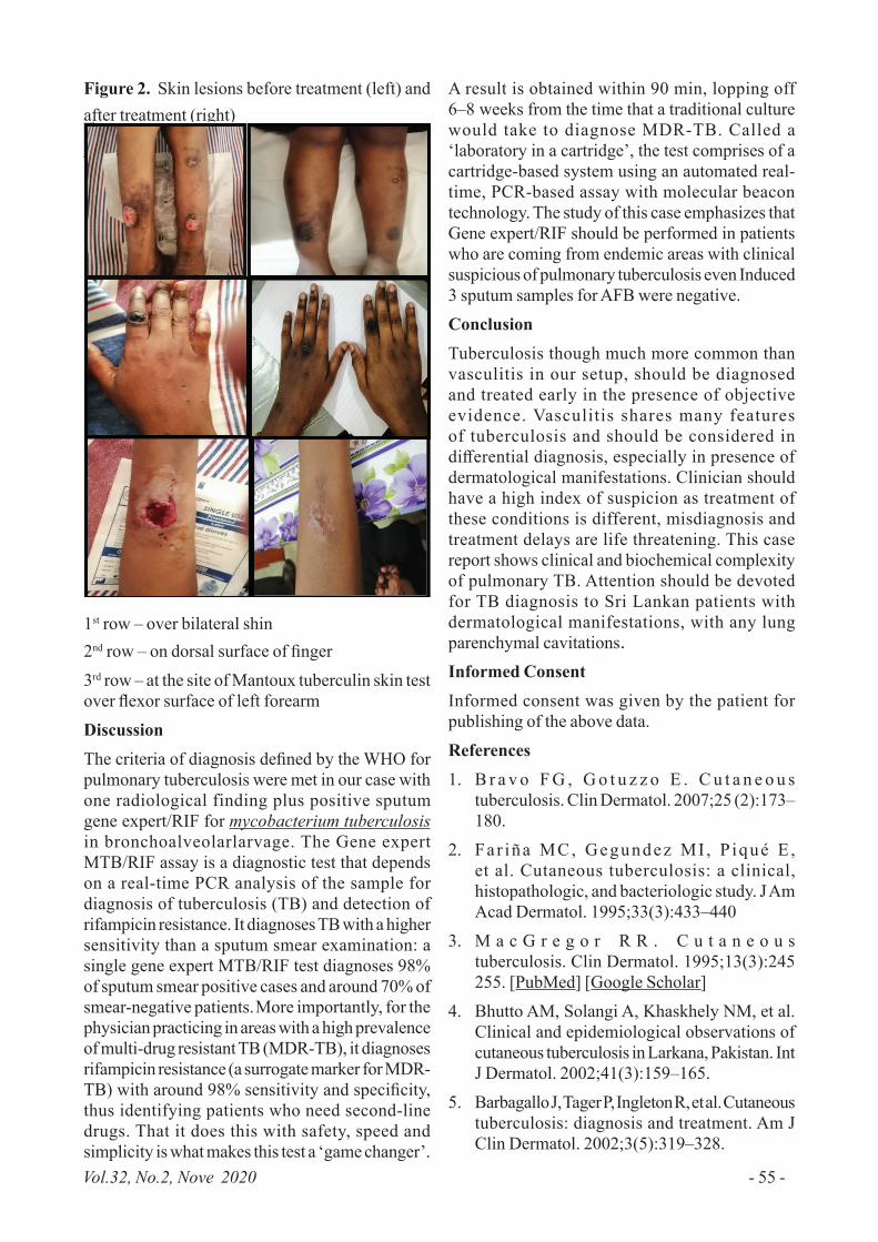
Tuberculin skin test had no erythema or induration until 72 hours, but patient developed same bullous lesion over the mantoux site in 96 hours (Figure 2). Skin biopsy over the bullous lesions revealed no evidence of vasculitis. ANA, C-ANCA, P-ANCA, Hepatitis B S/f antigen, Hepatitis C antibody were negative. VDRL and retroviral screening were also negative. Melioidosis antibody also was negative. Based on the complete radiological findings with relevant clinical and biochemical data a possibility of pulmonary tuberculosis was suggested. The diagnosis was confirmed by positive gene expert /RIF for Mycobacterium tuberculosis from broncho alveolar lavage. Patient was treated with intravenous levofloxacin because of deranged liver function test, fever responded and patient improved clinically and biochemically. Anti-tuberculosis CAT – 1 treatment was gradually started once liver function tests returned to normal. The cavities in chest X- ray cleared and the cutaneous manifestations responded well to ATT. (Figure 1 & 2)
Figure 1. CXRs show multiple cavities in both lung fields before treatment (left) and complete resolution after treatment with ATT (right)
Use of a novel diagnostic test in diagnosing an atypical presentation of pulmonary tuber-culosis
Sivakajani B 1, Ghetheeswaran S1
1Teaching hospital Jaffna
Corresponding Author: Sivakajani B. Email: [email protected], https://orcid.org/0000-0001-5546-4492, submitted June 2020 Accepted October 2020
This is an open-access article distributed under the terms of the Creative Commons Attribution 4.0 International License, which permits unrestricted use, distribution and reproduction in any medium provided the original author and source are credited
Case Report
Vol.32, No.2, Nove 2020 - 55 -
Figure 2. Skin lesions before treatment (left) and after treatment (right)
1st row – over bilateral shin2nd row – on dorsal surface of finger3rd row – at the site of Mantoux tuberculin skin test over flexor surface of left forearmDiscussionThe criteria of diagnosis defined by the WHO for pulmonary tuberculosis were met in our case with one radiological finding plus positive sputum gene expert/RIF for mycobacterium tuberculosis in bronchoalveolarlarvage. The Gene expert MTB/RIF assay is a diagnostic test that depends on a real-time PCR analysis of the sample for diagnosis of tuberculosis (TB) and detection of rifampicin resistance. It diagnoses TB with a higher sensitivity than a sputum smear examination: a single gene expert MTB/RIF test diagnoses 98% of sputum smear positive cases and around 70% of smear-negative patients. More importantly, for the physician practicing in areas with a high prevalence of multi-drug resistant TB (MDR-TB), it diagnoses rifampicin resistance (a surrogate marker for MDR-TB) with around 98% sensitivity and specificity, thus identifying patients who need second-line drugs. That it does this with safety, speed and simplicity is what makes this test a ‘game changer’.
A result is obtained within 90 min, lopping off 6–8 weeks from the time that a traditional culture would take to diagnose MDR-TB. Called a ‘laboratory in a cartridge’, the test comprises of a cartridge-based system using an automated real-time, PCR-based assay with molecular beacon technology. The study of this case emphasizes that Gene expert/RIF should be performed in patients who are coming from endemic areas with clinical suspicious of pulmonary tuberculosis even Induced 3 sputum samples for AFB were negative.Conclusion Tuberculosis though much more common than vasculitis in our setup, should be diagnosed and treated early in the presence of objective evidence. Vasculitis shares many features of tuberculosis and should be considered in differential diagnosis, especially in presence of dermatological manifestations. Clinician should have a high index of suspicion as treatment of these conditions is different, misdiagnosis and treatment delays are life threatening. This case report shows clinical and biochemical complexity of pulmonary TB. Attention should be devoted for TB diagnosis to Sri Lankan patients with dermatological manifestations, with any lung parenchymal cavitations.Informed ConsentInformed consent was given by the patient for publishing of the above data.References1. B r a v o F G , G o t u z z o E . C u t a n e o u s
tuberculosis. Clin Dermatol. 2007;25 (2):173–180.
2. Far iña MC, Gegundez MI , P iqué E ,et al. Cutaneous tuberculosis: a clinical,histopathologic, and bacteriologic study. J AmAcad Dermatol. 1995;33(3):433–440
3. M a c G r e g o r R R . C u t a n e o u stuberculosis. Clin Dermatol. 1995;13(3):245255. [PubMed] [Google Scholar]
4. Bhutto AM, Solangi A, Khaskhely NM, et al.Clinical and epidemiological observations ofcutaneous tuberculosis in Larkana, Pakistan. IntJ Dermatol. 2002;41(3):159–165.
5. Barbagallo J, Tager P, Ingleton R, et al. Cutaneoustuberculosis: diagnosis and treatment. Am JClin Dermatol. 2002;3(5):319–328.