Embed Size (px)
Citation preview

Hindawi Publishing CorporationCase Reports in PediatricsVolume 2013, Article ID 153239, 3 pageshttp://dx.doi.org/10.1155/2013/153239
Case ReportAcute MRSA Sinusitis with Intracranial Extension andMarginal Vancomycin Susceptibility
Parvathi S. Kumar and Kenji M. Cunnion
Department of Pediatrics, Division of Infectious Diseases, Children’s Hospital of the King’s Daughters and EVMS,Norfolk, VA 23507, USA
Correspondence should be addressed to Parvathi S. Kumar; [email protected]
Received 10 July 2013; Accepted 13 August 2013
Academic Editors: N.-C. Chiu and K.-H. Lue
Copyright © 2013 P. S. Kumar and K. M. Cunnion. This is an open access article distributed under the Creative CommonsAttribution License, which permits unrestricted use, distribution, and reproduction in any medium, provided the original work isproperly cited.
Methicillin resistant Staphylococcus aureus (MRSA) is increasingly being described as a cause of acute sinusitis. We presenta patient with acute MRSA sinusitis complicated by rapid intracranial extension, marginal vancomycin susceptibility (MIC =2mg/L), delayed drainage of intracranial abscess, and subsequent development of rifampin resistance. Given the relatively highrisk of intracranial extension of severe acute bacterial sinusitis and high mortality associated with invasive MRSA infections, wesuggest early surgical drainage of intracranial abscesses in these circumstances. We believe this is important given the limitedintracranial penetration of currently available treatment options forMRSA, especially those with a vancomycin minimal inhibitoryconcentration (MIC) of ≥2mg/L.
1. Introduction
Our patient, a previously healthy 12-year-old male, presentedwith acute MRSA sinusitis and rapid intracranial extension.The clinical case was complicated by a marginal vancomycinsusceptibility (MIC = 2mg/L), delayed drainage of intracra-nial abscess, and development of rifampin resistance. Thiscase is illustrative of community-associated MRSA as a pote-ntial cause of acute sinusitis leading to intracranial exten-sion, the challenges of antibiotic management of intracranialMRSA abscess, and the hazards of delayed drainage ofintracranial MRSA abscess.
2. Case Presentation
A previously healthy 12-year-old male with a history of inter-mittent migraines was admitted with acute onset of alteredmental status and facial swelling. The patient had symptomsof headache, “upset stomach,” increasing fatigue, and tactilefever for twodays prior to admission.On the day of admissionhe was found to be minimally responsive with significantswelling to the left aspect of the face, yellowish dischargefrom the left eye, and a protuberance from the forehead. A
noncontrast head CT scan done at an outside health carefacility demonstrated bilateral orbital cellulitis, pansinusitis,and possible venous sinus thrombosis prompting transfer toa pediatric hospital.
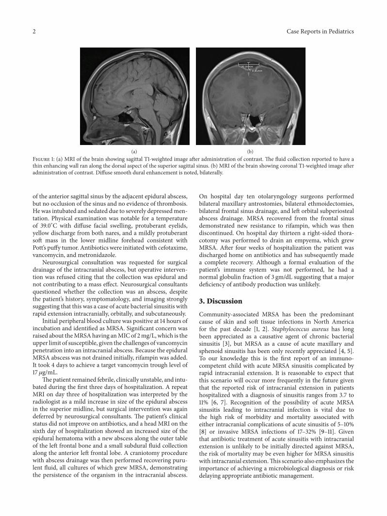
On admission, the complete blood count (CBC) revealeda white blood cell count of 8400 cells/𝜇L with a manual dif-ferential of 8% bands, 3% metamyelocytes, 74% neutrophilsand 9% lymphocytes, hemoglobin (gm/dL)/hematocrit (%)of 8.2/24.3, and platelet count of 101 × 103/𝜇L. Additionally,a coagulation panel revealed a prolonged prothrombin time(PT) and activated partial thromboplastin time (aPTT) of 19seconds and 42.3 seconds, respectively, as well elevated D-dimer levels of 11.11mg/L suggestive of disseminated intravas-cular coagulation in this patient. A brain MRI showed asuperior midline epidural fluid collection, measuring 8.6 cmanteroposterior × 3.1 cm transverse × 1.0 cm craniocaudal,following the dorsal aspect of the superior sagittal sinus(Figures 1(a) and 1(b)). The fluid collection demonstrated athin enhancing wall, and diffuse smooth dural enhancementwas noted bilaterally. Additionally, a tiny focus of intracranialair was present posterior and superior to the opacified frontalsinuses. Pansinusitis with bilateral orbital cellulitis was alsoreported. An MRA/MRV of the head noted mild narrowing
2 Case Reports in Pediatrics
(a) (b)Figure 1: (a) MRI of the brain showing sagittal T1-weighted image after administration of contrast. The fluid collection reported to have athin enhancing wall ran along the dorsal aspect of the superior sagittal sinus. (b) MRI of the brain showing coronal T1-weighted image afteradministration of contrast. Diffuse smooth dural enhancement is noted, bilaterally.
of the anterior sagittal sinus by the adjacent epidural abscess,but no occlusion of the sinus and no evidence of thrombosis.He was intubated and sedated due to severely depressedmen-tation. Physical examination was notable for a temperatureof 39.0∘C with diffuse facial swelling, protuberant eyelids,yellow discharge from both nares, and a mildly protuberantsoft mass in the lower midline forehead consistent withPott’s puffy tumor. Antibiotics were initiated with cefotaxime,vancomycin, and metronidazole.
Neurosurgical consultation was requested for surgicaldrainage of the intracranial abscess, but operative interven-tion was refused citing that the collection was epidural andnot contributing to a mass effect. Neurosurgical consultantsquestioned whether the collection was an abscess, despitethe patient’s history, symptomatology, and imaging stronglysuggesting that this was a case of acute bacterial sinusitis withrapid extension intracranially, orbitally, and subcutaneously.
Initial peripheral blood culture was positive at 14 hours ofincubation and identified as MRSA. Significant concern wasraised about theMRSAhaving anMICof 2mg/L, which is theupper limit of susceptible, given the challenges of vancomycinpenetration into an intracranial abscess. Because the epiduralMRSA abscess was not drained initially, rifampin was added.It took 4 days to achieve a target vancomycin trough level of17 𝜇g/mL.
Thepatient remained febrile, clinically unstable, and intu-bated during the first three days of hospitalization. A repeatMRI on day three of hospitalization was interpreted by theradiologist as a mild increase in size of the epidural abscessin the superior midline, but surgical intervention was againdeferred by neurosurgical consultants. The patient’s clinicalstatus did not improve on antibiotics, and a head MRI on thesixth day of hospitalization showed an increased size of theepidural hematoma with a new abscess along the outer tableof the left frontal bone and a small subdural fluid collectionalong the anterior left frontal lobe. A craniotomy procedurewith abscess drainage was then performed recovering puru-lent fluid, all cultures of which grew MRSA, demonstratingthe persistence of the organism in the intracranial abscess.
On hospital day ten otolaryngology surgeons performedbilateral maxillary antrostomies, bilateral ethmoidectomies,bilateral frontal sinus drainage, and left orbital subperiostealabscess drainage. MRSA recovered from the frontal sinusdemonstrated new resistance to rifampin, which was thendiscontinued. On hospital day thirteen a right-sided thora-cotomy was performed to drain an empyema, which grewMRSA. After four weeks of hospitalization the patient wasdischarged home on antibiotics and has subsequently madea complete recovery. Although a formal evaluation of thepatient’s immune system was not performed, he had anormal globulin fraction of 3 gm/dL suggesting that a majordeficiency of antibody production was unlikely.
3. Discussion
Community-associated MRSA has been the predominantcause of skin and soft tissue infections in North Americafor the past decade [1, 2]. Staphylococcus aureus has longbeen appreciated as a causative agent of chronic bacterialsinusitis [3], but MRSA as a cause of acute maxillary andsphenoid sinusitis has been only recently appreciated [4, 5].To our knowledge this is the first report of an immuno-competent child with acute MRSA sinusitis complicated byrapid intracranial extension. It is reasonable to expect thatthis scenario will occur more frequently in the future giventhat the reported risk of intracranial extension in patientshospitalized with a diagnosis of sinusitis ranges from 3.7 to11% [6, 7]. Recognition of the possibility of acute MRSAsinusitis leading to intracranial infection is vital due tothe high risk of morbidity and mortality associated witheither intracranial complications of acute sinusitis of 5–10%[8] or invasive MRSA infections of 17–32% [9–11]. Giventhat antibiotic treatment of acute sinusitis with intracranialextension is unlikely to be initially directed against MRSA,the risk of mortality may be even higher for MRSA sinusitiswith intracranial extension.This scenario also emphasizes theimportance of achieving a microbiological diagnosis or riskdelaying appropriate antibiotic management.

Case Reports in Pediatrics 3
This case additionally illustrates the importance of timelydrainage of an intracranial MRSA abscess. MRSA subpe-riosteal abscesses in orbital infections have been noted tobe increasing in incidence and are associate with a moreaggressive disease course than for other organisms, leading torecommendations for empiric antibiotic coverage with avery low threshold for surgical intervention [12–15]. It isreasonable that similar recommendations for timely surgicalmanagement be applied in the setting of MRSA sinusitis withintracranial extensions, given limited CNS penetration forvancomycin of 7–14% of serum concentration [16]. Limitedvancomycin penetration into abscesses [17] additionally com-promises antimicrobial effectiveness. These challenges areespecially daunting in the face of MRSA with a marginalvancomycin MIC of 2mg/L, which has been frequentlyassociated with antibiotic failure in a variety of clinicalsettings [18, 19]. For our patient, six days of vancomycintherapy did not sterilize or prevent extension of the abscess,emphasizing the importance of timely surgical debridement.Gallagher et al. concluded that optimal treatment of suppu-rative intracranial complications of sinusitis is debridementof the paranasal sinuses in combination with neurosurgicaldrainage of the intracranial focus and intravenous antibiotics[20].
Delayed surgical intervention has also been associatedwith increasing MIC values for vancomycin leading tothe development of resistance (VISA) and heteroresistance(hVISA) [21]. Although increasing vancomycin MIC did notoccur for this patient, his MRSA did develop rifampin resis-tance while on vancomycin and rifampin. This likely rep-resented inadequate vancomycin antimicrobial activity inpurulent fluid collections consistent with delayed surgicaldebridement and the marginal vancomycin MIC for thisMRSA.
References
[1] J. S. Gerber, S. E. Coffin, S. A. Smathers, and T. E. Zaoutis,“Trends in the incidence of methicillin-resistant Staphylococcusaureus infection in children’s hospitals in the United States,”Clinical Infectious Diseases, vol. 49, no. 1, pp. 65–71, 2009.
[2] J. Edelsberg, C. Taneja, M. Zervos et al., “Trends in US hospitaladmissions for skin and soft tissue infections,” Emerging Infec-tious Diseases, vol. 15, no. 9, pp. 1516–1518, 2009.
[3] T. T. Kingdom and R. E. Swain Jr., “The microbiology and anti-microbial resistance patterns in chronic rhinosinusitis,” Ameri-can Journal of Otolaryngology, vol. 25, no. 5, pp. 323–328, 2004.
[4] W.-H. Huang and P.-K. Hung, “Methicillin-resistant Staphylo-coccus aureus infections in acute rhinosinusitis,” Laryngoscope,vol. 116, no. 2, pp. 288–291, 2006.
[5] I. Brook, P. A. Foote, and J. N. Hausfeld, “Increase in thefrequency of recovery of meticillin-resistant Staphylococcusaureus in acute and chronic maxillary sinusitis,” Journal ofMedical Microbiology, vol. 57, no. 8, pp. 1015–1017, 2008.
[6] G. L. Clayman, G. L. Adams, D. R. Paugh, and C. F. Koop-mann Jr., “Intracranial complications of paranasal sinusitis: acombined institutional review,” Laryngoscope, vol. 101, no. 3, pp.234–239, 1991.
[7] R. W. Dolan and K. Chowdhury, “Diagnosis and treatmentof intracranial complications of paranasal sinus infections,”
Journal ofOral andMaxillofacial Surgery, vol. 53, no. 9, pp. 1080–1087, 1995.
[8] E. E. Lang,A. J. Curran,N. Patil, R.M.Walsh,D. Rawluk, andM.A.Walsh, “Intracranial complications of acute frontal sinusitis,”Clinical Otolaryngology and Allied Sciences, vol. 26, no. 6, pp.452–457, 2001.
[9] M. Pastagia, L. C. Kleinman, E. G. Lacerda de la Cruz, and S.G. Jenkins, “Predicting risk for death from MRSA bacteremia,”Emerging Infectious Diseases, vol. 18, no. 7, pp. 1072–1080, 2012.
[10] E. D. McCoul, D. N. Jourdy, M. R. Schaberg, and V. K.Anand, “Methicillin-resistant Staphylococcus aureus sinusitis innonhospitalized patients: a systematic review of prevalence andtreatment outcomes,” Laryngoscope, vol. 122, no. 10, pp. 2125–2131, 2012.
[11] Active Bacterial Core Surveillance Report, “Emerging Infec-tions Program Network, Methicillin-Resistant Staphylococcusaureus,” 2008, http://www.cdc.gov/abcs/reports-findings/surv-reports/mrsa08.html.
[12] S. Liao, M. L. Durand, and M. J. Cunningham, “Sinogenicorbital and subperiosteal abscesses: microbiology and methici-llin-resistant Staphylococcus aureus incidence,” Otolaryngology,vol. 143, no. 3, pp. 392–396, 2010.
[13] V. T. E. Soon, “Pediatric subperiosteal orbital abscess secondaryto acute sinusitis: a 5-year review,” American Journal of Oto-laryngology, vol. 32, no. 1, pp. 62–68, 2011.
[14] G. J. Harris, “Subperiosteal abscess of the orbit: older childrenand adults require aggressive treatment,”Ophthalmic Plastic andReconstructive Surgery, vol. 17, no. 6, pp. 395–397, 2001.
[15] S. Coenraad and J. Buwalda, “Surgical or medical managementof subperiosteal orbital abscess in children: a critical appraisalof the literature,” Rhinology, vol. 47, no. 1, pp. 18–23, 2009.
[16] I. Lutsar, G. H. McCracken Jr., and I. R. Friedland, “Antibioticpharmacodynamics in cerebrospinal fluid,” Clinical InfectiousDiseases, vol. 27, no. 5, pp. 1117–1129, 1998.
[17] E. Brauner, S. Gorbach, and P. Davey, “Comparative studyof clindamycin, imipenem, oxacillin and vancomycin in theinfected granuloma pouch model,” Journal of AntimicrobialChemotherapy, vol. 23, no. 6, pp. 891–898, 1989.
[18] J.-L. Wang, J.-T. Wang, W.-H. Sheng, Y.-C. Chen, and S.-C. Chang, “Nosocomial methicillin-resistant Staphylococcusaureus (MRSA) bacteremia in Taiwan: mortality analyses andthe impact of vancomycin, MIC = 2 mg/L, by the brothmicrodilution method,” BMC Infectious Diseases, vol. 10, article159, 2010.
[19] A. Soriano, F. Marco, J. A. Mart́ınez et al., “Influence of van-comycin minimum inhibitory concentration on the treatmentofmethicillin-resistant Staphylococcus aureus bacteremia,”Clin-ical Infectious Diseases, vol. 46, no. 2, pp. 193–200, 2008.
[20] R. M. Gallagher, C. W. Gross, and C. D. Phillips, “Suppurativeintracranial complications of sinusitis,” Laryngoscope, vol. 108,no. 11, pp. 1635–1642, 1998.
[21] B. P. Howden, P. D. R. Johnson, P. B. Ward, T. P. Stinear, andJ. K. Davies, “Isolates with low-level vancomycin resistanceassociated with persistent methicillin-resistant Staphylococcusaureus bacteremia,” Antimicrobial Agents and Chemotherapy,vol. 50, no. 9, pp. 3039–3047, 2006.

Submit your manuscripts athttp://www.hindawi.com
Stem CellsInternational
Hindawi Publishing Corporationhttp://www.hindawi.com Volume 2014
Hindawi Publishing Corporationhttp://www.hindawi.com Volume 2014
MEDIATORSINFLAMMATION
of
Hindawi Publishing Corporationhttp://www.hindawi.com Volume 2014
Behavioural Neurology
EndocrinologyInternational Journal of
Hindawi Publishing Corporationhttp://www.hindawi.com Volume 2014
Hindawi Publishing Corporationhttp://www.hindawi.com Volume 2014
Disease Markers
Hindawi Publishing Corporationhttp://www.hindawi.com Volume 2014
BioMed Research International
OncologyJournal of
Hindawi Publishing Corporationhttp://www.hindawi.com Volume 2014
Hindawi Publishing Corporationhttp://www.hindawi.com Volume 2014
Oxidative Medicine and Cellular Longevity
Hindawi Publishing Corporationhttp://www.hindawi.com Volume 2014
PPAR Research
The Scientific World JournalHindawi Publishing Corporation http://www.hindawi.com Volume 2014
Immunology ResearchHindawi Publishing Corporationhttp://www.hindawi.com Volume 2014
Journal of
ObesityJournal of
Hindawi Publishing Corporationhttp://www.hindawi.com Volume 2014
Hindawi Publishing Corporationhttp://www.hindawi.com Volume 2014
Computational and Mathematical Methods in Medicine
OphthalmologyJournal of
Hindawi Publishing Corporationhttp://www.hindawi.com Volume 2014
Diabetes ResearchJournal of
Hindawi Publishing Corporationhttp://www.hindawi.com Volume 2014
Hindawi Publishing Corporationhttp://www.hindawi.com Volume 2014
Research and TreatmentAIDS
Hindawi Publishing Corporationhttp://www.hindawi.com Volume 2014
Gastroenterology Research and Practice
Hindawi Publishing Corporationhttp://www.hindawi.com Volume 2014
Parkinson’s Disease
Evidence-Based Complementary and Alternative Medicine
Volume 2014Hindawi Publishing Corporationhttp://www.hindawi.com