Embed Size (px)
Citation preview

Case I

Patient Description
48 year old, black male with history of HIV and Kaposi’s sarcoma presents with left sided abdominal pain, fever and fatigue worsening over past month
On lamivudine/zidovudine, efavirenz, TMP-SMX, azithromycin, but on no KS therapy
Patient Description
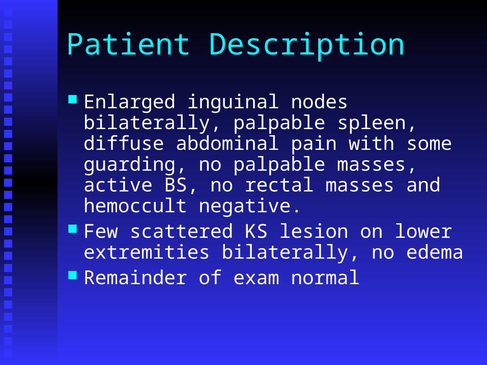
Enlarged inguinal nodes bilaterally, palpable spleen, diffuse abdominal pain with some guarding, no palpable masses, active BS, no rectal masses and hemoccult negative.
Few scattered KS lesion on lower extremities bilaterally, no edema
Remainder of exam normal

Patient Description
CD4+ count 56 cells/mm3; plasma HIV RNA 1000 copies/mL
Liver enzymes mildly elevated, creatinine normal, amylase normal
Hb 9.7 g/dL; Hct 29.3%; WBC 3000/mm3; platelet count 120,000; MCV 90 fL; retic 0.014

What is your initial diagnosis

Possible Diagnosis
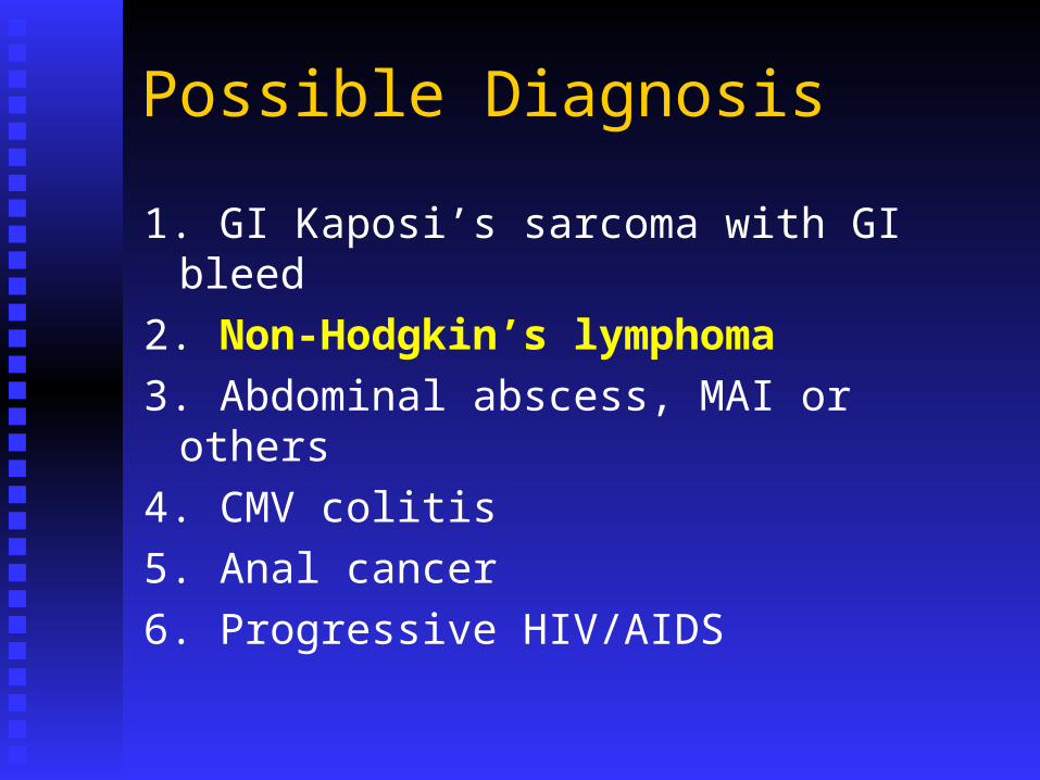
1. GI Kaposi’s sarcoma with GI bleed
2. Non-Hodgkin’s lymphoma
3. Abdominal abscess, MAI or others
4. CMV colitis
5. Anal cancer
6. Progressive HIV/AIDS
Possible Diagnosis
1. GI Kaposi’s sarcoma with GI bleed
2. Non-Hodgkin’s lymphoma
3. Abdominal abscess, MAI or others
4. CMV colitis
5. Anal cancer
6. Progressive HIV/AIDS

What additional test would you do first?

Additional Tests
1. CT abdomen
2. Biopsy lymph node
3. Repeat VL and HIV genotype
4. Culture blood and stool, endoscopy
5. Stool occult blood, bone marrow, further heme work up
6. Chest X-ray and Chest CT if appropriate
Additional Tests
1. CT abdomen
2. Biopsy lymph node
3. Repeat VL and HIV genotype
4. Culture blood and stool, endoscopy
5. Stool occult blood, bone marrow, further heme work up
6. Chest X-ray and Chest CT if appropriate
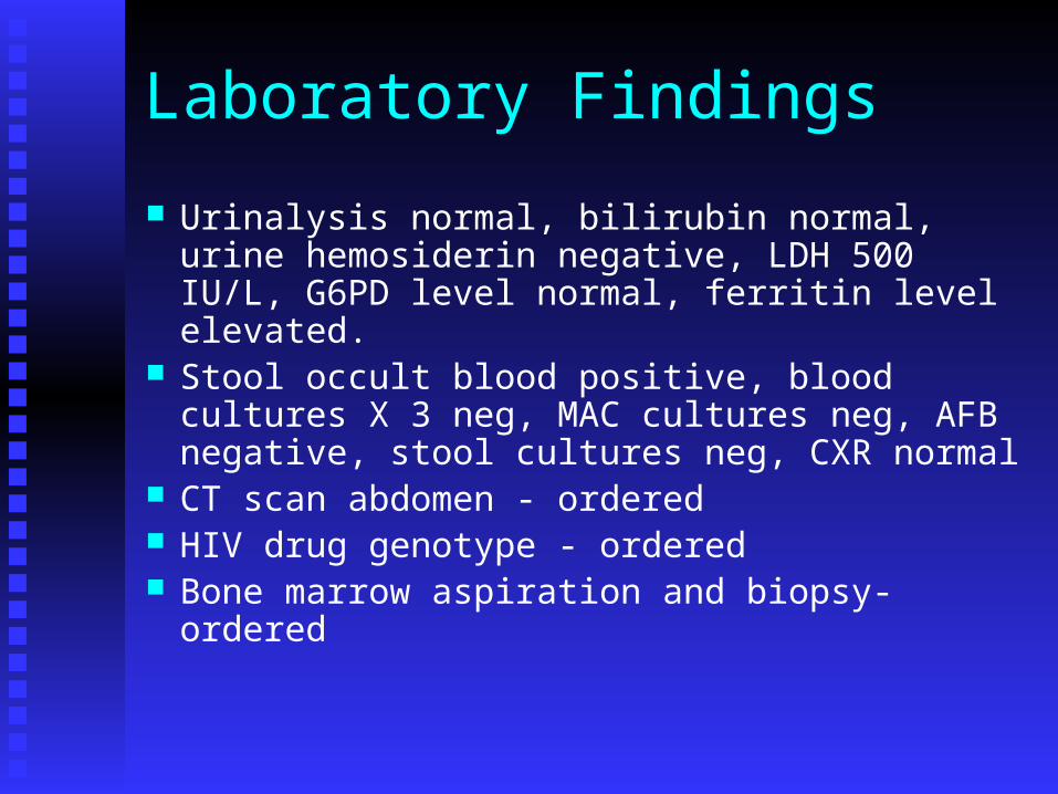
Laboratory Findings
Urinalysis normal, bilirubin normal, urine hemosiderin negative, LDH 500 IU/L, G6PD level normal, ferritin level elevated.
Stool occult blood positive, blood cultures X 3 neg, MAC cultures neg, AFB negative, stool cultures neg, CXR normal
CT scan abdomen - ordered HIV drug genotype - ordered Bone marrow aspiration and biopsy- ordered

Laboratory Findings
CT scan of abdomen shows large gastric mass, enlarged perigastric and periaortic nodes, a few liver nodules and small amount of ascites
Gastroscopy shows large gastric ulcerated mass, no active bleeding
Histology reveals large B-cell NHL, CD-20+, no CMV or KS
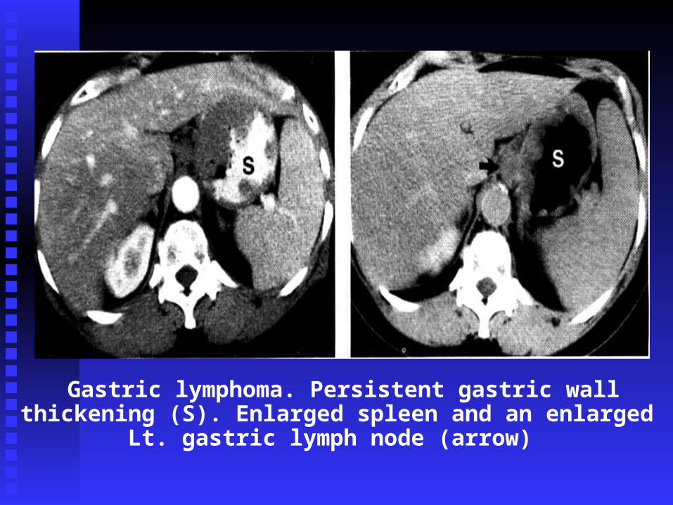
Gastric lymphoma. Persistent gastric wall thickening (S). Enlarged spleen and an enlarged Lt. gastric lymph node
(arrow)

Clinical Decision Point
The patient has no CNS symptoms, CBC has not changed. Staging work up includes:
CT of chest - no abnormalities LP with CSF cytology - neg for lymphoma Bone marrow aspiration and biopsy
hypocellular, normal iron stores, no lymphoid infiltrates, normal lymphocyte flow panel, no granuloma or infection seen

How would you proceed now?

Clinical Decision
1. Chemotherapy (e.g CHOP-R or EPOCH-R)2. Hydrate, allopurinol, follow electrolytes,
creatinine, phosphorus, calcium3. Intrathecal cytosine arabinoside X 44. Continue TMP-SMX, azithromycin5. Begin prophylaxis with ciprofloxacin6. Follow CBC, +/- G-CSF, +/- rEPO7. Change ART8. All of the above
Clinical Course
Patient tolerates chemotherapy with EPOCH-R, however after two cycles, Hb now 8.0 g/dL. What do you do?1. Repeat bone marrow aspirate and biopsy
2. Give iron and folic acid
3. Check EPO level and give recombinant erythropoietin alpha
4. Stop AZT and switch to new ARV
5. Transfuse 2 units of PRBC and schedule endoscopy
Clinical Course
EPO level 125, rEPO administered at 40,000 IU per week with supplemental iron and folic acid.
After three cycles of EPOCH, patient has achieved a complete remission. Treatment continues for 6 cycles. CBC returns to normal.

Clinical Course 6 months later, he is again noted to have Hb
9.5 g/dL, WBC 2300/mm3 and platelets 65,000/mm3 and enlarged femoral nodes
What do you do?

What do you do?1. Work up anemia, DC TMP-SMZ, retreat with
epoetin alfa and follow nodes2. Assume recurrent lymphoma and retreat with
EPOCH-R3. Assume lymphoma and treat with alternate
regimen4. Assume progressive KS and treat with liposomal
doxorubicin5. Biopsy lymph node and bone marrow
What do you do?1. Work up anemia, DC TMP-SMZ, retreat with
epoetin alfa and follow nodes2. Assume recurrent lymphoma and retreat with
EPOCH-R3. Assume lymphoma and treat with alternate
regimen4. Assume progressive KS and treat with liposomal
doxorubicin5. Biopsy lymph node and bone marrow

Clinical Course
Biopsy of lymph node and bone marrow shows high grade B-cell (CD20+) NHL, large cell type
Patient treated with ESHAP, G-CSF, epoetin alfa and prophylactic antibiotics (ciprofloxacin)
Complete remission achieved after 5 cycles, patient treated for 8 cycles and continues to be followed.

Key Points AIDS patients can have multiple cancers Evaluate for multiple etiologies for anemia in
advanced AIDS patients Consider use of Epoetin alfa when risk of
further myelosuppression is great Early relapse in AIDS/NHL should be treated
with non-cross resistant salvage chemotherapy Use prophylactic antibiotics and hematopoietic
growth factors in AIDS patients on chemotherapy, especially if receiving Rituximab

Case 2
Patient Description
57 year-old white male with HIV infection since 1985, currently on tenofovir/emtricitabline,darunavir/ritonavir,TMP-SMX, azithromycin, valgancyclovir and fluconazole
CD4 count 12, VL >150,000 copies/mL, Hb 9.2 g/dL, Hct 28 %, platelet count 111,000/mm3
Presents with low grade fevers, progressively worsening personality changes for past month and mental confusion and lethargy for past week
Patient Description
He suffers grand mal seizure on day of admission
MRI scan of brain shows single contrast-enhancing lesion in the basal ganglion with surrounding edema
What do you suspect is the most likely diagnosis?
Possible Diagnosis
1. CMV encephalitis
2. Toxoplasmosis
3. PML
4. CNS lymphoma
5. Fungal abscess
6. Infectious meningitis

Possible Diagnosis
1. CMV encephalitis
2. Toxoplasmosis
3. PML
4. CNS lymphoma
5. Fungal abscess
6. Infectious meningitis
Clinical Decision Point
Patient loaded with Phenitoin and started on dexamethasone 10 mg QID
Which diagnostics study would produce the greatest likelihood of a diagnosis?

Clinical Decision Point
1. Toxoplasma serology2. Brain biopsy3. LP with CSF cultures and cytology4. LP with toxo titer, CMV and EBV PCR5. Blood cultures for bacteria, fungi, AFB,
viruses6. Bone marrow aspiration and biopsy7. None of the above
Which diagnostics study would produce the greatest likelihood of a diagnosis?

Clinical Course
CSF cultures and cytologies negative; CMV and EBV CSF PCR sent
Blood cultures sent, preliminary negative Toxo IgG+ but IgM negative Sterotactic biopsy under MRI guidance
shows immunoblastic lymphoma, EBV+, CD20+, HHV-8 negative
Retic count 0.14, LDH 140 IU/L, ferritin normal

Clinical course
Bone marrow obtained, hypocellular with all marrow elements present, no granuloma or lymphoma, few intracellular inclusions seen, cultures pending
What is your preferred therapeutic approach?

Clinical CourseTherapeutic approach?
1. Refer for radiation therapy2. Call oncologist for high-dose MTX with
leukovorin rescue3. Change antiretroviral therapy, if possible,
continue TMP-SMX, azithromycin4. Begin GCV 5 mg/kg BID5. Begin epoetin alfa 40,000 IU QW with iron
and folate, and G-CSF6. Nothing, just continue dexamethasone and
anticonvulsants and provide palliation

Clinical Course
Patient responds to HDMTX with leukovorin rescue. Hct increases to 38% on EPO. GCV instituted for EBV-8 and with response dose reduced to 5 mg/kg TIW
ART changed to raltegravir, TMC-125 EAP and enfuvirtide
6 months later, he is still in remission. His mental status improves but not completely
12 months later he relapses, responds transiently to radiation therapy but ultimately succumbs to tumor progression and respiratory failure.

Key Points
CNS changes in AIDS may be due to multiple causes
PCNSL is rare in the HAART era, but can occur late in disease
Biopsy of brain lesion for diagnosis If inaccessible for biopsy, EBV PCR on CSF
and/or genetic studies on lymphocytes may be helpful
Treatment best with HDMTX w/wo XRT Prognosis is unfortunately very poor, but
improving

Case 3
Patient Description
49 year old, white male with recently diagnosed HIV and presumed Kaposi’s sarcoma presents to you for treatment of his KS
He has been treated by his primary physician with lamivudine/zidovudine, efavirenz for 6 months, but has received no specific KS therapy
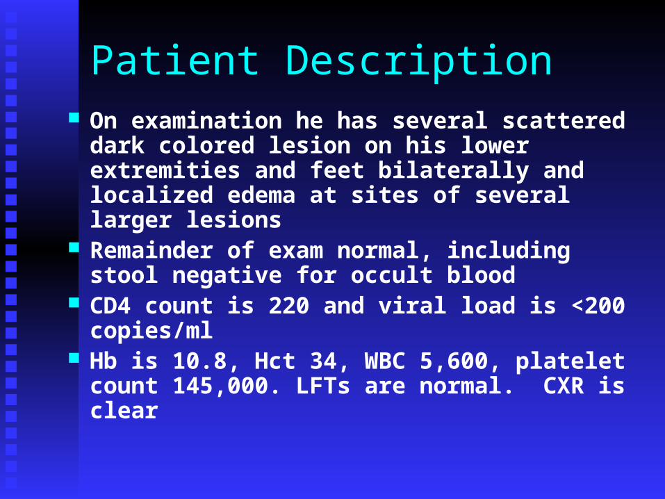
Patient Description On examination he has several scattered
dark colored lesion on his lower extremities and feet bilaterally and localized edema at sites of several larger lesions
Remainder of exam normal, including stool negative for occult blood
CD4 count is 220 and viral load is <200 copies/ml
Hb is 10.8, Hct 34, WBC 5,600, platelet count 145,000. LFTs are normal. CXR is clear

How would you proceed?
What would you do?
1. Order upper and lower endoscopy to r/o GI involvement with KS
2. Order whole body PET-CT
3. Biopsy the skin lesion
4. Begin liposomal doxorubicin for KS
5. Change efavirenz to lopinavir/ritonavir
6. 4 + 5
What would you do?
1. Order upper and lower endoscopy to r/o GI involvement with KS
2. Order whole body PET-CT
3. Biopsy the skin lesion
4. Begin liposomal doxorubicin for KS
5. Change efavirenz to lopinavir/ritonavir
6. 4 + 5

Laboratory Findings
Skin biopsy confirms Kaposi’s sarcoma CT scan of chest and abdomen show
small retroperitoneal lymphadenopathy but no visceral lesions
Repeat CD4 count 329, VL <50 copies Ferritin, Fe/TIBC, folate, and calcium
normal, corrected retic count 0.01 The patient says that he would like
something done for his leg lesions

How would you proceed now?

Clinical Decision PointWhat do you do?
1. You indicate that he should not do anything at this point as ART can cause KS to regress
2. Change efavirenz to lopinavir/ritonavir3. You begin treatment with liposomal
doxorubicin4. You begin topical 9-cis retinoic acid for the
larger lesions5. Refer to radiation therapy for treatment of
his large lesions and edema6. 2 + 4

Clinical Course Patient tolerates change in ART and notices
some local control of his KS lesions, but eventually he notices that the leg lesions have become more confluent and locally infiltrative with brawny edema
He also notices some lymphadenopathy in his groin and some mild testicular swelling
There are no new cutaneous lesions Six months later his testicular swelling is more
pronounced and he begins to have pain in his extremities

Clinical decision pointWhat do you do?
1. Recheck his viral load and CD4 count as his HIV disease must be progressing
2. Refer him for biopsy of his lymph node to R/0 NHL
3. Order CT or MRI of abdomen4. Biopsy his skin lesion5. Begin liposomal doxorubicin 6. 1 + 27. 3 + 5

Clinical decision pointWhat do you do?
1. Recheck his viral load and CD4 count as his HIV disease must be progressing
2. Refer him for biopsy of his lymph node to R/0 NHL
3. Order CT or MRI of abdomen4. Biopsy his skin lesion5. Begin liposomal doxorubicin 6. 1 + 27. 3 + 5

Clinical Course
CT of the abdomen shows retroperitoneal adenopathy and enlarged inguinal nodes
Testicular ultrasound shows testicular edema but no masses
Alpha fetoprotein and beta hCG are normal
Patient begun on liposomal doxorubicin q2 weeks with reduction in testicular and lower extremity edema and less pain after 2 cycles of therapy
Clinical Course
After 6 cycles of liposomal doxorubicin the leg lesions are under better control, but still present
He develops a non-productive cough, mild SOB and low grade fevers over course of a month
You order a chest X-ray which shows some mediastinal adenopathy, blunting of both costophrenic angles and slightly enlarged cardiac silhouette

What is your diagnosis and what do you do?1. Pulmonary KS, Order bronchoscopy for
endobronchial lesions and transbronchial biopsy2. Pulmonary TB or bacterial pneumonia or PCP,
Order bronchoscopy with broncho-alveolar lavage
3. Non-Hodgkin’s lymphoma, Order CT scan of chest and abdomen and biopsy inguinal lymph node
4. Non-Hodgkin’s lymphoma or KS, Order pericardiocentesis under ultrasonic guidance

Clinical Course Pericardiocentesis shows an exudate with many
lymphoblastic appearing cells which are CD20+, EBV+, HHV-8+ and culture negative for TB, bacteria or fungus
Flow cytometry confirm malignant anaplastic B-cells, negative for epithelial markers, c/w primary effusion lymphoma
PET CT showed hypermetabolic lymphadenopathy in the mediastinum but not in the abdomen
Bone marrow biopsy did not show NHL CSF was negative for malignant cells

Clinical Course
Patient was begun on EPOCH-R and GCV Tumor initially regressed and patient’s
symptoms improved However after 3 cycles of therapy, the
effusions returned, requiring 2 additional pericardiocentesis
Chemotherapy was changed to R-CODOX M, but the tumor continued to progress and the patient expired after deciding against further intervention
Key Points
Kaposi’s sarcoma continues to remain both a local and systemic problem in the HAART era
Progression of KS can occur even with good virologic control of HIV
Other HHV-8 related tumors may occur in individuals with KS
Treatment of Primary Effusion Lymphoma is difficult and may require more aggressive treatment

Case 4

Patient Description
42 year old, white gay male with recently diagnosed HIV infection presents to your office for the first time.
He is on efavirenz/tenofovir/emtricitabine
(Atripla) and has a CD4 count of 320 (22%) and a viral load <50 copies/ml
Patient Description
As part of your routine work up, in addition to a complete history and physical examination, what do you also order?
1. CXR and EKG2. Immunization with Pneumococcal vaccine,
hepatitis A and B vaccine, influenza vaccine3. PSA and flexible sigmoidoscopy4. Perform anal pap test5. All of the above

Patient Description
Routine labs, CXR and EKG are normal Anal pap test shows moderate
dysplastic changes You refer for high-resolution anoscopy
and biopsy which show grade 3 ASIL
How would you proceed?
1. Refer to surgery for AP resection and lymph node sampling
2. Refer to radiation therapy3. Perform infrared coagulation4. Do nothing and repeat HRA and biospy
in 6 months5. Refer for electrocauterization 6. Treat with imiquimod

How would you proceed?
1. Refer to surgery for AP resection and lymph node sampling
2. Refer to radiation therapy3. Perform infrared coagulation4. Do nothing and repeat HRA and biospy
in 6 months5. Refer for electrocauterization 6. Treat with imiquimod

Clinical Course
He undergoes IRC which he tolerates well. Follow up exam in 1 month shows good healing. He is asked to return in 3 month for repeat exam
He is lost to follow up and returns to you 3 years later stating he was in Iraq with a contract security firm
He has been poorly compliant with his HIV medications, as he did not want his employer and associates to know of his HIV status which would have threatened his employment

Patient Description
He appears in good health. His physical exam is normal.
CD4 count is 179 (15%) and his viral load is 230,000 copies/ml
You refer him for high resolution anoscopy and biopsy which reveals a 2.0 cm mass in the posterior anus, which is biopsied and found to be invasive poorly-differentiated squamous cell carcinoma
Patient description
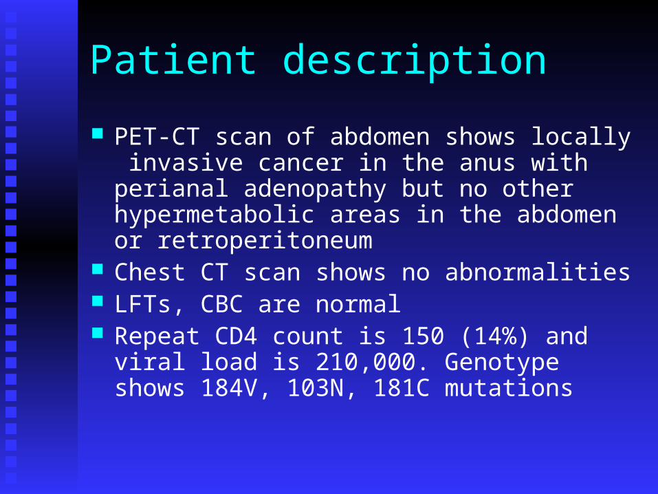
PET-CT scan of abdomen shows locally invasive cancer in the anus with perianal adenopathy but no other hypermetabolic areas in the abdomen or retroperitoneum
Chest CT scan shows no abnormalities LFTs, CBC are normal Repeat CD4 count is 150 (14%) and viral load
is 210,000. Genotype shows 184V, 103N, 181C mutations

Clinical Decision Point
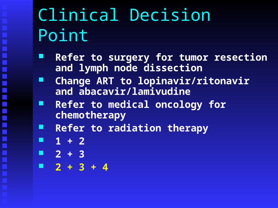
1. Refer to surgery for tumor resection and lymph node dissection
2. Change ART to lopinavir/ritonavir and abacavir/lamivudine
3. Refer to medical oncology for chemotherapy
4. Refer to radiation therapy 5. 1 + 26. 2 + 37. 2 + 3 + 4
Clinical Decision Point
Refer to surgery for tumor resection and lymph node dissection
Change ART to lopinavir/ritonavir and abacavir/lamivudine
Refer to medical oncology for chemotherapy
Refer to radiation therapy 1 + 2 2 + 3 2 + 3 + 4
Clinical Decision
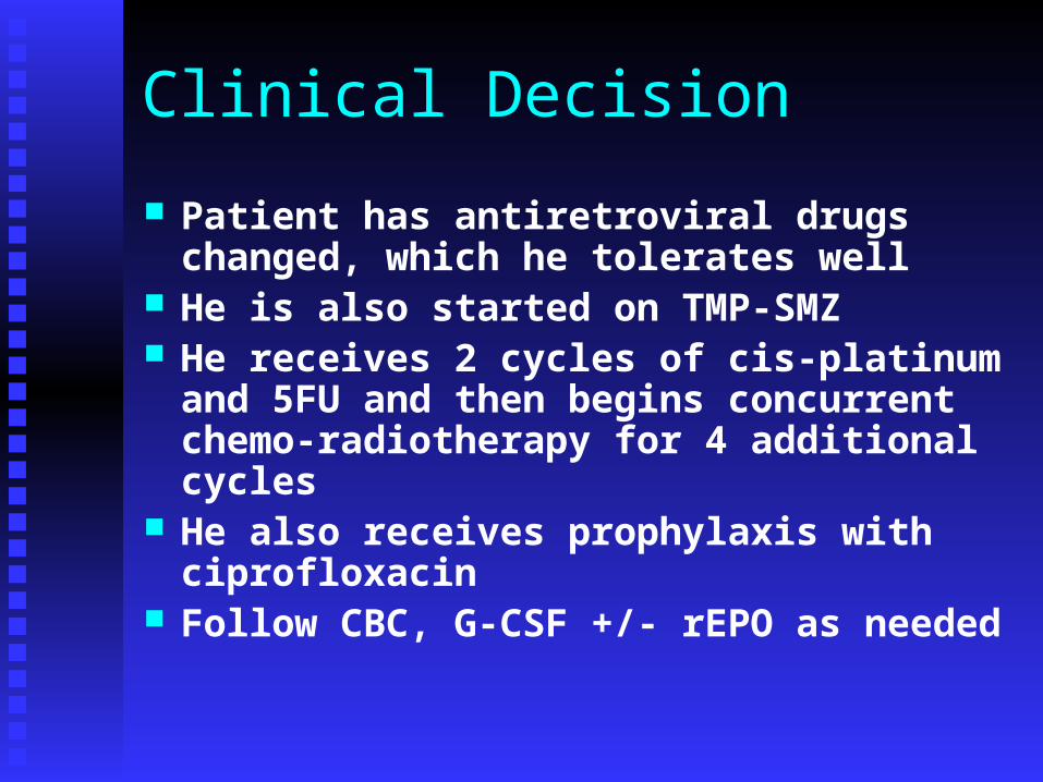
Patient has antiretroviral drugs changed, which he tolerates well
He is also started on TMP-SMZ He receives 2 cycles of cis-platinum and
5FU and then begins concurrent chemo-radiotherapy for 4 additional cycles
He also receives prophylaxis with ciprofloxacin
Follow CBC, G-CSF +/- rEPO as needed
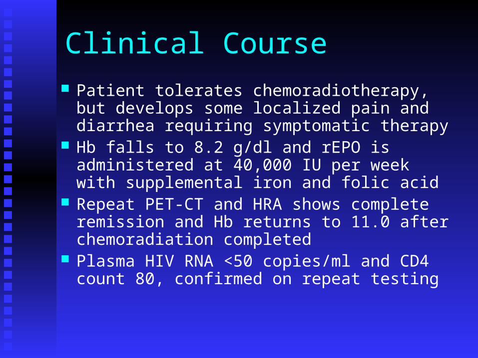
Clinical Course Patient tolerates chemoradiotherapy, but
develops some localized pain and diarrhea requiring symptomatic therapy
Hb falls to 8.2 g/dl and rEPO is administered at 40,000 IU per week with supplemental iron and folic acid
Repeat PET-CT and HRA shows complete remission and Hb returns to 11.0 after chemoradiation completed
Plasma HIV RNA <50 copies/ml and CD4 count 80, confirmed on repeat testing
What do you recommend now?1. Follow up every 3-6 months with repeat HRA and
PET-CT for 1 year2. Change antiretroviral therapy because of lower
CD4 count3. Institute additional prophylaxis for MAC and fungus4. Consolidation chemotherapy with 2 additional
cycles of chemothearpy5. 1 + 36. 2 + 37. 1 + 4
Clinical Course
Patient now works in the USA and returns regularly for follow up
CD4 counts gradually increases to 200 after 6 months
At 9 months patient develops a non-productive cough and some weight loss
CXR shows multiple diffusely scattered pulmonary nodules with hilar adenopathy
PET-CT shows retroperitoneal adenopathy and perianal adenopathy

Key Points
Patients with HIV should undergo anal PAP testing at first presentation and q6 month if abnormality detected
Local ablative therapy is effective for most HSIL, but progression can occur
Treatment for invasive anal CA consists of concurrent chemoradiotherapy
Recurrence after remission can occur and if disseminated, may have poor prognosis
Be on alert for possible chemo-ART drug interactions and plan accordingly


![Index [link.springer.com]978-3-642-17869-6/1.pdf · 410 Index. K Kaposi’s sarcoma, 90 ... Sarcoidosis Rosai-Dorfman disease, 335 Sarcoma, 2, ... Thalassemia, 268 Thyroglossal duct](https://img.pdfslide.us/doc/110x75/5b7c95787f8b9a9d078c2151/index-link-978-3-642-17869-61pdf-410-index-k-kaposis-sarcoma-90-.jpg)