Embed Size (px)
Citation preview

Carpentier-Edwards
PERIMOUNT MAGNA MitralPericardial Bioprosthesis
Model 7000TFX
Implantation Manual

This booklet illustrates my technique for mitral valve replacement with theCarpentier-Edwards PERIMOUNT Magna mitral bioprosthesis. The techniqueis based on sound surgical principles and on extensive personal experiencewith Carpentier-Edwards PERIMOUNT mitral bioprostheses over many years.This technique is employed by many cardiothoracic surgeons with excellentresults. It is my hope that this booklet proves valuable to the cardiac surgicaltrainee and cardiac surgeons interested in this implant technique.
Allen Morris, M.D.DirectorCardiovascular ServicesMercy General HospitalSacramento, CA USA
Carpentier-Edwards PERIMOUNT Magna Mitral Pericardial Bioprosthesis

Table of Contents
Introduction and Product Description ......................................1
Preparation ..............................................................................5
Technique ................................................................................9
Sizing and Bioprosthesis Preparation. ....................................13
Implantation ..........................................................................17
Post-implantation ..................................................................23

Carpentier-Edwards PERIMOUNT Magna Mitral Pericardial Bioprosthesis

Introduction and Product Description
1

2
Carpentier-Edwards PERIMOUNT Magna Mitral Pericardial Bioprosthesis
Introduction & Product Description In 1984 the Carpentier-Edwards PERIMOUNT mitral pericardial
bioprosthesis model 6900 was introduced for replacement of the mitral valve.The mitral bioprosthesis stent and leaflet attachment is similar to theCarpentier-Edwards PERIMOUNT aortic bioprosthesis but was bioengineeredto withstand the higher closing pressures encountered in the mitral position. A bioprosthesis has distinct advantages over a mechanical prosthesis in thatbioprostheses can allow freedom from lifelong anticoagulation, reducedthromboembolism risks, and reduced majorbleeding events1. Porcine bioprosthesesavailable in the early 1980s had limited long-term durability in the mitral position due tofailures from structural valve deterioration(SVD), primarily from leaflet tears andcalcification2. It was during this time that the PERIMOUNT mitral bioprosthesis wasintroduced using design based on computer-aided modeling to minimize leaflet stress(Figure 1), bovine pericardium, and improvedtissue processing*. One of the largest studies(435 patients) on long-term durability of amitral bioprosthesis has shown excellentresults for the PERIMOUNT mitralbioprosthesis, even with a young mean ageof patients at 60.7 years. The cumulativeincidence of explant due to SVD at 16 yearswas shown to be 11% in patients with aPERIMOUNT mitral bioprosthesis implantedat age ≥60 years with no SVD failures reported prior to six years (Figure 2)3.
Figure 1. PERIMOUNT leaflet and wireform computer-aided
stress analysis.

3
Introduction and Product Description
Introduced in 2005, the Carpentier-Edwards PERIMOUNT Magna mitralbioprosthesis model 7000TFX (presented in this manual) embodies over 40years of Edwards innovation in mitral valve replacement and repair. Buildingon the same stent and leaflet design of model 6900 PERIMOUNT mitralbioprosthesis, the PERIMOUNT Magna mitral bioprosthesis is designed withan asymmetric sewing cuff to maximize mitral annular conformity. Its sewingcuff is widened and has increased flexibility on the posterior aspect foraccommodating calcific degenerative processes commonly encountered inthat region when replacing a mitral valve4. In addition, by reducing thedimension of the effective profile, the stent projection into the ventricle hasbeen minimized to reduce any ventricular outflow tract obstruction or injury toventricular endocardial surfaces5 (Figure 3).
Figure 2. PERIMOUNT mitral bioprosthesis, model 6900, cumulative incidence of explantdue to SVD3.
Cu
mu
lati
ve In
cid
ence
o
f Ex
pla
nt
du
e to
SV
D
100%
80%
60%
40%
20%
0%
Cumulative incidence at 16 years for patients >_ 60 years is 11.3±2.4%
Cumulative incidence at 16 years for patients >_ 65 years is 7.6±2.2%
Cumulative incidence at 15 years for patients >_ 70 years is 1.1±1.1%
Years Postoperative
16-YEAR DURABILITY OF THE PERIMOUNT MITRAL VALVE MODEL 6900
N= 99188278
80160238
77153228
73147219
71143212
65131191
59120183
49104164
4698
157
3682
157
2972
123
205599
153766
24
1512
82141
31025
N=N=
11%8%1%
Figure 3. Profile and inflow views of PERIMOUNT Magna mitral bioprosthesis.

4
Carpentier-Edwards PERIMOUNT Magna Mitral Pericardial Bioprosthesis
The PERIMOUNT Magna mitral bioprosthesis includes the Carpentier-Edwards ThermaFix process*, the only tissue treatment thatconfronts both major causes of calcification – critical to achieving the long-term structural integrity necessary to mitigate the effects of SVD.
Edwards has also developed accessories specific for mitral replacement. The handle (model 1117) has the appropriate flexibility and length for mitralprocedures. The sizer (model 1177HP) dimensions accurately replicate thestent and cuff diameters of the bioprosthesis and are labeled with the clinicallyobtained effective orifice area (EOA) to help gauge the expected hemodynamicperformance (Figure 4). The accessories can be organized in a tray (modelTRAY1177HP) for easy sterilization (Figure 5). All PERIMOUNT mitralbioprostheses include the Tricentrix holder system, which aids in implantabilityand visualization, reduces the risk of strut capture with suture tie-down, andeases bioprosthesis insertion (Figure 6).
Figure 6.Tricentrix holder system.
Figure 5. Accessory tray, model TRAY1177HP.Figure 4. Sizer set, model 1177HP.

Preparation
5
Introduction and Product Description

6
Carpentier-Edwards PERIMOUNT Magna Mitral Pericardial Bioprosthesis
Bioprosthesis PreparationThe PERIMOUNT Magna mitral bioprosthesis requires preparation prior to
implantation. To prepare the bioprosthesis for implantation these steps mustbe followed (Figures 7-18).
Figure 8. Removethe assemblyfrom the jar.
Figure 9. Grasp the plastic sleeve (cage)and rotate the handle clockwise anadditional 1/4-turn or until the postreaches the unlock position;
Figure 10. then push on the handle untilthe post snaps into its fully deployedposition. This creates the protectivetenting structure that reduces the risk for looping a suture around a strut duringimplantation.
Figure 11.Remove thesleeve. Thesleeve isremoved bypulling awayfrom the rest ofthe assembly.
Figure 12. Removethe clip. The clip isremoved by slidingit away from theholder.
Figure 7. Insert thehandle 1111 or 1117(reusable) or 1126(disposable) and turnclockwise until snugfit. Once the handlehas been attached, it should not beremoved until thebioprosthesis is seated in the annulus.

7
Preparation
Figure 14. Rinse the bioprosthesistwo times, oneminute each, inseparate salinebowls filled with at least 500 mlsaline.
Figure 13. The ID tag is usedto verify size and serial numberand then removed by cuttingthe attachment thread.
Figure 18. and pushing on the handle untilthe tenting structure is produced and thepost snaps into position.
Figures 15 & 16. Check for properdeployment; the holder should belocked. There should be no spacebetween the base of the white postand the holder, and no slidingmovement. The green tenting threadswill form a protective barrier thatreduces the risk of looping a suturearound a strut.
Deployed Undeployed
Figure 17. If the surgeon suspects thatthe holder is not fully deployed whenhanded the bioprosthesis, deploymentcan be completed by gently holding thebioprosthesis sewing cuff, unlocking thepost (1/4-clockwise turn of the handle),

8
Carpentier-Edwards PERIMOUNT Magna Mitral Pericardial Bioprosthesis
NOTES:

Introduction and Product DescriptionPreparation
Technique
9

10
Carpentier-Edwards PERIMOUNT Magna Mitral Pericardial Bioprosthesis
Patient PreparationThe operation is typically preceded by administration of general
anesthesia of narcotic and/or inhalational agents, placement of a urinarycatheter, placement of a pulmonary artery catheter for monitoring ofpulmonary pressures and cardiac output, and intravenous antibiotics areadministered and generally continued for two days.
ExposureRoutinely, exposure for mitral valve replacement is achieved via a median
sternotomy. The pericardium is opened longitudinally in the midline from thereflection over the aorta to the diaphragm where it is T’ed to the pleuralreflections (Figure 19). The pericardial edges are sutured to the sternotomywound creating a pericardialcradle. The heart is prepared forbypass after systemicheparinization. Cannulation isperformed by using two rightangle cannulae for venous blooddrainage and placing an arterialcannula in the distal ascendingaorta. A small cannula is placed inthe superior vena cava above thesino-atrial node. The other cavalcannula is placed in the rightatrium at the junction of the atriaand the inferior vena cava.Cardiopulmonary bypass is initiated and flow is set to about 1.5 L/min/m2. Adequate decompression of the heart is assured andhypothermia is initiated by cooling to 30-34°C. The myocardium can beprotected using antegrade and retrograde cold (4°C) cardioplegia. Using aninitial loading dose of antegrade cardioplegia with subsequent retrogradedoses every fifteen to twenty minutes is useful to maintain myocardialprotection. Retrograde cardioplegia may be more effective as the aortic valvecan sometimes distort and become incompetent during retraction of theatrium. Retrograde cardioplegia also protects the ischemic left ventricle in thepresence of coronary artery disease and helps in de-airing of the heart andthe ascending aorta.
Figure 19. Median sternotomy with pericardium T-incision.

11
Exposure of the mitral valve is aided by developing Sondergaard’s planenear the inter-atrial groove. This is accomplished by careful cautery dissectionof the plane separating the right from the left atrium extending from thejunction of the superior vena cava to the junction of the inferior vena cava. The more medial that this dissection can be accomplished allows the left atrialincision to be made close to the atrial septum and greatly aids in the exposureof the mitral valve (Figure 20). Upon opening the left atrium, exposure of the mitral valve is accomplished by retraction of the left atrial wall at the 10-11 o’clock position and inferiorly at the 2-3 o’clock position. Retraction isassisted with the availability of many commercial mitral valve retractors (Figure 21).
Annular PreparationWith adequate exposure, identification of the important left atrial structures
such as the orifice of the left and right pulmonary veins, left atrial appendageand mitral valve apparatus is accomplished. One should first assess the degree of calcification of the anterior and posterior mitral valve leaflets andannulus. Severe calcification of the leaflets and annulus may lead to significantdifficulties in seating the bioprosthesis. As much as possible, debridement of the anterior and posterior annulus should be accomplished. If excessivecalcification is encountered along the anterior leaflet and annulus, excision of the anterior mitral valve leaflet leaving a 2-3 mm rim of tissue to bolsterpledgeted sutures may be helpful. If the posterior mitral valve annulus isseverely calcified, attempts to debride this tissue can be made but increase the risk of atrial ventricular disruption. In this case, the posterior mitral valveleaflet itself can be utilized as the anchors for the valve sutures. If extensive
Technique
Figure 20. Dissection of the Sondergaard’splane near the inter-atrial groove.
Figure 21. Exposure of the mitral valve with LA retraction at 10-11 o’clock and 2-3 o’clock positions.

12
Carpentier-Edwards PERIMOUNT Magna Mitral Pericardial Bioprosthesis
debridement is necessary, utilization of an ultrasonic surgical aspirator deviceor reconstruction of the posterior annulus using bovine pericardium may be necessary.
Beyond the above considerations regarding valve and annulardebridement, replacement of the mitral valve preferably involves some form ofnative leaflet and thus chordal-papillary muscle preservation. Studies haveshown improved outcomes in patients with leaflet and chordal-papillarymuscle preservation1. The anterior leaflet can be preserved by partiallyexcising it or folding it against anterior annulus using a polypropylene suture.Preservation of all or some of the posterior leaflet can preserve chordal-papillary support, and in the case of friable or calcified annular tissue, canprotect the atrial-ventricular groove from rupture6. If the anterior mitral valveleaflet is excised to aid in placement of a larger bioprosthesis, in most casesthe posterior mitral leaflet with its chordal attachments can be preserved.
If all the native leaflet tissue and subsequent chordal support must besacrificed, papillary muscle resuspension can still be accomplished. Routinely,a pledgeted 5-0 flouropolymer suture can be placed in the fibrous head of theanterior-lateral or posterior-medial papillary muscle and brought to the mitralvalve annulus. Correct length of the chord is usually aided by measuring itagainst remaining secondary chordal elements. Anchoring these sutures tothe annulus at the 8 o’clock position for the antero-lateral papillary muscleand at the 4 o’clock position for the posterior-medial papillary muscleprevents impingement on the struts or the outflow tract (Figure 22).
Figure 22. Papillary muscle resuspension usingflouropolymer suture.

Sizing and Bioprosthesis Preparation
13

14
Carpentier-Edwards PERIMOUNT Magna Mitral Pericardial Bioprosthesis
SizingThe suturing technique described in this guide for attaching the
PERIMOUNT Magna mitral bioprosthesis to the mitral annulus uses pledgetedmattress sutures placed from the ventricle to the atrium with pledgets on theventricular side, also called a non-everting technique. This technique mayallow the use of a larger size bioprosthesis while improving left ventricularoutflow by further reducing ventricular projection of the stent.
When sizing for non-evertingsuturing techniques the sizer barrelshould comfortably fit in the annuluswhile the lip of the sizer should beseated on top of the annulus (Figure 23). The dimensions of thesizer barrel match the external stentdiameter while the dimensions of thesizer lip match the bioprosthesis sewing cuff, giving an accurate reflection of bioprosthesis fit (Figure 24 a and b).
Figure 23. Sizer fit for non-everting suturingtechniques.
Figure 24a. Sizer dimensions.
Sizer lip
Sizer barrel
Valve sewing cuff
External stent diameter
Figure 24b. Valve dimensions

15
The sizer lip of model 1177HP is asymmetric in a similar manner to thePERIMOUNT Magna mitral bioprosthesis sewing cuff. The sizer has leftventricular outflow tract (LVOT) markings that match the bioprosthesis. The two black markings (one double and one single) help aid in the sizerorientation and the space between them should be positioned over the LVOT as the bioprosthesis will later be (Figure 25). Over-sizing should be avoided with the PERIMOUNT Magna mitral bioprosthesis as all sizes have excellenthemodynamics and over-sizing increases the risk of stent distortion (Figure 26).
Sizing and Valve Preparation
Figure 26. Clinical EOA at 1-2 years post-implant in cm2 7.
Figure 25. Sizer positioning with the space betweenthe black markers aligned over the LVOT.
0
0.9
1.8
2.7
3.6
2.32.6 2.6 2.5
1.8
Valve Size
25 mm 27 mm 29 mm 31 mm 33 mm
Clinical EOA at 1-2 yr post implant cm2
3 40 47 27 4n=

16
Carpentier-Edwards PERIMOUNT Magna Mitral Pericardial Bioprosthesis
Bioprosthesis Preparation The bioprosthesis should be properly prepared as described earlier and
irrigated frequently during implantation to prevent drying or dehydration of the tissue. The Tricentrix holder system should be properly deployed and thebioprosthesis should be adequately rinsed. If the holder system is handed to the surgeon in an undeployed fashion (the protective tenting system is not created) the surgeon or operative nurse can produce the deployment by gently holding the bioprosthesis sewing cuff, unlocking the post with a 1/4-clockwise turn to the handle (Figure 27), and pushing on the handle (Figure 28) until the tenting structure is produced and the post snaps intoposition. Do not implant the bioprosthesis if this tenting structure is notproduced as it protects from looping a suture around the stent as well as aids insertion into the annulus (Figure 29). The bioprosthesis should beirrigated frequently (every 1-2 minutes) on both sides with saline to keep itmoist during the implant procedure.
Figure 27. Unlock the post 1/4-clockwise turn.
Figure 29. Tenting structure protects againstsuture looping.
Figure 28. Push on the handle until tentingstructure is produced and post snaps intoposition.

Implantation
17

18
Carpentier-Edwards PERIMOUNT Magna Mitral Pericardial Bioprosthesis
ImplantationTo produce the most natural flow the PERIMOUNT Magna mitral
bioprosthesis should be oriented so that there is no obstruction of the LVOTby the post or strut of the bioprosthesis. In addition, the sewing cuff of thePERIMOUNT Magna mitral bioprosthesis is asymmetric and should beoriented so that the anterior saddle shape is positioned over the nativeanterior leaflet aiding in a natural distribution of chordal force if leafletpreservation techniques are used. This also positions the cuff regions ofhighest flexibility over the posterior annulus which is often calcified maximizingannular-cuff coaptation. To achieve proper orientation of the bioprosthesis, theanterior region of the sewing cuff (marked by the space between the two postor strut markers - one double and one single) should be positioned over theLVOT. This results in the double marking positioned near the anteriorcommissure of the native annulus, the single marking near the posteriorcommissure of the native valve, and the non-marked commissure of thebioprosthesis near the 6 o’clock position. Sutures are placed in the sewingcuff of the bioprosthesis making sure to properly orient the bioprosthesis(Figure 30).
Once all annular sutures are placed in the sewing cuff, the bioprosthesisis lowered using the handle and gentle traction on the sutures to maintaintension and avoid suture entrapment or tangling (Figure 31). The bioprosthesisis carefully inserted into the annulus by bending the handle sufficiently toenable the insertion of the posterior strut first and then the anterior portion
Figure 30. Proper orientation of the PERIMOUNTMagna mitral valve with the space between thetwo markers aligned over the LVOT.

19
of the bioprosthesis. This may help reduce the risk of damage to the ventricleor annulus. Once seated, approximation of the annulus and sewing cuff is checked.
The handle is released from the holder without unscrewing it by cuttingthe single green thread attaching the adapter to the post near the handle(Figure 32). This provides additional access and visibility for tying. Do not cutthe three green threads on the legs that attach the holder to the bioprosthesisuntil all sutures have been tied. Sutures are tied being careful to check thatseating does not shift and the sutures are seated normally. When cutting thesuture tails verify that the knots or any remaining suture does not touch theleaflet tissue of the bioprosthesis as these may interfere with its function orcause abrasion to the leaflet.
Implantation
Figure 31. Bioprosthesis insertion with gentle suture traction to avoid tangling.
Figure 32. The single central green thread can be cut to quickly release the handle.

Once all suture knots are tied, the holder is removed. In some cases one or two remaining sutures can be tied only by removing the holder first. In these cases all the sutures adjacent to each of the three frame struts must be tied down before cutting the holder attachment threads as the protectivetenting is no longer in place after the holder is removed. The holder isreleased by cutting the three green threads on the legs of the holder. This isachieved by placing a scissors or cutting blade in the cutting channels (Figure 33). The holder is removed from the bioprosthesis by securing thebioprosthesis to the annulus and grasping the holder with sterile gloved handsor protected forceps and pulling it away from the bioprosthesis (Figure 34).
20
Carpentier-Edwards PERIMOUNT Magna Mitral Pericardial Bioprosthesis
Figure 33. Three green threads are cut to remove the holder after all sutures are tied.
Figure 34. Holder is removed while securing thebioprosthesis to the annulus.
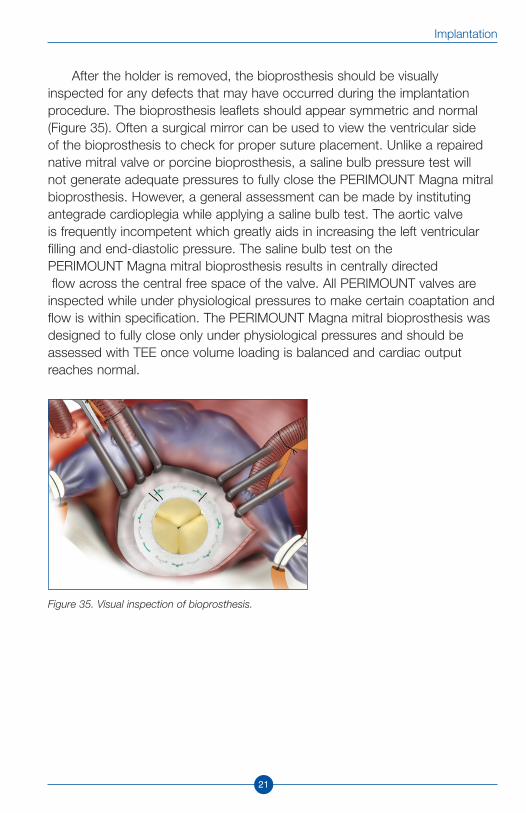
After the holder is removed, the bioprosthesis should be visuallyinspected for any defects that may have occurred during the implantationprocedure. The bioprosthesis leaflets should appear symmetric and normal(Figure 35). Often a surgical mirror can be used to view the ventricular side of the bioprosthesis to check for proper suture placement. Unlike a repairednative mitral valve or porcine bioprosthesis, a saline bulb pressure test will not generate adequate pressures to fully close the PERIMOUNT Magna mitralbioprosthesis. However, a general assessment can be made by institutingantegrade cardioplegia while applying a saline bulb test. The aortic valve is frequently incompetent which greatly aids in increasing the left ventricularfilling and end-diastolic pressure. The saline bulb test on the PERIMOUNT Magna mitral bioprosthesis results in centrally directedflow across the central free space of the valve. All PERIMOUNT valves are
inspected while under physiological pressures to make certain coaptation andflow is within specification. The PERIMOUNT Magna mitral bioprosthesis wasdesigned to fully close only under physiological pressures and should beassessed with TEE once volume loading is balanced and cardiac outputreaches normal.
21
Implantation
Figure 35. Visual inspection of bioprosthesis.

22
Carpentier-Edwards PERIMOUNT Magna Mitral Pericardial Bioprosthesis
NOTES:

Post-implantation
23

24
Carpentier-Edwards PERIMOUNT Magna Mitral Pericardial Bioprosthesis
Post-implantationThe mitral replacement procedure is often followed by a ligation of the left
atrial appendage to prevent clot formation in patients with atrial fibrillation orenlarged atrium. Closure of the atrium is accomplished using a runningpolypropylene suture. Mitral replacement can be done with a concomitantcoronary artery bypass graft. The bypass should be done first to reduce therisk of the bioprosthesis damaging tissue while lifting the heart and to aid indelivery of cardioplegia through the graft. On the other hand, tricuspid valveprocedures are usually performed after replacing the mitral valve. In casesrequiring both aortic and mitral valve replacement, generally the surgeon willexcise the aortic valve and proceed to the mitral replacement. The aorticbioprosthesis is then sewn in after the mitral bioprosthesis is in place.Attempting to expose the mitral valve with an aortic bioprosthesis in place canbe difficult and may require other techniques such as division of the atrial septum or the dome of the left atrium for adequate visualization.
AssessmentTrans-esophageal echocardiography (TEE) is particularly useful for
imaging the mitral valve to detect proper bioprosthesis function and to aid inremoval of intra-cardiac air prior to decannulation. Careful de-airing at the endof the operation is essential. Venting can be achieved through the left atrium,the aorta, and sometimes the left ventricle apex. Once de-airing maneuversare completed, the cross-clamp can be released and the patient is completelyre-warmed. The venous return can then be occluded partially and the heart isthen gradually loaded as any necessary pharmacological agents are given.Pulmonary artery pressures should be carefully monitored. Temporaryepicardial pacing wires may be placed at the right atrium and the rightventricle to suppress atrial and ventricular tachyarrhythmias.
Once volume loading is balanced and cardiac output approaches normal,the PERIMOUNT Magna mitral bioprosthesis can be imaged by TEE forassessment of bioprosthesis function. Similar to most prosthetic valves,PERIMOUNT bioprostheses exhibit a unique pattern of signature flow which isnormal (Figure 36). Normally functioning PERIMOUNT bioprosthesesdemonstrate on TEE trace or mild central regurgitation from the free space atthe center of the bioprosthesis. As the left ventricle returns to physiologicpressures following bypass, the appearance of any central jet should diminish

25
until only trivial or mild signature flow remains. Normal pressures may takemore than 45 minutes to be reached. Similarly, prior to heparin reversal, thebioprosthesis may show some temporary minor jets through the sewing ringcloth prior to administration of protamine. This type of flow will resolve asheparin is reversed.
If there is detection of 2+ or greater central regurgitation or the presenceof a moderate eccentric jet then this indicates a problem. Excessiveregurgitation (especially severe regurgitation), eccentric regurgitant jets, or arestricted appearance of the leaflets on echocardiographic assessment mayindicate an entrapped leaflet. In this case the bypass should be reinstitutedand the atrial incision should be reopened for further assessment.
Antithrombogenic TherapyAnticoagulation is prescribed for all patients undergoing mitral valve
replacement. The therapeutic international normalized ratio (INR) for patientsafter mitral valve replacement ranges from 2.5-3.5 and warfarin therapy isusually started on the second postoperative day. Various methods of heparinor dextran bridging therapy can be used until the warfarin has reachedtherapeutic level. The INR levels can be in the low range for patients in sinusrhythm with PERIMOUNT Magna mitral bioprosthesis. Patients can beevaluated in 6-12 weeks for any rhythm disturbances and if they arepredominantly in sinus rhythm, warfarin is stopped. All mitral replacementpatients are recommended aspirin therapy daily indefinitely.
Post-Implantation
Figure 36. Normal signature flow of PERIMOUNT mitral bioprosthesesappears as trace or mild central regurgitation on TEE.

26
Carpentier-Edwards PERIMOUNT Magna Mitral Pericardial Bioprosthesis
NOTES:

27
Introduction and Product Description
NOTES:

* No clinical data are available which evaluate the long-term impact of the Edwards Lifesciences tissue treatment in patients.
The surgical technique presented herein is the technique used by Allen Morris, M.D.Edwards Lifesciences does not endorse any particular surgical technique.
Allen Morris, M.D. is a paid consultant to Edwards Lifesciences.
Edwards Lifesciences devices placed on the European market meeting the essentialrequirements referred to in Article 3 of the Medical Device Directive 93/42/EEC bear the CE marking of conformity.
Edwards is a trademark of Edwards Lifesciences Corporation. Edwards Lifesciences, the stylized E logo, Carpentier-Edwards, PERIMOUNT, PERIMOUNT Magna, ThermaFixand Tricentrix are trademarks of Edwards Lifesciences Corporation and are registered in the United States Patent and Trademark Office.
© 2007 Edwards Lifesciences LLCAll rights reserved. ARXXXXXX
REFERENCES1. Bonow RO et al. AHA/ACC Guidelines for the Management of Patients with Valvular Herat
Disease. J Am Coll Cardiol and Circulation. August 2006.2. Bonow et al AHA/ACC Guidelines. J Am Coll Cardiol 1998;32(5):1486-588.3. Marchand MA, et al. Fifteen-year experience with the Mitral Carpentier-Edwards PERIMOUNT
Pericardial Bioprosthesis. Ann Thorac Surg 2001;71:S236-9. Carpentier-Edwards PERIMOUNT Mitral Pericardial Bioprosthesis 16-year Results. Data on file at Edwards Lifesciences, 2003.
4. Arounlangsy P, et al. Histopathogenesis of early-stage mitral annular calcification. J Med Dent Sci 2004 Mar;51(1):35-44.
5. Gudbjartsson T, Aranki S, Cohn LH. Mechanical/bioprosthetic mitral valve replacement. In: Cohn LH, Edmunds LH Jr., eds. Cardiac Surgery in the Adult. New York; McGraw-Hill 2003:951986.
6. Spencer FC, et al. A clinical Evaluation of the Hypothesis that Rupture of the Left Ventricle Following Mitral Valve Replacement Can Be Prevented By Preservation of the Chordae of the Mural Leaflet. Ann Surg. 1985 Dec; 202(6): 673–680.
7. Directions for Use: Carpentier-Edwards PERIMOUNT Magna mitral pericardial bioprosthesis, Model 7000TFX.
Edwards Lifesciences LLC · Irvine, CA 92614 USA · 949.250.2500 · www.edwards.comEdwards Lifesciences (Canada) Inc. · Mississauga, Ontario · Canada L5C 4R3 · 905.566.4220
Edwards Lifesciences Europe S.A. · 1162 Saint-Prex · Switzerland · 41.21.823.4300